healthy nutrition, healthy...
TRANSCRIPT

HEALTHY NUTRITION, HEALTHY BONESHOW NUTRITIONAL FACTORS AFFECT MUSCULOSKELETAL HEALTH THROUGHOUT LIFE
www.iofbonehealth.org

2
NORMAL BONE OSTEOPOROTIC BONE
Osteoporosis is a disease characterized by low bone mass and deterioration in the microarchitecture of bone tissue, leading to an increased risk of fracture. Osteoporosis occurs when bone mass decreases more quickly than the body can replace it, leading to a net loss of bone strength. As a result the skeleton becomes fragile, so that even a slight bump or fall can lead to a broken bone, (referred to as a fragility fracture). Osteoporosis has no signs or symptoms until a fracture occurs – this is why it is often called a ‘silent disease’.
Osteoporosis affects all bones in the body; however, fractures occur most frequently in the vertebrae (spine), wrist and hip. Osteoporotic fractures of the pelvis, upper arm and lower leg are also common. Osteoporosis itself is not painful but the broken bones can result in severe pain, significant disability and even mortality. Both hip and spine fractures are also associated with a higher risk of death - 20% of those who suffer a hip fracture die within 6 months after the fracture.
A COMMON DISEASE
It is estimated that worldwide an osteoporotic fracture occurs every three seconds. At 50 years of age, one in three women and one in five men will suffer a fracture in their remaining lifetime. For women, the risk of hip fracture is higher than the risk of breast, ovarian and uterine cancer combined. For men, the risk is higher than the risk for prostate cancer. Approximately 50% of people with one osteoporotic fracture will have another, with the risk of new fractures rising exponentially with each fracture.
A GROWING PUBLIC HEALTH PROBLEM
The risk of sustaining a fracture increases exponentially with age due not only to the decrease in bone mineral density, but also due to the increased rate of falls among the elderly. The elderly represent the fastest growing segment of the population. Thus, as life expectancy increases for the majority of the world’s population, the financial and human costs associated with osteoporotic fractures will increase dramatically unless preventive action is taken.
WHAT IS OSTEOPOROSIS?
TABLE OF CONTENTS § Foreword 3
§ Changing nutritional needs throughout life 4
§ Maternal nutrition: getting a head start in life 6
§ Building bone in childhood and adolescence: stocking up for the future 9
§ Maintaining bone mass in adulthood: stemming the tide of bone loss 15
§ The special nutritional needs of seniors: fighting frailty and reducing falls and fractures 21
§ Diseases and disorders that affect nutritional status 26
§ References 29

3
Our skeleton is formed before we are born, supports us throughout our lives, and can remain long after we die. Regardless of age, gender, race or nationality, we all have one. Yet this essential organ is so often taken for granted. This 2015 World Osteoporosis Day Report focuses on the nutritional needs of our skeleton throughout the life-course, from before the cradle, to the grave.
To begin at the beginning, we must ensure that expectant mothers are sufficiently well-nourished to support an infant’s development in utero. In this regard, surveys from around the world, which have identified low levels of calcium intake and vitamin D insufficiency as being very common in pregnancy, are a cause for concern.
Osteoporosis has been characterized as a paediatric disease with geriatric consequences. Childhood and adolescence are critical periods in skeletal development which will determine an individual’s peak bone mass. This is the time to maximize savings in the ‘bone bank’, because during the remainder of our lives, we will be making withdrawals against that peak balance. Whilst genetics plays a significant role in determining skeletal growth, decisions made by parents and their children regarding nutrition and exercise, can have a profound effect upon fracture risk later in life. We genuinely owe it to our children to ensure that they have a well-balanced diet, rich in calcium, vitamin D and protein, and take adequate exercise to set them up for a long, healthy and active life.
During the adult decades from our twenties to sixties, our goal must be to avoid premature bone loss and maintain a healthy skeleton. A clear consensus exists on the amount of dietary calcium that we should consume, which is shared by leading organizations across the world. However, as for pregnant women and adolescents in general, and particularly teenage girls in some countries, numerous reports from all regions of the world indicate that calcium intake is often far below the recommendations of national guidelines. With regard to vitamin D, IOF’s efforts to map vitamin D insufficiency and deficiency paint a truly alarming picture among all age groups in all regions. Other dietary factors which can adversely affect bone health include excessive consumption of alcohol and caffeinated beverages. Furthermore, as in all things, being at either extreme of the body mass distribution – whether underweight or overweight – is bad for bones.
FOREWORD
Cyrus CooperNIHR Musculoskeletal Biomedical Research Unit, Nuffield, Department of Orthopaedics, University of Oxford, Oxford, UK
MRC Lifecourse Epidemiology Unit, University of Southampton, Southampton General Hospital, Southampton, UK
Bess Dawson-HughesJean Mayer USDA Human Nutrition Research Center on Aging, Tufts University, Boston, MA, USA
Catherine M. GordonDivisions of Adolescent Medicine and Endocrinology, Hasbro Children’s Hospital, Alpert Medical School of Brown University, Providence, RI, USA
René RizzoliDivision of Bone Diseases, Geneva University Hospitals and Faculty of Medicine, Geneva, Switzerland
Osteoporosis, and the fragility fractures it causes, are most common among seniors in our society. While fragility fracture incidence is rising rapidly throughout the world, as the baby boomer generation ages, a recent study of current and future fracture incidence in China highlights the threat posed by osteoporosis. In 2010, approximately 2.3 million fractures occurred in the Chinese population aged 50 years and over, a figure which is set to rise to almost 6 million by 2050. Nutrition plays an important role for our seniors, as a component of broader efforts to ensure that individuals who are at high risk of fracture, particularly those who have suffered fragility fractures in the past, receive optimal treatment to prevent future fractures.
Although osteoporosis primarily affects older people, behaviours and decisions throughout the life-course, for better or worse, can dramatically contribute to an individual’s risk of suffering a fragility fracture. This report provides clear guidance on how nutrition can support musculoskeletal health at each stage of life.

4
Changing nutritional needs throughout life
GOOD NUTRITION, INCLUDING SUFFICIENT CALCIUM,
PROTEIN AND VITAMIN D, ARE ESSENTIAL TO BUILDING AND
MAINTAINING A HEALTHY SKELETON AT ALL AGES.

5
Throughout our lives the size of our skeleton, and the amount of bone contained within it, changes significantly. As illustrated in Figure 1, during the first 10–12 years of life, bone mass increases steadily for both boys and girls. At puberty, the rate of accrual of bone mass accelerates, increasing more rapidly in boys1, resulting in achievement of peak bone mass (PBM) by the mid-twenties. Thereafter, a gradual decline into old age occurs in men, and an accelerated period of bone loss for several years after the menopause occurs in women.
The primary objectives for good bone health at the various stages of life are:
§ Children and adolescents: achieve genetic potential for peak bone mass
§ Adults: avoid premature bone loss and maintain a healthy skeleton
§ Seniors: prevent and treat osteoporosis
Provision of adequate nutrition to build and maintain the skeleton is essential to the achievement of these objectives. The most important nutrients for bone health are calcium, vitamin D and protein. The role of other micronutrients is also discussed in this report, including vitamin A, B vitamins, vitamin K, magnesium and zinc.
Calcium
Calcium is a major building block of our skeleton; 99% of the 1 kg of calcium found in the average adult body resides in our bones. It is present in bone in the form of a mineral complex called
hydroxyapatite which confers strength to the skeleton. Calcium also plays an important role in nerve and muscle-functioning which requires blood calcium levels to be tightly controlled. As a result, the skeleton acts as a calcium ‘reservoir’. Should blood levels fall, parathyroid hormone (PTH) from the parathyroid glands in the neck causes the skeleton to release calcium into the blood stream to compensate for a fall in circulating calcium concentrations. Calcium is important for bone health throughout the life-course, particularly during the teenage years when about half of our bone mass is accumulated3.
Vitamin D
Vitamin D plays two key roles in the development and maintenance of healthy bones4:
§ Assists calcium absorption from food in the intestine
§ Ensures correct renewal and mineralization of bone
Notably, vitamin D was misnamed when it was discovered in 1922. It is not a true vitamin because an ongoing nutrient source is not required to sustain normal levels in the body. Vitamin D should properly be classified as a hormone precursor. A hormone is a chemical substance produced by one organ and then transported in the blood stream to a target organ, where it causes a specific biological action. Vitamin D is made in the skin when it is exposed to UV-B rays in sunlight, but can also be obtained from foods such as oily fish. Vitamin D deficiency in children can lead to growth retardation and bone deformities known as rickets. The same processes in adults lead to osteomalacia, which is a ‘softening’ of the bones due to poor mineralization. Milder degrees of vitamin D insufficiency are common, and can predispose individuals to osteoporosis5,6.
Protein
Dietary protein provides the body with a source of amino acids to support building of the bone matrix. It also has a favourable effect on bone by increasing blood levels of insulin-like growth factor I (IGF-I) which plays an important role in bone formation7. Variation in protein intake during childhood and adolescence can affect skeletal growth and may modulate the genetic potential for PBM attainment8. In older adults, low protein intake is associated with loss of bone mineral density (BMD) at the hip and the spine9. Protein supplementation of patients who have suffered a hip fracture has been shown to reduce post-fracture bone loss, medical complications and rehabilitation hospital stay10.
7050 604020 3010
pubertymenopause
male female
Years
Bo
ne
mas
s
peak bone mass
FIGURE 1 Bone mass throughout the life-course2

6
Maternal nutrition: getting a head start in life
POOR EARLY GROWTH DUE TO INADEQUATE MATERNAL
NUTRITION IS ASSOCIATED WITH REDUCED ADULT BONE
MINERAL CONTENT AT PEAK BONE MASS AND IN LATER LIFE,
AND ALSO INCREASED RISK OF HIP FRACTURE.

7
During the last two decades, the concept that environmental influences during intrauterine and early postnatal life could have long-term implications for adult health and disease has become well established. Correlations have been demonstrated between geographic areas with high rates of cardiovascular disease and those with high rates of infant mortality 50 years before11,12. This underpins the hypothesis that chronic noncommunicable diseases during later life may stem from a mismatch between the environment experienced in utero and that experienced in early postnatal life. It is becoming increasingly clear that the early life environment has long-term consequences for musculoskeletal development13,14, indeed poor early growth is associated with reduced adult bone mineral content (BMC) at peak bone mass and in later life15, and also increased risk of hip fracture16,17. Studies of mother-offspring cohorts have revealed particular influences during pregnancy which may mediate such associations, with roles demonstrated for maternal body build, lifestyle, physical activity, diet and vitamin D status18-21.
The role of maternal diet during pregnancy
The majority of bone development in the human foetus occurs during the third trimester, requiring a total of 30 g of calcium22. Intestinal absorption of calcium increases in the mother during pregnancy, and very low maternal intake may be a risk factor for lower bone mass in neonates, particularly in areas where dietary calcium content is chronically poor23. Although the general pattern of maternal diet during gestation appears related to offspring bone development, with more healthy maternal diets associated with greater offspring bone mass24, the gestational micronutrient that has been most strongly associated with offspring bone development is vitamin D.
The role of maternal vitamin D
Vitamin D insufficiency is common during pregnancy. A mother-offspring cohort study from Southampton, UK reported that 31% of mothers had insufficient (11–20 ng/mL) and 18% deficient (<11 ng/mL) circulating concentrations of 25-hydroxyvitamin D [25(OH)D] during late stage pregnancy20. Lower concentrations of gestational 25(OH)D were associated with reduced whole-body and lumbar spine BMC and BMD in children at 9 years of age. Another study from the same group of investigators reported a correlation between maternal vitamin D concentrations in pregnancy and neonatal bone mass25. To date, only one small-scale intervention study has considered the impact of vitamin D supplementation in pregnancy which included an assessment of bone mineralization in the offspring26. In order to address this gap in the
evidence-base, the UK Maternal Vitamin D Osteoporosis Study (MAVIDOS), a large-scale randomized-controlled trial, is testing whether offspring of mothers supplemented with vitamin D in pregnancy have higher bone mass at birth than those whose mothers were not supplemented27.
Dietary guidelines and the needs of expectant mothers
United States of America
In February 2015, the Dietary Guidelines Advisory Committee (DGAC) published an Advisory Report for the Secretary of Health and Human Services and the Secretary of Agriculture31. The DGAC found that several nutrients are under-consumed relative to the Estimated Average Requirement (EAR) or the Adequate Intake (AI) levels set by the Institute of Medicine (IOM)32. These so-called ‘shortfall nutrients’ are vitamin A, vitamin D, vitamin E, vitamin C, folate, calcium, magnesium, fibre and potassium. Among these, calcium, vitamin D, fibre and potassium have also been classified as ‘nutrients of public health concern’ because of well documented links to adverse health outcomes. With regard to calcium and vitamin D, the DGAC findings echo those of the U.S. Food and Drug Administration (FDA), which designated calcium and vitamin D as nutrients of ‘public health significance’ in its recent review of evidence in publishing a Proposed Rule on the Nutrition Facts label33. Notably, among pregnant women, 90% had intakes below the EAR for vitamin D and 24% had intakes below the EAR for calcium. This led the DGAC to note specifically that calcium is an under-consumed nutrient of public health concern among pregnant women.
Strategies have been proposed to achieve the Recommended Dietary Allowance (RDA) of
The gestational micronutrient that has been most strongly associated with offspring bone development is vitamin D

8
vitamin D by the American Academy of Pediatrics (AAP)34, the Endocrine Society35 and the National Osteoporosis Foundation36 which include:
§ Consumption of fortified foods § Broadening the range of fortified dairy products § In some cases, the use of a vitamin D
supplement or a multivitamin including vitamin D
Strategies to improve calcium intake include increased consumption of dairy or fortified products that are important sources of calcium.
United Kingdom
The National Health Service (NHS) in the UK recommends expectant mothers should take a supplement containing 10 µg (400 IU) vitamin D
each day throughout pregnancy and during breastfeeding37. The recommendation highlights that women who choose to take a multivitamin supplement to obtain vitamin D should not use any supplements containing vitamin A (retinol), as too much could be harmful to the foetus.
In 2014, the National Institute for Health and Clinical Excellence (NICE) published Public Health Guidance 56 on increasing vitamin D supplement use among at-risk groups, including pregnant women38. The guidance notes that the main natural source is from the action of sunlight on skin. However, from mid-October to the beginning of April in the UK there is no ambient ultraviolet sunlight of the appropriate wavelength for skin synthesis of vitamin D, resulting in a significant minority of adults and children having low levels39.
The biological mechanism considered to mediate such early influences on later adult disease is termed developmental plasticity13. A highly prevalent phenomenon in the natural world, developmental plasticity is caused by environmental determination of a phenotype through altered gene expression. Thus, a single genotype can generate multiple phenotypes dependent upon particular environmental exposures at critical periods of development. In mammals, developmental plasticity provides a mechanism for developmental cues before birth to allow the next generation to adjust aspects of their phenotype to fit well with their expected later environment14. Figure 2 illustrates how early modulation of environmental factors can impact on disease progression throughout the life-course.
The science of epigenetics – which literally means ‘outside conventional genetics’ – relates to information which can be passed to the next generation but which is not contained within the DNA code itself. Epigenetic mechanisms are critical in the regulation of gene expression during development28, and thus provide an organism with the ability to fine-tune gene expression in the offspring, so they are born appropriately adapted to the prevailing environment. The primary molecular processes which enable gene expression to be turned on or off are DNA methylation, chromatin histone modification and noncoding RNAs.
Two studies have provided evidence relating to epigenetic influences in the developmental origins of osteoporosis:
§ Endothelial nitric oxide synthase (eNOS) is important in bone metabolism, playing a mechanistic role in the function of osteocytes, osteoblasts and osteoclasts29. Investigators sought to relate the methylation status of the eNOS gene promoter in stored umbilical cord to bone size and mineral density in children aged 9 years. An association was apparent between methylation status at birth and bone size and density.
§ Retinoid-X Receptor-alpha (RXRA) is an essential cofactor in the action of 1,25-dihydroxyvitamin D30. Methylation of the RXRA gene promoter in umbilical cord was inversely associated with percentage bone mineral content (%BMC) and BMC corrected for body size at 4 years old.
In time, epigenetic studies may provide a basis for development of novel biomarkers to identify children who are at increased risk of poor bone health in later life.
Interaction between genes and the environment in utero
mother& infant
adulthood
earlier interventionimproves functionalcapacity and responsesto new challenges
late intervention impactfor vulnerable groups
Life course
Ch
ron
ic N
CD
ris
k
childhood
plasticity inadequate response to new challenges
FIGURE 2 Phenotypic diversion through the life-course and potential for early environ-mental modulation14
Adapted from J Bone Miner Res 2014; 29:1917-1925 with permission from John Wiley and Sons.

9
Building bone in childhood and
adolescence: stocking up for the future
THE PRIMARY OBJECTIVE FOR CHILDREN AND
ADOLESCENTS IS TO ACHIEVE THEIR GENETIC POTENTIAL
FOR PEAK BONE MASS.

10
To a great extent, the course for bone health throughout life is set during our first two decades. Actions taken, or not, during childhood and adolescence determine whether an individual achieves his or her own genetic potential for PBM. Analysis of the relative influences of peak BMD, age-related bone loss and menopause on the development of osteoporosis predicted that a 10% increase in peak BMD would delay the development of osteoporosis by 13 years40. While genetics contributes up to 80% of the variance of BMD observed within the population, a number of modifiable factors impact on the skeletal growth trajectory for an individual child.
From birth to adulthood, BMC increases 50-fold41. Approximately half of our bone mass is accumulated during adolescence41, with a quarter being acquired during the two-year period when peak height velocity is reached42. The age of peak calcium accretion for boys and girls has been reported as 14 years and 12.5 years, respectively42. Up to the age of 10–12 years, there are no significant differences in bone mass between boys and girls. But during puberty the duration of bone mass accumulation is longer in boys, hence resulting in bigger bones1. Sex steroids and the growth hormone/insulin-like growth factor I (IGF-I) axis of the endocrine system control bone mass accrual during childhood and adolescence43. Swedish investigators explored the relationship between free testosterone, oestradiol and the size of cortical bone – the hard ‘outer casing’ of bones - in young men44. They found that androgens increase, whereas oestrogens reduce, cortical bone size. Thus, during puberty, boys develop larger bones than girls and so accrue greater bone mass.
In addition to genetics and gender, ethnicity and race are non-modifiable factors which affect bone mass accrual45,46.
The impact of modifiable factors on skeletal development
A Clinical Report published in 2014 by the American Academy of Pediatrics highlighted the following modifiable factors which affect bone mass accrual in children and adolescents34:
§ Nutrition § Exercise and lifestyle § Body weight and composition § Hormonal status
NUTRITION
The nutrients of most importance to optimize bone health in children and adolescents are calcium, vitamin D and protein. Dietary choices which can adversely affect bone health include so-called ‘milk displacement’ – where carbonated beverages (sodas) are consumed in preference to milk – and diets high in sodium. Soda consumption is on the rise worldwide and meta-analysis has shown this to be associated with lower intakes of milk, calcium, and other nutrients51.
Calcium
The primary source of nutrition for infants during the first year of life is breast milk from the mother or infant formula. Inadequate calcium intake is a worldwide problem52 which has been reported specifically among women of childbearing age53 and among pregnant women31. Studies which have sought to address the impact of maternal calcium intake during late-stage pregnancy and lactation on breast milk calcium content have reported equivocal findings. Spanish investigators explored the relationship between calcium intake and serum calcium levels during the third trimester of pregnancy with calcium levels in transition milk (days 13–14 of lactation) and mature milk (day 40 of lactation)54. While mothers with a lower calcium intake (<1,100 mg/day) did not experience a drop in serum levels of calcium during pregnancy or lactation, nor
Milk and other dairy products are the source of up to 80% of dietary calcium intake for children from the second year of life onwards
‘Environmental factors, particularly dietary intakes and physical activity can modulate the tracking of bone mass accrual47-49. Diet-mediated favourable influences could thus be considered as a measure of primary prevention for osteoporosis later in life.’
Professor René Rizzoli50

11
were decreased levels of calcium observed in their transition milk, these mothers had 15% lower levels of calcium in their mature milk, as compared to women with higher calcium intakes (>1,100 mg/day). However, other studies have reported that calcium levels in breast milk are independent of maternal calcium intake, even among women with very low calcium intakes55,56. It would appear that physiological mechanisms, including changes in calcium metabolism, intestinal calcium absorption efficiency and renal calcium handling, operate to provide calcium for breast-milk production57.
Milk and other dairy products are the source of up to 80% of dietary calcium intake for children from the second year of life onwards. Studies based on national survey data collected in developed countries have evaluated milk consumption among children and adolescents over recent decades58. Downward trends since the 1970s have been reported in France59, Germany60 and the USA61. Predictably, a growing body of evidence suggests that a decrease in milk consumption is concomitant with increased consumption of sweetened beverages58.
The dietary reference intakes for calcium recommended by the IOM in the United States are shown by age range for children in Table 132. Given that a 240 mL glass of milk, a cup of yoghurt or 42 g of natural cheese provide about 300 mg of calcium, achieving the RDAs should not be difficult. However, less than 15% of adolescent girls in the United States consume the RDA, with the average intake being just 876 mg/day62. A 2014 study from the UK assessed calcium intake in the diets of preschool children63. While the UK Department of Health Reference Nutrient Intake (RNI) for calcium – which is a comparable measure to RDA – is significantly lower than the IOM’s RDA (350 mg/day versus 700 mg/day for children aged 18 months and 3.5 years), it is of concern that mean calcium intake decreased during this two-year period from 806 mg/day to 768 mg/day. When considered in light of the IOM’s RDA of 700 mg/day, these data suggest that one-third of children aged
18 months were below the RDA recommendation, increasing to 45% by age 3.5 years.
Vitamin D
Vitamin D3 is synthesised in the skin when 7-dehydrocholesterol is exposed to UV-B rays in sunlight. After transfer to the liver, it is metabolized to 25(OH)D, which is currently considered to be the best marker of vitamin D status. Subsequent secondary hydroxylation to 1,25-dihydroxyvitamin D [1,25(OH)2D] in the kidney produces the biologically active form of the hormone.
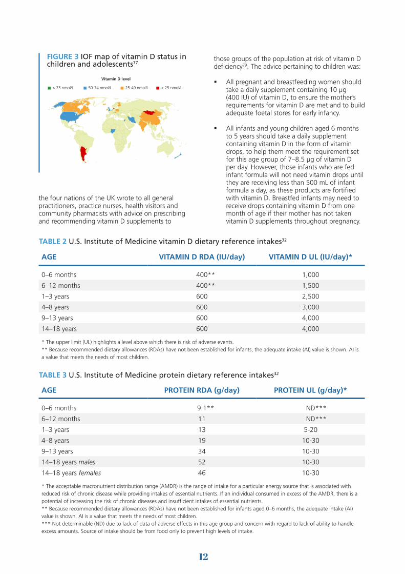
Vitamin D synthesis is dependent on several factors, including latitude, skin pigmentation and use of sunscreen. During the winter months, individuals living at latitudes higher than 33° in the northern or southern hemispheres do not receive adequate UV-B exposure to synthesize vitamin D in their skin. Consequently, vitamin D insufficiency is observed throughout the world52, including sunny countries such as Australia64, where sunscreen use to prevent skin cancer has become commonplace. Reports from Asia65, Europe66-71, the Middle East72, North America73 and Oceania74-76 suggest that low levels of vitamin D in children are highly prevalent, as illustrated by the IOF vitamin D status map in Figure 377.
The dietary reference intakes for vitamin D recommended by the IOM are shown by age range for children in Table 232. Only a small group of foods are naturally rich in vitamin D, which includes oily fish (e.g., salmon, mackerel and sardines) and liver. In some countries margarine and breakfast cereals are fortified with vitamin D. A recent pan-European study concluded that adolescent consumers of ready-to-eat-cereals (RTECs) had favourable micronutrient intake, including vitamin D, as compared to non-consumers of RTECS78.
Guidance from several countries recommend vitamin D supplementation for infants and young children34,64,79. In 2012, Chief Medical Officers for
AGE CALCIUM RDA (mg/day) CALCIUM UL (mg/day)*
0–6 months 200** 1,000
6–12 months 260** 1,500
1–3 years 700 2,500
4–8 years 1,000 2,500
9–13 years 1,300 3,000
14–18 years 1,300 3,000
* The upper limit (UL) highlights a level above which there is risk of adverse events.** Because recommended dietary allowances (RDAs) have not been established for infants, the adequate intake (AI) value is shown. AI is a value that meets the needs of most children.
TABLE 1 U.S. Institute of Medicine calcium dietary reference intakes32
12
the four nations of the UK wrote to all general practitioners, practice nurses, health visitors and community pharmacists with advice on prescribing and recommending vitamin D supplements to
those groups of the population at risk of vitamin D deficiency79. The advice pertaining to children was:
§ All pregnant and breastfeeding women should take a daily supplement containing 10 µg (400 IU) of vitamin D, to ensure the mother’s requirements for vitamin D are met and to build adequate foetal stores for early infancy.
§ All infants and young children aged 6 months to 5 years should take a daily supplement containing vitamin D in the form of vitamin drops, to help them meet the requirement set for this age group of 7–8.5 µg of vitamin D per day. However, those infants who are fed infant formula will not need vitamin drops until they are receiving less than 500 mL of infant formula a day, as these products are fortified with vitamin D. Breastfed infants may need to receive drops containing vitamin D from one month of age if their mother has not taken vitamin D supplements throughout pregnancy.
Copyright © Free Vector Maps.com
25-49 nmol/L < 25 nmol/L> 75 nmol/L 50-74 nmol/L
Vitamin D level
FIGURE 3 IOF map of vitamin D status in children and adolescents77
AGE VITAMIN D RDA (IU/day) VITAMIN D UL (IU/day)*
0–6 months 400** 1,000
6–12 months 400** 1,500
1–3 years 600 2,500
4–8 years 600 3,000
9–13 years 600 4,000
14–18 years 600 4,000
* The upper limit (UL) highlights a level above which there is risk of adverse events.** Because recommended dietary allowances (RDAs) have not been established for infants, the adequate intake (AI) value is shown. AI is a value that meets the needs of most children.
AGE PROTEIN RDA (g/day) PROTEIN UL (g/day)*
0–6 months 9.1** ND***
6–12 months 11 ND***
1–3 years 13 5-20
4–8 years 19 10-30
9–13 years 34 10-30
14–18 years males 52 10-30
14–18 years females 46 10-30
* The acceptable macronutrient distribution range (AMDR) is the range of intake for a particular energy source that is associated with reduced risk of chronic disease while providing intakes of essential nutrients. If an individual consumed in excess of the AMDR, there is a potential of increasing the risk of chronic diseases and insufficient intakes of essential nutrients.** Because recommended dietary allowances (RDAs) have not been established for infants aged 0–6 months, the adequate intake (AI) value is shown. AI is a value that meets the needs of most children.*** Not determinable (ND) due to lack of data of adverse effects in this age group and concern with regard to lack of ability to handle excess amounts. Source of intake should be from food only to prevent high levels of intake.
TABLE 3 U.S. Institute of Medicine protein dietary reference intakes32
TABLE 2 U.S. Institute of Medicine vitamin D dietary reference intakes32

13
Protein
Dietary protein is a source of amino acids which are needed to build the bone matrix. Milk is a source of high-quality protein, mostly casein, but also whey proteins which contain growth promoting elements80. Healthy children who were given extra servings of milk in their diets, and hence extra protein, experienced significant increases in IGF-l as compared to control subjects81. Variation in protein intake which is considered to be within the normal range for well-nourished children and adolescents can affect skeletal growth, and so impact on a child’s capacity to achieve their genetic potential for PBM80. The dietary reference intakes for protein recommended by the IOM are shown by age range for children in Table 332.
EXERCISE AND LIFESTYLE
In 2013, Osteoporosis Australia published Building healthy bones throughout life which included a comprehensive review of evidence pertaining to the impact of exercise on bone health64. This strategy recognized that childhood and adolescence may be the pivotal period in the life-course for exercise to most significantly impact on bone health in the long term. The primary recommendation relating to exercise needs in children and adolescents was:
§ ‘Encourage schools to incorporate a diverse and enjoyable battery of weight-bearing activities and sports into their school physical education programmes. This could include participation in short periods (5–10 minutes) of daily, targeted, multidirectional, moderate- to high-impact activities such as jumping, skipping and hopping.’
BODY WEIGHT AND COMPOSITION
A healthy body weight during childhood and adolescence is required for optimal bone health. Having a body mass index (BMI) at either end of the spectrum can pose a threat to development of the skeleton. Anorexia nervosa has been shown to have a profound negative impact on BMD in adolescent girls82 and boys83, as well as indices of skeletal strength84,85. Overweight and obese children have low bone mass and area for their weight86, and overweight children and adolescents are more likely to sustain repeated fracture at the wrist87.
Fracture trends in childhood and later life
The incidence of limb fractures in the population shows a bimodal distribution as a function of age, with the first peak being coincident with the pubertal growth spurt for both boys and girls88. Swiss investigators sought to determine whether PBM is low among girls who sustained fractures89.
Before and during early puberty, BMC and width of the radial diaphysis was lower in the fracture compared with the no-fracture group. At pubertal maturity, BMC at the ultradistal radius, femoral trochanter and lumbar spine were all significantly lower in girls with fractures. Throughout puberty, the gains in BMC at the various skeletal sites were also smaller for the girls with fractures. Accordingly, the investigators concluded that childhood fractures may be markers for low PBM and persistent bone fragility. A similar study from the UK found that children who fracture tend to have a smaller skeleton relative to their overall body size90.
A key question is whether a fracture in childhood predisposes individuals to higher fracture risk in adult life. In 2014, investigators from the Mayo Clinic in the United States evaluated bone strength and architecture parameters in children who had suffered wrist fractures as a result of mild trauma (e.g., a fall from standing height as opposed to a fall while riding a bicycle, which was classified as moderate trauma)91. They concluded that children with mild trauma fractures had thinning of cortical bone and deficits in bone microstructure at the distal radius and tibia compared to sex-matched, controls with no fracture history. This suggests that wrist fractures in children have two distinct causes:
1. Fractures resulting from mild trauma which suggest underlying skeletal fragility
2. Fractures resulting from moderate trauma when bone strength is normal
The same group subsequently investigated adult women and men who had sustained a mild trauma wrist fracture during childhood (prior to 18 years of age)92. These otherwise healthy young adults (age 20–40 years), had diminished bone strength, deficits of the cortical bone at the wrist, and lower BMD at the wrist, hip, and total body regions compared to controls. These results suggest that lifestyle interventions may need to be targeted to children and adolescents who suffer mild trauma wrist fractures to improve their bone health in the long-term.

14
Paediatric models of malnutrition-induced osteoporosis
In addition to the challenge posed by anorexia nervosa, a number of other childhood diseases which result in malnutrition impact negatively on the developing skeleton. These include:
§ Inflammatory bowel disease (IBD)
§ Cystic fibrosis
§ Coeliac disease
Several aspects of bone metabolism in children with IBD are inhibited, including bone modelling, remodelling and linear growth94. Biomarker studies indicate that both bone formation and bone resorption are decreased by 30–50% as compared to normal rates. At diagnosis, children with IBD, particularly those with Crohn’s disease, often have stunted growth. Control of inflammation, improved nutrition and regular physical activity are the key components of management of IBD in children with respect to bone health.
A significant proportion of children with cystic fibrosis (CF) have low BMD95. In 2011, the European Cystic Fibrosis Society published comprehensive guidelines on the assessment, prevention and treatment of bone disease in CF sufferers96.
A recent meta-analysis reported that fractures were almost twice as common in individuals with clinically diagnosed coeliac disease as compared to those without the disease97. A position statement published in Canada in 2012 recommended that BMD should be assessed one year after diagnosis of coeliac disease in children if gluten-free diet (GFD) adherence is not strict, and that a GFD is the most important treatment for bone loss98.
Assessment of bone health in a growing child or adolescent
In 2014, the International Society for Clinical Densitometry published Official Revised Positions on reporting of densitometry results in children93. The primary recommendations made were:
What are the most appropriate and reproducible sites for densitometry in children?
The posterior-anterior spine and total body less head (TBLH) are the preferred skeletal sites for performing BMC and areal BMD (aBMD) measurements in most paediatric subjects. Other sites may be useful depending on clinical need. Note that the hip is not a preferred site in growing children because of variability in skeletal development.
What is the best method for reporting aBMD in children; what corrections should be made for bone size, height, lean body mass, skeletal age, or pubertal stage?
In children with short stature or growth delay, spine and TBLH BMC and aBMD results should be adjusted. For the spine, adjust using either bone mineral apparent density (BMAD) or the height (for age) Z-score. For TBLH, adjust using the height Z-score.
What are the most appropriate normative databases for use in childhood?
An appropriate reference data set must include a sample of healthy representatives of the general population sufficiently large to capture variability in bone measures that takes into consideration gender, age, and race/ethnicity.
Detailed guidance is also provided on what elements should be included in a dual X-ray absorptiometry (DXA) report for a child or adolescent.

15
Maintaining bone mass in adulthood: stemming the
tide of bone loss
THE PRIMARY OBJECTIVE FOR ADULTS IS TO
AVOID PREMATURE BONE LOSS AND MAINTAIN A
HEALTHY SKELETON.
16
Bone is metabolically active throughout life
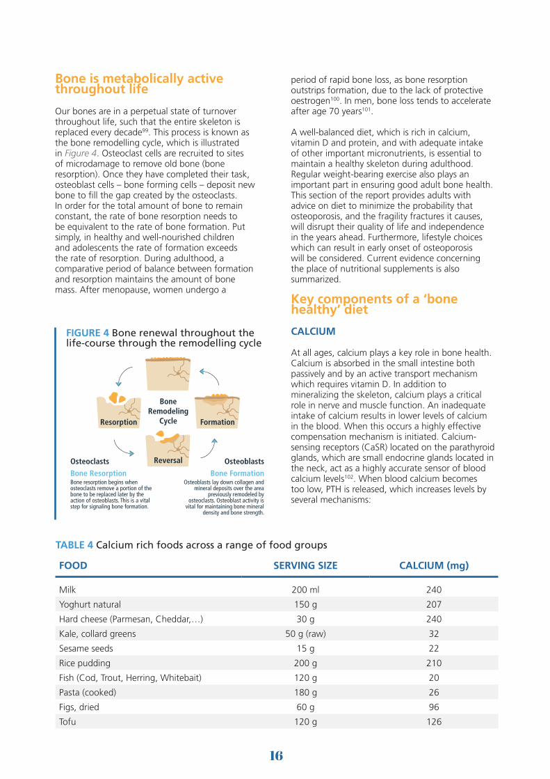
Our bones are in a perpetual state of turnover throughout life, such that the entire skeleton is replaced every decade99. This process is known as the bone remodelling cycle, which is illustrated in Figure 4. Osteoclast cells are recruited to sites of microdamage to remove old bone (bone resorption). Once they have completed their task, osteoblast cells – bone forming cells – deposit new bone to fill the gap created by the osteoclasts. In order for the total amount of bone to remain constant, the rate of bone resorption needs to be equivalent to the rate of bone formation. Put simply, in healthy and well-nourished children and adolescents the rate of formation exceeds the rate of resorption. During adulthood, a comparative period of balance between formation and resorption maintains the amount of bone mass. After menopause, women undergo a
period of rapid bone loss, as bone resorption outstrips formation, due to the lack of protective oestrogen100. In men, bone loss tends to accelerate after age 70 years101.
A well-balanced diet, which is rich in calcium, vitamin D and protein, and with adequate intake of other important micronutrients, is essential to maintain a healthy skeleton during adulthood. Regular weight-bearing exercise also plays an important part in ensuring good adult bone health. This section of the report provides adults with advice on diet to minimize the probability that osteoporosis, and the fragility fractures it causes, will disrupt their quality of life and independence in the years ahead. Furthermore, lifestyle choices which can result in early onset of osteoporosis will be considered. Current evidence concerning the place of nutritional supplements is also summarized.
Key components of a ‘bone healthy’ diet
CALCIUM
At all ages, calcium plays a key role in bone health. Calcium is absorbed in the small intestine both passively and by an active transport mechanism which requires vitamin D. In addition to mineralizing the skeleton, calcium plays a critical role in nerve and muscle function. An inadequate intake of calcium results in lower levels of calcium in the blood. When this occurs a highly effective compensation mechanism is initiated. Calcium-sensing receptors (CaSR) located on the parathyroid glands, which are small endocrine glands located in the neck, act as a highly accurate sensor of blood calcium levels102. When blood calcium becomes too low, PTH is released, which increases levels by several mechanisms:
BoneRemodeling
Cycle
OsteoclastsBone ResorptionBone resorption begins when osteoclasts remove a portion of the bone to be replaced later by the action of osteoblasts. This is a vital step for signaling bone formation.
OsteoblastsBone Formation
Osteoblasts lay down collagen and mineral deposits over the area
previously remodeled by osteoclasts. Osteoblast activity is
vital for maintaining bone mineral density and bone strength.
Resorption
Reversal
Formation
FIGURE 4 Bone renewal throughout the life-course through the remodelling cycle
FOOD SERVING SIZE CALCIUM (mg)
Milk 200 ml 240
Yoghurt natural 150 g 207
Hard cheese (Parmesan, Cheddar,…) 30 g 240
Kale, collard greens 50 g (raw) 32
Sesame seeds 15 g 22
Rice pudding 200 g 210
Fish (Cod, Trout, Herring, Whitebait) 120 g 20
Pasta (cooked) 180 g 26
Figs, dried 60 g 96
Tofu 120 g 126
TABLE 4 Calcium rich foods across a range of food groups

17
§ Stimulating osteoclast cells to resorb bone and release calcium by acting on the osteoblast
§ Increasing gastrointestinal calcium absorption by activating vitamin D
§ Reabsorption of calcium by the kidneys
Milk and other dairy foods are the most readily available sources of calcium in the diet. Other good food sources of calcium include certain green vegetables (e.g., broccoli, curly kale, bok choy); whole canned fish with soft, edible bones such as sardines or pilchards; nuts (almonds and Brazil nuts in particular); and tofu set with calcium. The amounts of calcium available from these foods is shown in Table 4.
It should also be noted that some mineral waters and tap waters provide a valuable source of calcium, which can also be helpful to people who are lactose intolerant. For example, calcium concentration of tap water varies from 1 mg/L to 135 mg/L across the USA and Canada, and
filtration can remove almost 90% of calcium103. Some mineral waters have been shown to have more than 200 mg/L. For people who choose to obtain a proportion of their dietary calcium intake from water, it is important to know precisely how much calcium is present in the water they drink.
Recommendations on dietary calcium intake for adults from leading organizations are consistent:
§ Australia: National Health and Medical Research Council Recommended Dietary Intake (RDI) for calcium for adults aged 19–50 years is 1,000 mg/day104
§ USA: IOM Dietary Reference Intake for calcium for adults aged 19–50 years is 1,000 mg/day32
§ World Health Organization/Food and Agriculture Organization of the United Nations: WHO/FAO Dietary Reference Intake for calcium for adults aged 25–50 years is 1,000 mg/day105
REGION & COUNTRY
AGE(years)
DRIa (mg/day)
CALCIUM INTAKE (mg/day)STUDY
male female
Europe
Austria
19-79 >1,000 561 (±290)b 576 (±309)b
Kudlacek et al106<40 1,000 604 (±345)b 560 (±299)b
40-60 >1,000 590 (±318)b 561 (±287)b
Germany
18-79 >1,000 1,181 (902-1,535)
1,082 (849-1,379) Hintzpeter et al107
Adults 1,000 619 (213-1,025) 705 (313-1,094) Anke108
40-64 >1,000 774 (334-1,330)c 707 (287-1,225)c Schulze et al109
UKf 45-55 1,000 1,133 (950-1,316)
1,063 (931-1,195) Vyas et al110
North America
USA 19-50 1,000 812 (788-837) 626 (596-659) Ma et al111
South-East Asia
Bangladesh 16-40 1,000 180d Islam et al112
Indonesia 18-40 1,000 270 (239-302)e Green et al113
Malaysia 18-40 1,000 386 (353-420)e Green et al113
South Pacific
Australia 20-94 >1,000 643 (±340)b Pasco et al114
New Zealand 40-64 >1,000 794 (8-1,580)d 794 (8-1,580)d Metcalf et al115
aDaily recommended intake FAO/WHO105 • bMean (±SD) • cMean (10th and 90th centiles) • dMean (range) • eMean (95% CI) • fWhite, urban population
TABLE 5 Nutritional calcium deficit in selected countries (reproduced with kind permission of Professor Meinrad Peterlik)52

18
Accordingly, it is of considerable concern that a major shortfall in calcium intake has been reported from many countries, as illustrated in Table 5.
The observed deficit in calcium intakes raises the question of the role of calcium supplements in healthy individuals. An ongoing debate in the scientific community has sought to establish the risk-benefit ratio of calcium supplementation with respect to beneficial effects on bone health as compared to adverse impacts on the cardiovascular system. Supplements should be used only as needed to bring total calcium intake to the recommended level in healthy adults.
VITAMIN D
Vitamin D has a beneficial effect on bone health throughout life, operating through a number of physiological mechanisms:
§ Assisting calcium absorption from food in the intestine116
§ Ensuring correct renewal and mineralization of bone117
§ Down regulation of PTH so reducing PTH-induced bone loss118,119
§ Increasing BMD120
§ Directly stimulating muscle tissue and so reducing falls risk121,122
The primary source of vitamin D comes from sun exposure, which triggers synthesis in the skin. However, as highlighted earlier, vitamin D insufficiency has become a global problem due to factors such as increasingly indoor lifestyles. Very few foods are naturally rich in vitamin D but some
good sources are highlighted in Table 6. The IOM RDA for vitamin D for adults aged 19–70 years (as cholecalciferol) is 600 IU per day (15 µg/day)32.
In 2009, an IOF Working Group published a review of global vitamin D status and determinants of hypovitaminosis D123. As reported previously for children and adolescents, low levels of vitamin D are also highly prevalent for adults throughout the world, as illustrated by the IOF vitamin D status map in Figure 577.
Adults at elevated risk of having inadequate levels of vitamin D include:
§ Inhabitants of latitudes with minimal exposure to sunlight
§ Individuals who are obese
§ Individuals with a darker skin tone
FOOD VITAMIN D (IU/100 g)*
Wild salmon 600-1000
Farmed salmon 100-250
Sardines, canned 300-600
Mackerel, canned 250
Tuna, canned 236
Cod liver oil 400-1000 per tablespoon
Shiitake mushrooms, fresh 100
Shiitake mushrooms, sun dried 1600
Egg yolk 20 per yolk
*per 100g unless otherwise statedIU: International Unit
TABLE 6 Natural nutritional sources of vitamin D
Copyright © Free Vector Maps.com
25-49 nmol/L < 25 nmol/L> 75 nmol/L 50-74 nmol/L
Vitamin D level
FIGURE 5 IOF map of vitamin D status in adults

19
§ Individuals who cannot expose their skin to the sun for medical or cultural reasons
§ Individuals with diseases that reduce uptake of vitamin D from the intestine
While population screening for vitamin D deficiency is not recommended, measurement of serum 25(OH)D in high risk individuals enables assessment of response to supplementation and the need for dose adjustment124. In 2013, the U.S. Preventive Services Task Force (USPSTF) evaluated the effects of vitamin D supplementation, with or without calcium, on bone health outcomes in community-dwelling adults125. The USPSTF reached the following conclusions:
§ The current evidence is insufficient to assess the balance of the benefits and harms of combined vitamin D and calcium supplementation for the primary prevention of fractures in healthy premenopausal women or in men
§ The current evidence is insufficient to assess the balance of the benefits and harms of daily supplementation with greater than 400 IU of vitamin D3 and greater than 1000 mg of calcium for the primary prevention of fractures in noninstitutionalized postmenopausal women
PROTEIN
Protein is a source of amino acids which are needed to maintain bone structure. Protein also stimulates IGF-l release, which may increase production of bone matrix by increasing osteoblast activity. In 2009, the first systematic review and meta-analysis of the relationship between dietary protein and bone health in healthy human adults was published126. The investigators reported a positive association between protein intake and BMD and BMC, and a reduction in markers of bone resorption. Whilst the effect size was small, and a relationship between dietary protein and fracture risk was not identified, current healthy eating guidelines were considered appropriate with respect to bone health.
VITAMIN K
Vitamin K is required to make osteocalcin, which is the second most abundant protein in bone after collagen. Epidemiological studies have suggested that diets high in vitamin K are associated with a lower risk of hip fractures in older people127. Good food sources of vitamin K include leafy green vegetables such as lettuce, spinach, cabbage and kale, liver, some fermented foods such as fermented cheeses and natto (fermented soybeans) and dried fruit (e.g. prunes). Randomized controlled trials of vitamin K1 or K2 supplementation did not increase BMD at major sites128. Accordingly, further studies
are needed to determine the role of vitamin K supplements for the prevention and treatment of osteoporosis.
B VITAMINS AND HOMOCYSTEINE
Homocysteine is an amino acid which can interfere with collagen synthesis, the main protein in bone. When blood levels of vitamin B6, vitamin B12 and folic acid are low, homocysteine levels can rise. Accordingly, inadequacy of B vitamins could compromise bone health, a notion supported by observational studies which found an association between high homocysteine levels and lower BMD129, and increased hip fracture risk in older people130. However, a 2014 review concluded that inconsistencies within the current evidence base necessitate definitive studies to be conducted to evaluate the role of B vitamins in prevention of osteoporosis131.
VITAMIN A
The role of vitamin A in bone health is controversial132. High intake of preformed vitamin A, which can be obtained from animal food sources such as liver, other offal and fish oils, has been associated with osteoporosis and hip fracture. However, carotenoids, which are precursors to vitamin A, have been associated with improved bone health. Carotenoids can be obtained from green leafy vegetables, carrots, pumpkins, red and yellow peppers, mangoes, papaya and apricots. Taking fish oil supplements and a multivitamin
For both men and women, more than two units per day of alcohol can increase the risk of suffering a fragility fracture, while more than four units per day can double fracture risk

20
supplement concurrently could lead to excessive intake of vitamin A, so many countries caution against doing so.
MAGNESIUM
Approximately half of total body magnesium is stored in the skeleton133. Magnesium plays an important role in bone formation through stimulating proliferation of osteoblasts. Magnesium deficiency is rare in well-nourished populations. However, because magnesium absorption decreases with age, the elderly can be at risk of mild magnesium deficiency. Good sources of magnesium include green vegetables, legumes, nuts, seeds, unrefined grains, fish and dried fruit (apricots, prunes, raisins).
ZINC
Zinc plays a role in bone tissue renewal and mineralisation. Zinc deficiency is usually associated with calorie and protein malnutrition, and has been reported to be common in community-dwelling older people134. While vegetarian diets do not necessarily have lower zinc intakes, the bioavailability of zinc may be lower for vegetarians, so higher intakes may
be required135. Sources of zinc include lean red meat, poultry, whole grain cereals, pulses, legumes and dried fruit (peaches, prunes, apricots).
Acid-base balance of the diet
The notion that high dietary acid content may cause bone loss has been the subject of debate in the lay media in recent years. In 2011, a systematic review and meta-analysis of the scientific literature sought to evaluate causal relationships between dietary acid load and osteoporosis136. The investigators concluded that no association was evident and, conversely, that there is no evidence that an alkaline diet is protective of bone health. A limitation of this meta-analysis was that the primary studies were not weighted for sample size, and that young and older subjects were included. More recent studies have evaluated the impact of potassium citrate supplementation on calcium balance137, bone density, microarchitecture, and fracture risk prediction by FRAX®138. Positive findings from these comparatively small-scale studies make the case for larger and longer studies evaluation to determine the impact of supplementation with alkaline salts of potassium (i.e. citrate or bicarbonate) on fracture incidence.
Lifestyle factors that can negatively affect bone health
Alcohol
For both men and women, more than two units per day of alcohol can increase the risk of suffering a fragility fracture, while more than four units per day can double fracture risk139. If an individual chooses to drink, moderation is the bone healthy choice. Up to two 120 mL glasses of wine per day do not negatively impact on bone health.
Caffeine
Caffeine increases urinary and faecal calcium losses and so, in combination with a diet low in calcium, has the potential to adversely affect bone health140. A Swedish study suggests that caffeine intake at 330 mg per day (i.e. four cups /600 mL) could be associated with a 20% increase in risk of osteoporotic fractures as compared to caffeine intake of less than 200 mg per day141. However, increasing calcium intake by 40 mg for every cup of caffeine containing coffee consumed counter balances the potential for loss142.
Under-nutrition and over-nutrition
BMI is a measure of how lean someone is and can be used to assess risk for osteoporosis143. A BMI between 20–25 kg/m2 is generally considered to be ideal. A BMI below 19 is considered underweight and is a risk factor for osteoporosis. Emerging data also suggests that the belief that obesity is protective against osteoporosis may be flawed144. In 2010, analysis from Addenbrookes Hospital in the UK reported a surprisingly high prevalence of obesity in postmenopausal women presenting with fragility fractures145. Furthermore, the Global Longitudinal Study of Osteoporosis in Women (GLOW) has reported that obese women with fracture undergo a longer period of hospitalization for treatment and have poorer functional status and health-related quality of life than non-obese women146.
21
The special nutritional needs of seniors: fighting frailty and reducing falls
and fractures
THE PRIMARY OBJECTIVE FOR SENIORS IS TO PREVENT AND
TREAT OSTEOPOROSIS.

22
Malnutrition in the elderly
Malnutrition is highly prevalent in the elderly147. With regards to calcium, vitamin D and protein, a number of factors can contribute to inadequate availability of these key nutrients for bone health:
Calcium
§ Decreased overall dietary energy intake, including sources of calcium
§ Decreased intestinal absorption of calcium, exacerbated by low vitamin D status
§ Decreased capacity of intestinal cells to adapt to low calcium intake
§ Decreased retention of calcium by the kidneys
Vitamin D
§ Less frequent exposure to sunlight for the housebound
§ Decreased capacity of the skin to synthesize vitamin D
§ Decreased renal capacity to convert vitamin D to its active form
Protein
§ Decreased anabolic responses to ingested protein
§ Increased need for protein to offset inflammatory and catabolic conditions
Calcium, vitamin D and protein requirements
A number of expert groups have published guidance on nutrients important to bone health in the elderly. The dietary reference intakes for calcium, vitamin D and protein recommended by the IOM are shown for the general population aged 50–70 years and over 70 years in Table 7. IOM
concluded that a 25(OH)D level of 40 nmol/L (16ng/mL) covers the requirements of approximately half the population, while a 25(OH)D level of 50 nmol/L (20ng/mL) covers the requirements of ≥97.5% of the population. The IOM protein recommendations are based on an intake of 0.8 g/kg/day for adults.
The following recommendations from learned societies are focused on care of patients with or at increased risk for osteoporosis. These generally recommend higher intakes or 25(OH)D levels than the IOM.
In 2010, IOF published a position statement on vitamin D recommendations for older adults148. In order to determine the level of serum 25(OH)D that was required to prevent falls and fractures, results from meta-analyses were considered122,149. With respect to falls, a mean serum level of 25(OH)D of at least 60 nmol/L (24 ng/mL) is needed for optimal falls risk reduction122. With respect to fractures, the mean serum levels of 25(OH)D associated with reduced risk of non-vertebral fracture and hip fractures were 66 nmol/L (26.4 ng/mL) and 74 nmol/L (29.6 ng/mL), respectively149. Accordingly, the IOF Working Group proposed a target level of 25(OH)D for older individuals of 75 nmol/L (30 ng/mL).
The estimated average vitamin D requirement for older adults to achieve a serum 25(OH)D level of 75 nmol/L (30 ng/mL) is 20 to 25 µg per day (800–1,000 IU per day). However, considerably higher doses would be needed to ensure that almost all older adults achieved the target level. In high-risk individuals, measurement of serum 25(OH)D is recommended. The required dose of vitamin D could be estimated based upon the notion that each 2.5 µg (100 IU) per day added will increase serum 25(OH)D by about 2.5 nmol/L (1 ng/mL)150. Re-testing after three months of supplementation is recommended for high-risk individuals to confirm that target levels have been achieved.
In 2011, an Endocrine Society Task Force published a Clinical Practice Guideline on the evaluation, treatment, and prevention of vitamin D deficiency35. The key recommendations of this guideline of relevance to older people were:
AGE GENDER CALCIUM RDA (mg/day)
VITAMIN D RDA (IU/day)
PROTEIN RDA (g/day)
51-70 yearsFemale 1,200 600 46
Male 1,000 800 56
>70 yearsFemale 1,200 600 46
Male 1,200 800 56
TABLE 7 U.S. Institute of Medicine, vitamin D and protein dietary reference intakes for older people and the elderly32

23
§ Screening for vitamin D deficiency in individuals at risk for deficiency is recommended, while population screening in individuals who are not at risk is not recommended
§ Adults aged 50–70 years and over 70 years require at least 600 and 800 IU per day, respectively, of vitamin D. However, to raise the blood level of 25(OH)D above 75 nmol/L (30 ng/mL) may require at least 1500–2000 IU per day of supplemental vitamin D
§ Adults who are vitamin D deficient should be treated with 50,000 IU of vitamin D2 or vitamin D3 once a week for 8 weeks or its equivalent of 6000 IU of vitamin D2 or vitamin D3 daily to achieve a blood level of 25(OH)D above 75 nmol/L (30 ng/mL), followed by maintenance therapy of 1500–2000 IU per day
In 2012, the European Union Geriatric Medicine Society (EUGMS), in cooperation with other scientific organizations, established an International Study Group (the PROT-AGE Study Group) to review dietary protein needs with ageing. In 2013, the PROT-AGE Study Group published a position paper which provided the following key recommendations151:
§ To maintain physical function, older people need more dietary protein than do younger people; older people should consume an average daily intake at least in the range of 1–1.2 g/kg body weight/day
§ Most older adults who have an acute or chronic disease need even more dietary protein (i.e. 1.2–1.5 g/kg body weight/day); people with severe illness or injury or with marked malnutrition may need as much as 2.0 g/kg body weight/day
§ Older people with severe kidney disease who are not on dialysis (i.e. estimated Glomerular filtration rate (GFR) < 30 mL/min/1.73m2) are an exception to the high-protein rule; these individuals need to limit protein intake
§ Protein quality, timing of intake, and amino acid supplementation may be considered so as to achieve the greatest benefits from protein intake, but further studies are needed to make explicit recommendations
§ In combination with increased protein intake, exercise is recommended at individualized levels that are safe and tolerated
The Position Paper also comments specifically on the subject of protein requirements for hip fracture patients and people with osteoporosis.
Supplementary protein or higher dietary intake of protein by older people who have been hospitalized with hip fracture has been shown to improve bone density126,152, reduce the risk of complications153-155 and reduce rehabilitation time152. Among older people with osteoporosis, higher BMD has been reported when protein intake was at levels higher than 0.8 g/kg body weight/day or was 24% of total energy intake126,156-158.
In 2014, the European Society for Clinical and Economic Aspects of Osteoporosis and Osteoarthritis (ESCEO) published a consensus statement on the role of dietary protein and vitamin D in maintaining musculoskeletal health in postmenopausal women159. Key recommendations included:
§ Optimal dietary protein intake of 1.0–1.2 g/kg body weight/day with at least 20–25 g of high-quality protein at each main meal
§ Vitamin D intake of 800 IU per day to maintain serum 25(OH)D levels greater than 50 nmol/L (20 ng/mL)
§ Calcium intake of 1,000 mg per day
§ Regular physical activity/exercise 3–5 times per week combined with protein intake in close proximity to exercise
A common theme is evident among all of these guidelines; adequate dietary intake of calcium, vitamin D and protein is an essential component of effective bone care for older people.
Preventing muscle wasting is important because it lowers the risk of falls and associated injuries, including fragility fractures

24
Impact of nutrition on muscle in seniors
As we age, in addition to declining bone mass, we can lose muscle mass and strength. This process is known as sarcopenia, from Greek sarx (flesh) and penia (poverty), and commonly referred to as age-related muscle wasting. Akin to the trajectory for development and subsequent loss of bone mass, skeletal muscle mass and strength reach a peak in the early adult years and then gradually decline as illustrated in Figure 6. Preventing muscle wasting is important because it lowers the risk of falls and associated injuries, including fragility fractures.
In 2010, the European Working Group on Sarcopenia in Older People (EWGSOP) developed a practical clinical definition and consensus diagnostic criteria for age-related sarcopenia161. A diagnosis of sarcopenia is dependent on the presence of both low muscle mass and low muscle function (i.e. strength or performance). EWGSOP applied these characteristics to further define conceptual stages as ‘presarcopenia’, ‘sarcopenia’ and ‘severe sarcopenia’.
A wide range of tools were reviewed that can be used to measure the specific variables of muscle mass, muscle strength and physical performance.
One of the first applications of the EWGSOP recommendations was to determine the prevalence of sarcopenia in the Hertfordshire Cohort Study (HCS) population in the UK162. Two techniques were used to assess individuals: using the lowest third of DXA lean mass (LM) and the lowest third of skin-fold-based fat-free mass (FFM) as markers of low muscle mass. Among 103 community-dwelling men participating in the Hertfordshire Sarcopenia Study (HSS, average age 73 years), the prevalence of sarcopenia was 6.8% and 7.8% when using the lowest third of DXA LM and FFM, respectively. The prevalence of sarcopenia among the 765 HCS male and 1,022 HCS female participants (mean age 67 years) was 4.6% and 7.9%, respectively.
In 2013, the IOF Nutrition Working Group published a position paper on the impact of nutrition on muscle mass, strength and performance in older adults160. As protein plays an important role in muscle health, an intake of 1–1.2 g/kg of body weight per day was recommended. Review of the scientific literature identified a moderate relationship between vitamin D and muscle strength, suggesting a role for vitamin D in the development and preservation of muscle mass and function. Furthermore, the authors concluded that the acid-base balance of the diet plays an important role in maintaining muscle mass.
Treatment of osteoporosis
It is among seniors that the majority of pharmacological treatment for osteoporosis should be deployed. As highlighted by the IOF Capture the Fracture® Campaign163,164, a pervasive and persistent osteoporosis care gap is evident among the most obvious group of older people who are at high risk of suffering fragility fractures in the future, namely those who have suffered fragility fractures in the past. Major efforts are ongoing throughout the world to eliminate this care gap through widespread implementation of Fracture Liaison Services (FLS)165-170.
early life adult life older life
Age
Mu
scle
mas
s an
d s
tren
gth
disability threshold
range in individuals
FIGURE 6 Life course changes in muscle mass and strength160
Adapted from Osteoporos Int 2013;24:1555-1566 with permission from Springer.

25
Meta-analysis of the principal agents licensed for the treatment of osteoporosis suggests that a 30–50% reduction in fracture incidence can be achieved during 3 years of pharmacotherapy171. We now have a broad choice of effective agents that can be taken as daily, weekly or monthly tablets, or as daily, quarterly, six-monthly or annual injections. The 2013 and 2014 World Osteoporosis Day Reports considered in detail how these treatments should be used to prevent fractures in women and men, respectively100,101. As health systems take steps to eliminate the secondary prevention care gap, the advent of absolute fracture risk calculators such as FRAX® provide health professionals with an opportunity to deliver primary fracture prevention in a systematic way172,173.
Given the human and economic scale of the threat that fragility fractures pose to mankind, urgent action is required on a global scale. In China alone, the number of fragility fractures occurring annually is projected to increase from 2.33 million cases in 2010 to 5.99 million cases by 2050174. As the population of the world ages, failure to implement the evidence base we have for fracture prevention is not an option.
The need for pharmacological management
Although bone healthy nutrition, exercise, and avoidance of negative lifestyle habits are important throughout the life-course, drug therapies are critical for fracture protection in high-risk patients.
The anti-fracture efficacy of approved treatments for postmenopausal women and men with osteoporosis when given with calcium and vitamin D varies from between 30-50%. When prescribing a specific pharmacologic treatment, the physician will take into account the patient’s individual risk profile including the risk for a specific type of fractures (spine versus hip), co-morbid conditions, poly-pharmacy, and the patient’s preference. Finally, cost and cost-effectiveness considerations, insurance plans, and national health policies will undoubtedly affect the choice of therapeutic options.
Common types of drug treatments, not all available in all countries, include:
§ Bisphosphonates (alendronate, risedronate, ibandronate, zoledronic acid )
§ Denosumab
§ Hormone Replacement Therapy
§ SERMs: Raloxifene
§ Strontium ranelate
§ Parathyroid Hormones: Teriparatide and PTH(1-84)
Poor adherence to osteoporosis medication is a major challenge. Unfortunately, up to half of patients stop their treatment after only one year. Patients need to be encouraged to remain on their prescribed treatment and to consult their doctors if they are experiencing problems taking their medication.
Although adopting bone-healthy behaviours are important throughout the life-course, drug therapies are critical for fracture protection in high risk patients

26
Diseases and disorders that affect nutritional
status
DISEASES OF THE GASTROINTESTINAL SYSTEM CAN AFFECT
NUTRIENT ABSORPTION PUTTING PEOPLE AT INCREASED RISK
OF OSTEOPOROSIS AND FRACTURES. IT IS RECOMMENDED
THAT IN SUCH CASES INDIVIDUALS HAVE THEIR NUTRIENT
STATUS CHECKED.

27
Inflammatory bowel disease
IBD refers to a number of diseases which are characterized by inflammation of the bowel. The most common of such disorders are Crohn’s disease and ulcerative colitis:
§ Crohn’s disease causes ulcers throughout the small and large intestine
§ Ulcerative colitis usually causes ulcers in the lower part of the large intestine
Symptoms of these disorders tend to occur intermittently, and include diarrhoea, abdominal cramps and pains, fever and weight loss. A number of factors predispose sufferers to be at increased risk of bone loss and fragility fractures:
§ Poor food intake and nutritional status
§ Poor absorption of nutrients by the damaged intestine (including calcium, vitamin D, protein and calories)
§ Surgery to remove parts of the intestine
§ Treatment with glucocorticoid medications to reduce the inflammation
§ Hormonal modifications induced by the gastrointestinal disease
§ Release of cytokines as part of the inflammatory process, which increase the loss of calcium from bone
A large-scale analysis of the Taiwan National Health Insurance Research Database identified specific predictors for osteoporosis and fractures among IBD sufferers175. As compared to age- and sex-matched controls, IBD sufferers had significantly higher rates of osteoporosis (approx. 30%), with those requiring hospitalization for IBD having considerably increased risk for osteoporosis and fractures.
Osteoporosis prevention measures need to be included in the overall care strategy for patients with these disorders, including ensuring an adequate calcium and vitamin D intake either through diet or supplements. Other measures to prevent bone loss include avoidance of excessive alcohol intake and smoking, and taking regular weight-bearing exercise. Osteoporosis medications may be recommended for some patients, for example older patients taking long-term glucocorticoid therapy and those with prior fragility fractures, as determined by the doctor.
Coeliac disease
Coeliac disease (CD) is a genetically mediated autoimmune disease characterized by intolerance to gluten (a protein group) found in wheat, rye and barley. It is also sometimes referred to as coeliac sprue, gluten-sensitive enteropathy, or simply gluten intolerance, and is a relatively common disorder thought to affect about 0.5–1% of the population. A 2014 multicentre study from the UK reported that CD accounts for 1 in 31 referrals in secondary care to unselected gastroenterology clinics176. Those affected suffer damage to the villi, the tiny finger-like protrusions lining the surface of the intestine that are involved in the absorption of nutrients from food. Symptoms include diarrhoea, weight loss, anaemia, fatigue, muscle cramps and nutritional deficiencies, and the disorder has to be controlled by strict adherence to a gluten-free diet.
People with CD may be at increased risk of osteoporosis if the disorder goes undiagnosed or is poorly controlled, due to inadequate nutrient absorption from food (including calcium and vitamin D), sometimes leading to frank malnutrition. Rates of CD are commonly found to be higher among patients with osteoporosis than those without osteoporosis. Consequently, as CD can sometimes have no symptoms, doctors may test for the condition when an individual is found to be osteoporotic. It may also be ‘discovered’ when a patient who is vitamin D-deficient shows no response (i.e. blood levels don’t change) to being given a large therapeutic dose of vitamin D.
In 2014, the British Society of Gastroenterology (BSG) published guidelines on the diagnosis and management of adult CD177. Recommendations relating to bone health include:
§ Bone density should be measured after one year of diet in patients who have additional risk factors for osteoporosis or if over the age of 55 years
§ Adult patients with CD should have a calcium intake of at least 1,000 mg per day
§ A gluten-free diet is the core management strategy for prevention of osteoporosis
Lactose maldigestion and intolerance
When people are unable to digest all the lactose they have eaten, they are said to have lactose maldigestion. It results from a deficiency in the enzyme lactase, produced in the small intestine, which is responsible for breaking down lactose (the principal sugar found in milk) into simpler

28
sugars, which are then absorbed by the body. The term lactose intolerance refers to the abdominal symptoms (e.g., cramps, bloating) resulting from the inability to digest lactose. The prevalence of lactose intolerance varies significantly between races and as a function of age. A systematic literature review published in 2010 reported178:
§ Prevalence of lactose intolerance is very low in children and remained low into adulthood among individuals of Northern European descent
§ Prevalence of lactose intolerance in African American, Hispanic, Asian, and American Indian populations may be 50% higher in late childhood and adulthood
In 2010, the U.S. National Institutes of Health (NIH) published a consensus development conference statement on lactose intolerance and health179. Key components of the statement included:
§ Lactose intolerance is a real and important clinical syndrome, but its true prevalence is not known
§ The majority of people with lactose malabsorption do not have clinical lactose intolerance. Many individuals who think they are lactose intolerant are not lactose malabsorbers
§ Many individuals with real or perceived lactose intolerance avoid dairy and ingest inadequate amounts of calcium and vitamin D, which may predispose them to decreased bone accrual, osteoporosis, and other adverse health
outcomes. In most cases, individuals do not need to eliminate dairy consumption completely
§ Evidence-based dietary approaches with and without dairy foods and supplementation strategies are needed to ensure appropriate consumption of calcium and other nutrients in lactose-intolerant individuals
§ Educational programmes and behavioural approaches for individuals and their health-care providers should be developed and validated to improve the nutrition and symptoms of individuals with lactose intolerance and dairy avoidance
As stated by the NIH group, being lactose intolerant does not necessarily preclude all dairy products from the diet; some people with this disorder can still drink small quantities of milk without suffering any symptoms. In some countries lactose-reduced milks are available. Yoghurt with live cultures can often be tolerated, because the bacteria in the cultures produce the enzyme lactase, and some hard cheeses contain only negligible amounts of lactose. Another alternative is to take lactase tablets or drops along with dairy foods. Other foods and drinks can provide good sources of calcium, such as green leafy vegetables, nuts, canned fish with soft, edible bones such as salmon and sardines, calcium-fortified beverages and calcium-rich mineral waters.
People who are lactose intolerant should consult with their doctor to discuss the best way of ensuring adequate calcium intake, either through diet, or if necessary, through the use of supplements.

REFERENCES1. Bonjour JP, Chevalley T, Ferrari S, Rizzoli R (2009)
The importance and relevance of peak bone mass in the prevalence of osteoporosis. Salud publica Mex. 51 Suppl 1:S5-17.
2. Cooper C, Melton LJ, 3rd (1992) Epidemiology of osteoporosis. Trends Endocrinol Metab.: TEM 3:224-229.
3. Institute of Medicine (2011) Dietary Reference Intakes for Calcium and Vitamin D. In.
4. Lips P, van Schoor NM (2011) The effect of vitamin D on bone and osteoporosis. Best Pract Res Clin Endocrinol Metab. 25:585-591.
5. Wahl DA, Cooper C, Ebeling PR, et al. (2012) A global representation of vitamin D status in healthy populations. Arch Osteoporos 7:155-172.
6. Hilger J, Friedel A, Herr R, Rausch T, Roos F, Wahl DA, Pierroz DD, Weber P, Hoffmann K (2014) A systematic review of vitamin D status in populations worldwide. Br J Nutr. 111:23-45.
7. Heaney RP (2009) Dairy and bone health. J Am Coll Nutr. 28 Suppl 1:82S-90S.
8. Conigrave AD, Brown EM, Rizzoli R (2008) Dietary protein and bone health: roles of amino acid-sensing receptors in the control of calcium metabolism and bone homeostasis. Annu Rev Nutr. 28:131-155.
9. Hannan MT, Tucker KL, Dawson-Hughes B, Cupples LA, Felson DT, Kiel DP (2000) Effect of dietary protein on bone loss in elderly men and women: the Framingham Osteoporosis Study. J Bone Miner Res. 15:2504-2512.
10. Bonjour JP (2011) Protein intake and bone health. Int J Vitam Nutr Res. 81:134-142.
11. Barker DJ, Winter PD, Osmond C, Margetts B, Simmonds SJ (1989) Weight in infancy and death from ischaemic heart disease. Lancet. 2:577-580.
12. Barker DJ, Eriksson JG, Forsen T, Osmond C (2002) Fetal origins of adult disease: strength of effects and biological basis. Int J Epidemiol. 31:1235-1239.
13. Holroyd C, Harvey N, Dennison E, Cooper C (2012) Epigenetic influences in the developmental origins of osteoporosis. Osteoporos Int. 23:401-410.
14. Harvey N, Dennison E, Cooper C (2014) Osteoporosis: a lifecourse approach. J Bone Miner Res. 29:1917-1925.
15. Baird J, Kurshid MA, Kim M, Harvey N, Dennison E, Cooper C (2011) Does birthweight predict bone mass in adulthood? A systematic review and meta-analysis. Osteoporos Int. 22:1323-1334.
16. Javaid MK, Eriksson JG, Kajantie E, Forsen T, Osmond C, Barker DJ, Cooper C (2011) Growth in childhood predicts hip fracture risk in later life. Osteoporos Int. 22:69-73.
17. Cooper C, Eriksson JG, Forsen T, Osmond C, Tuomilehto J, Barker DJ (2001) Maternal height, childhood growth and risk of hip fracture in later life: a longitudinal study. Osteoporos Int. 12:623-629.
18. Harvey NC, Javaid MK, Arden NK, et al. (2010) Maternal predictors of neonatal bone size and geometry: the Southampton Women’s Survey. J Dev Orig Health Dis. 1:35-41.
19. Godfrey K, Walker-Bone K, Robinson S, Taylor P, Shore S, Wheeler T, Cooper C (2001) Neonatal bone mass: influence of parental birthweight, maternal smoking, body composition, and activity during pregnancy. J Bone Miner Res. 16:1694-1703.
20. Javaid MK, Crozier SR, Harvey NC, Gale CR, Dennison EM, Boucher BJ, Arden NK, Godfrey KM, Cooper C, Princess Anne Hospital Study G (2006) Maternal vitamin D status during pregnancy and childhood bone mass at age 9 years: a longitudinal study. Lancet. 367:36-43.
21. Harvey NC, Holroyd C, Ntani G, et al. (2014) Vitamin D supplementation in pregnancy: a systematic review. Health Technol Assess. 18:1-190.
22. Namgung R, Tsang RC (2003) Bone in the pregnant mother and newborn at birth. Clin Chim Acta. 333:1-11.
23. Ganpule A, Yajnik CS, Fall CH, et al. (2006) Bone mass in Indian children--relationships to maternal nutritional status and diet during pregnancy: the Pune Maternal Nutrition Study. J Clin Endocrinol Metab. 91:2994-3001.
24. Cole ZA, Gale CR, Javaid MK, Robinson SM, Law C, Boucher BJ, Crozier SR, Godfrey KM, Dennison EM, Cooper C (2009) Maternal dietary patterns during pregnancy and childhood bone mass: a longitudinal study. J Bone Miner Res. 24:663-668.
25. Harvey NC, Javaid MK, Poole JR, Taylor P, Robinson SM, Inskip HM, Godfrey KM, Cooper C, Dennison EM, Southampton Women’s Survey Study G (2008) Paternal skeletal size predicts intrauterine bone mineral accrual. J Clin Endocrinol Metab. 93:1676-1681.
26. Congdon P, Horsman A, Kirby PA, Dibble J, Bashir T (1983) Mineral content of the forearms of babies born to Asian and white mothers. BMJ. 286:1233-1235.
27. Harvey NC, Javaid K, Bishop N, Kennedy S, Papageorghiou AT, Fraser R, Gandhi SV, Schoenmakers I, Prentice A, Cooper C (2012) MAVIDOS Maternal Vitamin D Osteoporosis Study: study protocol for a randomized controlled trial. The MAVIDOS Study Group. Trials. 13:13.
28. Jaenisch R, Bird A (2003) Epigenetic regulation of gene expression: how the genome integrates intrinsic and environmental signals. Nature. 33 Suppl:245-254.
29. Harvey NC, Lillycrop KA, Garratt E, et al. (2012) Evaluation of methylation status of the eNOS promoter at birth in relation to childhood bone mineral content. Calcif Tissue Int. 90:120-127.
30. Harvey NC, Sheppard A, Godfrey KM, et al. (2014) Childhood bone mineral content is associated with methylation status of the RXRA promoter at birth. J Bone Miner Res. 29:600-607.
31. Dietary Guidelines Advisory Committee (2015) Scientific Report of the 2015 Dietary Guidelines Advisory Committee: Advisory Report to the Secretary of Health and Human Services and the Secretary of Agriculture. In U.S. Department of Agriculture, Department of Health and Human Services - USA (eds). Washington DC.
32. Institute of Medicine (2015) Dietary Reference Intakes Tables and Application. http://www.iom.edu/Activities/Nutrition/SummaryDRIs/DRI-Tables.aspx Accessed 24 February 2015.
33. U.S. Department of Health and Human Services (2014) Food Labeling: Revision of the Nutrition and Supplement Facts Labels; Proposed Rule. Federal Register [Internet]. In Food and Drug Administration (ed).
34. Golden NH, Abrams SA, Committee on N (2014) Optimizing bone health in children and adolescents. Pediatrics. 134:e1229-1243.
35. Holick MF, Binkley NC, Bischoff-Ferrari HA, Gordon CM, Hanley DA, Heaney RP, Murad MH, Weaver CM, Endocrine S (2011) Evaluation, treatment, and prevention of vitamin D deficiency: an Endocrine Society clinical practice guideline. J Clin Endocrinol Metab. 96:1911-1930.
36. National Osteoporosis Foundation (2015) Calcium and Vitamin D: What You Need to Know. http://nof.org/articles/10 Accessed 24 February 2015.
37. Choices N (2015) Vitamins and nutrition in pregnancy: Vitamin D in pregnancy. http://www.nhs.uk/conditions/pregnancy-and-baby/pages/vitamins-minerals-supplements-pregnant.aspx#close Accessed 24 February 2015.
38. National Institute for Health and Clinical Excellence (2014) Vitamin D: increasing supplement use among at-risk groups. NICE public health guidance 56. In. London.
39. Food Standards Agency (2007) Update on Vitamin D: Position statement by the Scientific Advisory Committee on Nutrition. In Department of Health (ed). Norwich.
40. Hernandez CJ, Beaupre GS, Carter DR (2003) A theoretical analysis of the relative influences of peak BMD, age-related bone loss and menopause on the development of osteoporosis. Osteoporos Int. 14:843-847.
41. Parfitt AM (1997) Genetic effects on bone mass and turnover-relevance to black/white differences. J Am Coll Nutr. 16:325-333.
42. Bailey DA, Martin AD, McKay HA, Whiting S, Mirwald R (2000) Calcium accretion in girls and boys during puberty: a longitudinal analysis. J Bone Miner Res. 15:2245-2250.
43. Russell M, Breggia A, Mendes N, Klibanski A, Misra M (2011) Growth hormone is positively associated with surrogate markers of bone turnover during puberty. Clin Endocrinol (Oxf). 75:482-488.
44. Lorentzon M, Swanson C, Andersson N, Mellstrom D, Ohlsson C (2005) Free testosterone is a positive, whereas free estradiol is a negative, predictor of cortical bone size in young Swedish men: the GOOD study. J Bone Miner Res. 20:1334-1341.
45. Burrows M, Baxter-Jones A, Mirwald R, Macdonald H, McKay H (2009) Bone mineral accrual across growth in a mixed-ethnic group of children: are Asian children disadvantaged from an early age? Calcif Tissue Int. 84:366-378.
46. Hui SL, Perkins AJ, Harezlak J, Peacock M, McClintock CL, Johnston CC, Jr. (2010) Velocities of bone mineral accrual in black and white American children. J Bone Miner Res. 25:1527-1535.
47. Rizzoli R, Bonjour JP (2010) Osteoporosis: pathophysiology and clinical management. In Adler RA (ed) Determinants of peak bone mass acquisition, 2nd edn. Humana Press, New York, p 1e22.
48. Rizzoli R, Bonjour JP (1999) Determinants of peak bone mass and mechanisms of bone loss. Osteoporos Int. 9 Suppl 2:S17-23.
49. Rizzoli R, Bianchi ML, Garabedian M, McKay HA, Moreno LA (2010) Maximizing bone mineral mass gain during growth for the prevention of fractures in the adolescents and the elderly. Bone. 46:294-305.
50. Rizzoli R (2014) Nutritional aspects of bone health. Best Pract Res Clin Endocrinol Metab. 28:795-808.
51. Vartanian LR, Schwartz MB, Brownell KD (2007) Effects of soft drink consumption on nutrition and health: a systematic review and meta-analysis. Am J Public Health. 97:667-675.
52. Peterlik M, Boonen S, Cross HS, Lamberg-Allardt C (2009) Vitamin D and calcium insufficiency-related chronic diseases: an emerging world-wide public health problem. Int J Environ Res Public Health. 6:2585-2607.
53. Jung ME, Stork MJ, Stapleton J, Bourne JE, Martin Ginis KA (2014) A systematic review of behavioural interventions to increase maternal calcium intake. Matern Child Nutr.
54. Ortega RM, Martinez RM, Quintas ME, Lopez-Sobaler AM, Andres P (1998) Calcium levels in maternal milk: relationships with calcium intake during the third trimester of pregnancy. Br J Nutr. 79:501-507.
55. Kalkwarf HJ, Specker BL, Bianchi DC, Ranz J, Ho M (1997) The effect of calcium supplementation on bone density during lactation and after weaning. N Engl J Med. 337:523-528.
56. Prentice A, Jarjou LM, Cole TJ, Stirling DM, Dibba B, Fairweather-Tait S (1995) Calcium requirements of lactating Gambian mothers: effects of a calcium supplement on breast-milk calcium concentration, maternal bone mineral content, and urinary calcium excretion. Am J Clin Nutr. 62:58-67.
57. Olausson H, Goldberg GR, Laskey MA, Schoenmakers I, Jarjou LM, Prentice A (2012) Calcium economy in human pregnancy and lactation. Nutr Res Rev. 25:40-67.
58. Dror DK, Allen LH (2014) Dairy product intake in children and adolescents in developed countries: trends, nutritional contribution, and a review of association with health outcomes. Nutr Rev. 72:68-81.
59. Lioret S, Dubuisson C, Dufour A, Touvier M, Calamassi-Tran G, Maire B, Volatier JL, Lafay L (2010) Trends in food intake in French children from 1999 to 2007: results from the INCA (etude Individuelle Nationale des Consommations Alimentaires) dietary surveys. Br J Nutr. 103:585-601.
60. Alexy U, Kersting M (2003) Time trends in the consumption of dairy foods in German children and adolescents. Eur J Clin Nutr. 57:1331-1337.
61. Nielsen SJ, Popkin BM (2004) Changes in beverage intake between 1977 and 2001. Am J Prev Med. 27:205-210.
29

62. Bailey RL, Dodd KW, Goldman JA, Gahche JJ, Dwyer JT, Moshfegh AJ, Sempos CT, Picciano MF (2010) Estimation of total usual calcium and vitamin D intakes in the United States. J Nutr. 140:817-822.
63. Cribb VL, Northstone K, Hopkins D, Emmett PM (2014) Sources of vitamin D and calcium in the diets of preschool children in the UK and the theoretical effect of food fortification. J Hum Nutr Diet.
64. Ebeling PR, Daly RM, Kerr DA, Kimlin MG (2013) An evidence-informed strategy to prevent osteoporosis in Australia. Med J Aust. 198:90-91.
65. Choi YJ, Kim MK, Jeong SJ (2013) Vitamin D deficiency in infants aged 1 to 6 months. Korean J Pediatr. 56:205-210.
66. Zamora SA, Rizzoli R, Belli DC, Slosman DO, Bonjour JP (1999) Vitamin D supplementation during infancy is associated with higher bone mineral mass in prepubertal girls. J Clin Endocrinol Metab. 84:4541-4544.
67. Outila TA, Karkkainen MU, Lamberg-Allardt CJ (2001) Vitamin D status affects serum parathyroid hormone concentrations during winter in female adolescents: associations with forearm bone mineral density. Am J Clin Nutr. 74:206-210.
68. Guillemant J, Le HT, Maria A, Allemandou A, Peres G, Guillemant S (2001) Wintertime vitamin D deficiency in male adolescents: effect on parathyroid function and response to vitamin D3 supplements. Osteoporos Int. 12:875-879.
69. Lehtonen-Veromaa MK, Mottonen TT, Nuotio IO, Irjala KM, Leino AE, Viikari JS (2002) Vitamin D and attainment of peak bone mass among peripubertal Finnish girls: a 3-y prospective study. Am J Clin Nutr. 76:1446-1453.
70. Cheng S, Tylavsky F, Kroger H, et al. (2003) Association of low 25-hydroxyvitamin D concentrations with elevated parathyroid hormone concentrations and low cortical bone density in early pubertal and prepubertal Finnish girls. Am J Clin Nutr. 78:485-492.
71. Tylavsky FA, Cheng S, Lyytikainen A, Viljakainen H, Lamberg-Allardt C (2006) Strategies to improve vitamin D status in northern European children: exploring the merits of vitamin D fortification and supplementation. J Nutr. 136:1130-1134.
72. El-Hajj Fuleihan G, Nabulsi M, Choucair M, Salamoun M, Hajj Shahine C, Kizirian A, Tannous R (2001) Hypovitaminosis D in healthy schoolchildren. Pediatrics. 107:E53.
73. Looker AC, Dawson-Hughes B, Calvo MS, Gunter EW, Sahyoun NR (2002) Serum 25-hydroxyvitamin D status of adolescents and adults in two seasonal subpopulations from NHANES III. Bone. 30:771-777.
74. Munns C, Zacharin MR, Rodda CP, et al. (2006) Prevention and treatment of infant and childhood vitamin D deficiency in Australia and New Zealand: a consensus statement. Med J Aust. 185:268-272.
75. Rockell JE, Green TJ, Skeaff CM, et al. (2005) Season and ethnicity are determinants of serum 25-hydroxyvitamin D concentrations in New Zealand children aged 5-14 y. J Nutr. 135:2602-2608.
76. Rockell JE, Skeaff CM, Williams SM, Green TJ (2006) Serum 25-hydroxyvitamin D concentrations of New Zealanders aged 15 years and older. Osteoporos Int. 17:1382-1389.
77. International Osteoporosis Foundation (2015) Vitamin D status around the world in children, adolescents and adults. http://www.iofbonehealth.org/facts-and-statistics/vitamin-d-studies-map Accessed 10 March 2015.
78. Michels N, De Henauw S, Breidenassel C, et al. (2014) European adolescent ready-to-eat-cereal (RTEC) consumers have a healthier dietary intake and body composition compared with non-RTEC consumers. Eur J Nutr.
79. Davies SC, Jewell T, McBride M, Burns H (2012) Vitamin D - Advice on supplements for at risk: Gateway reference 17193. In Welsh Government, Department of Health Social Services and Public Safety, The Scottish Government, Department of Health (eds). Cardiff Belfast Edinburgh London.
80. Rizzoli R (2008) Nutrition: its role in bone health. Best Pract Res Clin Endocrinol Metab. 22:813-829.
81. Cadogan J, Eastell R, Jones N, Barker ME (1997) Milk intake and bone mineral acquisition in adolescent girls: randomised, controlled intervention trial. BMJ 315:1255-1260.
82. Misra M, Aggarwal A, Miller KK, Almazan C, Worley M, Soyka LA, Herzog DB, Klibanski A (2004) Effects of anorexia nervosa on clinical, hematologic, biochemical, and bone density parameters in community-dwelling adolescent girls. Pediatrics. 114:1574-1583.
83. Misra M, Katzman DK, Cord J, Manning SJ, Mendes N, Herzog DB, Miller KK, Klibanski A (2008) Bone metabolism in adolescent boys with anorexia nervosa. J Clin Endocrinol Metab. 93:3029-3036.
84. DiVasta AD, Feldman HA, Beck TJ, LeBoff MS, Gordon CM (2014) Does hormone replacement normalize bone geometry in adolescents with anorexia nervosa? J Bone Miner Res. 29:151-157.
85. Misra M, Katzman DK, Clarke H, Snelgrove D, Brigham K, Miller KK, Klibanski A (2013) Hip structural analysis in adolescent boys with anorexia nervosa and controls. J Clin Endocrinol Metab. 98:2952-2958.
86. Goulding A, Taylor RW, Jones IE, McAuley KA, Manning PJ, Williams SM (2000) Overweight and obese children have low bone mass and area for their weight. Int J Obes Relat Metab Disord. 24:627-632.
87. Goulding A, Grant AM, Williams SM (2005) Bone and body composition of children and adolescents with repeated forearm fractures. J Bone Miner Res. 20:2090-2096.
88. Heaney RP, Abrams S, Dawson-Hughes B, Looker A, Marcus R, Matkovic V, Weaver C (2000) Peak bone mass. Osteoporos Int. 11:985-1009.
89. Ferrari SL, Chevalley T, Bonjour JP, Rizzoli R (2006) Childhood fractures are associated with decreased bone mass gain during puberty: an early marker of persistent bone fragility? J Bone Miner Res. 21:501-507.
90. Clark EM, Ness AR, Bishop NJ, Tobias JH (2006) Association between bone mass and fractures in children: a prospective cohort study. J Bone Miner Res. 21:1489-1495.
91. Farr JN, Amin S, Melton LJ, 3rd, Kirmani S, McCready LK, Atkinson EJ, Muller R, Khosla S (2014) Bone strength and structural deficits in children and adolescents with a distal forearm fracture resulting from mild trauma. J Bone Miner Res. 29:590-599.
92. Farr JN, Khosla S, Achenbach SJ, Atkinson EJ, Kirmani S, McCready LK, Melton LJ, 3rd, Amin S (2014) Diminished bone strength is observed in adult women and men who sustained a mild trauma distal forearm fracture during childhood. J Bone Miner Res. 29:2193-2202.
93. Crabtree NJ, Arabi A, Bachrach LK, Fewtrell M, El-Hajj Fuleihan G, Kecskemethy HH, Jaworski M, Gordon CM, International Society for Clinical D (2014) Dual-energy X-ray absorptiometry interpretation and reporting in children and adolescents: the revised 2013 ISCD Pediatric Official Positions. J Clin Densitom. 17:225-242.
94. Pappa H, Thayu M, Sylvester F, Leonard M, Zemel B, Gordon C (2011) Skeletal health of children and adolescents with inflammatory bowel disease. J Pediatr Gastroenterol Nutr. 53:11-25.
95. Bianchi ML, Romano G, Saraifoger S, Costantini D, Limonta C, Colombo C (2006) BMD and body composition in children and young patients affected by cystic fibrosis. J Bone Miner Res. 21:388-396.
96. Sermet-Gaudelus I, Bianchi ML, Garabedian M, et al. (2011) European cystic fibrosis bone mineralisation guidelines. J Cyst Fibros. 10 Suppl 2:S16-23.
97. Heikkila K, Pearce J, Maki M, Kaukinen K (2015) Celiac disease and bone fractures: a systematic review and meta-analysis. J Clin Endocrinol Metab. 100:25-34.
98. Fouda MA, Khan AA, Sultan MS, Rios LP, McAssey K, Armstrong D (2012) Evaluation and management of skeletal health in celiac disease: position statement. Can J Gastroenterol. 26:819-829.
99. Office of the Surgeon General (2004) Bone Health and Osteoporosis: A Report of the Surgeon General. In US Department of Health and Human Services (ed). Washington.
100. Dawson-Hughes B, El-Hajj Fuleihan G, Clark P (2013) Bone care for the postmenopausal woman. In Mitchell PJ, Misteli L (eds) World Osteoporosis Day Thematic Report. International Osteoporosis Foundation, Nyons.
101. Ebeling PR (2014) Osteoporosis in men: Why change needs to happen. In Mitchell PJ (ed) World Osteoporosis Day Thematic Report. International Osteoporosis Foundation, Nyon.
102. Centeno V, de Barboza GD, Marchionatti A, Rodriguez V, Tolosa de Talamoni N (2009) Molecular mechanisms triggered by low-calcium diets. Nutr Res Rev. 22:163-174.
103. Morr S, Cuartas E, Alwattar B, Lane JM (2006) How much calcium is in your drinking water? A survey of calcium concentrations in bottled and tap water and their significance for medical treatment and drug administration. HSS J. 2:130-135.
104. National Health and Medical Research Council (2006) Nutrient Reference Values for Australia and New Zealand including Recommended Dietary Intakes. In.
105. Food and Agriculture Organization (2002) Human vitamin and mineral requirements: Report of a Joint FAO/WHO Expert Consultation. In. Rome.
106. Kudlacek S, Schneider B, Peterlik M, Leb G, Klaushofer K, Weber K, Woloszczuk W, Willvonseder R, Austrian Study Group on Normative Values of Bone M (2003) Assessment of vitamin D and calcium status in healthy adult Austrians. Eur J Clin Invest. 33:323-331.
107. Hintzpeter B, Mensink GB, Thierfelder W, Muller MJ, Scheidt-Nave C (2008) Vitamin D status and health correlates among German adults. Eur J Clin Nutr. 62:1079-1089.
108. Anke M (2006) Lebensnotwendigkeit, Toxizität, Versorgung, scheinbare Absorption und Bilanz der Alkali- und Erdalkalimetalle beim Menschen. Sitzungsberichte der Mathematisch - Naturwissenschaftlichen Klasse der Akademie gemeinnütziger Wissenschaften. Verlag der Akademie gemeinnütziger Wissenschaften, Erfurt, Germany, pp 9-58.
109. Schulze MB, Linseisen J, Kroke A, Boeing H (2001) Macronutrient, vitamin, and mineral intakes in the EPIC-Germany cohorts. Ann Nutr Metab. 45:181-189.
110. Vyas A, Greenhalgh A, Cade J, Sanghera B, Riste L, Sharma S, Cruickshank K (2003) Nutrient intakes of an adult Pakistani, European and African-Caribbean community in inner city Britain. J Hum Nutr Diet. 16:327-337.
111. Ma J, Johns RA, Stafford RS (2007) Americans are not meeting current calcium recommendations. Am J Clin Nutr. 85:1361-1366.
112. Islam MZ, Lamberg-Allardt C, Karkkainen M, Ali SM (2003) Dietary calcium intake in premenopausal Bangladeshi women: do socio-economic or physiological factors play a role? Eur J Clin Nutr. 57:674-680.
113. Green TJ, Skeaff CM, Rockell JE, et al. (2008) Vitamin D status and its association with parathyroid hormone concentrations in women of child-bearing age living in Jakarta and Kuala Lumpur. Eur J Clin Nutr. 62:373-378.
114. Pasco JA, Henry MJ, Nicholson GC, Brennan SL, Kotowicz MA (2009) Behavioural and physical characteristics associated with vitamin D status in women. Bone. 44:1085-1091.
115. Metcalf PA, Scragg RK, Tukuitonga CF, Dryson EW (1998) Dietary intakes of middle-aged European, Maori and Pacific Islands people living in New Zealand. N Z Med J. 111:310-313.
116. Heaney RP, Dowell MS, Hale CA, Bendich A (2003) Calcium absorption varies within the reference range for serum 25-hydroxyvitamin D. J Am Coll Nutr. 22:142-146.
117. Priemel M, von Domarus C, Klatte TO, et al. (2010) Bone mineralization defects and vitamin D deficiency: histomorphometric analysis of iliac crest bone biopsies and circulating 25-hydroxyvitamin D in 675 patients. J Bone Miner Res. 25:305-312.
118. Steingrimsdottir L, Gunnarsson O, Indridason OS, Franzson L, Sigurdsson G (2005) Relationship between serum parathyroid hormone levels, vitamin D sufficiency, and calcium intake. JAMA 294:2336-2341.
119. Dawson-Hughes B, Harris SS, Krall EA, Dallal GE (1997) Effect of calcium and vitamin D supplementation on bone density in men and women 65 years of age or older. N Engl J Med. 337:670-676.
30

120. Bischoff-Ferrari HA, Kiel DP, Dawson-Hughes B, Orav JE, Li R, Spiegelman D, Dietrich T, Willett WC (2009) Dietary calcium and serum 25-hydroxyvitamin D status in relation to BMD among U.S. adults. J Bone Miner Res. 24:935-942.
121. Ceglia L, da Silva Morais M, Park LK, Morris E, Harris SS, Bischoff-Ferrari HA, Fielding RA, Dawson-Hughes B (2010) Multi-step immunofluorescent analysis of vitamin D receptor loci and myosin heavy chain isoforms in human skeletal muscle. J Mol Histol. 41:137-142.
122. Bischoff-Ferrari HA, Dawson-Hughes B, Staehelin HB, Orav JE, Stuck AE, Theiler R, Wong JB, Egli A, Kiel DP, Henschkowski J (2009) Fall prevention with supplemental and active forms of vitamin D: a meta-analysis of randomised controlled trials. BMJ 339:b3692.
123. Mithal A, Wahl DA, Bonjour JP, et al. (2009) Global vitamin D status and determinants of hypovitaminosis D. Osteoporos Int. 20:1807-1820.
124. Bischoff-Ferrari HA (2011) Three steps to unbreakable bones: Vitamin D, calcium and exercise. In Stenmark J, Misteli L (eds) World Osteoporosis Day Thematic Report. International Osteoporosis Foundation, Nyons.
125. Moyer VA, Force* USPST (2013) Vitamin D and calcium supplementation to prevent fractures in adults: U.S. Preventive Services Task Force recommendation statement. Ann Intern Med. 158:691-696.
126. Darling AL, Millward DJ, Torgerson DJ, Hewitt CE, Lanham-New SA (2009) Dietary protein and bone health: a systematic review and meta-analysis. Am J Clin Nutr. 90:1674-1692.
127. Feskanich D, Weber P, Willett WC, Rockett H, Booth SL, Colditz GA (1999) Vitamin K intake and hip fractures in women: a prospective study. Am J Clin Nutr. 69:74-79.
128. Hamidi MS, Gajic-Veljanoski O, Cheung AM (2013) Vitamin K and bone health. J Clin Densitom. 16:409-413.
129. Morris MS, Jacques PF, Selhub J (2005) Relation between homocysteine and B-vitamin status indicators and bone mineral density in older Americans. Bone. 37:234-242.
130. McLean RR, Jacques PF, Selhub J, Tucker KL, Samelson EJ, Broe KE, Hannan MT, Cupples LA, Kiel DP (2004) Homocysteine as a predictive factor for hip fracture in older persons. N Engl J Med. 350:2042-2049.
131. Clarke M, Ward M, Strain JJ, Hoey L, Dickey W, McNulty H (2014) B-vitamins and bone in health and disease: the current evidence. Proc Nutr Soc. 73:330-339.
132. Tanumihardjo SA (2013) Vitamin A and bone health: the balancing act. J Clin Densitom. 16:414-419.
133. de Baaij JH, Hoenderop JG, Bindels RJ (2015) Magnesium in man: implications for health and disease. Physiol Rev. 95:1-46.
134. Kvamme JM, Gronli O, Jacobsen BK, Florholmen J (2014) Risk of malnutrition and zinc deficiency in community-living elderly men and women: the Tromso Study. Public Health Nutr. 1-7.
135. Tucker KL (2014) Vegetarian diets and bone status. Am J Clin Nutr. 100 Suppl 1:329S-335S.
136. Fenton TR, Tough SC, Lyon AW, Eliasziw M, Hanley DA (2011) Causal assessment of dietary acid load and bone disease: a systematic review & meta-analysis applying Hill’s epidemiologic criteria for causality. Nutr J. 10:41.
137. Moseley KF, Weaver CM, Appel L, Sebastian A, Sellmeyer DE (2013) Potassium citrate supplementation results in sustained improvement in calcium balance in older men and women. J Bone Miner Res. 28:497-504.
138. Jehle S, Hulter HN, Krapf R (2013) Effect of potassium citrate on bone density, microarchitecture, and fracture risk in healthy older adults without osteoporosis: a randomized controlled trial. J Clin Endocrinol Metab. 98:207-217.
139. Kanis JA, Johansson H, Johnell O, Oden A, De Laet C, Eisman JA, Pols H, Tenenhouse A (2005) Alcohol intake as a risk factor for fracture. Osteoporos Int. 16:737-742.
140. Barger-Lux MJ, Heaney RP (1995) Caffeine and the calcium economy revisited. Osteoporos Int. 5:97-102.
141. Hallstrom H, Wolk A, Glynn A, Michaelsson K (2006) Coffee, tea and caffeine consumption in relation to osteoporotic fracture risk in a cohort of Swedish women. Osteoporos Int. 17:1055-1064.
142. Barrett-Connor E, Chang JC, Edelstein SL (1994) Coffee-associated osteoporosis offset by daily milk consumption. The Rancho Bernardo Study. JAMA 271:280-283.
143. De Laet C, Kanis JA, Oden A, et al. (2005) Body mass index as a predictor of fracture risk: a meta-analysis. Osteoporos Int. 16:1330-1338.
144. Migliaccio S, Greco EA, Wannenes F, Donini LM, Lenzi A (2014) Adipose, bone and muscle tissues as new endocrine organs: role of reciprocal regulation for osteoporosis and obesity development. Horm Mol Biol Clin Investig. 17:39-51.
145. Premaor MO, Pilbrow L, Tonkin C, Parker RA, Compston J (2010) Obesity and fractures in postmenopausal women. J Bone Miner Res. 25:292-297.
146. Compston JE, Flahive J, Hooven FH, et al. (2014) Obesity, health-care utilization, and health-related quality of life after fracture in postmenopausal women: Global Longitudinal Study of Osteoporosis in Women (GLOW). Calcif Tissue Int. 94:223-231.
147. Hickson M (2006) Malnutrition and ageing. Postgrad Med J. 82:2-8.
148. Dawson-Hughes B, Mithal A, Bonjour JP, Boonen S, Burckhardt P, Fuleihan GE, Josse RG, Lips P, Morales-Torres J, Yoshimura N (2010) IOF position statement: vitamin D recommendations for older adults. Osteoporos Int. 21:1151-1154.
149. Bischoff-Ferrari HA, Willett WC, Wong JB, Giovannucci E, Dietrich T, Dawson-Hughes B (2005) Fracture prevention with vitamin D supplementation: a meta-analysis of randomized controlled trials. JAMA 293:2257-2264.
150. Heaney RP, Davies KM, Chen TC, Holick MF, Barger-Lux MJ (2003) Human serum 25-hydroxycholecalciferol response to extended oral dosing with cholecalciferol. Am J Clin Nutr. 77:204-210.
151. Bauer J, Biolo G, Cederholm T, et al. (2013) Evidence-based recommendations for optimal dietary protein intake in older people: a position paper from the PROT-AGE Study Group. J Am Med Dir Assoc. 14:542-559.
152. Schurch MA, Rizzoli R, Slosman D, Vadas L, Vergnaud P, Bonjour JP (1998) Protein supplements increase serum insulin-like growth factor-I levels and attenuate proximal femur bone loss in patients with recent hip fracture. A randomized, double-blind, placebo-controlled trial. Ann Intern Med. 128:801-809.
153. Milne AC, Potter J, Vivanti A, Avenell A (2009) Protein and energy supplementation in elderly people at risk from malnutrition. Cochrane Database Syst Rev. CD003288.
154. Avenell A, Handoll HH (2010) Nutritional supplementation for hip fracture aftercare in older people. Cochrane Database Syst Rev. CD001880.
155. Botella-Carretero JI, Iglesias B, Balsa JA, Arrieta F, Zamarron I, Vazquez C (2010) Perioperative oral nutritional supplements in normally or mildly undernourished geriatric patients submitted to surgery for hip fracture: a randomized clinical trial. Clin Nutr. 29:574-579.
156. Devine A, Dick IM, Islam AF, Dhaliwal SS, Prince RL (2005) Protein consumption is an important predictor of lower limb bone mass in elderly women. Am J Clin Nutr. 81:1423-1428.
157. Meng X, Zhu K, Devine A, Kerr DA, Binns CW, Prince RL (2009) A 5-year cohort study of the effects of high protein intake on lean mass and BMC in elderly postmenopausal women. J Bone Miner Res. 24:1827-1834.
158. Dawson-Hughes B, Harris SS, Rasmussen H, Song L, Dallal GE (2004) Effect of dietary protein supplements on calcium excretion in healthy older men and women. J Clin Endocrinol Metab. 89:1169-1173.
159. Rizzoli R, Stevenson JC, Bauer JM, et al. (2014) The role of dietary protein and vitamin D in maintaining musculoskeletal health in postmenopausal women: a consensus statement from the European Society for Clinical and Economic Aspects of Osteoporosis and Osteoarthritis (ESCEO). Maturitas. 79:122-132.
160. Mithal A, Bonjour JP, Boonen S, et al. (2013) Impact of nutrition on muscle mass, strength, and performance in older adults. Osteoporos Int. 24:1555-1566.
161. Cruz-Jentoft AJ, Baeyens JP, Bauer JM, et al. (2010) Sarcopenia: European consensus on definition and diagnosis: Report of the European Working Group on Sarcopenia in Older People. Age Ageing 39:412-423.
162. Patel HP, Syddall HE, Jameson K, Robinson S, Denison H, Roberts HC, Edwards M, Dennison E, Cooper C, Aihie Sayer A (2013) Prevalence of sarcopenia in community-dwelling older people in the UK using the European Working Group on Sarcopenia in Older People (EWGSOP) definition: findings from the Hertfordshire Cohort Study (HCS). Age Ageing 42:378-384.
163. Akesson K, Mitchell PJ (2012) Capture the Fracture: A global campaign to break the fragility fracture cycle. In Stenmark J, Misteli L (eds) World Osteoporosis Day Thematic Report. International Osteoporosis Foundation, Nyon.
164. Akesson K, Marsh D, Mitchell PJ, McLellan AR, Stenmark J, Pierroz DD, Kyer C, Cooper C, Group IOFFW (2013) Capture the Fracture: a Best Practice Framework and global campaign to break the fragility fracture cycle. Osteoporos Int.
165. Mitchell PJ, Ganda K, Seibel MJ (2015) Australian and New Zealand Bone and Mineral Society Position Paper on Secondary Fracture Prevention Programs. In. In press.
166. Osteoporosis Canada (2013) Make the FIRST break the LAST with Fracture Liaison Services. In.
167. Osteoporosis New Zealand (2012) Bone Care 2020: A systematic approach to hip fracture care and prevention for New Zealand. In. Wellington.
168. Chandran M, Tan MZ, Cheen M, Tan SB, Leong M, Lau TC (2013) Secondary prevention of osteoporotic fractures--an “OPTIMAL” model of care from Singapore. Osteoporos Int. 24:2809-2817.
169. Gittoes N, McLellan AR, Cooper A, et al. (2014) Effective Secondary Prevention of Fragility Fractures: Clinical Standards for Fracture Liaison Services. In. National Osteoporosis Society, Camerton.
170. National Bone Health Alliance (2014) Fracture Prevention CENTRAL. http://www.nbha.org/fpc Accessed 18 August 2014.
171. Cranney A, Guyatt G, Griffith L, Wells G, Tugwell P, Rosen C, Osteoporosis Methodology G, The Osteoporosis Research Advisory G (2002) Meta-analyses of therapies for postmenopausal osteoporosis. IX: Summary of meta-analyses of therapies for postmenopausal osteoporosis. Endocr Rev. 23:570-578.
172. McCloskey E (2009) FRAX® Identifying people at high risk of fracture: WHO Fracture Risk Assessment Tool, a new clinical tool for informed treatment decisions. In. Nyon, Switzerland.
173. World Health Organization Collaborating Centre for Metabolic Bone Diseases University of Sheffield UK (2013) FRAX® WHO Fracture Risk Assessment Tool. http://www.shef.ac.uk/FRAX/ Accessed 20 August 2014.
174. Si L, Winzenberg TM, Jiang Q, Chen M, Palmer AJ (2015) Projection of osteoporosis-related fractures and costs in China: 2010-2050. Osteoporos Int.
175. Tsai MS, Lin CL, Tu YK, Lee PH, Kao CH (2015) Risks and predictors of osteoporosis in patients with inflammatory bowel diseases in an Asian population: a nationwide population-based cohort study. Int J Clin Pract. 69:235-241.
176. Mooney PD, Leeds JS, Libzo N, et al. (2014) Case-finding for coeliac disease in secondary care: a prospective multicentre UK study. Dig Liver Dis. 46:32-35.
177. Ludvigsson JF, Bai JC, Biagi F, et al. (2014) Diagnosis and management of adult coeliac disease: guidelines from the British Society of Gastroenterology. Gut. 63:1210-1228.
178. Wilt TJ, Shaukat A, Shamliyan T, Taylor BC, MacDonald R, Tacklind J, Rutks I, Schwarzenberg SJ, Kane RL, Levitt M (2010) Lactose intolerance and health. Evid Rep Technol Assess. 1-410.
179. Suchy FJ, Brannon PM, Carpenter TO, et al. (2010) NIH consensus development conference statement: Lactose intolerance and health. NIH Consens State Sci Statements. 27:1-27.
31

32
AUTHORS Cyrus Cooper University of Oxford, UK Bess Dawson-Hughes Tufts University, USA Catherine M. Gordon Brown University, USA René Rizzoli Geneva University Hospitals, SwitzerlandWRITER Paul Mitchell Synthesis Medical Limited and University of Notre Dame, AustraliaEDITORS Charanjit K. Jagait IOF Laura Mistelli IOFREVIEWERS Mark Edwards, Nick Harvey MRC Lifecourse Epidemiology Unit, University of Southampton, UK Dominique Pierroz IOFDESIGN Gilberto D Lontro IOF
International Osteoporosis Foundationrue Juste-Olivier, 9 • CH-1260 NyonSwitzerlandT +41 22 994 01 00 F +41 22 994 01 [email protected]
COVER PHOTO GILBERTO LONTRO
©2015 International Osteoporosis Foundation D150724 P3000
World Osteoporosis Day 2015 Official Partners Nutrition Supporters
This report highlights the role that nutrition plays in the
development and maintenance of a healthy skeleton throughout
the life-course. It addresses the public health challenges
associated with insufficient intake of calcium, vitamin D and
protein as well as other micronutrients crucial to musculoskeletal
health, while outlining strategies to address these shortfalls.
Beginning with the importance of maternal nutrition to the
growing foetus the report continues through the life stages to
put the spotlight on the necessity for: children and adolescents to
build maximum peak bone mass; adults to avoid premature bone
loss; and seniors to prevent osteoporosis and get appropriate
treatment, where necessary.
PROF CYRUS COOPER Chair of the Committee of Scientific Advisors, IOF