harmonisation of the dento-facial complex a result of combination a orthodontic and orthognathic...
TRANSCRIPT

IOSR Journal of Dental and Medical Sciences (IOSR-JDMS) e-ISSN: 2279-0853, p-ISSN: 2279-0861.Volume 14, Issue 2 Ver. VII (Feb. 2015), PP 128-139 www.iosrjournals.org
DOI: 10.9790/0853-1427128139 www.iosrjournals.org 128 | Page
Harmonisation of the Dento-Facial Complex A Result Of
Combination A Orthodontic and Orthognathic Surgical Therapy
Prof, Dr. Nezar Watted1 Dr. Muhamad Abu-Hussein2 Prof, Dr. Josip Bill3
Prof, Dr. Peter Proff4 Department of Orthodontics, Arab American University, Palestine
Department Pediatric Dentistry, University of Athens, Greece
Clinic for Cranio-maxillo-facial surgery Würzburg, Germany
Department of Orthodontics, University of Regensburg, Germany
Abstract: Studies of motivation, expectation and satisfaction show that patients seek orthognathic surgery not
for functional but rather for esthetic reasons wherby the weighing of the reasons for the individual depends e.g.
on the extraoral extent of the skeletal deformity. Independent from that, the orthodontist has to set the treatment
goals so that the esthetic and function for the individual is optimal. For example the solely occlusion orientated
therapy might not be related to a satisfying facialesthetic result.
Keywords: Cephalometrics, lower face height, skeletal deep bite, short face syndrome, mandibular
advancement, splint therapy, lateral open bite
I. Introduction One of the main objectives of orthodontics, in addition to the diagnosis of dysgnathy, is to determine
the status of indication for orthodontic treatment, for which treatment necessity and prognosis are evaluated.
Occlusion, function and esthetics are considered equivalent parameters in modern orthodontics,
particularly in combined orthodontic and orthognathic surgical treatment. This was achieved through the
optimisation of diagnostic tools and advancements and increasing experience in orthopaedic surgery.
The objectives of orthodontic and orthognathic surgical treatments are:
the establishment of a neutral, stable and functional occlusion with physiological condylar positioning;
the optimization of facial esthetics;
the optimization of dental esthetics, considering the periodontal situation;
the assurance of the stability of the results achieved; and
the fulfillment of the patient’s expectations.
The following factors are to be considered in assessing the prospects of success of orthodontic therapy:
the degree of the dysgnathy;
the growth configuration and potential;
the individual reaction of the periodontal and skeletal structures;
the general condition of the teeth;
the patient’s age;
the patient’s compliance;
the patient’s wishes and expectations; and
the dentist’s the ability and experience.
In many cases, the objectives of dentoalveolar treatment measures — the achievement of the functional
and esthetic optimum for the patient — can be achieved using modern treatment methods.
While minor dysgnathies can be treated using dentoalveolar measures only, successful treatment of
prominent sagittal discrepancies, such as Class II dysgnathies, is far more difficult.
Correction can be achieved through dental movement, if the jaw proportion is correct and if the
dysgnathy is purely dentoalveolar. However, dental movements are possible only up to a certain degree and are
thus limited.
A correction or stable dental compensation of a skeletal dysgnathy (for example, the correction of a
frontal cross-bite in a Class III or the correction of an extremely enlarged sagittal overjet in a Class II) is
doubtful in some cases and, in general, shows a compromise in esthetics and/or function.
In order to determine the options available for the therapy of a Class II dysgnathy, the remaining growth
of the patient must be assessed 35. Functional orthodontic treatment is a therapy form that can influence growth

Harmonisation of the dento-facial complex A Result of combination a orthodontic…
DOI: 10.9790/0853-1427128139 www.iosrjournals.org 129 | Page
and is considered a causal therapy in adolescents [8,51,57,67,70,71, 78, 79, 80]. If the growth is completed,
orthognathic surgery to correct the position discrepancy between both jaws is a causal therapy form (Fig. 1).
A premise for the successful realization of a combined therapy is that less invasive treatment options (for
example, growth influence, as mentioned above) can no longer be used or do not achieve the treatment
objectives or even worsen the situation (for example, extraction in a flat profile) [33, 34, 77]
The second option for the causal therapy of a skeletal dysgnathia (Class II) using combined orthodontic
and orthognathic surgical correction is discussed in this article, with a special focus on Class II dysgnathias with
skeletal deep occlusion [ 1, 2, 3, 6, 7, 44, 45, 55].
clinical implementation of the treatment concept
A 21-year-old female patient presented at our Clinic, complaining of temporomandibular joint pain
when chewing and poor esthetics, due to the malpositioning of her maxillary incisors.
The lateral image shows a short face, a deepened supramentale and, in comparison to the mid-face, a
short lower face — 53% : 47% instead of 50% : 50% (Table I; Figs. 2a, b) [4, 5, 14, 15, 43, 63] (Fig 2a-c, Fig
3a-c) Owing to the enlarged overjet (13 mm), there was a dysfunctioning of the lower lips in occlusion, owing
to which lip closure was not possible without habitual, ventral positioning of the mandible.
Furthermore, the frontal image shows a Class II/1-, a deep bite (6 mm) with a bite into the palatal
mucosa. The upper dental arch shows a deficit in the transversal dimension relative to the lower arch. The curve
of Spee in the lower arch is more distinct (Figs. 4a-e).
The Cephalometric analysis (Tables I, II) clearly shows sagittal and vertical dysgnathy in the soft-tissue
profile and the skeletal profile.
The parameters indicated a skeletal deep bite with the typical extra-oral symptoms of the short face
syndrome: disto-basal jaw relationship, decreased Gonion angle, decreased interbase angle due to the anterior
rotation of the mandible, increased ratio between anterior and posterior facial height.
The vertical arrangement of the soft-tissue profile showed a disharmony between the mid-face and the
lower face (G’-Sn:Sn-Me’; 53% : 47%), which was expressed in the bony structures (N-Sna:Sna-Me; 48% :
52%). Disharmony in the region of the lower face was also evident (Sn-Stm:Stm-Me’; 30% : 70%).
These discrepancies in the ratio are the result of the deficient lower face, rather than the length of the
upper lip. An additional assessment of the lower face indicated that the ratio between the subnasal-labral inferius
(Sn-Li) and the soft-tissue menton (Li-Me’), which should have been 1:0.9, was shifted in the favor of Sn-Li
(1:0.7). This larger ratio was primarily caused by the short mandible (Fig. 5a-c, Fig. 6).
Therapeutic objectives and treatment planning
An improvement of the facial esthetics, not only in the sagittal but also in the vertical dimention, was a
specific treatment objective [ 21, 25]. This was to be achieved through the lengthening of the lower face without
extreme amplifying the prominence of the chin.
Lengthening of the lower face as causal therapy and the subsequent effect on the facial esthetics could
be achieved in those cases using orthognathic surgical treatment. It would not have been possible to achieve the
treatment objectives with respect to esthetics using orthodontic procedures alone.
The decisive step for the desired functional and esthetic results was taken during surgery. The surgical
enlargement of the mandibular angle (Gonion angle) was decisive for the improvement of the extraoral
appearance through a posterior
rotation of the dentigerous segment.
The three-point support on the incisors and molars was a prerequisite for a stable enlargement of the
jaw angle and thus a posterior rotation of the horizontal mandibular ramus.
Through the rotation, the menton was shifted caudally, so that the skeletal situation and the soft -tissue
profile of the lower face were improved in the vertical Dimention. Accordingly, the interbase angle was
enlarged, while the ratio between the posterior and anterior facial height was reduced (Fig. 7a).
A translation of the dentigerous segment led to the correction of the sagittal dysgnathy without the
improvement of the vertical Dimention. In addition, the translation resulted in an enhancement of the prominent
chin, which led to a flattened mouth profile and thus to a maturation of the patients appearance (Fig. 7b).
These above-established treatment goals are reached decisively with surgery. The necessary
lengthening of the lower face is achieved by rotation of the tooth -bearing segment of the mandible during
surgery. The orthodontist plans and controls the extent of these movements [72, 73, 74]. Prerequisite for the
stability of the enlarged gonial angle by this move, is a three-point support at the incisors and molars at the time
of the surgery. Mandible can be rotated posteriorly only when the lower front teeth are in contact with the
palatal surfaces of the maxillary incisors. Once the incisors are occluded, the skeletal correction is
accomplished by a clockwise rotation of the mandible. This maneuver increases the lower face height and
enlarges the gonial angle (Fig.7a-c). Although the mandible is displaced anteriorly, the chin prominence is

Harmonisation of the dento-facial complex A Result of combination a orthodontic…
DOI: 10.9790/0853-1427128139 www.iosrjournals.org 130 | Page
accentuated only a little because the posterior rotation compensates for the ventral movement, which in these
patients is an advantage. The flattening of the sublabial sulcus can also be observed.
A straightforward advancement of the mandible when the curve of Spee is leveled, might correct the
sagittal discrepancy, but not the vertical; chin will become prominent, and the face concave. It will then be
necessary to perform genioplasty (Fig. 7b).
Therapeutic procedure
The correction of the dysgnathy was done in six phases:
1. Splint therapy: An occlusal splint was inserted in the lower arch for six weeks, to determine the
physiological condylar position or centric position before the final treatment planning. The forced bite could
thus be demonstrated to its full extent [75, 76].
2. Orthodontic therapy: The aim of the orthodontic preparation is to align the dental arches, harmonize in three
dimensions of space and to eliminate the dental compensations. Special care must be taken for the transversal
dimension at the canines to prevent premature contacts during surgery - it forces the mandible distally. The
upper dental arch in Class II deformities usually shows a deficit in the transversal dimension relative to the
lower arch. The correction of this discrepancy by widening the upper arch can be impeded or even made
impossible when there is a stable occlusion. In the short-face syndrome patients the lower arch is not leveled
prior to surgery so that the curve of Spee and the deep bite is left uncorrected. For this purpose arch wires with
corresponding bends are inserted (Fig. 8a-e). Leveling of the lower arch by incisor intrusion increases the
overjet. In turn, mandible requires greater advancement at the expense of rotational movement. In the event
teeth have compensated for the skeletal deformity, and the curve of Spee nonexistent, extrusion of the lower
anteriors might be necessary [71, 72, 73].
3. Splint therapy: Four to six weeks prior to surgery, splint therapy was performed to determine the condylar
centric and thus register the temporomandibular joint in a physiological position (centric).
4. Orthognathic surgery: Orthognathic surgery was performed in order to correct skeletal dysgnathia. After a
model operation, determination of the translocation path and production of the splint in the target occlusion, the
preliminary surgical mandibular translocation was carried out by means of sagittal split according to
Obwegeser–Dal Pont [9, 10, 12, 13, 23, 24, 36-38, 52-54, 56, 58, 61, 62, 69], after the positioning of the TMJ,
the jaw segments were fixed with positioning screws [ 22, 46-50, 59, 65]
5. Orthodontic therapy: As a consequence of the posterior rotation of the segment with 3-point contact, a
laterally open bite results after surgery which requires the earliest possible correction (Fig. 9). Therefore, the
post-surgical orthodontic treatment starts only a few days after surgery:
Its objectives are the correction of the laterally open bite without any loss of skeletal height and
Finishing of occlusion
According to our concept, the open bite s hould be corrected only by extrusion of the upper posterior
teeth and not by intrusion of the anteriors. In the later case, an anterior rotation of the mandible would result,
and consequently a reduced height of the lower face, which in turn would partly cancel out the surgical gain of
lower face height.
The closure of the lateral open bite is done in two phases:
1. The maxillary steel archwire is replaced by 0.018 x 0.025 NiTi. Extrusive bends for the premolars and the
first molar are incorporated in this archwire and vertical elastics are used to augment the extrusive effect while
minimizing the intrusive reaction to the remaining teeth. The elastics are placed in such a manner that one tooth
in the upper jaw and two teeth in the lower jaw are loaded. Some days later, the extrusive step in the open bite
area – mostly the first or second premolar – is increased, and the elastic use continued (Fig 10a).
2. After the NiTi-wire is passive in the upper jaw a NiTi wire replaces the lower steel archwire. A gain, up-and-
down elastics are used to close the residual open bite by extrusion of the premolars, and as little as possible by
intrusion of the anterior teeth. Now one tooth in the lower is loaded against two teeth in the upper jaw (Fig.
10b).
6. Retention: Following surgery reorientation of skeletal parts by the muscle pull could be a significant problem
for the soft tissues balance [ 40]. This strain on the muscles is reduced significantly by rotation of the mandible
as described above. To allow the muscles to adapt to the new situation, we suggest a bimaxillary appliance for
retention e.g., a bionator. The construction bite must be taken with teeth only slightly disoccluded. If the
mandibular advancement was significant, especially in patients with tense or short muscles of the suprahyoid

Harmonisation of the dento-facial complex A Result of combination a orthodontic…
DOI: 10.9790/0853-1427128139 www.iosrjournals.org 131 | Page
complex, a physiotherapeutic treatment is prescribed to rehabilitate and reorient the muscles to their new
positions [ 17, 26, 27, 28, 60]. Furthermore, a bonded canine to canine retainer might be recommended,
especially in patients with severely malposed teeth prior to treatment.
II. Results Figures 11a to e show the situation in occlusion and after closure of the lateral open bite, a neutral
occlusion and correct midline with physiological overjet. and overbite. The extra-oral photos show a harmonic
face in the vertical Dimention which was achieved through the surgical lenghthening of the lower face, and a
harmonic profile in the sagittal Dimention. The mouth profile is harmonious, with relaxed lip closure and a
relaxed supramental sulcus (Fig. 12a-c). The Cephalometric X-ray shows the changes in the parameters that
arose as a result of the enlargement of the Gonion Angle. The Gonion angle was increased surgically by 5
degrees. Accordingly, the mandibular inclination was increased, which led to an enlargement of the interbase
angle (around 5 degrees).
Due to the enlargement of the anterior facial height by the posterior rotation of the mandible, there was
a reduction of the ratio PFH / AFH (around 5%) (Table II).
The vertical analysis of the bony and soft tissue profile shows a harmonization. The disharmony in the lower
third of the face is improved (Figure 13 a-c, Table I.).
Table I: Parameter Mean Before treatment After treatment
G΄-Sn/G΄-Me΄ 50% 53% 51% Sn-Me΄/G΄-Me΄ 50% 47% 49%
Sn-Stm/Stm-Me΄ 1:2 (33%:67%) (30%:70%) (31%:69%)
Sn-Li/Li-Me´ 1:0.9 1:0.7 1:9
Table II: Parameter Mean Before treatment After treatment
SNA 82° 76.5° 76.5°
SNB 80° 72° 75,5°
ANB 2° 4,5° (indl. 1°) 1.5° (indl. 1.5°)
WITS-Wert 1mm 3.5mm 1,3 mm
ML-SNL 32° 27,5° 32,5°
NL-SNL 9° 7,5° 7,5°
ML-NL 23° 20° 25°
Gonion-< 130° 121° 126°
SN-Pg 81° 76,5° 79°
PFH/AFH 63% 69% 64%
N-Sna/N-Me 45% 48% 45%
Sna-Me/N-Me 55% 52% 55%
III. Discussion Skeletal facial deformations, beside posing functional risks, jeopardize psycho-social well-being due to
impaired aesthetics. Negative social experiences often arise from personality stereotypes often attributed to the
jaw profile. Little astounding, therefore, that psycho-aesthetic motives play a crucial role in the patient’s
decision to undergo orthognathic surgery and his subjective evaluation of treatment outcome [ 11, 29, 30, 31,
32, 39, 41, 42, 64].
It is often necessary to insert an initial splint in these patients either for th e treatment of
temporomandibular joint problems or for diagnostics [66, 68, 75, 76]. Celenza [18, 19, 20] and Calagna et al.
[16] have showed that by tiring the muscles with a splint, the mandible could be retruded more than the hinge -
axis position. In Class II div.1 patients a ventral positioning of the mandible is normally observed. Habitually,
these patients protrude the mandible to make lip-closure possible. If there is a discrepancy of centric occlusion
and centric position of the condyles (centric relation) after splint therapy, all diagnostic records (cephalograms,
facial photographs, study casts and articulated casts) need to be re-taken in centric relation to be able to set up
the final treatment plan [71, 72, 73, 74]
We demonstrate in this report that patients with the short-face-syndrome are best treated by posterior
rotation of the mandible. The clinician is cautioned that leveling of the lower dental arch prior to surgery
reduces the effectiveness of the surgical move. The leveled dental arch will force a straightforward
advancement. As discussed earlier, without the rotational component, the chin becomes undesirably prominent.
It may even stretch the suprahyoid muscles beyond their natural tolerance and elicit relapse.
From a dental perspective it is important to realize that labial tipping of the lower anteriors promotes
posterior rotation of the mandibular segment during surgery. Naturally, the surgical rotation as described gives
rise to a lateral open bite. The three-point-contact (molars and anteriors) could potentially overload the anterior

Harmonisation of the dento-facial complex A Result of combination a orthodontic…
DOI: 10.9790/0853-1427128139 www.iosrjournals.org 132 | Page
teeth and can harm (root resorption) teeth. Thus, we advocate closure of the created lateral open bite as soon as
possible following surgery. This can be done by extrusion of the buccal teeth and as little as possible by
intrusion of the anterior teeth. A strategic plan for treatment based on the concepts outlined in this report, in our
experience, ensures satisfactory facial esthetics and stability.
IV. Conclusion The focus of this Article is on patients with a class II deformity and skeletal deep bite, i.e., Short Face
Syndrome. Those patients who experience their shortened lower faces as a particular aesthetic shortcoming,
pose a special challenge to a subtle and conclusive treatment concept. The expected harmonization of the soft
tissue structures and proportions in both sagittal and vertical dimension was achieved after systematic
application of the treatment concept including surgical posterior rotation of the mandible.
Literatur [1]. Alemann O: Ny operation för progeni (facies progenaea). Svensk Tandläk Tskr 4: 181 -185, 1921.
[2]. Albino J E, Tedesco L: Esthetic need for orthodontic treatment. In Melsen B, editor: Current controversies in orthodontics. Chicago, , Quintessence Publishing, pp. 11-24, 1994.
[3]. Angle E H: Double resection of the lower maxilla. Dent Cosmos Philadelphia. 40:, 635-638, 1898.
[4]. Arnett G W, Bergmann R T: Facial keys to orthodontic diagnosis and treatment planning - Part I. Am J Orthod Dentofac Orthop 103: 299-312, 1993.
[5]. Arnett, G W, Bergmann R T: Facial keys to orthodontic diagnosis and treatment planning- Part II. Am J Orthod Dentofac Orthop 103:395-411,1993.
[6]. Auffenberg F von: Osteoplastische Verlängerung des Unterkiefers bei Mikrognathie, Langenbeck ́s Arch. klin Chir 79: 594-605, 1901.
[7]. Babcock W W: Advancement of the receding lower jaw. Ann Surg 106: 1105-1108, 1937. [8]. Bass, N. M.: Dento-facial orthopaedics in der correction of the skeletal II malocclusion. Br J Orthod 9: 3-8, 1982.
[9]. Bell W H, Scheidemann G B: Correction of vertical maxillary deficiency: Stability and soft t issue changes. J. Oral Surg 39: 666, 1981.
[10]. Blair, V P: Report of a case of double resection for the correction of protrusion of the mandible. Dent Cosmos Philadelphia. 48: 817-819, 1906.
[11]. Berscheid E, Gangestade S: The social psychological implications of facial physical attractiveness, Clin Plast Surg 9: 289 -296, 1982.
[12]. Bruhn, Ch., Über die Beseitigung der Progenie durch chirurgische und zahnärztlich-orthopädische Maßnahmen, Dtsch. Zahnheilk., Sonderheft (Walkhoff-Festschrift), Leipzig S.: 1-65, 1920.
[13]. Bruhn Ch: Über die Beseitigung der Makrognathie und Mikrognathie des Unterkiefers. Dtsch Mschr Zahnheilk 39: 385-409, 1921. [14]. Burstone G J: The integumental profile. Am J Orthod 44: 1-25, 1958. [15]. Burstone G J: Lip posture and its significance in treatment planning. Am J Orthod 53: 262-84, 1967.
[16]. Calangna L, Silverman S, Garfinkel L: Influence of neuromuscular conditioning on centric relation registrations. J Prosthet Dent 30: 598-604, 1973.
[17]. Carlson D S, Ellis E, Dechow P C: Adptation of the suprahyoid muscle complex to mandibular advancement surgery. Am J Orthod 92: 134-143, 1987.
[18]. Celenza, F V: The centric position. replacement and character. J Prosthet Dent 30: 591-598 1973 [19]. Celenza F V, Nasedkin J N. Occlusion: State of the Art. Quintessence Publishing Co Inc, Chicago 1978. [20]. Celenza F V. Physiologie und Pathologie der Kondylenposition. Internationales Journal für Parodontologie und restaurative
Zahnheilkunde 2: 39-51, 1985.
[21]. Canut, J. (1996): Eine Analyse der dentofazialen Ästhetik [22]. Champy M, Lodde J P, Wilk A, Grasset D: Plattenosteosynthesen bei Mittelgesichtsfrakturen und Osteotomien. Dtsch Z Mund
Kifer Gesichtschir 2: 26-38, 1978. [23]. Dal Pont G: L`osteotomia retromolare per la correzione della progenia. Minerva chir 18: 1138-1141, 1959.
[24]. Dal Pont G.: Die retromolare Osteotomie zur Korrektur der Progenie, der Retrogenie und des Mordex apertus. Öst Z Stoma 58: 8 -10, 1961
[25]. Dryland-Vig KWL, Ellis III E: Diagnosis and treatment planning for the surgical-orthodontic Patient. Cli Plast Surg 16: 645-658,
1989. [26]. Ellis E; Hinton R j: Histologic examination of the temporomandibular joint after mandibular advancement with and without rigid
fixation: An experimental investigation in adult Maccaca mulatta. J Oral Maxillofac Syrg; 49: 1316-1328. 1991 [27]. Ellis E: Condylar Positioning Devices for Orthodontic Surgery: Are They Necessary. J Oral Maxillofac Syrg, 52: 536-552, 1994.
[28]. Epker B.N.: Modification in the sagittal osteotomy of the mandible. J Oral Surg 34: 157-159, 1977. [29]. Farkas L G: Anthropometry of the head and face in medicine. New York: Elsevier Nor Holland Inc., 1981. [30]. Farkas L G, Kolar J C: Anthropometry and art in the aesthetics of women´s face. Clin Plast Surg 14: 599-615, 1987. [31]. Flanary C M, Barnwell G M, Alexander J M: Patient perceptions of orthognathic surgery, Am J Orthod 88: 137 -145, 1985.
[32]. Floris F: Korrektur einer Progenie durch chirurgischen Eingriff. In: Verhandlungen des V. Internationalen zahnärztlichen Kongresses, Band II: 363-366, 1909.
[33]. Gianelly, A. A.: A Strategy for Nonexraction Class II Treatment. Prespectives on Class II Treatment. Seminar in Orthod 4:, 26-32, 1998.
[34]. Gianelly, A. A., Bendnar J., Dietz V.S.: Japanese NiTi coils used to muve molars distally. Am J Orthod Dentofac Orthop 99: 564-566, 1991
[35]. Helm, S., S. Siersbaek-Nielsen, V. Skieller, A. Björk: Skelatal maturation of the hand in relation to maximum puberal growth in
body height, Tandlaegebladet (Danish Dental Jornal) 75: 1223-1234, 1971. [36]. Hogemann K-E: Surgical orthopaedic correction of mandibular protrusion. Acta chir Scand 159: 58-129, 1951. [37]. Hullihen, S.R. Case of elongationof the under jaw and distorsion of the face and neck, caused by a burn. Dent.Cosmos,
Philadelphia. 42: 287-293, 1900. (Reprintdes Artikels von 1849).
[38]. Jaboulay, M. et aI. Bérard, Traitement chirurgical du prognathisme inférieur. Presse med Paris. 6: 173-176, 1898.

Harmonisation of the dento-facial complex A Result of combination a orthodontic…
DOI: 10.9790/0853-1427128139 www.iosrjournals.org 133 | Page
[39]. Jacobson A: The influence of children´s dentofacial appearance on their social attractiveness as judged by peers and lay adults, Am J Orthod 79: 399-415, 1981.
[40]. Karabouta I, Matris C: The TMJ dysfunction syndrome bevor and after sagittal splint Osteotomy of the rami. J Oral Maxillofac Surg 43: 185-188, 1985.
[41]. Kiyak H A, Hohl T , West R A: Psychologie changes in orthognathic surgery patients: a 24-month follow-up, J Oral Maxillofac Surg 42: 506-512, 1984.
[42]. Kazanjian V H: The treatment of mandibular prognathism. with special reference to edenzulous patients. Oral Surg Med Path 4: 680-691, 1951.
[43]. Legan H L, Burstone G J: Soft t issue cephalometric analysis for orthognathic surgery. J Oral Surg 38: 744-51, 1980. [44]. Lexer E: Die gesamte Wiederherstellungschirurgie. 2. Aufl., Leipzig: 140-152, 1931.
[45]. Lindemann A: Die Wehrchirurgie des Gesichtsschädels –Nachbehandlung und Nachoperation. Dtsch Z Zahn Mund Kieferheilk 3: 105, 1936
[46]. Lindemann A., Hofrath H: Die Kieferosteotomie. Chirurg. 10: 745 -770, 1938.
[47]. Lindorf H H: Funktionsstabile Tandem-Verschraubung der sagittalen Ramusosteotomie –Operationsthenik, neue Instrumente und Erfahrungen. Dtsch Z Mund Kiefer Gesichtschir 8: 367-373, 1984
[48]. Lindorf H H: Sagittal ramus osteotomy with tandem screw fixation –technique and results. J Maxillofac Surg 14: 311-316, 1986. [49]. Luhr, H. G.: Skelettverlagernde Operationen zur Harmonisierung des Gesichtsprofils –Probleme der stabilen Fixation von
Osteotomiesegmenten. In : G. Pfeifer: Die Ästhetik von Form und Funktion in der plastischen und Wiederherstellungs-Chirurgie. Springer, Berlin, S. 87-92, 1985
[50]. Magnusson R, Ahlborg D, Finner K, et al.: Changes in temporomandibular joint pain-dysfunction after surgical correction of dentofacial anomalies. J Oral Maxillofac Surg 15: 707-714, 1986.
[51]. McNamara, J.A., Jr., P.D. McDougall, J.M. Dierks: Arch with development in Class II patients treated with ex traoral force and functional jaw orthodontics. Am J Orthodont 52: 353-359 1966
[52]. Obwegeser, H., Trauner, R.: Zur Operationstechnik bei der Progenie und anderen Unterkieferanomalien. Dtsch Zahn Mund Kieferheilk 23: H 1 und 2, 1955.
[53]. Obwegeser, H.: The suegical correction of mandibular prognathims and retrognathia with consideration of genioplasty. J Oral Surg 10: 687, 1957.
[54]. Obwegeser, H.: The indication for surgical correction of mandibular deformity by sagittal splitt ing technique. Br J Surg 1: 157,
1963. [55]. Perthes G: Operative Korrektur der Progenie. Zbl Chir 49: 1540-1541, 1922. [56]. Peterson R G Bilateral osteotomy of the mandibular rami for correction of prognathism in an edentulous mouth: report of case. J
Oral Surg 4: 203-206, 1946.
[57]. Petrovic, A.G., J. Stutzmann: Reaktionsfähigkeit des tierischen und menschlichen Kondylenknorpels auf Zell- und Molekularebene im Lichte einer kybernetischen Auffassung des faszialen Wachstums. Fortschr Kieferorthop 49: 405 -425, 1988.
[58]. Pichler H: Über Knochenplastik am Unterkiefer. Vjschr Zahnheilk. Berlin 33: 348-385, 1917. [59]. Proffit WR, White RP: Surgical-Orthodontic Treatment, Mosby Year Book, Inc., St. Louis, Missouri, 1991.
[60]. Reynolds ST, Ellis E, Carlson D S. Adaption of the suprahyoid muscle complex to larger mandibular advancement . J Oral Maxillofac Surg 46: 1077-1085 1988.
[61]. Robert E: Surgical correction of mandibular protrusion. Dent Cosmos, Philadelphia 75: 1112-1117, 1933. [62]. Schuchardt K: Die Chirurgie als Helferin in der Kieferorthopädie, Fortschr. Kieferorthop., 15: 1 -25, 1954.
[63]. Schwarz A M: Die Röntgendiagnostik. Urban & Schwarzenberg, Wien 1958. [64]. Scott, O, Kijak, H A: Treatment expectation versus outcomes among orthodontic surgery patients. Int J Adult Orthod Orthognath
Surg 6: 247-255, 1991. [65]. Spiessl B: Osteosynthese bei sagittaler Osteotonie nach Obwegeser-Dal Pont. Fortschr Kiefer Gesichtschir 18: 145-148, 1974.
[66]. Storum K A, Bell W H: The effect of physical rehabilitation on mandibular function after ramus osteotomies. J Oral Maxillofac Surg 44: 94-99, 1986.
[67]. Stutzmann, J., A. Petrovic: Durch Bionator verursachtes zusätzliches Längenwachstum des Unterkiefers beim Kind. Stellungnahme
zur Wirkungsweise von funktionskieferorthopädischen Geräten. Fortschr Kieferorthop 48: 556 -558, 1987. [68]. Tucker M R, Thomas P M: Temporomandibular pain and dysfunction in the orthodontic surgical patient: rational for evaluation and
treatment sequencing. Int J Adult Orthod Orthognath Surg 1: 11-22, 1986. [69]. Wassmund M: Lehrbuch der praktischen Chirurgie des Mundes und der Kiefer; Bd. I, Leipzig S.: 245 -308, 1935.
[70]. Watted, N., E. Witt: NMR study of TNJ changes following functional orthopaedic treatment using the "Würzburg approach", Europen Orthodontic Society (EOS) 74 th Congress, 1998.
[71]. Watted, N.: Behandlung von Klasse II-Dysgnathien- Funktionskieferorthopädisch Therapie unter besondrer Berücksichtgung der dentofazialen Ästhetik, Kieferorthop 13: 193-208, 1999.
[72]. Watted N, Bill J, Witt E:Therapy Concept for the Combined Orthodontic-Surgical Treatment of Angle Class II Deformities with Short Face Syndrome New Aspects for Surgical Lengthening of the Lower Face. Clinc. Orthod. Resear. 3:78-93, 2000.
[73]. Watted N, Bill J, Witt E, Reuther J. Lengthening of the lower face Angle class II patients with skelettaly deep bite (short -face-syndrome) through combined orthodontic-surgical treatment. 75th Congress of the European Orthodontic Society Strasbuorg,
France1999. [74]. Watted N, Teuscher T , Wieber, M. vertikaler Gesichtaufbau und Planung kieferorthopädisch-kieferchirurgischer
Kombinationsbehandlungen unter besonderer Berücksichtigung der dentofazialen Ästhetik . Kieferorthop 16: 29-44, 2002.
[75]. Williamsone E H, Caves S A, Edenfield R J, Morse P K. Cephalometric analysis: comparisons between maximum intercuspation and centric relation, Am J Orthod 74: 672-677, 1978.
[76]. Williamsone E H, Steinke R M, Morse P K, Swit T R. Centric relation: a comparison of musle-detemined position and operator guidance, Am J Orthod 77: 133-145, 1980.
[77]. Witt, E: Extraktion im Rahmen der Kieferorthopädie. In Schmuth, G.: Kieferorthopädie II, Praxis der Zahnheilkunde. Urban u. Schwarzenberg München. 107-149, 1988.
[78]. Witt E: Möglichkeiten und Grenzen der kieferorthopädischen Behandlung Erwachsener. Fortschr Kieferorthop 52: 1 -7, 1991 [79]. Witt, E.: Behandlungskonzepte. In Miethke, R.R., D. Drescher (Hrsg.): Kleines Lehrbuch der Angel-Klasse II,1 unter besonderer
Berücksichtigung der Behandlung. Quintessenz, Berlin 1996.
[80]. Zaytoun H Jr, Phillips C, Terry B C: Skelttal alterations following TOVRO or BSSO procedures. J Dent Res 64: 215-222, 1985.
Harmonisation of the dento-facial complex A Result of combination a orthodontic…
DOI: 10.9790/0853-1427128139 www.iosrjournals.org 134 | Page
Legend
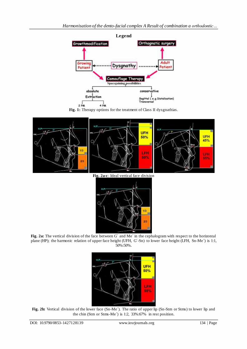
Fig. 1: Therapy options for the treatment of Class II dysgnathias.
Fig. 2a-c: Ideal vertical face division
Fig. 2a: The vertical division of the face between G´ and Me´ in the cephalogram with respect to the horizontal
plane (HP); the harmonic relation of upper face height (UFH, G´-Sn) to lower face height (LFH, Sn-Me´) is 1:1,
50%:50%.
Fig. 2b: Vertical division of the lower face (Sn-Me´). The ratio of upper lip (Sn-Stm or Stms) to lower lip and
the chin (Stm or Stms-Me´) is 1:2, 33%:67% in rest position.

Harmonisation of the dento-facial complex A Result of combination a orthodontic…
DOI: 10.9790/0853-1427128139 www.iosrjournals.org 135 | Page
Fig. 2c: Skeletal division of the face in the vertical dimension, the relation of mid- to lower face with respect to
Anterior nasal spine (N-Ans:Ans-Me) is 45% to 55%.
Fig. 3a-c: Lateral and frontal facial photograph picture of a Class II patient with short -face-syndrome, short
lower face, deepened sublabial sulcus with prominent lower lip and chin.
Fig. 4a-e:Clinical situation before the start of treatment
Fig. 4a-c: The Modells shows a distal occlusion, deep bite and malpositioned teeth.

Harmonisation of the dento-facial complex A Result of combination a orthodontic…
DOI: 10.9790/0853-1427128139 www.iosrjournals.org 136 | Page
Fig. 4d, e: A The upper dental arch shows a deficit in the transversal dimension relative to the lower arch.
Discrepancy of corresponding points of occlusion of the canines in the upper (27mm) and lower dental arch
(30mm)
Fig. 5a-c: Fig. 5a-b: Disharmonic vertical proportions of the soft tissue profile of upper face (G´-Sn) to lower
face (Sn-Me´). The lower face shows a deficit of 6% compared to the upper face. There is a disharmony of the
proportions of the lower face as well.
Fig. 5c: The cephalometric image shows the disharmonious skeletal arrangement in the vertical axis. The lower
face shows a deficit of 6percent in relation to the upper face. The mandibular angle and the interbase angle are
small.
Fig. 6: Orthopantomogram before treatment
Harmonisation of the dento-facial complex A Result of combination a orthodontic…
DOI: 10.9790/0853-1427128139 www.iosrjournals.org 137 | Page
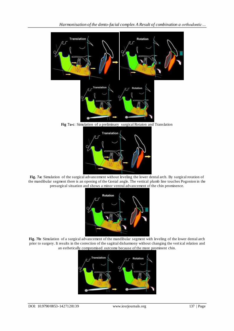
Fig 7a-c: Simulation of a preliminary surgical Rotaion and Translation
Fig. 7a: Simulation of the surgical advancement without leveling the lower dental arch. By surgical rotation of
the mandibular segment there is an opening of the Gonial angle. The vertical plumb line touches Pogonion in the
presurgical situation and shows a minor ventral advancement of the chin prominence.
Fig. 7b: Simulation of a surgical advancement of the mandibular segment with leveling of the lower dental arch
prior to surgery. It results in the correction of the sagittal disharmony without changing the vert ical relation and
an esthetically compromised outcome because of the more prominent chin.

Harmonisation of the dento-facial complex A Result of combination a orthodontic…
DOI: 10.9790/0853-1427128139 www.iosrjournals.org 138 | Page
Fig. 7c: Demonstration of the effects of the different advancements: The rotation results in a minimal stretching
of the digastric muscle (right) whereas, it is rather extensive with translation (left).
Fig. 8a-e: Clinical situation after orthodontic preparation. Deep bite and curve of Spee are almost unchanged.
Fig. 9:Cephalogram after surgical advancement of the mandibular segment and its rigid fixation with screws,
consequence of the advancement with posterior rotation of the segment with three point contact is a lateral open
bite.
Fig. 10a, b: Phases and mechanics to close the lateral open bite.
Fig. 11a-e:Clinical Situation at the End of the Treatment. Class I and stable occlusion with correct overjet and
overbite.
Fig. 12a-c:Extraoral appearance of treatment results. The sagittal deficit was corrected without increasing the
chin prominence. At the same time the vertical dimension was harmonized. The sublabial sulcus was flattened.
Harmonisation of the dento-facial complex A Result of combination a orthodontic…
DOI: 10.9790/0853-1427128139 www.iosrjournals.org 139 | Page
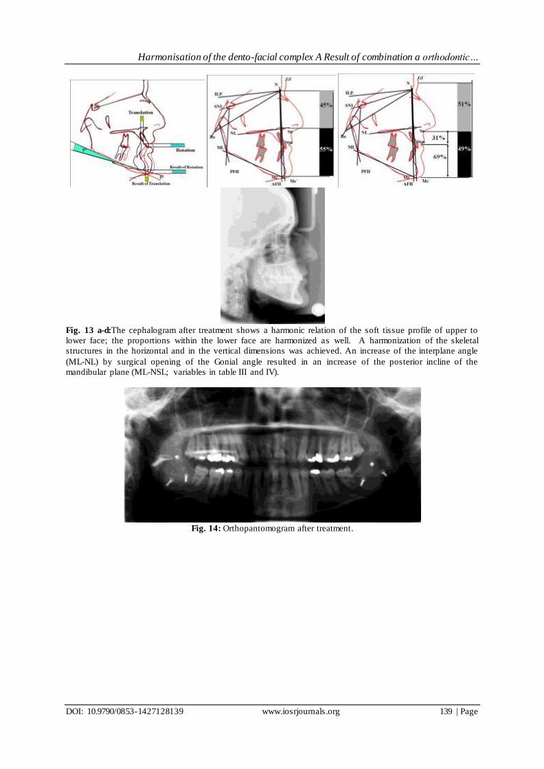
Fig. 13 a-d:The cephalogram after treatment shows a harmonic relation of the soft tissue profile of upper to
lower face; the proportions within the lower face are harmonized as well. A harmonization of the skeletal
structures in the horizontal and in the vertical dimensions was achieved. An increase of the interplane angle
(ML-NL) by surgical opening of the Gonial angle resulted in an increase of the posterior incline of the
mandibular plane (ML-NSL; variables in table III and IV).
Fig. 14: Orthopantomogram after treatment.