hallux valgus.pptx
TRANSCRIPT

Hallux Valgus
Siwaporn KhureerungRoi-Et Medical School3 Sep 2014

Introduction
• Hueter defined the deformity as an abduction contracture in which the great toe is turned away from the mid-line of the body.
• The adjective valgus implies a static deformity and should not be used interchangeably with abductuswhich refers to movement caused by muscle function

Lateral deviation of the great toe and medial deviation of the first metatarsal
Progressive subluxation of the first metatarsophalangeal (MTP) joint
Introduction

AnatomyFour groups that encircle the first MTP
joint 1) Extensor hallucis longus and brevis2) Flexor hallucis longus and brevis3) Abductor4) Adductor
Deforming Musculature1. Abductor Hallucis
-Inserts in the plantar aspect of the proximal phalanx-Can draw the phalanx medial and push metatarsal
head lateral2. Adductor Hallucis
-2 origins-common tendon to plantar aspect of proximal
phalanx and lateral aspect of plantar plate/sesamoid complex

AnatomyPlantar Plate
• 2 seasmoids incorporated into tendons of FHB
• Plantar Plate formed by tendons of Adductor Hallucis, Abductor Hallucis, FHL and Joint Capsule

Collateral Ligaments
Sesamoid Ligaments
Hood Ligament


Dermographic
• Age of oset : >20y• Gender : female• Bilaterality :> 84%

Footwear
Occupation
Heredity- 60% to 90%
Pes Planus
Hypermobility of
Metatarsocuneiform joint
Ligamentous laxity
Achilles Contracture
Neuromuscular disorders
Systemic conditions like RA
Misc factors: 2nd toe amputation;
Cystic degneration of medial capsule


Windlass Mechanism

This windlass mechanism is responsible for:
• Depression of 1st Metatarsal Head
• Weight transfer to hallux.
In HV this mech is disrupted
Transfer of weight laterally
*Surgery must minimize disruption of the windlass.

Patho anatomy
• Increased metatarsophalangeal angle
-plantar shift of abd.hallucis
-unopposed action of add.hallucis pulls greater toe
to further valgus
-medial capsular stuctures stretched and attenuated
-medial shift of metatarsal head

CONT….
• FHL,FHB&EHL increase valgus stress
• Lateral sesamoid displaced into first webspace

normal Hallux valgus

•

Clinical PresentationPAIN over the medial eminence (Bunion).
• Pressure from footwear is the most frequent cause of this discomfort.
• Bursal inflammation• Irritation of the skin• Breakdown of the skin may be noted.
• Bunion consists of:• Bony exostosis / prominence of the metatarsal head• Overlying subcutaneous bursa• Hyperkeratosis of dermis

Signs and Symptoms• Asymptomatic• Pain- The primary
symptom of hallux valgus is PAIN over the medial eminence.
• Pressure from footwear is the most frequent cause of this discomfort.
• deformity• Tenderness• Aesthetic• Look for presence of:
– neurologic disorder– ligamentous laxity
Sources of Pain in Hallux Valgus• Medial Eminence• 2nd Toe• Metatarsosesamoid Articulation• Dorsomedial Cutaneous Nerve• Transfer Metatarsalgia

Pronated Toe Fig 6

PHYSICAL EXAM
• Skin– calluses, areas of redness
• Sites of pain• Motion of 1st MTP joint-increased or decreased• Mobility and structure of foot in general• Gait analysis
• The patient sitting and standing – accentuated with weightbearing
• Pes planus deformity • Contracture of the Achilles tendon • Magnitude of the Hallux Valgus deformity • Pronation of the great toe

• Passive and active range of motion of the MTP joint is measured – Pain or crepitus, or both, with motion of the MTP
joint • Metatarsocuneiform joint for hypermobility
– Examiner grasps the first metatarsal with the thumb and index finger and pushes it in a plantar lateral-to-dorsomedial direction.
– Mobility of more than 9 mm represents hypermobility

Radiologic assesment
• Antero-posterior- wt bearing• Lateral- wt bearing• Medial Oblique wt bearing• Sesamoid view.

Standing dorsoplantar view
Non-standing lateral oblique view
Standing lateral view Axial sesamoid view

Radiographic Examination
Weightbearing AP/Lateral non weightbearing oblique view and axial views (sesamoid)
• Assess for bone and joint deformity• Length and shape of 1st MT• Congruent vs. Incongruent joint• Osteoarthrosis • Forefoot alignment is evaluated for
metatarsus Adductus• Hindfoot is Inspected for Pes Planus or Pes
Cavus.

IMA (normal <9) [8-9]HVA (normal <15) [15-20]DMAA (normal <10) [10-15]
Hallux valgus angle
Intermetatarsal angle
Distal metatarsal articular
angle

Measure Angles–Hallux Valgus angle: Intersection of longitudinal axis
of 1st MT and proximal phalanx. Normal < 150
–Intermetatarsal angleIntersection of 1st and 2nd MT.
Normal < 90 ; increased with metatarsus primus varus

Radiographic measurements
• Distal Metatarsal Articular Angle(DMMA)
Defines the relationship of the distal articular surface of the 1st MT to the longitudinal axis. Quantities the magnitude of lateral slope of articular surface.
With subluxation, the articular surface deviates laterally in relationship to the 1st Metatarsal. Usually < 60 .


Radiographic measurements• Hallux Interphalyngeal angle

CLASSIFICATION MILD MODERATE
SEVERE
Hallux valgus angle < 20° 20° to 40° >40°
1-2 intermetatarsal angle
11° or less. 12- 15° 16° or more
Subluxation of the lateral sesamoid, as measured on an AP radiograph
< 50% 50% to 75% > 75%
SEVERITY OF DEFORMITY


TREATMENT• Non-operative vs. Operative
• All patients should be treated non-operatively first.
Despite conservative measures, some patients eventually need surgical intervention.

Nonoperative
Footwear modification• Widen toe box
– decrease lateral deviation of great toe– decrease inflammation and pain
• Decrease heel height– prevent forward slide of the foot
• Arch support– may negate effects of pes planus
• Contracture of the Achilles tendon – Stretching exercises – Lengthening of the Achilles tendon


Painful joint ROMPainful joint ROMDeformity of the joint complexDeformity of the joint complexPain or difficulty with footwearPain or difficulty with footwearInhibition of activity or lifestyleInhibition of activity or lifestyle
Indications for surgeryIndications for surgery

Associated foot disordersAssociated foot disorders - Neuritis/nerve entrapment - Overlapping/underlapping 2nd digit - Hammer digits - First metatarsocuneiform joint exostosis - Sesamoiditis - Ulceration - Inflammatory conditions (bursitis, tendinitis)
of 1st metatarsal head
Indications for surgeryIndications for surgery

Extensive peripheral vascular disease Extensive peripheral vascular disease Active infection Active infection Active osteoarthropathy Active osteoarthropathy Septic arthritis Septic arthritis Lack of pain or deformity Lack of pain or deformity Advanced age Advanced age Lack of complianceLack of compliance
ContraindicationsContraindications

MI MI within the previouswithin the previous 6 6 months months Comorbid conditions that place the patieComorbid conditions that place the patie
nt at significant nt at significant CVCV or respiratory risk or respiratory risk
ContraindicationsContraindications

Relieve pain Correct deformity Preserve MTP joint motion
Surgical GoalsSurgical Goals

1. Valgus deviation of the great toe 2. Varus deviation of the 1st metatarsal 3. Pronation of hallux and/or 1st metatarsal 4. Hallux valgus interphalangeus 5. Arthritis and limitation of motion of the
1st metatarsophalangeal joint 6. Length of the 1st metatarsal relative to lesser metatarsals
Preoperative evaluationPreoperative evaluation
7. Excessive mobility or obliquity of the 1st metatarsomedial cuneiform joint
8. The medial eminence (bunion) 9. The location of the sesamoid apparatus 10. Intrinsic and extrinsic muscle-tendon
balance and synchrony
Preoperative evaluationPreoperative evaluation

Hallux Valgus <25Hallux Valgus <25Congruent Joint Chevron osteotomy Mitchell osteotomyIncongruent Joint Distal soft-tissue realignment (subluxation) Chevron osteotomy Mitchell osteotomy
Modified from Mann RA: Decision making in bunion surgery, ICL 1990.
Treatment of Hallux ValgusTreatment of Hallux Valgus

Hallux Valgus 25Hallux Valgus 25-40-40Congruent Joint Chevron osteotomy + Akin procedure Mitchell osteotomyIncongruent Joint Distal soft-tissue realignment + proximal osteotomy Mitchell osteotomy
Modified from Mann RA: Decision making in bunion surgery, ICL 1990.
Treatment of Hallux ValgusTreatment of Hallux Valgus

Severe Hallux Valgus >40Severe Hallux Valgus >40Congruent Joint Double osteotomy Akin + chevron osteotomy Akin + 1st metatarsal osteotomy Akin + 1st cuneiform opening wedge osteotomy
Modified from Mann RA: Decision making in bunion surgery, ICL 1990.
Treatment of Hallux ValgusTreatment of Hallux Valgus
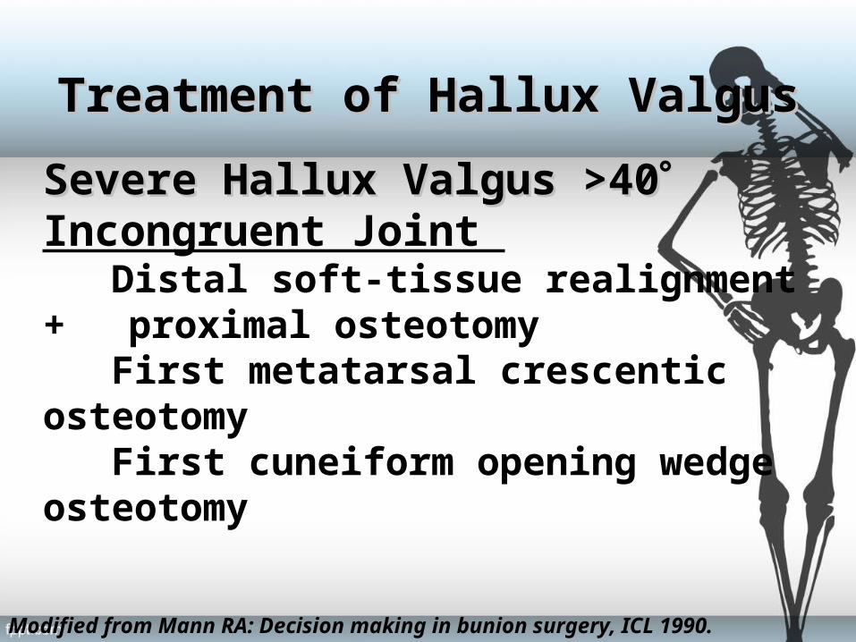
Severe Hallux Valgus >40Severe Hallux Valgus >40Incongruent Joint Distal soft-tissue realignment + proximal osteotomy First metatarsal crescentic osteotomy First cuneiform opening wedge osteotomy
Modified from Mann RA: Decision making in bunion surgery, ICL 1990.
Treatment of Hallux ValgusTreatment of Hallux Valgus

Hypermobile 1Hypermobile 1stst MTC Joint MTC Joint Distal soft-tissue realignment + fusion 1st metatarsocuneiform joint
Degenerative joint diseaseDegenerative joint disease Fusion or Keller procedure or prosthesis
Modified from Mann RA: Decision making in bunion surgery, ICL 1990.
Treatment of Hallux ValgusTreatment of Hallux Valgus

Surgical Algorhythm HVA IMA Procedure
< 40° < 13° to 15° modified McBride or distal chevron osteotomy
< 40 ° > 13° to 15° modified McBride and proximal osteotomy
>40° > 20° modified McBride and proximal osteotomy or arthrodesis

DSTPDSTPModified McBride bunionectomy
DuVries & Mann

Procedure
• Medial approach
• L-shaped capsulotomy
• Medial eminance removed
• Adductor tenotomy &lat.capsular release
• Lat.sesamoidectomy(Dorsal Approach/Plantar Approach)
• Medial capsular imbrication&wound closure

• Mitchell osteotomy
Removal of medial eminance
Osteotomy of distal portion of 1st MT shaft
Lateral displacement&angulation of capital fragment
Medial capsulorrrhaphy

Metatarsal OsteotomyMetatarsal OsteotomyMitchell osteotomy

Metatarsal OsteotomyMetatarsal OsteotomyModified Chevron osteotomy

Metatarsal OsteotomyMetatarsal OsteotomyJohnson modified Chevron osteotomy

Post-operative managementPost-operative management
Immobilization ~2 weeks Weight bearing as tolerated or NWB

Post-operative managementPost-operative management
HV night splint to be worn for 6-8 wks after dressing changes are completed

ComplicationsSURGERY
• Recurrent deformity 20-30%• Hallux Varus• Pronation deformity• Pain• Neurologic Injury• Osteonecrosis• Physeal injury/arrest• Nonunion/malunion