guidelines for platelet function testing - semantic scholar · pdf file1 guidelines for the...
TRANSCRIPT
1
Guidelines for the Laboratory Investigation of Heritable
Disorders of Platelet Function
British Committee for Standards in Haematology
Writing group:
Paul Harrison1, Ian Mackie2, Andrew Mumford3, Carol Briggs4,
Ri Liesner5, Mark Winter6, Sam Machin2
Address for correspondence:
BCSH Secretary
British Society for Haematology
100 White Lion Street
London N1 9PF
e-mail [email protected]
www.bcshguidelines.com
Published in August 2011
The guideline will be reviewed in August 2013 unless BCSH considers that new
evidence or information necessitates an earlier review. Any amendments to the
guideline and the date when the guideline was last reviewed will be published on the
BCSH website.
Disclaimer
While the advice and information in these guidelines is believed to be true and
accurate at the time of going to press, neither the authors, the British Society for
Haematology nor the publishers accept any legal responsibility for the content of
these guidelines.
1Oxford Haemophilia & Thrombosis Centre, Churchill Hospital, Oxford
2Haemostasis Research Unit, Department of Haematology, University College London
3Bristol Haemophilia Centre, Bristol Haematology and Oncology Centre, Bristol
4Department of Haematology, University College London Hospitals NHS Trust, London, W1T 4EU.
5Haemophilia Comprehensive Care Centre, Great Ormond Street Hospital for Children NHS Trust,
London
6Kent Haemophilia Centre, Kent and Canterbury Hospital

2
Table of contents:
Methodology ...................................................................................................3
1 Introduction ..............................................................................................4
2 Pre-analytical variables ...........................................................................5
3 Tests and assays .....................................................................................7
4 Specific assays of platelet function .......... 1Error! Bookmark not defined.
5 Other tests .............................................................................................. 19
6 Diagnostic features ................................................................................ 20
7 Diagnosis in infants and small children ............................................... 21 Table 1: drugs, compounds, foods that affect platelet function ............. 23
Table 2: agonists used for LTA and receptor targets .............................. 25
Table 3: minimun diagnostic criteria for platelet defects ......................... 26
Figure 1: LTA patterns and diagnosis ....................................................... 29
Appendix 1: Grading of evidence and recommendations ....................... 33
Acknowledgements and declarations of interests .................................... 34
References .................................................................................................... 35
3
METHODOLOGY
The guideline writing group was selected to be representative of UK-based medical
experts. MEDLINE was systematically searched for publications in English up to the
Summer of 2009 using key words platelet, platelet function testing and platelet
aggregometry. Relevant references generated from initial papers and published
guidelines/reviews were also examined. Meeting abstracts were not included. The
writing group produced the draft guideline which was subsequently revised and
agreed by consensus. Further comment was made by members of the Haemostasis
and Thrombosis Task Force of the British Committee for Standards in Haematology.
The guideline was then reviewed by a sounding board of approximately 40 UK
haematologists, the BCSH (British Committee for Standards in Haematology) and
the British Society for Haematology Committee and comments incorporated where
appropriate. Criteria used to quote levels and grades of evidence are as outlined in
appendix 1 of this guideline. The objective of this guideline is to provide healthcare
professionals with clear guidance on platelet function testing in patients with
suspected bleeding disorders. The guidance may not be appropriate to patients
receiving antiplatelet therapy and in all cases individual patient circumstances may
dictate an alternative approach.
Guideline update
A previous BCSH guideline was published in 1988 (The British Society for
Haematology BCSH Haemostasis and Thrombosis Task Force, 1988) and the new
guideline is designed to completely replace this.
4
1 INTRODUCTION
The diagnostic evaluation of platelet disorders is complex, poorly standardised and
time consuming. This coupled with the wide spectrum of a known range of disorders
some of which are very rare, presents a significant challenge to even the best
diagnostic laboratory. (Bolton-Maggs et al, 2006; Hayward & Favaloro, 2009; Pai &
Hayward, 2009; Watson et al, 2010). Many new tests (e.g. use of the PFA-100 and
flow cytometry) have become available since the last BCSH guideline (The British
Society for Haematology BCSH Haemostasis and Thrombosis Task Force, 1988)
and the bleeding time (BT) is now used less frequently. A number of recent surveys
have shown large variations between laboratories in platelet function testing practice
and clearly demonstrate that new guidelines are urgently required (Jennings et al,
2008;Moffat et al, 2005;Cattaneo et al, 2009). These surveys have revealed why
many types of mild platelet defects, (e.g. primary secretion defects) may be missed.
This is not only because of the heterogeneity and rarity of some defects, but is also
probably related to the failure to apply certain key platelet tests. This document
outlines a new standardised approach which could be adopted by most clinical
laboratories for the investigation of heritable platelet bleeding disorders. When the
clinical picture and/or laboratory results suggest an inherited platelet disorder,
referral to an expert reference centre should also be considered. Platelet function
tests used specifically for monitoring antiplatelet drugs and/or detecting platelet
hyperfunction will not be discussed in these guidelines.
An evaluation of patients with abnormal bleeding requires objective clinical
assessment of bleeding history, any family history and physical examination
followed, when appropriate, by laboratory investigations. During this process it is
essential to recognise that numerical and/or functional platelet disorders are
prevalent amongst patients with abnormal bleeding and may be clinically
indistinguishable from other haemostatic disorders, particularly von Willebrand
disease (VWD).(Hayward, 2008;Cattaneo, 2003) Platelet disorders can also
sometimes co-exist with other coagulation factor defects or VWD. (Daly et al,
2009;Quiroga et al, 2007) Laboratory investigations of platelet number and function
are therefore recommended in any patient where bleeding symptoms are not fully
explained by standard clinical laboratory investigations. Further information on the
clinical presentation of patients with platelet disorders and the differential diagnosis

5
is available in detail elsewhere.(Bolton-Maggs et al, 2006) The current guideline
focuses on the laboratory investigation of suspected platelet function disorders that
should be performed in UK haematology laboratories. However, laboratory tests
should ultimately be interpreted in terms of the clinical information.
2 PRE-ANALYTICAL VARIABLES
2.1 Specimen collection
2.1.1 Venipuncture
Ideally, samples for platelet function studies should only be collected from fasting
and resting subjects who have refrained from smoking and caffeine ingestion on the
day of testing. If the patient is taking medication known to affect platelet function, e.g.
non-steroidal anti-inflammatory drugs (George & Shattil, 1991), testing should, if
possible, be deferred for 10-14 days after the last dose. Herbal remedies, garlic,
alcohol and certain foods may also cause acquired platelet dysfunction. (George &
Shattil, 1991) See table 1 for a list of drugs and other agents that are known to
affect platelet function. In normal clinical practice it is difficult to avoid some of these
patient related variables and so a pragmatic approach is to consider proceeding with
platelet function tests, but if they are abnormal, collecting a fresh sample under more
suitable conditions and repeating the tests.
Blood should be collected by experienced phlebotomists using a standardised,
atraumatic protocol, from the antecubital fossa, by clean venipuncture using
minimum tourniquet pressure. Needles should be 19-21 gauge (butterfly cannulae
are suitable, providing blood flow is not restricted) and either evacuated tube
systems or plastic syringes may be used. A discard tube should be used before
collecting successive citrate tubes. Where tubes with a variety of anticoagulant types
are required, the citrate tubes should be collected before EDTA- or heparin-
containing tubes wherever possible to avoid the potential for carryover.(Favaloro et
al, 2008)
6
2.1.2 Anticoagulants
Blood should be collected into a 1/10 volume of trisodium citrate (105-109 mmol/L
final concentration) for clinical platelet function testing. Buffered citrate solutions that
maintain the sample pH are preferred. Care must be taken to ensure that tubes are
correctly filled.
2.1.3 Specimen processing
All specimens must be maintained at room temperature (20-25°C) and should not be
placed on ice, in a refrigerator or a water bath. Immediately after blood collection, all
tubes should be mixed by gentle inversion at least 6 times (and discarded if there is
any evidence of clotting). Tubes should be kept capped at room temperature and not
subjected to any vibration, shaking, vortexing, continuous mixing or agitation; they
should not be transported via pneumatic tube systems. The time delay between
collection, transport and analysis should ideally be preferably between 30 minutes
and 2 hours but not more than 4 hours.
Recommendations
A complete record of current medication taken by patients or controls
should be taken prior to blood collection to either prevent unwanted
drug interference or help interpretation of test results (1A)
Collect blood using a standardised, atraumatic protocol, with minimal
stasis (2C)
Use needles between 19 and 21 gauge; evacuated tube systems or
syringes are acceptable (2C)
The first 3-5ml of blood should not be used for platelet function tests
(2C)
Use 105-109 mmol/L buffered trisodium citrate tubes (2C)
Maintain specimens at room temperature (1B)
Keep tubes upright and capped; do not subject to excessive mixing or
agitation; do not use pneumatic transport systems (2C)
7
Samples should be tested between 30 minutes and no more than 4
hours from blood collection (2C)
3 TESTS AND ASSAYS
Laboratory tests for platelet disorders comprise:
i. Measurement of platelet number and size
ii. Global screening tests of platelet haemostatic function
iii. Specific assays of platelet haemostatic function
3.1 Platelet number, size and morphology
Performance of the modern “Full Blood Count” investigation on whole blood is an
essential investigation in patients with abnormal bleeding. The measurement of
platelet number and size using automated cell counters and blood film analysis is
highly sensitive and specific for numerical platelet disorders and is therefore valuable
early in the investigation. Normal results will eliminate thrombocytopenia and
anaemia as potential causes of bleeding and ensure that subsequent platelet
function tests are not going to be affected by low platelet counts. Low platelet counts
indeed affect most platelet function tests discussed below except flow cytometry
Thrombocytosis, which may underlie abnormal bleeding, will also be revealed. If
abnormalities in either platelet count, size (mean platelet volume, MPV) or
distribution are flagged by the instrument then it is recommended that a blood film be
examined to look for abnormalities in platelet number, size and/or granule
content.(Briggs et al, 2007)(Althaus & Greinacher, 2009) More recently, multiple light
scatter parameters and/or fluorescence, rather than impedance sizing alone have
been introduced into commercial analysers. This has improved their ability to
distinguish large platelets from red cells and can sometimes provide more accurate
counts (e.g. in samples from patients with macrothrombocytopenia where counts are
usually underestimated).(Harrison et al, 2000) Immunocounting by flow cytometry
should also be considered when accurate counts are required in
mactothrombocytopenia. (Harrison et al, 2000)
8
3.2 Global tests of platelet haemostatic function
Global tests of platelet function are often used during the investigation of individuals
with pathological bleeding. Since global tests do not enable a diagnosis of a specific
platelet disorder, they are normally performed as the first part of a strategy which
requires further testing with more specialised assays of platelet function. (Zeidan et
al, 2007)(Harrison & Mumford, 2009) Normal test results may therefore theoretically
be used to exclude the diagnosis of platelet function disorder so that further
specialised testing can be avoided. For this reason, global platelet function tests are
usually performed at the same time as global assays of coagulation pathway function
(prothrombin time (PT) and activated partial thromboplastin time (aPTT), von
Willebrand Factor (VWF) screening tests (VWF:Ag, VWF:RCo and F:VIII:C) and
measurement of platelet number). Guidelines for the systematic investigation of
patients with suspected VWD and other coagulation factor deficiencies have recently
been published elsewhere and are not discussed further in this review.(Laffan et al,
2004;Bolton-Maggs et al, 2004) The most widely performed tests for screening
platelet function disorders are currently the template BT and the Platelet Function
Analyser (PFA-100®; Siemens Diagnostics) closure time. Other commercial platelet
function assay systems are also available, including those designed to measure the
effect of antiplatelet drugs. (Harrison et al, 2007) Thromboelastography (TEG) and
Rotational Thromboelastometry (ROTEM) ROTEM and TEG provide global tests of
haemostasis and platelet function and are mainly used withinthe surgical
setting.(Perry, 2010) The utility of most of these assay systems including
TEG/ROTEM for the screening and diagnosis of platelet function defects has not yet
been examined systematically and their use for this application is therefore not
currently recommended.
3.2.1 Template bleeding time
The bleeding time described by Duke in 1910 is the oldest test of platelet
function.(Duke, 1910) Although the BT was previously recommended as a clinically
useful test of platelet function,(The British Society for Haematology BCSH
Haemostasis and Thrombosis Task Force, 1988) and surprisingly it remains in wide

9
use in the UK (Jennings et al, 2008), there is considerable variation in methodology
between laboratories.
The BT is highly dependent on operator technique, is subjective and is influenced by
patient variables unrelated to haemostasis, such as age, gender, haematocrit,
vascular pattern, skin thickness and skin temperature.(Rodgers & Levin,
1990;Peterson et al, 1998) The BT therefore has poor reproducibility, sensitivity and
specificity, as well as being invasive; for these reasons it is not recommended.
3.2.2 Closure time by the Platelet Function Analyser
Assay principle
The PFA-100® device is a test system in which citrated whole blood is aspirated at
high shear rates (5000-6000s-1) through disposable cartridges containing an
aperture coated with either collagen and epinephrine (CEPI) or collagen and ADP
(CADP).(Kundu et al, 1995;Jilma, 2001;Kratzer & Born, 1985) These agonists
trigger platelet adhesion, activation and aggregation leading to rapid occlusion of the
aperture and cessation of blood flow.(Kundu et al, 1995) The end-points for each test
are time to occlusion of blood flow (closure time [CT]) or non-closure if the CT
exceeds 300 seconds. The PFA-100® assay system requires small quantities of
citrated venous blood (0.8 mL per cartridge) and is therefore useful for studying
paediatric samples. Accordingly, the PFA-100® device is widely used as a screening
tool to measure global platelet haemostatic function.(Jennings et al, 2008;Moffat et
al, 2005)
Factors that influence PFA-100® closure time
The choice of anticoagulant, specimen collection and transportation techniques and
time between sampling and analysis (see Specimen Collection section above for
guidance) all have critical effects on CT results.(Jilma, 2001) (Harrison et al,
1999;Heilmann et al, 1997). Recent evidence reinforces the need for a discard tube
during blood collection for PFA-100 testing (Kunicki et al, 2009). It is important that
each laboratory establishes a reference range preferably within 105-109 mmol/L
buffered trisodium citrate tubes. Further guidance for the quality control of the PFA-

10
100® is published elsewhere. (Christie et al, 2008;Harrison, 2004;Hayward &
Eikelboom, 2007)(Favaloro, 2009) There are extensive general reviews of the clinical
utility of the PFA-100. (Favaloro, 2008)(Hayward et al, 2006)
Knowledge of the full blood count is critical for interpreting CT results from the PFA-
100®. Thrombocytopenia (< 100 x 109/l) and anaemia (< 20% haematocrit) often
results in prolongation of the CT. (Kundu et al, 1995;Harrison et al, 1999) CT also
correlates inversely with plasma VWF activity in normal subjects and may therefore
be longer in patients with blood group O.(Lippi et al, 2001) The Collagen/Epinephrine
(CEPI) CT, but not the Collagen/ADP (CADP) CT, is usually prolonged by COX-1
inhibitors such as aspirin.(Jilma, 2001)
PFA-100® CT and VWD
Abnormal CT on both cartridges are typical for types 2A, 2B, 2M and 3 VWD with a
sensitivity of > 98%.(Franchini, 2005) When type 1 VWD is included, the overall
sensitivity of CT to VWD is reported to be lower (85-90%),(Favaloro, 2006) but there
is a clear relationship between VWF level and CT,(Moeller et al, 2001). Type 2N
gives normal results. The PFA-100® may also be useful for monitoring desmopressin
therapy in VWD patients.(Cattaneo et al, 1999;Favaloro et al, 2001;Franchini et al,
2002;Hayward et al, 2006;van Vliet et al, 2008)
PFA-100® CT and diagnosis of heritable platelet function disorders
Greater abnormalities in CT in both cartridges occur with the severe platelet function
defects, such as Glanzmann thrombasthenia (GT), Bernard–Soulier syndrome (BSS)
and platelet type or pseudo-VWD in which non-closure is typical.(Harrison et al,
1999;Harrison, 2005;Hayward et al, 2006; Mammen et al, 1998) In many less severe
platelet function defects, the CT may be either normal or prolonged; abnormal results
are more frequently reported with the CEPI than the CADP cartridge.(Hayward et al,
2006;Harrison et al, 2002) There are rare reports of abnormal CADP but with normal
CEPI CTs suggesting that the CEPI cartridge cannot be used exclusively as a
screening test. It is not currently possible to accurately determine the sensitivity of

11
the PFA-100® for most mild, heritable platelet function defects since most reported
studies comprise small patient numbers, with varying mixtures of these
defects.(Hayward et al, 2006;Harrison, 2005) The PFA-100® CT exhibits poor
sensitivity for mild platelet defects in a small number of prospective studies in
patients with an unequivocal personal and family history of mucocutaneous
bleeding.(Quiroga et al, 2004;Cattaneo, 2004;Podda et al, 2007) Other retrospective
cohort studies of patients with previously diagnosed platelet function defects indicate
sensitivities up to >80% for prolonged CT, although many of these studies included
subjects with severe phenotypes (e.g. GT, BSS) and VWD. (Harrison et al,
1999;Harrison et al, 2004;Kerenyi et al, 1999;Posan et al, 2003) A recent meta-
analysis concluded that the overall sensitivity and specificity of the CEPI cartridge for
disorders in primary haemostasis was 83% and 89% respectively. CADP sensitivity
was lower at 67% with an equivalent specificity of 86%. (Karger et al, 2007). The
PFA-100® has shown good sensitivity (> 90%) in screening patients with
menorrhagia for VWD and platelet function defects.(James et al, 2004;Philipp et al,
2005;Acharya et al, 2008)
Guidelines on the utility and practice of using the PFA-100 for clinical assessment of
platelet disorders are provided by various international and national organisations.
(Hayward et al, 2006;Bolton-Maggs et al, 2006; Christie et al, 2008).
It is reasonable to use normal PFA closure times to rule out a significant platelet
defect in patients who have a low clinical suspicion of such a defect, however if the
clinical suspicion of a platelet defect is high, then a normal PFA result should not be
used to rule out this possibility and specific assays of platelet function are indicated.
Recommendations
Perform a full blood count on all patients (1A)
In samples with abnormalities in platelet count or size distribution (as
indicated by an automated analyser), a blood film should be examined
(1B)
The bleeding time is not recommended (1B)

12
The PFA-100 provides an optional screening test, but this must be
interpreted with caution and in the context of the clinical background, as
the test is not diagnostic or sensitive for mild platelet disorders (1B)
Both PFA-100 CADP and CEPI cartridges should be used for screening
(1B)
4 SPECIFIC ASSAYS OF PLATELET FUNCTION
4.1 Light transmission aggregometry
Light transmission aggregometry (LTA) was invented in the early 1960s and is still
regarded as the gold standard for platelet function testing. Despite its widespread
use, the test is poorly standardised and there are wide variations in laboratory
practice(Jennings et al, 2008;Moffat et al, 2005;Cattaneo et al, 2009). Guidelines
specific for LTA have also recently been published (Christie et al, 2008; Cattaneo et
al, 2011; Hayward et al, 2010)
Sample preparation for LTA
Citrated blood samples obtained as described above are centrifuged to prepare
platelet rich plasma (PRP) and platelet poor plasma (PPP). To prepare PRP, whole
blood tubes should be centrifuged at 170-200 g for 10 minutes in a swing-out rotor at
room temperature (RT) without application of the brake. Autologous PPP is prepared
by centrifugation (after removal of PRP or using whole samples) at least 1500 g for
at least 15 minutes at RT (Christie et al, 2008). At the end of the centrifugation steps
a plastic pipette should be used to separate the top 2/3rds of PRP or PPP should be
carefully removed without disturbing the buffy coat layer and red cells. PRP or PPP
should then be transferred into separate polypropylene tubes capped and stored
upright at RT. The PRP should then be left for at least 30 minutes prior to testing.
Visual inspection of the samples is important as icteric, lipaemic, red cell
contaminated and haemolysed samples should not be tested. A platelet count should
be performed on the PRP and unless it is greater than 600 x 109/L, the platelet count
should not be adjusted using PPP, as this may cause artefactual inhibition of platelet
aggregation (Linnemann et al, 2008;Cattaneo et al, 2007). Analysis of PRP with a

13
platelet count <150 x 109/L is possible, but the results should be treated with caution
(ideally a normal control should be analysed, where the PRP count is adjusted to
equal that of the test, by dilution with buffer instead of PPP) (to prevent artefacts).
PRP with low counts can still be tested to exclude severe platelet disorders such as
BSS and type 2B and platelet type VWD.
Agonists for LTA
ADP, epinephrine, collagen (type I, tendon), arachidonic acid and ristocetin are the
traditional baseline panel of agonists for LTA. (see Table 2) (The British Society for
Haematology BCSH Haemostasis and Thrombosis Task Force, 1988) An extended
panel of agonists can include gamma Thrombin, Thrombin Receptor Activating
Peptides (TRAP), Collagen-Related Peptide, endoperoxide analogue U46619 and
calcium ionophore A23187, which may all be useful when a more detailed
investigation of the exact nature of the defect is required (see Table 2). Most
laboratories perform dose response curves for ADP (0.5 – 20 M), collagen (1.0 –
5.0 g/ml) and epinephrine (0.5 -10 M). Although this provides a detailed
pharmacological approach, more recent evidence supports the use of single doses
of a panel of agonists, which significantly increases the likelihood of detecting a
platelet defect (odds ratio 32).(Hayward et al, 2009b) (Dawood et al, 2007) A
recommended baseline example panel therefore comprises: 2.5 M ADP, 1.25 g/ml
collagen, 5 M epinephrine, 1.2 mg/ml ristocetin, and 1.0 mM arachidonic acid (all
final concentrations in PRP). If the initial aggregation results with ADP, collagen or
epinephrine are abnormal then retesting should be performed at higher
concentrations of the agonist(s) and even to supranormal concentrations to confirm a
specific defect. It should be noted that a significant proportion of normal samples
may not always give a full aggregation response to epinephrine (due to natural
variations in adrenoreceptor numbers) and have no related platelet defect. If the
aggregation to ristocetin is normal then retesting should additionally be performed
with low dose (0.5-0.7 mg/ml) ristocetin to check for hyperfunction or gain of function
(associated with Type 2B and platelet type VWD). If results with 1.2 mg/ml ristocetin
are absent then retesting can be performed with addition of an external source of
VWF (e.g. cryoprecipitate or a VWF concentrate) to confirm either a VWF or GpIb
14
defect. If arachidonic acid aggregation is abnormal then further testing should be
performed with 1.0 M U46619 to test for any thromboxane receptor abnormalities.
An extended panel of tests (usually only available within more specialised centres)
could also be considered including gamma thrombin (which does not cause clotting),
PAR-1 (SFLLRN) and PAR-4 (AYPGKF) TRAP peptides (if gamma thrombin is
abnormal) Collagen-Related Peptide (CRP), calcium ionophore and PMA (Phorbol
12-myristate 13 acetate) if abnormalities in the thrombin receptors, GpVI, calcium
mobilisation and protein kinase C respectively, are suspected.
Performing aggregometry
A maximum of 1/10 volume of agonist is added to PRP to initiate aggregation and
the final concentration of agonist within PRP is recorded (taking into account the 10
fold dilution factor). It is imperative that new batches of agonist are checked against
the previous batch for performance, using normal control samples.
Platelet aggregometers measure the change in optical density (or light
transmittance) over time of stirred PRP in cuvettes at 37°C after addition of the
agonists. They are calibrated for transmission using autologous PPP (100%) and
PRP (0%). A stir speed of 1000 - 1200 rpm is normally recommended. It is important
that samples are pre-incubated for at least 5 minutes at 37°C prior to assay to obtain
stable baseline traces. The appropriate agonists must then be added directly to the
PRP and not pipetted onto the side of the tube. It is important that no air bubbles are
introduced at any stage of the procedure as these can interfere with transmission
measurement. The aggregation tracing should be observed for at least 5 minutes,
but preferably 10 minutes to monitor the lag phase, shape change (negative
deflection), primary and secondary aggregation and any delayed platelet responses
e.g. reversible or spontaneous aggregation. The assay is then terminated and results
printed and stored for visual inspection.
It is recommended that local, normal cut-off values are established, using non-
parametric statistics. However, it is recognised that this is not possible for most
clinical laboratories due to the inherent variability of the test and the large number of
subjects that would be required (i.e. >40). For this reason, most clinical laboratory
staff subjectively evaluate the shape of the aggregation curves. The following

15
parameters should always be considered: lag phase, maximal amplitude, primary
aggregation slope, and disaggregation, for each commonly used agonist
concentration.(Hayward et al, 2009b;Hayward et al, 2008). It is important that the
overall shape of the aggregation responses obtained with each agonist are fully
described and interpreted by experienced staff (e.g. is the response fully reversible
and is there a significant lag phase; what is the maximal amplitude of the response).
See Figure 1 for typical examples of normal and abnormal aggregometry curves in
various classical defects.
Recommendations
Platelet counts >600x109/L (1B) in platelet rich plasma should be
diluted.
Repeat all unexpected, abnormal light transmission aggregometry tests
with a fresh sample, in parallel with a normal control sample (2C)
Only experienced individuals should interpret tracings and results (1C)
Assess performance of new batches of agonists by comparison with a
previous batch (1A)
4.2 Flow cytometry
The most commonly used flow cytometry tests relevant to platelet function are the
quantification of glycoprotein receptor density in the diagnosis of defects such as GT
and BSS, and detecting their heterozygous states. Flow cytometry can also be used
to measure the collagen (GpIa/IIa and GpVI) and PAR-1 receptor densities if LTA
testing suggests any abnormalities in these receptors. There are also tests available
to measure: platelet activation in response to classical agonists, dense granule
content, and exposure of anionic phospholipids As flow cytometry is expensive, time
consuming and requires specialised training, only those patients with an appropriate
clinical history and/or abnormalities of other platelet function tests should be
assessed for receptor defects. Guidelines and protocols on flow cytometry of

16
platelets have been published elsewhere.(Schmitz et al, 1998;Goodall & Appleby,
2004;Michelson et al, 2007) It is recommended that analysis should be performed
using fresh , citrated whole blood, to avoid platelet activation and loss of platelet
subpopulations during centrifugation. If measuring platelet activation and function it
is important to control for ex-vivo activation caused by delays in analysis from blood
sampling for example. Fluorescently labelled antibodies are added to the blood
samples and after incubation at room temperature, in the dark, the samples are
diluted to a final volume of between 1-2 ml with buffer (e.g. Hepes buffered saline,
pH 7.4), or a mild fixative before analysis. All buffers must be filtered (e.g. using a
0.2 µm filter) and tubes should not be vortexed, but mixed gently by tapping,
otherwise platelet aggregation will occur. Matched isotype control fluorescent
antibodies should be tested at the same time in control tubes. It is recommended
that normal positive control samples are analysed in parallel to verify assay
performance and that the antibodies are efficiently binding to their respective
receptors particularly if a receptor is completely absent in GT or BS for example.
Some commercial assays are now available that can give absolute quantification of
the copy number of individual receptors of interest. Normal ranges can be
established for either fluorescence or copy number of individual glycoproteins
Neonates may also have significantly lower receptor densities than adults. The lower
limit of detection if ~500 receptors/platelet so the test cannot always be used reliably
to detect low copy number receptors. It is possible to measure platelet procoagulant
activity, apoptosis (and microparticles) by incubating samples with high affinity
probes against phosphatidyl serine (e.g. Annexin-V) and activating the cells with
calcium ionophore, collagen-related peptide or combinations of thrombin and
collagen. This enables the diagnosis of Scott syndrome and related disorders
although these defects are indeed very rare.
Recommendations
Flow cytometry should be used in the investigation or confirmation of
Glanzmann thrombasthenia, Bernard–Soulier Syndrome (1B) and Scott
syndrome (1C); and may also be used to investigate abnormalities in the
collagen (GpVI and GpIa/IIa) and thrombin receptors (PAR-1) (1B)

17
Whole blood platelet assays are preferable although PRP can be used
for BSS diagnosis (1B)
Analyse normal controls in parallel with test samples (1A)
4.3 Measurement of total and released nucleotides
The measurement of total and/or released adenine nucleotides provides an
important additional diagnostic tool usually in conjunction with aggregometry for
determining whether there is any specific deficiency in dense granule numbers or
their content (e.g. storage pool disease), or specific defect(s) in degranulation (e.g.
release defects). There is evidence to suggest that these defects can be
misdiagnosed if relying on platelet aggregometry alone.(Nieuwenhuis et al,
1987;Israels et al, 1990)(Hayward et al, 2009b)(Cattaneo, 2009) It is therefore
recommended that laboratories perform an independent measurement of the release
reaction. However, although nucleotide measurement is very straightforward and
normally involves measuring ATP by simple bioluminescent assays (using firefly
luciferin/luciferase assays), recent surveys indicate that many laboratories do not
measure platelet nucleotides.(Jennings et al, 2008;Moffat et al, 2005) This suggests
that many platelet storage and secretion defects are potentially being
underdiagnosed with current practice.
The simplest assay of released platelet nucleotides can be performed in real time
with a Lumi-Aggregometer (either LTA or whole blood aggregometry, WBA).
(Dawood et al, 2007; Christie et al, 2008; Watson et al, 2010)These instruments
provide a rapid assessment of ATP levels during platelet aggregation and normally
demonstrate release of ATP during the secondary aggregation phase in LTA. The
amount of ATP released is easily calibrated using commercially available ATP
standards analysed in the same channels of the aggregometer. However, it is
impossible to distinguish between storage and release defects using this approach.
Many laboratories therefore determine the total platelet content of both ADP and
ATP with lysed platelet preparations (at standardised platelet counts) and sometimes
after a degranulation step to induce release. Adenine nucleotides are measured in
platelet lysates using either luminometers (Summerfield et al, 1981 or by HPLC
(Greaves & Preston, 1985), with conversion of ADP to ATP (using pyruvate kinase).

18
Calibration is performed using an ATP standard. These assays have the advantage
that samples can be frozen and shipped to more specialised laboratories that
regularly perform nucleotide measurements.
There are two nucleotide pools within the platelet:- the metabolic pool and the dense
granular/storage pool, the latter comprising about 60% of the total content. The ratio
of ATP:ADP is therefore of fundamental diagnostic importance as there are
pronounced differences between the relative concentrations in the two pools. Any
storage defects are associated with a decrease in the amount of stored and released
ADP with an increased ratio of ATP:ADP. Normal ADP levels and ATP:ADP ratios
but decreased ADP release are indicative of a release-defect.
Normal ranges should be established locally, but typical values are 19-38 and 41-61
nMol/109 platelets for total ADP and ATP respectively (ATP:ADP ratio 1.24-2.56).
Typical normal ranges for released nucleotides are: 18-28 and 8-20 nMol/109
platelets for ADP and ATP respectively (ATP:ADP ratio 0.43-0.79) (Chanarin, 1989).
Serotonin (5-HT) is actively taken up and stored within the platelet dense granules
and it is possible to measure the uptake and release of radiolabelled serotonin into
and from the platelets with standardised assays.(The British Society for
Haematology BCSH Haemostasis and Thrombosis Task Force, 1988;Zhou &
Schmaier, 2005) ELISA assays for platelet serotonin content are also available.
Mepacrine uptake and release by the dense granules can be measured by flow
cytometry(Wall et al, 1995;Gordon et al, 1995).
Recommendations
When there is a high clinical suspicion of a platelet function defect,
adenine nucleotides should be measured even if the aggregation is
normal (1B)
If aggregation results suggest storage pool disease or a release defect,
measure stored and released nucleotides (1B)
19
4.4 Whole blood aggregometry
In impedance aggregometry, whole blood is stirred at 37°C and aggregation is
detected by the accretion of platelets to the surface of two fine, precious metal, wire
electrodes.(Fritsma, 2007) Adherent platelets increase the electrical impedance
between the electrodes, which can be displayed as a wave of aggregation.(Cardinal
& Flower, 1980) Impedance aggregation measurements in whole blood may be
influenced by: haematocrit (>0.35 L/L), platelet count, and elevated white cell count,
while the agonist responsiveness differs from LTA. (Mackie et al, 1984;Ingerman-
Wojenski et al, 1983;Sweeney et al, 1989) Impedance and LTA methods show similar
dose responsiveness to equine tendon collagen, but higher ADP concentrations are
required to induce aggregation by the impedance technique and low doses (e.g. 1 M)
give no impedance response. Reversible aggregation and biphasic responses to ADP
cannot be demonstrated by whole blood impedance,(Mackie et al, 1984;Ingerman-
Wojenski et al, 1983) while epinephrine responses tend to be absent or very
weak,(Mackie et al, 1984;Swart et al, 1984). A study by UK NEQAS surveyed 169
haemostasis centres, (119 UK and 50 non-UK) and found that only 4/88 performed
whole blood platelet aggregation studies.(Jennings et al, 2008) Some of the
technical problems of whole blood impedance aggregation have been overcome by the
development of disposable electrodes, standardised reagents and the availability of a
5 channel multiple electrode platelet aggregometer (Dynabyte, Munich, Germany)).
(Toth et al, 2006) There is very sparse peer review literature comparing impedance and
LTA methods and a lack of clinical validation in the diagnosis of heritable platelet
function defects.
5 OTHER TESTS
Platelet alpha granule proteins (e.g. Platelet Factor 4 (PF4) and Beta-
Thromboglobulin (TG)) can be measured by ELISA, RIA or western blotting and
may be helpful for the diagnosis of Quebec platelet disorder (Kahr et al, 2001).
Electron microscopy has also proven very useful for defining ultrastructural
abnormalities associated with a variety of platelet defects.(Clauser & Cramer-Borde,
2009). The simpler whole mount electron microscopy technique has proven useful
for confirming dense granule defects(Hayward et al, 2009a).

20
Molecular genetic diagnosis of heritable platelet disorders may offer valuable
confirmation of diagnosis in affected individuals, in family members where
phenotypic testing of platelets is impractical and for ante-natal diagnosis. Molecular
diagnosis is most feasible in GT and BSS where the number of candidate genes is
small and there are already accessible databases containing large patient groups to
help confirm that observed nucleotide variations are pathogenic (http://www.b-
ss.org/1.html and sinaicentral.mssm.edu). Clinical diagnostic services for GT and
BSS by direct sequencing of PCR-amplified genomic DNA are now offered in a small
number of clinical genetic laboratories in the UK. For mild platelet function or platelet
number disorders individual candidate genes can occasionally be identified using
clinical and laboratory phenotypic features (e.g. MYH9 related disorder, CAMT, TAR,
WAS) or by laboratory phenotype alone (e.g. thromboxane and P2Y12 ADP receptor
defects, GPVI defects). (Nurden et al, 2009 ; Watson et al, 2010). However,
molecular genetic analysis of these disorders is currently available only in research
laboratories. Since there is limited repertoire of reported mutations, it is usually
difficult to assign pathogenicity to observed nucleotide variations without expression
studies. In some patient populations where a specific pathogenic mutation is
prevalent (e.g. 16bp deletion in HPS1 in Peurto Rican descent patients with
Hermansky-Pudlak syndrome), allele specific mutation detection strategies may
enable rapid molecular diagnosis of selected disorders. In all cases when molecular
genetic diagnosis is considered, families should undergo careful genetic counselling
and provide written consent in accordance with current best practice guidelines.
(Ludlam et al, 2005).
6 DIAGNOSTIC FEATURES
Typical clinical and laboratory findings of platelet function tests in many different
platelet defects are detailed in Table 3. For more diagnosis and treatment of all
platelet disorders including the inherited thrombocytopenias see Bolton-Maggs et al,
2006(Bolton-Maggs et al, 2006).

21
7 DIAGNOSING PLATELET FUNCTION DISORDERS IN INFANTS AND SMALL
CHILDREN
Severe platelet disorders such as GT and BSS usually present in infancy or early
childhood but the diagnosis of these disorders in the very young is more challenging
than in older children or adults for a number of reasons, mainly pre-analytical. Junior
paediatricians commonly take blood using heelpricks, fingerpricks or a standard
venipuncture needle inserted into a vein, collecting the drops from the end of the
needle. None of these methods is appropriate for assessment of platelet function
which should be done on a free-flowing venepuncture sample though in practice
indwelling arterial and central venous catheters have also been used to facilitate
getting the necessary volumes from small children. The minimum volume of blood
required for full platelet LTA and nucleotide testing is usually 20 ml; this may be 8-
10% of the blood volume in neonates and could cause hypovolaemic symptoms. The
usual needle gauge for blood sampling in infants and smaller children is 23G as the
recommended size of 19-21G can be too big for small peripheral veins and is also
more likely to cause trauma to subcutaneous tissues which may be significant if a
severe platelet disorder is present. It is therefore recommended that the control
sample is also taken with a 23G needle to ensure that the patient sample is
processed along with a similarly taken sample.
Validation of platelet function tests in age-matched normal control populations of
infants and children is rarely possible due to ethical issues of taking large volumes of
blood from healthy normals. Although there are scanty data on LTA in „normal‟
neonates and infants, there are no known comprehensive studies looking at
nucleotide values at different ages through the 1st year of life. The available
literature suggests that in infancy the platelets are generally hyporeactive (except to
ristocetin and variably collagen) but beyond infancy reactiveness of platelets, both
aggregation and nucleotide release reactions, is very similar to that in adults (Knofler
et al, 1998 ; Bonduel et al, 2007). Therefore the usual and pragmatic approach is to
assess platelet function in children > 1 year of age using adult controls and normal
ranges (Hayward et al, 2010). Family testing may also be useful not only to confirm a
given defect but to discern the potential inheritance pattern. As there is few data,
results of investigations taken in the first year should be viewed with more caution
and should always be repeated, particularly if the apparent abnormalities are
22
relatively subtle and if the putative diagnosis is one of the usually milder disorders
such as a granule or secretion defect. Results of investigations in infants with the
severe function defects – GT or BSS – are usually very clear-cut at all ages and it
could be argued that the safest way to diagnose these disorders in an infant is to
limit the investigations to those that can be performed on relatively small volumes of
blood; with a full blood count, PFA-100 (which will reliably show non-closure with
both cartridges in both GT and BSS), and flow cytometry. Flow cytometry if
performed carefully (see above section) can also be utilised to study platelet
function/activation in small volumes of blood by determining responsiveness to
various agonists at differing concentrations. Confirmatory LTA can then be done
when possible but demonstration of a severe defect of primary haemostasis using
the PFA-100 in combination with absent or very low levels of the affected receptor,
and macro-thrombocytopenia in BSS, is highly suggestive and enough to guide
appropriate treatment for bleeding. Conversely, both GT and BSS can effectively be
excluded in infants if the PFA-100 shows normal closure times – this can be of
crucial clinical use in unexplained severe bleeding such as intracranial haemorrhage
when there is a query as to whether this is an inflicted injury or it is due to
„spontaneous‟ bleeding in association with a severe bleeding diathesis. A detailed
diagnostic approach to platelet disorders in children has recently been published
(Israels et al, 2011)

23
TABLE 1 A list of drugs, compounds and dietary components/herbs that can affect platelet function (Reprinted and modified with permission from Kottke-Marchant and Corcoran G. The laboratory diagnosis of platelet disorders. Arch Pathol Lab Med 2002:126:133-146 with permission from Archives of Pathology & Laboratory Medicine. Copyright 2002. College of American Pathologists. Cyclo-oxygenase (COX)-1 inhibitors (irreversible) Aspirin and all proprietary or over-the-counter preparations containing acetylsalicylic acid. COX-1 and COX-2 inhibitors (reversible) (Nonsteroidal anti-inflammatory drugs (NSAIDs) Ibuprofen Indomethacin, naproxen Mefenamic acid Inhibitors of Platelet Receptors
Abciximab, tirofiban, eptifibatide (IIb3) Ticlopidine, clopidogrel, prasugrel (irreversible), cangrelor (reversible), ticagrelor (reversible) (P2Y12) Phosphodiesterase Inhibitors Dipyridamole Cilostazole Anticoagulants Heparinoids, vitamin K antagonists and direct thrombin inhibitors may indirectly influence platelet function due to inhibition of thrombin. Cardiovascular Agents
-adrenergic blockers (propranolol) Vasodilators (nitroprusside, nitroglycerin) Diuretics (furosemide) Calcium channel blockers Antimicrobials
lactams (penicillins, cephalosporins) Amphotericin (antifungal) Hydroxychloroquine (antimalarial) Nitrofurantoin Chemotherapeutic agents Asparaginase Plicamycin Vincristine Psychotropics and Anaesthetics

24
Tricyclic antidepressants (imipramine) Phenothiazines (chloropromazine) Local and general anaesthesia (halothane) Thrombolytic Agents Streptokinase Urokinase Tissue Plasminogen Activator (TPA) Miscellaneous Clofibrate Dextrans Guaifenesin (expectorant) Radiographic contrast media Food/Herbs (at high concentrations) Alcohol Caffeine (methylxanthine) Cumin Dong quai Fenugreek Garlic, onion, ginger Ginseng Fish Oil Tamarind Turmeric Willow Vitamins C and E Black Tree Fungus (“Chinese mushroom”). N.B.: This is only a partial list and many other agents are also known to affect platelet function. A full drug and relevant dietary history should always be taken for each subject tested for platelet function. If abnormal results are obtained then retesting can confirm if any defect is transiently acquired or not.

25
TABLE 2 A list of the basic and extended panels of platelet agonists used for LTA recommended starting concentrations and the range of final concentrations (after dilution into the PRP) normally used, the receptor target and effect of defects on the aggregation response
Agonist Recommend Starting concentrations In PRP
Range of Final concentrations in PRP
Receptor Targets
Baseline Panel
ADP 0.5 – P2Y1 and P2Y12
Epinephrine
0.5 - Adrenoreceptors
Arachidonic acid
1 mM 0.5- 1.0 mM (single dose)
Testing Thromboxane generation and TX receptor
Collagen (type I tendon)
1.0 – GpVI and GpIa/IIa receptors
Ristocetin 1.2-1.5 mg/ml 1.2-1.5 and 0.5-0.7 mg/ml (single doses)
GpIb/VWF axis
Extended Panel
Gamma-thrombin
50-200 ng/ml Thrombin receptors but without clotting
U-46619 dose
Thromboxane receptor
Collagen-related peptide Convulxin
10-1000 ng/ml 1-1000 ng/ml
GpVI stimulation
TRAP peptides
SFLLRN (PAR-1) 10-100
AYPGKF (PAR-4) 100-
PAR-1 and PAR-4
Calcium ionophore – A23187
1.25- Calcium mobilisation and procoagulant function
PMA (Phorbol 12-myristate 13 acetate
30 nM Protein kinase C
26
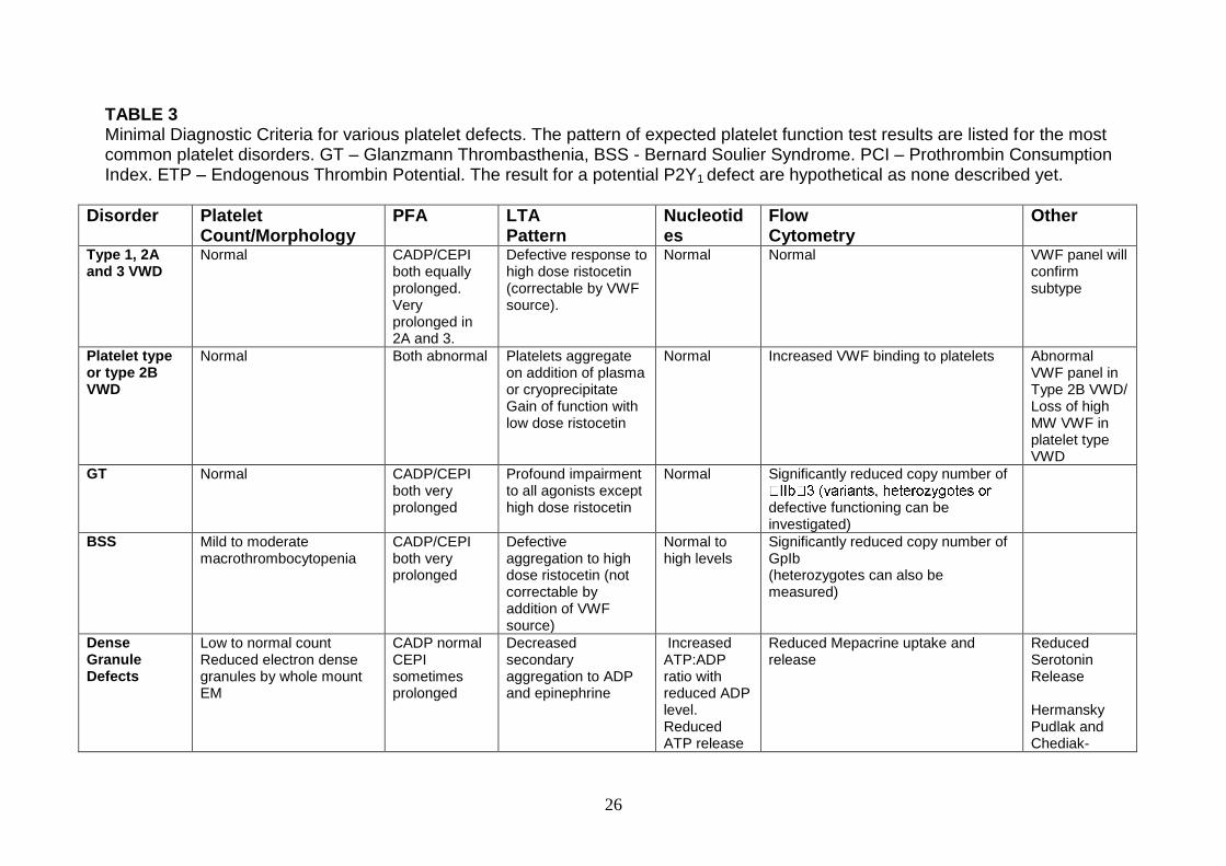
TABLE 3 Minimal Diagnostic Criteria for various platelet defects. The pattern of expected platelet function test results are listed for the most common platelet disorders. GT – Glanzmann Thrombasthenia, BSS - Bernard Soulier Syndrome. PCI – Prothrombin Consumption Index. ETP – Endogenous Thrombin Potential. The result for a potential P2Y1 defect are hypothetical as none described yet.
Disorder Platelet Count/Morphology
PFA LTA Pattern
Nucleotides
Flow Cytometry
Other
Type 1, 2A and 3 VWD
Normal CADP/CEPI both equally prolonged. Very prolonged in 2A and 3.
Defective response to high dose ristocetin (correctable by VWF source).
Normal Normal VWF panel will confirm subtype
Platelet type or type 2B VWD
Normal Both abnormal Platelets aggregate on addition of plasma or cryoprecipitate Gain of function with low dose ristocetin
Normal Increased VWF binding to platelets Abnormal VWF panel in Type 2B VWD/ Loss of high MW VWF in platelet type VWD
GT Normal CADP/CEPI both very prolonged
Profound impairment to all agonists except high dose ristocetin
Normal Significantly reduced copy number of
defective functioning can be investigated)
BSS Mild to moderate macrothrombocytopenia
CADP/CEPI both very prolonged
Defective aggregation to high dose ristocetin (not correctable by addition of VWF source)
Normal to high levels
Significantly reduced copy number of GpIb (heterozygotes can also be measured)
Dense Granule Defects
Low to normal count Reduced electron dense granules by whole mount EM
CADP normal CEPI sometimes prolonged
Decreased secondary aggregation to ADP and epinephrine
Increased ATP:ADP ratio with reduced ADP level. Reduced ATP release
Reduced Mepacrine uptake and release
Reduced Serotonin Release Hermansky Pudlak and Chediak-
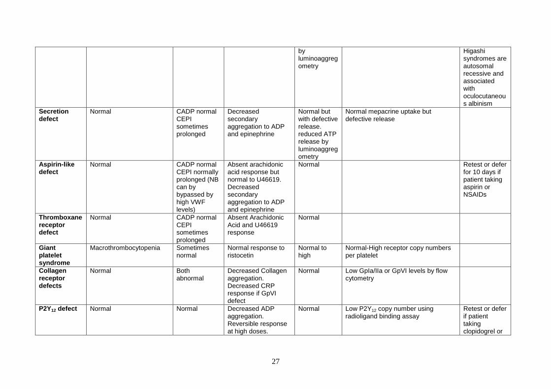
27
by luminoaggregometry
Higashi syndromes are autosomal recessive and associated with oculocutaneous albinism
Secretion defect
Normal CADP normal CEPI sometimes prolonged
Decreased secondary aggregation to ADP and epinephrine
Normal but with defective release. reduced ATP release by luminoaggregometry
Normal mepacrine uptake but defective release
Aspirin-like defect
Normal CADP normal CEPI normally prolonged (NB can by bypassed by high VWF levels)
Absent arachidonic acid response but normal to U46619. Decreased secondary aggregation to ADP and epinephrine
Normal Retest or defer for 10 days if patient taking aspirin or NSAIDs
Thromboxane receptor defect
Normal CADP normal CEPI sometimes prolonged
Absent Arachidonic Acid and U46619 response
Normal
Giant platelet syndrome
Macrothrombocytopenia Sometimes normal
Normal response to ristocetin
Normal to high
Normal-High receptor copy numbers per platelet
Collagen receptor defects
Normal Both abnormal
Decreased Collagen aggregation. Decreased CRP response if GpVI defect
Normal Low GpIa/IIa or GpVI levels by flow cytometry
P2Y12 defect Normal Normal Decreased ADP aggregation. Reversible response at high doses.
Normal Low P2Y12 copy number using radioligand binding assay
Retest or defer if patient taking clopidogrel or

28
Reduced secondary responses
other anti-P2Y12 drugs
P2Y1 defect Unknown Unknown Decreased response to ADP – no shape change and curves not reversible
Normal
Scott syndrome
Normal Normal Normal Normal Reduced expression of phosphatidyl serine on activated platelets by flow cytometry using Annexin-V
Reduced PCI And ETP
29
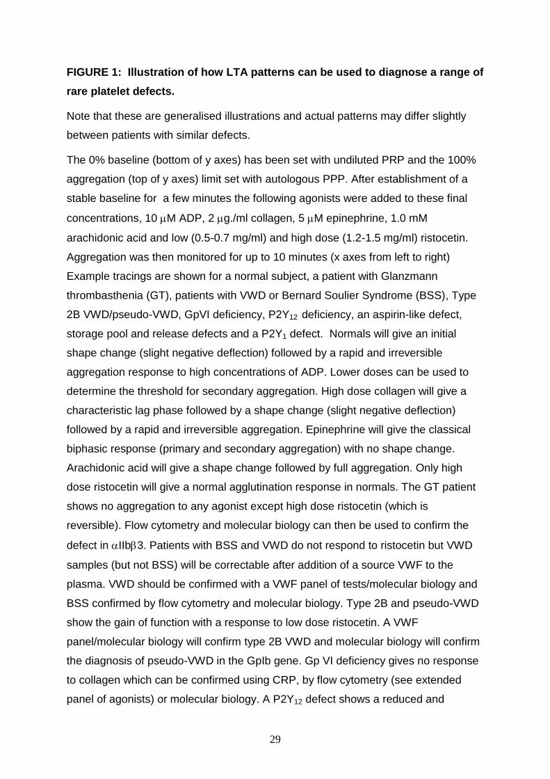
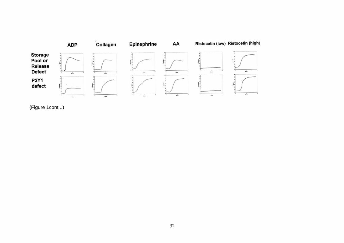
FIGURE 1: Illustration of how LTA patterns can be used to diagnose a range of
rare platelet defects.
Note that these are generalised illustrations and actual patterns may differ slightly
between patients with similar defects.
The 0% baseline (bottom of y axes) has been set with undiluted PRP and the 100%
aggregation (top of y axes) limit set with autologous PPP. After establishment of a
stable baseline for a few minutes the following agonists were added to these final
concentrations, 10 M ADP, 2 g./ml collagen, 5 M epinephrine, 1.0 mM
arachidonic acid and low (0.5-0.7 mg/ml) and high dose (1.2-1.5 mg/ml) ristocetin.
Aggregation was then monitored for up to 10 minutes (x axes from left to right)
Example tracings are shown for a normal subject, a patient with Glanzmann
thrombasthenia (GT), patients with VWD or Bernard Soulier Syndrome (BSS), Type
2B VWD/pseudo-VWD, GpVI deficiency, P2Y12 deficiency, an aspirin-like defect,
storage pool and release defects and a P2Y1 defect. Normals will give an initial
shape change (slight negative deflection) followed by a rapid and irreversible
aggregation response to high concentrations of ADP. Lower doses can be used to
determine the threshold for secondary aggregation. High dose collagen will give a
characteristic lag phase followed by a shape change (slight negative deflection)
followed by a rapid and irreversible aggregation. Epinephrine will give the classical
biphasic response (primary and secondary aggregation) with no shape change.
Arachidonic acid will give a shape change followed by full aggregation. Only high
dose ristocetin will give a normal agglutination response in normals. The GT patient
shows no aggregation to any agonist except high dose ristocetin (which is
reversible). Flow cytometry and molecular biology can then be used to confirm the
defect in IIb3. Patients with BSS and VWD do not respond to ristocetin but VWD
samples (but not BSS) will be correctable after addition of a source VWF to the
plasma. VWD should be confirmed with a VWF panel of tests/molecular biology and
BSS confirmed by flow cytometry and molecular biology. Type 2B and pseudo-VWD
show the gain of function with a response to low dose ristocetin. A VWF
panel/molecular biology will confirm type 2B VWD and molecular biology will confirm
the diagnosis of pseudo-VWD in the GpIb gene. Gp VI deficiency gives no response
to collagen which can be confirmed using CRP, by flow cytometry (see extended
panel of agonists) or molecular biology. A P2Y12 defect shows a reduced and

30
reversible response to ADP. In a homozygous P2Y12 defect, only minimal primary
aggregation to all concentrations of ADP will be present. In a heterozygous P2Y12
defect there will be absence of secondary aggregation but disaggregation tends to
begin < 10 M ADP with a biphasic response to epinephrine. An aspirin-like defect
will show reduced secondary aggregation responses to ADP and epinephrine and
absent arachidonic acid aggregation. A defect in the thromboxane receptor can then
be checked for using U46619 (see extended panel of agonists). Patients with
storage pool disease or release defects will give an identical pattern as the aspirin-
like defect except giving a good primary aggregation response to arachidonic acid.
The P2Y defect pattern is based upon in vitro response to anti-P2Y1 antagonists as
no patients with a P2Y1 defect have ever been described. This figure was
significantly modified with permission from Figure 12.3, page 115 in Chapter 12 –
Diagnostic Assessment of platelet function by P.Nurden and A. Nurden in Quality in
Laboratory Hemostasis and Thrombosis edited by Kitchen S, Olson JD & Preston FE
and published by Wiley-Blackwell in 2009.

31
(Figure 1)
32
(Figure 1cont...)

33
Appendix 1: Grading of recommendations and levels of evidence: The GRADE Nomenclature:
STRENGTH OF RECOMMENDATIONS:
Strong (grade 1): Strong recommendations (grade 1) are made when there is confidence
that the benefits do or do not outweigh harm and burden. Grade 1 recommendations can be
applied uniformly to most patients. Regard as 'recommend'.
Weak (grade 2): Where the magnitude of benefit or not is less certain a weaker grade 2
recommendation is made. Grade 2 recommendations require judicious application to
individual patients. Regard as „suggest‟.
QUALITY OF EVIDENCE
The quality of evidence is graded as high (A), moderate (B) or low (C). To put this in context
it is useful to consider the uncertainty of knowledge and whether further research could
change what we know or our certainty.
(A) High Further research is very unlikely to change confidence in the estimate of effect.
Current evidence derived from randomised clinical trials without important limitations.
(B) Moderate Further research may well have an important impact on confidence in the
estimate of effect and may change the estimate. Current evidence derived from randomised
clinical trials with important limitations (e.g. inconsistent results, imprecision - wide
confidence intervals or methodological flaws - e.g. lack of blinding, large losses to follow up,
failure to adhere to intention to treat analysis),or very strong evidence from observational
studies or case series (e.g. large or very large and consistent estimates of the magnitude of
a treatment effect or demonstration of a dose-response gradient).
(C) Low Further research is likely to have an important impact on confidence in the
estimate of effect and is likely to change the estimate. Current evidence from observational
studies, case series or just opinion.
More information on the GRADE nomenclature can be found at
http://www.gradeworkinggroup.org/index.htm
34
ACKNOWLEDGEMENTS AND DECLARATIONS OF CONFLICTS OF INTEREST
Paul Harrison is a consultant for Sysmex UK and has received a research grant from
Siemens Diagnostics and Eli Lilly. Carol Briggs has received an unrestricted educational
grant from Sysmex Europe and is a consultant to Beckman Coulter. The authors would also
like to thank Dr. Elizabeth Chalmers, Professor Steve Watson, Dr. Ban Dawood, the ISTH
platelet physiology SSC and the CLSI subcommittee on platelet function testing for helpful
discussions and suggestions. Paul Harrison, Ian Mackie, Andrew Mumford, Carol Briggs, Ri
Liesner, Mark Winter & Sam Machin all contributed to the design, writing and editing of this
guideline.
The BCSH Haemostasis and Thrombosis Task Force at the time of publication of this
guideline consists of:
David Keeling (Chair), Henry Watson (Secretary), Mike Laffan, Andrew Mumford, Ian
Jennings, Isobel Walker, Elaine Gray, Campbell Tait, Mike Makris
35
REFERENCES Acharya,S., Barraclough,J., Ibrahim,M.S., Oxby,C., Jones,S.E., Parapia,L., & O'donovan,P. (2008) The usefulness of the platelet function analyser (PFA-100) in screening for underlying bleeding disorders in women with menorrhagia. J Obstet.Gynaecol., 28, 310-314.
Althaus,K. & Greinacher,A. (2009) MYH9-related platelet disorders. Semin.Thromb.Hemost., 35, 189-203.
Bolton-Maggs,P.H., Chalmers,E.A., Collins,P.W., Harrison,P., Kitchen,S., Liesner,R.J., Minford,A., Mumford,A.D., Parapia,L.A., Perry,D.J., Watson,S.P., Wilde,J.T., & Williams,M.D. (2006) A review of inherited platelet disorders with guidelines for their management on behalf of the UKHCDO. Br.J Haematol., 135, 603-633.
Bolton-Maggs,P.H., Perry,D.J., Chalmers,E.A., Parapia,L.A., Wilde,J.T., Williams,M.D., Collins,P.W., Kitchen,S., Dolan,G., & Mumford,A.D. (2004) The rare coagulation disorders--review with guidelines for management from the United Kingdom Haemophilia Centre Doctors' Organisation. Haemophilia., 10, 593-628.
Bonduel, M., Frontroth, J.P., Hepner, M., Sciuccati, G., Feliu-Torres, A. (2007) Platelet aggregation and adenosine triphosphate release values in children and adults. J.Thromb.Haemost 5 1782-1787
Briggs,C., Harrison,P., & Machin,S.J. (2007) Platelet Counting. Platelets (ed. by A. D. Michelson), pp. 475-483. Academic Press, San Diego.
Cardinal,D.C. & Flower,R.J. (1980) The electronic aggregometer: a novel device for assessing platelet behavior in blood. J.Pharmacol.Methods, 3, 135-158.
Cattaneo, M et al (2011) ISTH platelet physiology SSC guidelines on LTA. Manuscript in Preparation
Cattaneo,M. (2009) Light transmission aggregometry and ATP release for the diagnostic assessment of platelet function. Semin.Thromb.Hemost., 35, 158-167.
Cattaneo,M. (2003) Inherited platelet-based bleeding disorders. J.Thromb.Haemost., 1, 1628-1636.
Cattaneo,M. (2004) Are the bleeding time and PFA-100 useful in the initial screening of patients with mucocutaneous bleedings of hereditary nature? J.Thromb.Haemost., 2, 890-891.
Cattaneo,M., Federici,A.B., Lecchi,A., Agati,B., Lombardi,R., Stabile,F., & Bucciarelli,P. (1999) Evaluation of the PFA-100 system in the diagnosis and therapeutic monitoring of patients with von Willebrand disease. Thromb.Haemost., 82, 35-39.
Cattaneo,M., Hayward,C.P., Moffat,K.A., Pugliano,M.T., Liu,Y., & Michelson,A.D. (2009) Results of a worldwide survey on the assessment of platelet function by light transmission aggregometry: a report from the platelet physiology subcommittee of the SSC of the ISTH. J Thromb.Haemost., 7, 1029.

36
Cattaneo,M., Lecchi,A., Zighetti,M.L., & Lussana,F. (2007) Platelet aggregation studies: autologous platelet-poor plasma inhibits platelet aggregation when added to platelet-rich plasma to normalize platelet count. Haematologica, 92, 694-697.
Chanarin, I. (1989) Platelet Function Tests. In : Laboratory Haematology. Churchill Livingstone, Edinburgh, 371-399.
Christie,D.J., Avari,T., Carrington,LR., Cohen,E., DeBiase,B., Harrison,P., Kickler,T., Kottke-Marchant,K., Ledford-Kraemer,M.R., Rand,M.L., Schmaier,A.H., & McCabe White,M. (2008) Platelet Function Testing by Aggregometry: Approved Guideline. Wayne PA: Clinical and Laboratory Standards Institute, 28, 1-45.
Clauser,S. & Cramer-Borde,E. (2009) Role of platelet electron microscopy in the diagnosis of platelet disorders. Semin.Thromb.Hemost., 35, 213-223.
Daly,M.E., Dawood,B.B., Lester,W.A., Peake,I.R., Rodeghiero,F., Goodeve,A.C., Makris,M., Wilde,J.T., Mumford,A.D., Watson,S.P., & Mundell,S.J. (2009) Identification and characterization of a novel P2Y 12 variant in a patient diagnosed with type 1 von Willebrand disease in the European MCMDM-1VWD study. Blood, 113, 4110-4113.
Dawood,B.B., Wilde,J., & Watson,S.P. (2007) Reference curves for aggregation and ATP secretion to aid diagnose of platelet-based bleeding disorders: effect of inhibition of ADP and thromboxane A(2) pathways. Platelets., 18, 329-345.
Duke,W.W. The relation of blood platelets to hemorrhagic disease. Description of a method for determining the bleeding time and the coagulation time and report of three cases of hemorrahagic disease relieved by blood transfusion. JAMA 55, 1185-1192. 1910.
Favaloro,E.J. (2006) The utility of the PFA-100 in the identification of von Willebrand disease: a concise review. Semin.Thromb.Hemost., 32, 537-545.
Favaloro,E.J. (2008) Clinical utility of the PFA-100. Semin.Thromb.Hemost., 34, 709-733.
Favaloro,E.J. (2009) Internal quality control and external quality assurance of platelet function tests. Semin.Thromb.Hemost., 35, 139-149.
Favaloro,E.J., Kershaw,G., Bukuya,M., Hertzberg,M., & Koutts,J. (2001) Laboratory diagnosis of von Willebrand disorder (vWD) and monitoring of DDAVP therapy: efficacy of the PFA-100 and vWF:CBA as combined diagnostic strategies. Haemophilia., 7, 180-189.
Favaloro,E.J., Lippi,G., & Adcock,D.M. (2008) Preanalytical and postanalytical variables: the leading causes of diagnostic error in hemostasis? Semin.Thromb.Hemost., 34, 612-634.
Franchini,M. (2005) The platelet function analyzer (PFA-100): an update on its clinical use. Clin.Lab, 51, 367-372.
Franchini,M., Gandini,G., Manzato,F., & Lippi,G. (2002) Evaluation of the PFA-100 system for monitoring desmopressin therapy in patients with type 1 von Willebrand's disease. Haematologica, 87, 670.
Fritsma,G.A. (2007) Platelet function testing: aggregometry and lumiaggregometry. Clin.Lab Sci., 20, 32-37.

37
George,J.N. & Shattil,S.J. (1991) The clinical importance of acquired abnormalities of platelet function. N.Engl.J Med, 324, 27-39.
Goodall,A.H. & Appleby,J. (2004) Flow-cytometric analysis of platelet-membrane glycoprotein expression and platelet activation. Methods Mol.Biol., 272, 225-253.
Gordon,N., Thom,J., Cole,C., & Baker,R. (1995) Rapid detection of hereditary and acquired platelet storage pool deficiency by flow cytometry. Br.J.Haematol., 89, 117-123.
Greaves, M., & Preston, F.E. (1985) The laboratory investigation of acquired and platelet disorders. In Thomson J.M. (ed) Blood Coagulation and Haemostasis: A Practical Guide. Churchill Livingstone, Edinburgh, 56-134.
Harrison,P. (2005) The role of PFA-100 testing in the investigation and management of haemostatic defects in children and adults. Br.J.Haematol., 130, 3-10.
Harrison,P. (2004) In vitro measurement of high-shear platelet adhesion and aggregation by the PFA-100. Methods Mol.Biol., 272, 215-223.
Harrison,P., Frelinger,A.L., III, Furman,M.I., & Michelson,A.D. (2007) Measuring antiplatelet drug effects in the laboratory. Thromb.Res., 120, 323-336.
Harrison,P., Horton,A., Grant,D., Briggs,C., & Machin,S. (2000) Immunoplatelet counting: a proposed new reference procedure. Br.J Haematol., 108, 228-235.
Harrison,P. & Mumford,A. (2009) Screening tests of platelet function: update on their appropriate uses for diagnostic testing. Semin.Thromb.Hemost., 35, 150-157.
Harrison,P., Robinson,M., Liesner,R., Khair,K., Cohen,H., Mackie,I., & Machin,S. (2002) The PFA-100: a potential rapid screening tool for the assessment of platelet dysfunction. Clin.Lab Haematol., 24, 225-232.
Harrison,P., Robinson,M.S., Mackie,I.J., Joseph,J., McDonald,S.J., Liesner,R., Savidge,G.F., Pasi,J., & Machin,S.J. (1999) Performance of the platelet function analyser PFA-100 in testing abnormalities of primary haemostasis. Blood Coagul.Fibrinolysis, 10, 25-31.
Harrison,P., Segal,H., Furtado,C., Verjee,S., Sukhu,K., & Murphy,M.F. (2004) High incidence of defective high-shear platelet function among platelet donors. Transfusion, 44, 764-770.
Hayward,C.P. (2008) Diagnostic approach to platelet function disorders. Transfus.Apher.Sci., 38, 65-76.
Hayward,C.P. & Eikelboom,J. (2007) Platelet function testing: quality assurance. Semin.Thromb.Hemost., 33, 273-282.
Hayward,C.P. & Favaloro,E.J. (2009) Diagnostic evaluation of platelet disorders: the past, the present, and the future. Semin.Thromb.Hemost., 35, 127-130.

38
Hayward,C.P., Harrison,P., Cattaneo,M., Ortel,T.L., & Rao,A.K. Platelet function analyser (PFA-100) closure time in the evaluation of platelet disorders and platelet function. Journal of Thrombosis and Haemostasis 4, 1-8. 2006.
Hayward,C.P., Moffat,K.A., Pai,M., Liu,Y., Seecharan,J., McKay,H., Webert,K.E., Cook,R.J., & Heddle,N.M. (2008) An evaluation of methods for determining reference intervals for light transmission platelet aggregation tests on samples with normal or reduced platelet counts. Thromb.Haemost., 100, 134-145.
Hayward,C.P., Moffat,K.A., Spitzer,E., Timleck,M., Plumhoff,E., Israels,S.J., & White,J. (2009a) Results of an external proficiency testing exercise on platelet dense-granule deficiency testing by whole mount electron microscopy. Am.J Clin.Pathol., 131, 671-675.
Hayward,C.P., Pai,M., Liu,Y., Moffat,K.A., Seecharan,J., Webert,K.E., Cook,R.J., & Heddle,N.M. (2009b) Diagnostic utility of light transmission platelet aggregometry: results from a prospective study of individuals referred for bleeding disorder assessments. J Thromb.Haemost. 7, 676-684
Hayward CP, Moffat KA, Raby A, Israels S, Plumhoff E, Flynn G & Zehnder (2010) Development of North American Consensus Guidelines for Medical Laboratories that Perform and Interpret Platelet Function Testing Using Light Transmission Aggregometry. Am J Clin Path, 134, 955-963.
Heilmann,E.J., Kundu,S.K., Sio,R., Garcia,C., Gomez,R., & Christie,D.J. (1997) Comparison of four commercial citrate blood collection systems for platelet function analysis by the PFA-100 system. Thromb.Res., 87, 159-164.
Ingerman-Wojenski,C., Smith,J.B., & Silver,M.J. (1983) Evaluation of electrical aggregometry: comparison with optical aggregometry, secretion of ATP, and accumulation of radiolabeled platelets. J Lab Clin.Med, 101, 44-52.
Israels,S.J., McNicol,A., Robertson,C., & Gerrard,J.M. (1990) Platelet storage pool deficiency: diagnosis in patients with prolonged bleeding times and normal platelet aggregation. Br.J.Haematol., 75, 118-121.
Israels, S.J., Kahr, W.H.A, Blanchette, V.S., Luban, N.L.C., Rivard, G.E., & Rand, M.L. (2011) Platelet disorders in children : A diagnostic approach. Pediatr. Blood Cancer, 56, 975-983.
James,A.H., Lukes,A.S., Brancazio,L.R., Thames,E., & Ortel,T.L. (2004) Use of a new platelet function analyzer to detect von Willebrand disease in women with menorrhagia. Am.J.Obstet.Gynecol., 191, 449-455.
Jennings,I., Woods,T.A., Kitchen,S., & Walker,I.D. (2008) Platelet function testing: practice among UK National External Quality Assessment Scheme for Blood Coagulation participants, 2006. J Clin.Pathol., 61, 950-954.
Jilma,B. (2001) Platelet function analyzer (PFA-100): a tool to quantify congenital or acquired platelet dysfunction. J.Lab Clin.Med., 138, 152-163.
Kahr,W.H., Zheng,S., Sheth,P.M., Pai,M., Cowie,A., Bouchard,M., Podor,T.J., Rivard,G.E., & Hayward,C.P. (2001) Platelets from patients with the Quebec platelet disorder contain

39
and secrete abnormal amounts of urokinase-type plasminogen activator. Blood, 98, 257-265.
Karger,R., Donner-Banzhoff,N., Muller,H.H., Kretschmer,V., & Hunink,M. (2007) Diagnostic performance of the platelet function analyzer (PFA-100) for the detection of disorders of primary haemostasis in patients with a bleeding history-a systematic review and meta-analysis. Platelets., 18, 249-260.
Kerenyi,A., Schlammadinger,A., Ajzner,E., Szegedi,I., Kiss,C., Pap,Z., Boda,Z., & Muszbek,L. (1999) Comparison of PFA-100 closure time and template bleeding time of patients with inherited disorders causing defective platelet function. Thromb.Res., 96, 487-492.
Knofler, R., Weissbach, G., Kuhlisch, E. (1998) Platelet function tests in childhood. Measuring aggregation and release reaction in whole blood. Semin Thromb Hemost 24 513-521
Kratzer,M.A. & Born,G.V. (1985) Simulation of primary haemostasis in vitro. Haemostasis, 15, 357-362.
Kundu,S.K., Heilmann,E.J., Sio,R., Garcia,C., Davidson,R.M., & Ostgaard,R.A. (1995) Description of an in vitro platelet function analyzer--PFA-100. Semin.Thromb.Hemost., 21 Suppl 2, 106-112.
Laffan,M., Brown,S.A., Collins,P.W., Cumming,A.M., Hill,F.G., Keeling,D., Peake,I.R., & Pasi,K.J. (2004) The diagnosis of von Willebrand disease: a guideline from the UK Haemophilia Centre Doctors' Organization. Haemophilia., 10, 199-217.
Linnemann,B., Schwonberg,J., Mani,H., Prochnow,S., & Lindhoff-Last,E. (2008) Standardization of light transmittance aggregometry for monitoring antiplatelet therapy: an adjustment for platelet count is not necessary. J Thromb.Haemost.
Lippi,G., Franchini,M., Brocco,G., & Manzato,F. (2001) Influence of the ABO blood type on the platelet function analyzer PFA-100. Thromb.Haemost., 85, 369-370.
Ludlam, C.A., Pasi, K.J., Bolton-Maggs, P., Collins, P.W., Cumming, A.M., Dolan, G., Fryer, A., Harrington, C., Hill, F.G., Peake, I.R., Perry, D.J., Skirton, H., & Smith, M. (2005) UK Haemophilia Centre Doctors' Organisation. A framework for genetic service provision for haemophilia and other inherited bleeding disorders. Haemophilia. 2, 145-63.
Mackie,I.J., Jones,R., & Machin,S.J. (1984) Platelet impedance aggregation in whole blood and its inhibition by antiplatelet drugs. J Clin.Pathol., 37, 874-878.
Mammen,E.F., Comp,P.C., Gosselin,R., Greenberg,C., Hoots,W.K., Kessler,C.M., Larkin,E.C., Liles,D., & Nugent,D.J. (1998) PFA-100 system: a new method for assessment of platelet dysfunction. Semin.Thromb.Hemost., 24, 195-202.
Michelson,A.D., Linden,M., Barnard,M.R., Furman,M.I., & Frelinger,A.L. (2007) Flow Cytometry. Platelets (ed. by A. D. Michelson), pp. 545-563. Academic Press.
Moeller,A., Weippert-Kretschmer,M., Prinz,H., & Kretschmer,V. (2001) Influence of ABO blood groups on primary hemostasis. Transfusion, 41, 56-60.

40
Moffat,K.A., Ledford-Kraemer,M.R., Nichols,W.L., & Hayward,C.P. (2005) Variability in clinical laboratory practice in testing for disorders of platelet function: results of two surveys of the North American Specialized Coagulation Laboratory Association. Thromb.Haemost., 93, 549-553.
Nieuwenhuis,H.K., Akkerman,J.W., & Sixma,J.J. (1987) Patients with a prolonged bleeding time and normal aggregation tests may have storage pool deficiency: studies on one hundred six patients. Blood, 70, 620-623.
Nurden,A.T., Fiore,M., Pillois,X., & Nurden,P. (2009) Genetic testing in the diagnostic evaluation of inherited platelet disorders. Semin.Thromb.Hemost., 35, 204-212.
Pai,M. & Hayward,C.P. (2009) Diagnostic assessment of platelet disorders: what are the challenges to standardization? Semin.Thromb.Hemost., 35, 131-138.
Perry,D.J., Fitzmaurice, D.A., Kitchen, S., Mackie, I.J., & Mallet, S. (2010) Point of care testing in Haemostasis . Br.J.Haematol, 150 (5) 501-14.
Peterson,P., Hayes,T.E., Arkin,C.F., Bovill,E.G., Fairweather,R.B., Rock,W.A., Jr., Triplett,D.A., & Brandt,J.T. (1998) The preoperative bleeding time test lacks clinical benefit: College of American Pathologists' and American Society of Clinical Pathologists' position article. Arch.Surg., 133, 134-139.
Philipp,C.S., Miller,C.H., Faiz,A., Dilley,A., Michaels,L.A., Ayers,C., Bachmann,G., Dowling,N., & Saidi,P. (2005) Screening women with menorrhagia for underlying bleeding disorders: the utility of the platelet function analyser and bleeding time. Haemophilia., 11, 497-503.
Podda,G.M., Bucciarelli,P., Lussana,F., Lecchi,A., & Cattaneo,M. (2007) Usefulness of PFA-100 testing in the diagnostic screening of patients with suspected abnormalities of hemostasis: comparison with the bleeding time. J Thromb.Haemost., 5, 2393-2398.
Posan,E., McBane,R.D., Grill,D.E., Motsko,C.L., & Nichols,W.L. (2003) Comparison of PFA-100 testing and bleeding time for detecting platelet hypofunction and von Willebrand disease in clinical practice. Thromb.Haemost., 90, 483-490.
Quiroga,T., Goycoolea,M., Munoz,B., Morales,M., Aranda,E., Panes,O., Pereira,J., & Mezzano,D. (2004) Template bleeding time and PFA-100 have low sensitivity to screen patients with hereditary mucocutaneous hemorrhages: comparative study in 148 patients. J.Thromb.Haemost., 2, 892-898.
Quiroga,T., Goycoolea,M., Panes,O., Aranda,E., Martinez,C., Belmont,S., Munoz,B., Zuniga,P., Pereira,J., & Mezzano,D. (2007) High prevalence of bleeders of unknown cause among patients with inherited mucocutaneous bleeding. A prospective study of 280 patients and 299 controls. Haematologica, 92, 357-365.
Rodgers,R.P. & Levin,J. (1990) A critical reappraisal of the bleeding time. Semin.Thromb.Hemost., 16, 1-20.
Schmitz,G., Rothe,G., Ruf,A., Barlage,S., Tschope,D., Clemetson,K.J., Goodall,A.H., Michelson,A.D., Nurden,A.T., & Shankey,T.V. (1998) European Working Group on Clinical

41
Cell Analysis: Consensus protocol for the flow cytometric characterisation of platelet function. Thromb.Haemost., 79, 885-896.
Summerfield, G.P., Keenan, J.P., Brodie, N.J., & Bellingham, A.J. (1981) Bioluminescent assay of adenine nucleotides: rapid analysis of ATP and ADP in red cells and platelets using the LKB luminometer. Clin Lab Haem., 3, 257-271
Swart,S.S., Pearson,D., Wood,J.K., & Barnett,D.B. (1984) Effects of adrenaline and alpha adrenergic antagonists on platelet aggregation in whole blood: evaluation of electrical impedance aggregometry. Thromb.Res., 36, 411-418.
Sweeney,J.D., Hoernig,L.A., Michnik,A., & Fitzpatrick,J.E. (1989) Whole blood aggregometry. Influence of sample collection and delay in study performance on test results. Am.J Clin.Pathol., 92, 676-679.
The British Society for Haematology BCSH Haemostasis and Thrombosis Task Force (1988) Guidelines on platelet function testing. J.Clin.Pathol., 41, 1322-1330
Toth, O., Calatzis, A., Losonczy, H., Siess, W. (2006) Multiple electrode aggregometry: a new device to measure platelet aggregation in whole blood. Thromb. Haemost., 96, 781-788
van Vliet,H.H., Kappers-Klunne,M.C., Leebeek,F.W., & Michiels,J.J. (2008) PFA-100 monitoring of von Willebrand factor (VWF) responses to desmopressin (DDAVP) and factor VIII/VWF concentrate substitution in von Willebrand disease type 1 and 2. Thromb.Haemost., 100, 462-468.
Wall,J.E., Buijs-Wilts,M., Arnold,J.T., Wang,W., White,M.M., Jennings,L.K., & Jackson,C.W. (1995) A flow cytometric assay using mepacrine for study of uptake and release of platelet dense granule contents. Br.J.Haematol., 89, 380-385.
Watson S., Daly M., Dawood B., Gissen P., Makris M., Mundell S., Wilde J., & Mumford A. (2010) Phenotypic approaches to gene mapping in platelet function disorrders. Hamostaseologie., 30, 29-38.
Zeidan,A.M., Kouides,P.A., Tara,M.A., & Fricke,W.A. (2007) Platelet function testing: state of the art. Expert.Rev.Cardiovasc.Ther., 5, 955-967.
Zhou,L. & Schmaier,A.H. (2005) Platelet aggregation testing in platelet-rich plasma: description of procedures with the aim to develop standards in the field. Am.J.Clin.Pathol., 123, 172-183.