general approach to the investigation of haemostasis
TRANSCRIPT

General approach to the investigation of haemostasis
Jan‐Gert NelDept. of HaematologyUniversity of Pretoria
2013

Clinical reasons to investigate haemostasis
• Investigating a clinically suspected bleeding tendency
• Following up an abnormal first‐line test.• Investigation of acute haemostatic failure

The components of a general coagulation screen:
•Structured clinical history
• Clinical findings• Laboratory tests
– FBC and peripheral blood smear– Prothrombin time (PT)– Activated Partial Thromboplastin Time (aPTT)

Just a quick revision of the basics

Present thinking on the working of this system:

Haemostasis
• Platelets• Vessels• Coagulation factors• Fibrinolysis


An Approach to the patient presenting with a bleeding diathesis
• Good structured clinical history• Good clinical examination• Formulation of a differential diagnosis, and choosing how you are going to go about confirming/ruling out diagnoses
• Planning laboratory investigations• Management

• In all cases comprehensive clinical evaluation:• patient's history• the family history and family tree• details of:
– the site,– frequency– character (purpura, bruising, large haematomata, haemarthroses,)
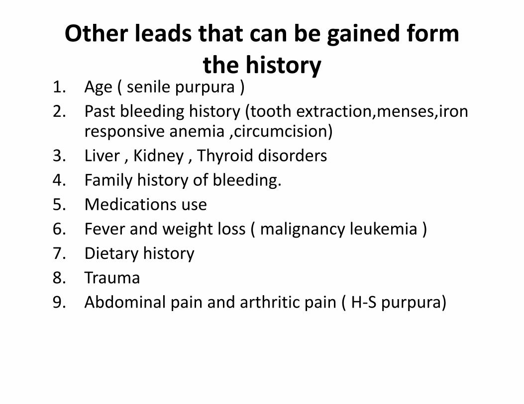
Other leads that can be gained form the history
1. Age ( senile purpura )2. Past bleeding history (tooth extraction,menses,iron
responsive anemia ,circumcision)3. Liver , Kidney , Thyroid disorders4. Family history of bleeding.5. Medications use6. Fever and weight loss ( malignancy leukemia )7. Dietary history 8. Trauma 9. Abdominal pain and arthritic pain ( H‐S purpura)

Vague questions such as “Do you bruise easily?’’ should be avoided.
It is particularly helpful to know whether the patient has had a prior haemostatic challenge

A negative family history of bleeding does not preclude congenital disorders such as Haemophillia

Clinical Examination1. Petechiae ( less than 2
mm)2. Purpura ( more than 2
mm and less than 20 mm)
3. Ecchymoses ( more than 20 mm)
4. Their Distribution. 5. Hepatosplenomegaly6. Lymphadenopathy7. Evidence of underlying
liver disease

Pre‐analytical variables
• Ratio of plasma to anticoagulant– 1:9– Influenced by over/ under filling
• Time from specimen collection until testing
• Therapy

Laboratory haemostatic evaluation
• Full blood count with smear• PT• aPTT• Thrombin time• Bleeding time

The full blood count
• White cell count
• Haemoglobin
• Platelet count

The smear
• ?True thrombocytopenia
• ?Artefactual– Clumping– Satelitism– Clots
• Platelet morphology

Generally Available Screening Assays of coagulation
• PT• aPTT

The PT• Measures EXTRINSIC pathway
• Used to calculate the INR

The aPTT• Measures the INTRINSIC pathway

PT• Extrinsic pathway
– TF, FVII
• Common pathway
aPTT• Intrinsic pathway
– VIII, IX, XI, XII
• Common pathway
II, V, X fibrinogen

Drawbacks of the PT and APTT
• In vitro assays• Normal biological variation
• Insensitivity to clinically important bleeding disorders– Mild but clinical sign Haemophilia A
– vWD– Factor XIII defic
• Artefact due to sample collection or pathological conditions
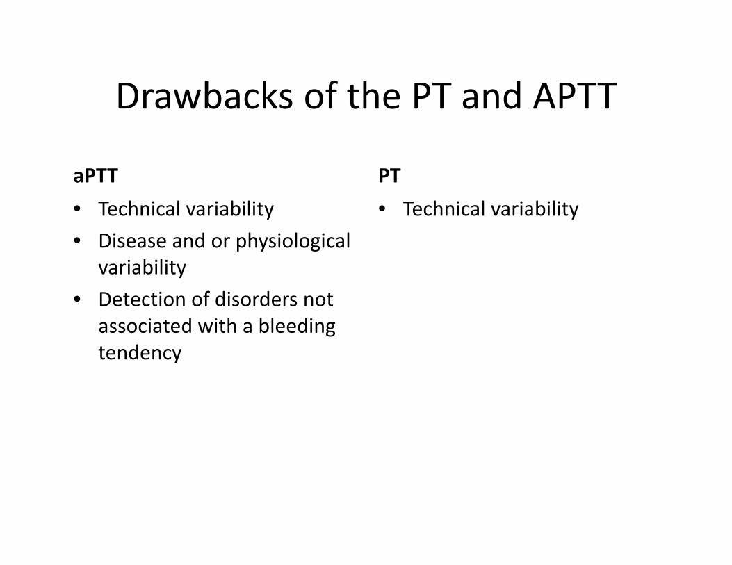
Drawbacks of the PT and APTT
aPTT• Technical variability• Disease and or physiological
variability• Detection of disorders not
associated with a bleeding tendency
PT• Technical variability

Different scenarios

Scenario 1.
• High PT normal aPTT

Scenario 1.
• Step 1– Is this result true?
• Or is it artefactualy prolonged– Tube underfilled– Specimen contaminated with IV fluid– Patient’s haematokrit elevated
• Step 2– Which conditions should I think about?

Scenario 1, Step 3
• What test next:– Mixing studies
• 50/50 mix patient plasma with “normal”• Should correct within a few seconds
– Factor VII assay if MS corrects

Scenario no. 2
• High PTT normal PT

Scenario 2
• Step 1– Is this result true?
• Step 2– Which conditions should I think about?

What test next
• Mixing studies• Factor assays for Factor 8 and 9

Prolonged PT and aPTT in combination

Normal PT and aPTT and bleeding:
• Consider evaluating for:1.Platelet disorder 2.Mild factor deficiency3. Factor XIII4.Monoclonal gammopathy5.Abnormal fibrinolysis6. a2 anti‐plasmin deficiency7.Vascular disorders8.Dysfibrinogenemia

BT
PLATELET VESSEL WALL
A normal BT does not predict the safety of surgical procedures, nor does an abnormal BT
predict for excessive bleeding
