gellan gum microspheres containing a novel α-amylase from marine nocardiopsis sp. strain b2 for...
TRANSCRIPT

B
GN
SQ1
Ca
b
c
a
ARRAA
KN�GII
1
btcsttcAiie
csiov
h0
1
2
3
4
5
6
7
8
9
10
11
12
13
14
15
16
17
18
19
20
21
22
23
24
25
26
27
28
29
30
31
32
33
34
35
36
37
38
39
40
41
42
ARTICLE IN PRESSG ModelIOMAC 4447 1–8
International Journal of Biological Macromolecules xxx (2014) xxx–xxx
Contents lists available at ScienceDirect
International Journal of Biological Macromolecules
j ourna l h o mepa ge: www.elsev ier .com/ locate / i jb iomac
ellan gum microspheres containing a novel �-amylase from marineocardiopsis sp. strain B2 for immobilization
amrat Chakrabortya, Sougata Janaa,∗, Arijit Gandhia, Kalyan Kumar Sena, Wang Zhiangb,handrakant Kokarec
Department of Pharmaceutics, Gupta College of Technological Sciences, Asansol 713301, W.B., IndiaInstitute of Microbiology, Chinese Academy of Sciences, No. 13 Beiyitiao, Zhongguancun, Beijing 100080, PR ChinaDepartment of Pharmaceutical Biotechnology, Bharati Vidyapeeth University, Poona College of Pharmacy, Pune 411 038, India
r t i c l e i n f o
rticle history:eceived 28 May 2014eceived in revised form 13 June 2014ccepted 14 June 2014vailable online xxx
eywords:
a b s t r a c t
A Nocardiopsis sp. stain B2 with an ability to produce stable �-amylase was isolated from marine sedi-ments. The characterization of microorganism was done by biochemical tests and 16S rDNA sequencing.The �-amylase was purified by gel filtration chromatography by using sephadex G-75. The molec-ular mass of the amylase was found to be 45 kDa by SDS-PAGE and gel filtration chromatography.The isolated �-amylase was immobilized by ionotropic gelation technique using gellan gum (GG).These microspheres were spherical with average particle size of 375.62 ± 21.76 to 492.54 ± 32.18 �m.
ocardiopsis sp.-Amylaseellan gum
mmobilizationn vitro release
The entrapment efficiency of these �-amylase loaded GG microspheres was found 74.76 ± 1.32 to87.64 ± 1.52%. Characterization of �-amylase–gellan gum microspheres was confirmed using FTIR andSEM analysis. The in vitro amylase release kinetic have been studied by various mathematical modelsthat follow the Korsmeyer–Peppas model (R2 = 0.9804–0.9831) with anomalous (non-Fickian) diffusionrelease mechanism.
43
44
45
46
47
48
49
50
51
52
53
54
55
56
57
58
59
. Introduction
Enzymes are exploited as biocatalysts in many industrial,iomedical, and analytical processes. Compared to organic syn-hesis, biocatalysts often have far better chemical precision, whichan lead to more efficient production of single stereoisomer, feweride reactions and a lower environmental burden. Up to now, morehan 3000 different enzymes have been identified and many ofhem have been applied in biotechnology and industry. But theurrent enzyme toolbox is still not sufficient to meet all demands.
major reason is that many available enzymes do not withstandndustrial reaction conditions. Therefore, researchers are now try-ng to exploit extremophiles which are the valuable source of novelnzymes [1–3].
�-amylases (EC 3.2.1.1, 1,4-�-d-glucan glucanohydrolase) werelassified in family 13 of glycosyl hydrolases and hydrolyzetarch, glycogen and related polysaccharides by randomly cleav-
Please cite this article in press as: S. Chakraborty, et al., Int. J. Biol. Ma
ng internal �-1,4-glucosidic linkages to produce different sizesf oligosaccharides. They have diverse applications in a wideariety of industries such as food, fermentation, textile, paper,
∗ Corresponding author. Tel.: +91 9434896683.E-mail address: [email protected] (S. Jana).
ttp://dx.doi.org/10.1016/j.ijbiomac.2014.06.046141-8130/© 2014 Published by Elsevier B.V.
60
61
62
© 2014 Published by Elsevier B.V.
detergent, pharmaceutical and sugar industries [4,5]. Over the pastfew decades, considerable researches have been undertaken withthe extracellular �-amylase being produced by a wide variety ofmicroorganisms. The major advantages of using microorganismsfor the production of amylases are the economical bulk produc-tion capacity of microbes and their easier manipulation to obtainenzymes with desired characteristics [3]. The major concern in anenzymatic process is the instability of the enzyme under repeti-tive or prolonged use and inhibition by high substrate and productconcentration.
In the present study, we have isolated the characterization ofthermostable, oxidant, detergent and surfactant stable �-amylasesecreted by a haloalkaliphilic Nocardiopsis sp. strain B2 that wasisolated from west coast of India.
There has been considerable interest in the developmentof carrier systems for enzyme immobilization because immo-bilized enzymes have enhanced stability compared to solubleenzymes, and can easily be separated from the reaction. The useof immobilized cells offers several advantages over free cells,such as relative ease of product separation, re-use of biocata-
cromol. (2014), http://dx.doi.org/10.1016/j.ijbiomac.2014.06.046
lysts, prevention of washout, reduced risk of Contamination andoperational stability. Furthermore, using the entrapment tech-nique, a dense cell culture can be established leading to improvedproductivity [6,7].
63
64
65
66

ING ModelB
2 of Biol
eAiowtcgoivtfhmi
2
2
afctk(bwwc
2
baaifeaedswNClo1I(
2
(tmt45
67
68
69
70
71
72
73
74
75
76
77
78
79
80
81
82
83
84
85
86
87
88
89
90
91
92
93
94
95
96
97
98
99
100
101
102
103
104
105
106
107
108
109
110
111
112
113
114
115
116
117
118
119
120
121
122
123
124
125
126
127
128
129
130
131
132
133
134
135
136
137
138
139
140
141
142
143
144
145
146
147
148
149
150
151
152
153
154
155
156
157
158
159
160
161
162
163
164
165
166
167
168
169
170
171
172
173
174
175
176
177
178
179
180
ARTICLEIOMAC 4447 1–8
S. Chakraborty et al. / International Journal
In this research work we employed a ion tropic technique forntrapment of isolated �-amylase into gellan gum microspheres.mong naturally occurring biodegradable polymers, gellan gum
s widely used polymer candidates for the designing and devel-pment of various drug delivery systems. Gellan gum (GG) is aater-soluble linear anionic polysaccharide obtained as a fermen-
ation product by a pure culture of Pseudomonas elodea. Gellan gumonsists of linear structure of repeating saccharide units of glucose,lucuronic acid and rhamnose in a molar ratio of 2:2:1 [8]. By virtuef its gelating potential, gellan gum can be extensively introducedn the formulation of sustained release matrix. In presence of multi-alent cations, such as Ca2+, Zn2+, Pb2+, Cd2+, Al3+, etc, it exhibits ionropic gelation. In pharmaceutical and biomedical research, the gelorming properties of gellan gum in presence of multivalent cationsave been exploited for sustained release of drug molecules [9]. Theechanical stability of the ion tropically cross-linked gel systems
s provided by multivalent cations.
. Materials and methods
.1. Materials
Starch, peptone, malt extract, glycerol, yeast extract, glucose,sparagine, maltose, casein, dintrosalicylic acid were purchasedrom Hi-media laboratory, India. Amylopectin, amylose, �/�yclodextrin, maltooligosaccharide, pullulan glycogen and mal-otriose were procured from Sigma, USA. Protein electrophoresisit and molecular mass markers comprising of Phosphorylase b97 kDa), bovine serum albumin (66 kDa), ovalbumin (43 kDa), car-onic anhydrase (29 kDa) and soyabean trypsin inhibitor (20.1 kDa)ere purchased from Genei private limited, India. Gellan gum(GG)as purchased from Sisco Research Laboratories Pvt. Ltd., India. All
hemicals, and reagents used were of analytical grade.
.2. Strain isolation and identification
Sediment samples were collected from Goa, Alibagh and Mum-ai coastal region of India at the time of low tide. Heat pretreatmentt 40 ◦C for 30 to 60 days was employed for isolation of marinectinomycetes [10,11]. Marine soil samples were suspended in ster-le sea water and thoroughly mixed on rotary shaker at 150 rpmor 20 min. Marine actinomycetes were isolated by using differ-nt selective media such as glycerol yeast extract agar, glucosespargine agar, starch casein agar, maltose yeast extract and yeastxtract malt extract agar. They were screened for amylase pro-uction by using starch agar medium consisted of following (g/l):tarch 20 g; beef extract 5 g, peptone 3 g prepared in artificial seaater (ASW). The artificial sea water contains (g/l): NaCl 23.37 g;a2SO4 3.91 g; NaHCO3 0.19 g; KCl 0.66 g; KBr 0.09 g; MgCl2 4.98 g;aCl2 1.10 g, SrCl20.02 g, and H3B03 0.02 g. The maximum amy-
ase producing marine actinomycetes species B2 was maintainedn maltose yeast extract agar medium containing (g/l): maltose0 g; yeast extract 4 g; and K2HPO4 0.5 g prepared in ASW at 4 ◦C.dentification of strain was done by scanning electron microscopySEM), 16S r-DNA sequencing, and biochemical tests.
.3. Inoculum preparation and culture condition
The maltose yeast extract broth prepared in artificial sea waterASW) was used for development of inoculum. The seed cul-ure was prepared in 100 ml of conical flasks containing 50 ml of
Please cite this article in press as: S. Chakraborty, et al., Int. J. Biol. Ma
edium by inoculating 2.0 ml of spore suspension containing 3.5o 4.0 × 10⁶ CFU/ml and cultivated under agitation at 200 rpm at5 ◦C for 4 days. Then 50 ml of seed culture was inoculated in the00 ml of fermentation medium containing (w/v): 2% starch; 0.4%
PRESSogical Macromolecules xxx (2014) xxx–xxx
peptone; 0.2% of malt extract; 11% of NaCl and 10 mM Ca2+ pre-pared in artificial sea water. The pH of the medium was adjusted to9 and fermentation was carried for 12 days at the same conditions aspre-culture medium. Cell-free supernatant containing extracellu-lar amylase was harvested by centrifugation at 5000 × g for 45 minand used for amylase purification and characterization.
2.4. Purification of amylase
The enzyme was precipitated by bringing the culture filtrateto 90% saturation with solid ammonium sulfate and kept at 4 ◦Cfor overnight. The precipitate was centrifuged at 12,000 × g for30 min. The precipitate was dissolved in glycine–NaOH buffer (pH9) and dialyzed for 48 h against the same buffer. The dialyzedsample was assayed for �-amylase activity and protein content.Enzyme obtained (2 ml) from the above step was loaded ontoa Sephadex G-75 column (1.2 × 135 cm), pre-equilibrated with50 mM glycine–NaOH buffer (pH 9) at flow rate of 10 ml/h. Frac-tions (3.5 ml) were collected and those having specific activitiesmore than 200 U/mg in the void volume were pooled for next step ofpurification. The enzyme obtained from Sephadex G-75 was loadedonto an equilibrated DEAE-Cellulose column (2 × 10 cm). The frac-tions with specific activities more than 500 U/mg were pooled andloaded onto insoluble corn starch column [12].
The enzyme obtained from the above step was loaded onto aninsoluble corn starch column (2.5 × 10 cm), pre-equilibrated with50 mM glycine–NaOH buffer (pH 9). The column was washed withcold water and then the bound enzyme was eluted by incubationin 50 mM of glycine–NaOH Buffer of pH 9 at 45 ◦C. Fractions withhigh amylase activities (above 1200 U/mg) were pooled and dia-lyzed against 50 mM of glycine–NaOH buffer (pH 9). Enzyme (2 ml)from starch column was loaded onto a Sephacryl S-400 column(1.2 × 130 cm). The fractions (3.5 ml) were collected and checkedfor the enzyme activities and protein concentrations [13].
2.5. Assay of amylase
The activity of �-amylase was estimated by determining theamount of reducing sugar released from starch. 1 ml of enzymesolution was added to 1 ml of starch solution (1% w/v). The mixturewas incubated at 45 ◦C for 60 min. The reaction was stopped bythe addition 2 ml of 3,5-dinitrosalicylic acid and A540nm was mea-sured in Jasco V-530 spectrophotometer. One unit (U) of enzymeactivity is defined as the amount of enzyme required for the libera-tion of 1 �mol of reducing sugar as glucose per minute under assaycondition [14].
2.6. Effect of NaCl, temperature and pH
Enzyme was pre-incubated in presence of NaCl of different con-centrations (2–20% w/v) in glycine–NaOH buffer (pH 9) containing10 mM of Ca2+ at 45 ◦C to the determine the influence of NaCl onamylase activity. Enzyme activity was expressed as percentage rel-ative activity. The highest enzyme activity was considered as 100%and with respect to that percentage relative activities were calcu-lated.
To determine optimum temperature for enzyme activity, theenzyme was pre-incubated at 4, 28, 37, 45, 55, 65, 75 and 85 ◦C,respectively, for 60 min in glycine–NaOH buffer containing 11%(w/v) of NaCl and 10 mM of Ca2+. Amylase activity was expressedas percentage relative activity as compared to control which wasconsidered as 100% of amylase activity. Enzyme activity after pre-
cromol. (2014), http://dx.doi.org/10.1016/j.ijbiomac.2014.06.046
incubation at 45 ◦C for 60 min at pH 9 in presence of 11% of NaCland 10 mM Ca2+ was considered as control.
Amylase was pre-incubated in various buffers (0.1 M) with dif-ferent pH values containing 11% (w/v) of NaCl and 10 mM of Ca2+
181
182
183
184
ING ModelB
of Biol
auaaw
2
apattTa
2
u[Pbiaibw(ow
2m
dsqmsosuoat
ae
2
efltsaf
E
185
186
187
188
189
190
191
192
193
194
195
196
197
198
199
200
201
202
203
204
205
206
207
208
209
210
211
212
213
214
215
216
217
218
219
220
221
222
223
224
225
226
227
228
229
230
231
232
233
234
235
236
237
238
239
240
241
242
243
244
245
246
247
248
249
250
251
252
253
254
255
256
257
258
259
260
261
262
263
264
265
266
267
268
269
270
271
272
273
274
275
276
277
278
279
280
281
282
283
284
285
286
287
288
289
290
ARTICLEIOMAC 4447 1–8
S. Chakraborty et al. / International Journal
t 45 ◦C to investigate the effect of pH on enzyme activity. Bufferssed were citrate buffer (pH 4, 6), phosphate buffer (pH 6, 6.5, 7nd 7.5), Tris buffer (pH 8 and 8.4), glycine–NaOH buffer (pH 9, 10nd 11) and disodium hydrogen phosphate (pH12). Enzyme activityas determined by performing the assay as described above.
.7. Substrate specificity
A substrate solution (0.9 ml) containing 1.0% (w/v) each ofmylopectin, amylose, �/� cyclodextrin, maltooligosaccharide,ullulan and glycogen in 50 mM glycine–NaOH buffer (pH 9) wasdded in reaction mixture consisted of 0.1 ml of the enzyme solu-ion, 11% (w/v) of NaCl and 10 mM Ca2+ at 45 ◦C for 30 min. Finally,he enzyme assay was performed under the standard conditions.he value for hydrolyzing starch 1.0% (w/v) was considered as 100%ctivity.
.8. Electrophoretic methods
SDS-PAGE was used to determine protein purity and the molec-lar mass of the purified enzyme using a 10% acrylamide gel15]. After electrophoresis, gel was stained with Coomassie R-250.hosphorylase b (97 kDa), bovine serum albumin (66 kDa), oval-umin (43 kDa), carbonic anhydrase (29 kDa) and soyabean trypsin
nhibitor (20.1 kDa) were used as molecular mass markers. Forctivity staining, gel was suspended in 20% (v/v) isopropanol andncubated for 30 min. It is then transferred to 0.1 M glycine–NaOHuffer (pH 9) and again incubated for 30 min. Amylolytic activityas determined by placing the gel onto agarose gel containing 1%
w/v) starch and incubating at 45 ◦C for 3 h. The transparent bandn the amylase containing agarose gel was observed after floodingith I2–KI solution.
.9. Method of preparation of ˛-amylase-loaded gellan gumicrospheres
Aqueous gellan gum solution was prepared in deionized double-istilled water by using magnetic stirrer. The pH value of theolution was adjusted to 7.0 ± 0.1 using 0.1 (N) NaOH. The requireduantity of enzyme (100 mg �-amylase) was dissolved in aboveixture. Polymer-enzyme mixture was introduced into a glass
yringe with an 18 G hypodermic needle. The droplets were shearedff at a flow rate of 5 ml/min into 50 ml of calcium chloride (3%w/v)olution. The resulting beads were allowed to harden for 30 minnder gentle stirring (100 rpm) with a small magnetic bar, decantedn a Buchner funnel, rinsed with de-ionized double-distilled water,nd dried to a constant weight in a vacuum desiccators at roomemperature over 48 h.
Different gellan gum microsphere formulations along withmounts of gellan gum, �-amylase and calcium chloride arenlisted in Table 1.
.10. Determination of entrapment efficiency
The entrapment efficiency was determined by dissolving thenzyme-loaded beads in a magnetically stirred simulated gastricuid (SGF) without enzyme for about 90 min. The resulting solu-ion was centrifuged at 2500 rpm for 10 min and aliquots from theupernatant were adjusted to pH 6.8 using 0.01 M NaOH and dilutedppropriately with 0.2 M phosphate buffer of pH 6.8 and assayed
Please cite this article in press as: S. Chakraborty, et al., Int. J. Biol. Ma
or enzyme content. The entrapment efficiency was calculated as
ntrapment efficiency (%) =(
actual drug content in microspheretheoretical drug content in microsphere
)× 100.
PRESSogical Macromolecules xxx (2014) xxx–xxx 3
2.11. Particle size measurements
The size of the prepared microsphere was measured by usingdigital slide callipers (CD-6 CS, Mitutoyo Corporation, Japan). Onehundred particles were taken and inserted in between the space oftwo metallic plates. Diameters of resultant particles were displayedin the digital screen of the previously calibrated equipment.
2.12. Surface morphology analysis
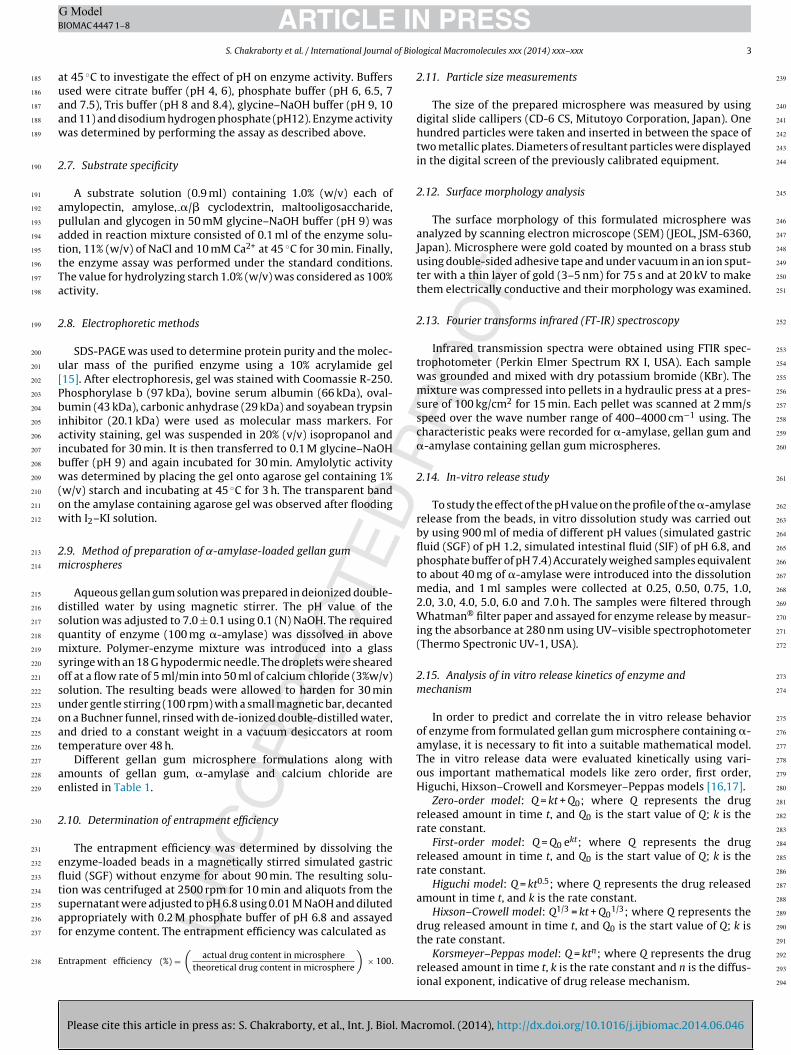
The surface morphology of this formulated microsphere wasanalyzed by scanning electron microscope (SEM) (JEOL, JSM-6360,Japan). Microsphere were gold coated by mounted on a brass stubusing double-sided adhesive tape and under vacuum in an ion sput-ter with a thin layer of gold (3–5 nm) for 75 s and at 20 kV to makethem electrically conductive and their morphology was examined.
2.13. Fourier transforms infrared (FT-IR) spectroscopy
Infrared transmission spectra were obtained using FTIR spec-trophotometer (Perkin Elmer Spectrum RX I, USA). Each samplewas grounded and mixed with dry potassium bromide (KBr). Themixture was compressed into pellets in a hydraulic press at a pres-sure of 100 kg/cm2 for 15 min. Each pellet was scanned at 2 mm/sspeed over the wave number range of 400–4000 cm−1 using. Thecharacteristic peaks were recorded for �-amylase, gellan gum and�-amylase containing gellan gum microspheres.
2.14. In-vitro release study
To study the effect of the pH value on the profile of the �-amylaserelease from the beads, in vitro dissolution study was carried outby using 900 ml of media of different pH values (simulated gastricfluid (SGF) of pH 1.2, simulated intestinal fluid (SIF) of pH 6.8, andphosphate buffer of pH 7.4) Accurately weighed samples equivalentto about 40 mg of �-amylase were introduced into the dissolutionmedia, and 1 ml samples were collected at 0.25, 0.50, 0.75, 1.0,2.0, 3.0, 4.0, 5.0, 6.0 and 7.0 h. The samples were filtered throughWhatman® filter paper and assayed for enzyme release by measur-ing the absorbance at 280 nm using UV–visible spectrophotometer(Thermo Spectronic UV-1, USA).
2.15. Analysis of in vitro release kinetics of enzyme andmechanism
In order to predict and correlate the in vitro release behaviorof enzyme from formulated gellan gum microsphere containing �-amylase, it is necessary to fit into a suitable mathematical model.The in vitro release data were evaluated kinetically using vari-ous important mathematical models like zero order, first order,Higuchi, Hixson–Crowell and Korsmeyer–Peppas models [16,17].
Zero-order model: Q = kt + Q0; where Q represents the drugreleased amount in time t, and Q0 is the start value of Q; k is therate constant.
First-order model: Q = Q0 ekt; where Q represents the drugreleased amount in time t, and Q0 is the start value of Q; k is therate constant.
Higuchi model: Q = kt0.5; where Q represents the drug releasedamount in time t, and k is the rate constant.
Hixson–Crowell model: Q1/3 = kt + Q01/3; where Q represents the
drug released amount in time t, and Q0 is the start value of Q; k is
cromol. (2014), http://dx.doi.org/10.1016/j.ijbiomac.2014.06.046
the rate constant.Korsmeyer–Peppas model: Q = ktn; where Q represents the drug
released amount in time t, k is the rate constant and n is the diffus-ional exponent, indicative of drug release mechanism.
291
292
293
294

ING ModelB
4 of Biol
it(t[
2
(C
3
3
yawmgrgacsacr
3
dpsscamr
3
ct
75 ◦C and 85 ◦C, respectively (Fig. 3b). This clearly indicates ther-mostable nature of the enzyme. The optimum temperature foractivity for halophilic �-amylases from Micrococcus sp., Haloarculahispanica and H. Haloarcula sp. strain S-1 was found to be 50 ◦C [25].
295
296
297
298
299
300
301
302
303
304
305
306
307
308
309
310
311
312
313
314
315
316
317
318
319
320
321
322
323
324
325
326
327
328
329
330
331
332
333
334
335
336
337
338
339
340
341
342
343
344
345
346
347
348
349
350
351
352
353
354
355
356
357
358
359
360
361
362
363
364
365
366
367
368
ARTICLEIOMAC 4447 1–8
S. Chakraborty et al. / International Journal
Again, the Korsmeyer–Peppas model was employed in then vitro drug release behavior analysis of these formulations to dis-inguish between competing release mechanisms: Fickian releasediffusion-controlled release), non-Fickian release (anomalousransport), and case-II transport (relaxation-controlled release)18,19].
.16. Statistical analysis
All measured data are expressed as mean ± standard deviationS.D.). The simple statistical analyses were conducted using Med-alc software version 11.6.1.0.
. Results and discussion
.1. Identification of strain B2
The strain shows good growth at 45 ◦C in 7 days on maltoseeast extract medium. Aerial and substrate mycelium was observednd both the mycelia fragmented into short rods. Spore chainas found to be short and spiral; and it was observed on aerialycelium. Meso-diaminopimelic acid (meso-DAP), arabinose and
alactose were found to be major constituents of cell wall. 16SDNA sequencing showed that strain B2 was closely related to theenus Nocardiopsis with highest level of similarity to Nocardiopsisntarctica DSM 43884T(X97885). Different strains of actinomycetesapable of producing bioactive secondary metabolites from marineediments collected from west coast of India by pre-heat treatmentt 40 ◦C. Among them, isolate B2 was chosen for further studies,onsidering its salient features which are conducive for furtheresearch.
.2. Growth kinetics and amylase production
Actinomycetes species are slow growing in nature. Enzyme pro-uction started in early log phase but there was drastic increase inroduction of enzyme at late logarithmic growth phase and earlytationary phase; and amylase production continued up to latetationary phase, after that it declined (Fig. 1). Thus, it can be con-luded that the kinetics of enzyme production is more of the growthssociated rather than non-growth associated. Effective inductionay not occur until the stationary phase has been reached and the
eadily available carbon source was depleted.
.3. Purification of amylase
Please cite this article in press as: S. Chakraborty, et al., Int. J. Biol. Ma
The enzyme was precipitated by ammonium sulphate foroncentration and followed by gel filtration (Sephadex G-75)o remove low molecular weight proteins and desalting. Ion
0
1000
2000
3000
4000
5000
6000
7000
1 2 3 4 5 6 7 8 9 10 11 13 14 15 16 17 18
Time ( in d ays)
Enzy
me
prod
uc�o
n (U
/l)
0
0.5
1
1.5
2
2.5
3
3.5
Op�
cal d
ensit
y at
600
nm
Enzyme produc�on (U/l)
Op�cal density at 600nm
Fig. 1. Amylase production (�) and growth characteristics (�) of strain B2.
PRESSogical Macromolecules xxx (2014) xxx–xxx
exchange chromatography and starch column were used to removecontaminated proteins and induction of specific activity up to1457.14 U/mg. Finally, the enzyme was subjected to SephacrylS-400 column to obtain a homogeneous amylase with specific activ-ity of 1754.9 U/mg. It corresponds to 43.92-fold purification with20.27% of yield. The purified enzyme showed a single band about45 kDa (Fig. 2) on SDS-PAGE and activity staining of gel confirmedhigh enzyme purity. Together results suggest that amylase is com-posed of single polypeptide chain. The mass of �-amylases fromvarious microbial sources vary from 22.5 to 184 kDa but the mostof them are in the range of 50–60 kDa [20,21].
3.4. Effect of NaCl and temperature on amylase activity
Maximum enzyme activity was found in presence of 11% (w/v)of NaCl. Amylase was found to be stable over wide range of NaClconcentrations as it retained almost 60% and 50% of its activity inpresence of 17% (w/v) and 6% (w/v) of NaCl, respectively (Fig. 3a).Enzyme completely lost its activity in absence of NaCl. This behav-ior is close to that reported for �- amylases from other halophilessuch as Natronococcus sp. strain Ah-36 [22]. Haloarchaeon Haloar-cula sp. strain S-1 [23] and archaeon Haloferax mediterranei [24].Amylases with this characteristics may have wide spread applica-tions in treatment of saline waters or waste solutions with starchresidues and high salt content. There was no growth and amylaseproduction in absence of NaCl (data not shown), suggesting thatstrain B2 is an obligate halophile. Since salt dilution is a commonstress factor for microorganisms living in environments where thesalt concentration constantly changes, this behavior may allow themicroorganisms to adapt to their natural habitat, mainly salterns.
Maximum enzyme activity was obtained at 45 ◦C and enzymeretained 75% and 69% of its activity after 1 h of pre-incubation at
cromol. (2014), http://dx.doi.org/10.1016/j.ijbiomac.2014.06.046
Fig. 2. SDS-PAGE and zymogram activity staining analysis of amylase enzyme. Lane1, molecular mass standard, lane 2, partially purified amylase (ammonium sulfatefractionation, lane 3, pure amylase enzyme, lane 4, activity staining of enzyme.

ARTICLE IN PRESSG ModelBIOMAC 4447 1–8
S. Chakraborty et al. / International Journal of Biological Macromolecules xxx (2014) xxx–xxx 5
amyla
Tmfp�PcsflsblharEsob
3
asanltfii
369
370
371
372
373
374
375
376
377
378
379
380
381
382
383
384
385
386
387
388
389
390
391
392
393
394
395
396
397
398
399
400
401
402
403
404
405
406
407
408
409
410
411
412
413
414
415
416
417
418
419
420
421
422
Fig. 3. Effect of (a) NaCl, (b) temperature, (c) pH on
his reveals that the purified amylase has a slightly lower opti-um temperature than that from other halophiles. �-Amylases
rom Nocardiopsis sp. was found to have wide variations in tem-erature optima and thermostability [26]. Reported cold-adaptive-amylase from psychrophilic Nocardiopsis 7326 isolated fromrydz Bay, Antartic. In contrast, Stamford et al. [27] isolated andharacterized thermo stable �-amylase from endophyte Nocardiop-is isolated from yam bean. Since, thermostability is an importanteature for use of amylolytic enzymes in starch processing, amy-ases from thermophilic and hyperthermophilic bacteria are ofpecial interest as a source of novel thermostable enzymes. Till date,acteria belong to genus Bacillus such as Bacillus substilis var. amy-
oliquefaciens. Bacillus stearothermophilus and Bacillus licheniformisave been exploited for commercial production of thermostablemylase enzyme. To best our knowledge, this is probably the firsteport on thermostable �-amylase from marine Nocardiopsis sp.nzyme may have wide spread applications in various industrialectors such as baking and food processing for the manufacturingf low caloric drinks where temperatures of pasteurization coulde used to inactivate the enzymes after fermentation.
.5. Effect of pH on enzyme
Haloalkaliphilic microorganisms not only require high salt butlso alkaline pH for growth and enzyme production. Good growth oftrain was observed between pH ranges of 7–11. However, growtht pH 9 and 10 was quite comparable. This indicates alkaliphilicature of isolate B2. The strain B2 showed gradual increase in amy-
ase production and optimum pH for enzyme production was found
Please cite this article in press as: S. Chakraborty, et al., Int. J. Biol. Ma
o be 9 (data not shown). Enzyme was found to be stable in dif-erent buffer systems as it retained almost 75% (244.54 U/ml) ofts activity in phosphate buffer (pH 7) and 64% (209.80 U/ml) ofts activity in disodium hydrogen phosphate buffer (pH 12) but
se activity and (d) substrate specificity of amylase.
maximum enzyme activity was obtained in glycine–NaOH bufferof pH 9 (Fig. 3c). As a comparison, the optimal pH of a thermostable�-amylase from Nocardiopsis sp. endophyte was at pH 5.0 [27]and that of cold- adaptive �-amylase from Nocardiopsis sp. 7326[26] was at around 8. Research is focused on the isolation of alka-line enzymes from microbes because alkaline enzymes have hugepotential in detergent industry.
3.6. Substrate specificity
Among the tested substrates, purified amylase showed asignificant activity toward amylose (134% relative to that for sol-uble starch). The activity toward amylopectin, maltodextrins andglycogen was 73%, 85% and 14%, respectively (Fig. 3d), relative tothat of soluble starch. No hydrolytic activity toward �/� cyclodex-trin and pullulan was observed. Therefore, it can be concluded that�-amylase from strain B2 has a high affinity for hydrolysis of amy-lose than the other linear �-glycosides.
3.7. Entrapment efficiency of ˛-amylase in gellan gummicrosphere
Entrapment efficiency was expressed as the percentage of �-amylase entrapped in these prepared gellan gum microspherescompared to the initial amount of �-amylase included in the for-mulation. The amylase entrapment efficiency of these particles wasachieved 74.76 ± 1.32 to 87.64 ± 1.52% (Table 1). Highest amylase
cromol. (2014), http://dx.doi.org/10.1016/j.ijbiomac.2014.06.046
entrapment efficiency was found in case of formulation F-4, pre-pared using 2% w/v gellan gum, 3% w/v Calcium chloride and 100 mg�-amylase. Increasing amylase entrapment was observed with thehigher amount of gellan gum.
423
424
425
426

ARTICLE IN PRESSG ModelBIOMAC 4447 1–8
6 S. Chakraborty et al. / International Journal of Biological Macromolecules xxx (2014) xxx–xxx
spher
3
m3oSf
3
gIalc
imkFa
427
428
429
430
431
432
433
434
435
436
437
438
439
440
441
442
443
444
445
446
447
448
449
450
451
452
453
454
455
456
457
458
459
460
461
462
463
464
465
Fig. 4. SEM photograph of (a) blank gellan gum micro
.8. Particle size and surface morphology
The average particle size of �-amylase loaded gellan gumicrosphere were measured and found within the range of
75.62 ± 21.76 to 492.54 ± 32.18 �m. The morphological analysisf �-amylase loaded gellan gum microspheres were visualized byEM (Fig. 4). They were found almost spherical in shape and freerom agglomeration.
.9. FT-IR analysis
FT-IR spectra of �-amylase, gellan gum and �-amylase loadedellan gum micrsphere are shown in Fig. 5. The rationale behindR interpretation was to confirm no such interaction between thective compound that is �-amylase and its principal aid that is gel-an gum. From the spectral information, it has been found that theharacteristic of any enzyme is its amide linkage. The frequency forCOO as amide functionalize lie within the default range, which
s 1656 cm−1. In the final formulation, the characteristic peak as
Please cite this article in press as: S. Chakraborty, et al., Int. J. Biol. Ma
ention earlier occurred with the same frequency. As it has beennown that a formulation cannot be established simply with theT-IR data, therefore it was a preliminary evaluation through FT-IRbout the probability of the formation of formulation.
Fig. 5. FTIR spectra of (a) �-amylase, (b) gellan gum an
e and (b) �-amylase loaded gellan gum microsphere.
3.10. In-vitro release of ˛-amylase from microsphere
In-vitro amylase release from these prepared microspheres wasevaluated in SGF of pH 1.2, phosphate buffer of pH 6.8 and 7.4.The cumulative percentage amylase release from �-amylase loadedgellan gum microsphere was found sustained over a period of 7 h(Fig. 6). The percentage released from �-amylase loaded gellangum microsphere in 7 h was within the range of 46.52 ± 1.92 (F-4) to 50.25 ± 2.02 (F-1)% at SGF of pH 1.2. The percentage amylasereleased from �-amylase-loaded gellan gum microsphere in 7 hwas within the range of 74.44 ± 2.12 (F-4) to 81.36 ± 1.86 (F-1) %in phosphate buffer of pH 6.8. The percentage amylase releasedfrom �-amylase-loaded gellan gum microsphere in 7 h was withinthe range of 92.67 ± 1.94 (F-4) to 89.78 ± 1.88 (F-1) % in phosphatebuffer of pH 7.4. Decreasing cumulative percent amylase releasefrom these newly prepared �-amylase-loaded gellan gum micro-sphere was found with the increasing amount of gellan gum.
The in-vitro amylase release data from various �-amylase-loaded gellan gum microsphere were evaluated kinetically usingvarious important mathematical models like zero order, first order,
cromol. (2014), http://dx.doi.org/10.1016/j.ijbiomac.2014.06.046
Higuchi, Hixson–Crowell and Korsmeyer–Peppas models. The R2
values of these models were determined for evaluation of accuracy.The result of the curve fitting into various mathematical models isgiven in Table 2.
d (c) �-amylase loaded gellan gum microsphere.
466
467
468
469

ARTICLE IN PRESSG ModelBIOMAC 4447 1–8
S. Chakraborty et al. / International Journal of Biological Macromolecules xxx (2014) xxx–xxx 7
Fig. 6. The in vitro release of �-amylase from various gellan gum microsphere at (a) SGF of pH 1.2, (b) phosphate buffer of pH 6.8 and (c) phosphate buffer of pH 7.4.
Table 1Formulation chart of gellun gum microparticles containing �-amylase with entrapment efficiency and particle size.
Formulation code Gellun gum (%w/v) �-amylase (mg) Calcium chloride (%w/v) Entrapment efficiency (%)a Particle size (�m)b
F-1 0.5 100 3 74.76 ± 1.32 375.62 ± 21.76F-2 1 100 3 80.45 ± 1.46 492.54 ± 32.18F-3 1.5 100 3 84.12 ± 1.64 442.36 ± 27.54F-4 2 100 3 87.64 ± 1.52 481.24 ± 28.72
a Mean ± S.D.; n = 3.b Mean ± S.D.; n = 100.
Table 2Results of curve fitting of the in vitro release profile of gellan gum microparticles containing �-amylase.
Formulation code Zero order model (R2) 1st order model (R2) Higuchi model (R2) Hixson–Crowell model (R2) Korsmeyer–Peppas model
R2 n
F-1 0.8413 0.9234 0.9662 0.8366 0.9828 0.680.91440.86220.9226
gK7�mFdc
4
g
470
471
472
473
474
475
476
477
478
479
480
481
482
483
484
485
486
487
488
489
490
491
F-2 0.8642 0.8852
F-3 0.9268 0.9648
F-4 0.8604 0.9412
When the respective R2 of these �-amylase-loaded gellanum microsphere were compared, it was found to follow theorsmeyer–Peppas model (R2 = 0.9804–0.9831) over a period of
h. The value of release exponent (n) determined from in vitro-amylase release data of various �-amylase-loaded gellan gumicrosphere ranged from 0.52 to 0.72, indicating anomalous (non-
ickian) diffusion mechanism for drug release. The anomalousiffusion mechanism of drug release demonstrates both diffusionontrolled, and swelling controlled drug release.
Please cite this article in press as: S. Chakraborty, et al., Int. J. Biol. Ma
. Conclusion
The adaptation and survival abilities of halophilic microor-anisms in a wide range of salinities (0.5–25%) offer potential
0.9774 0.9804 0.52 0.9563 0.9816 0.62 0.9458 0.9831 0.72
applications in various fields of biotechnology. The strain showsgood growth at 45 ◦C in 7 days on maltose yeast extract medium.Meso-diaminopimelic acid (meso-DAP), arabinose and galactosewere found to be major constituents of cell wall. 16S rDNAsequencing showed that strain B2 was closely related to the genusNocardiopsis with highest level of similarity to N. antarctica DSM43884T(X97885). In our laboratory, we have isolated forty actino-mycetes strains from marine sediments collected from west coastof India by pre- heat treatment at 40 ◦C. Isolated B2 was selectedfor further studies because it was appeared to be the one of the best
cromol. (2014), http://dx.doi.org/10.1016/j.ijbiomac.2014.06.046
producers of extracellular amylase in both liquid and solid media.Amylase exhibited specific activity of 1754.9 U/mg and molecu-lar mass of enzyme was found to be 45 kDa. Gellan gum beadsexhibited a promising sustained release of entrapped �-amylase
492
493
494
495

ING ModelB
8 of Biol
aspmwNfim�bemsswucwdwe
R
[[
[[
[[[
[
[
[
[[
[
[
[
496
497
498
499
500
501
502
503
504
505
506
507
508
509
510
511
512
513
514
515
516
517
518
519
520
521
522
523
524
525
526
527
528
529
530
531
532
533
534
535
536
537
538
539
540
541
542
543
544
545
546
547
548
549
550
551
552
ARTICLEIOMAC 4447 1–8
S. Chakraborty et al. / International Journal
nd can find a place in the design of multiparticulate deliveryystems. These microspheres were almost spherical with averagearticle size of 375.62 ± 21.76 to 492.54 ± 32.18 �m. The entrap-ent efficiency of these �-amylase loaded gellan gum microsphereas found within the range of 74.76 ± 1.32 to 87.64 ± 1.52%. So interaction of �-amylase in the gellan gum matrix was con-rmed using FTIR analyses. The in vitro dissolution of gellan gumicrosphere containing �-amylase showed sustained release of-amylase over 7 h. The kinetics of in-vitro release profiles haseen studied by computing R2 values of various important math-matical models that suggested to follow the Korsmeyer–Peppasodel (R2 = 0.9804–0.9831) with anomalous (non-Fickian) diffu-
ion drug release mechanism. The results of the present studyeem to be of value for pharmaceutical industries associatedith digestive enzyme formulations, and there is a probability ofsing it for a number of pharmaceutical and biomedical appli-ations. From this work we can conclude that stable �-amylaseas successfully isolated from a haloalkaliphilic marine Nocar-
iopsis sp. stain B2 and entrapped into gellan gum microsphere,hich may be a promising carrier for immobilization of �-amylase
nzyme.
eferences
Please cite this article in press as: S. Chakraborty, et al., Int. J. Biol. Ma
[1] M.N. Gupta, I. Roy, Indian J. Biochem. Biophys. 39 (2002) 220–228.[2] R.A. Herbert, Trends Biotechnol. 7 (1992) 395–401.[3] R. Patel, M. Dodia, S.P. Singh, Process Biochem. 40 (2005) 3569–3575.[4] R. Gupta, P. Gigras, H. Mohapatra, V.K. Goswami, B. Chauhan Microbial, Process
Biochem. 38 (2003) 1599–1616.
[[[
PRESSogical Macromolecules xxx (2014) xxx–xxx
[5] S. Sivaramakrishnan, D. Gangadharan, K.M. Nampoothiri, C.R. Soccol, A. Pandey,Food Technol. Biotechnol. 44 (2006) 173–184.
[6] D.S. Clark, Trends Biotechnol. 12 (1994) 439–443.[7] J.F. Liang, Y.T. Li, V.C. Yang, J. Pharm. Sci. 89 (2000) 979–990.[8] W.H. Chengyi, M. Ming, R. Jiang, Acta Pharmacol. Sin. 32 (1999) 400–404.[9] S. Jana, A. Das, A.K. Nayak, K.K. Sen, S.K. Basu, Int. J. Biol. Macromol. 57 (2013)
129–137.10] S.T. Williams, F.L. Davies, J. Gen. Microbiol. 38 (1965) 251–261.11] C.R. Kokare, K.R. Mahadik, S.S. Kadam, B.A. Chopade, Curr. Sci. 58 (2004)
283–289.12] M.F. Najaw, D. Deobagkar, Protein Expression Purif. 41 (2005) 349–354.13] O.H. Lowry, N.J. Rosenbrough, A.L. Farr, D. Randall, J. Biol. Chem. 48 (1951)
17–25.14] P. Bernfeld, Enzymology 1 (1955) 149–158.15] S.E. D’Souza, W. Altekar, S.F. D’Souza, Arch. Microbiol. 168 (1997) 68–71.16] S. Jana, S. Manna, A.K. Nayak, K.K. Sen, S.K. Basu, Colloids Surf., B: Biointerfaces
114 (2014) 36–44.17] A.K. Nayak, S. Khatua, M.S. Hasnain, K.K. Sen, DARU J. Pharm. Sci. 19 (2011)
356–366.18] S. Jana, A. Saha, A.K. Nayak, K.K. Sen, S.K. Basu, Colloids Surf., B: Biointerfaces
105 (2013) 303–309.19] S. Jana, N. Maji, A.K. Nayak, K.K. Sen, S.K. Basu, Carbohydr. Polym. 98 (2013)
870–876.20] A. Asoodeh, J.K. Chamani, M. Lagzian, Int. J. Biol. Macromol. 46 (2010) 289–297.21] N. Mollania, K. Khajeh, S. Hosseinkhani, B. Dabirmanesh, Int. J. Biol. Macromol.
46 (2010) 27–36.22] T. Kobayashi, H. Kanai, T. Hayashi, T. Akiba, R. Akaboshi, K. Horikoshi, J. Bacteriol.
174 (1992) 3439–3444.23] T. Fukushima, T. Mizuki, A. Echigo, A. Inoue, R. Usami, Extremophiles 9 (2005)
85–90.24] F. Perez-Pomares, V. Bautista, J. Ferrer, C. Pire, F.C. Marhuenda-Egea, M.J. Bonete,
cromol. (2014), http://dx.doi.org/10.1016/j.ijbiomac.2014.06.046
Extremophiles 7 (2003) 299–306.25] M. Shafie, A.A. Ziaee, M.A. Amoozegar, Process Biochem. 45 (2010) 694–699.26] J.W. Zhang, Y.R. Zeng, Mar. Biotechnol. 10 (2007) 75–82.27] T.L.M. Stamford, N.P. Stamford, L.C.B.B. Coelho, J.M. Araujo, Bioresour. Technol.
76 (2001) 137–141.
553
554
555
556
557