preparation and evaluation of mucoadhesive gellan gum …
TRANSCRIPT

Elazreg et al., IJPSR, 2015; Vol. 6(9): 3761-3774. E-ISSN: 0975-8232; P-ISSN: 2320-5148
International Journal of Pharmaceutical Sciences and Research 3761
IJPSR (2015), Vol. 6, Issue 9 (Research Article)
Received on 06 March, 2015; received in revised form, 10 April, 2015; accepted, 24 June, 2015; published 01 September, 2015
PREPARATION AND EVALUATION OF MUCOADHESIVE GELLAN GUM IN-SITU GELS
FOR THE OCULAR DELIVERY OF CARBONIC ANHYDRASE INHIBITOR NANOVESICLES
Rania Elazreg 1, 2
, Mahmoud Soliman *1
, Samar Mansour
1 and
Abdelhameed El Shamy
1
Department of Pharmaceutics and Industrial Pharmacy 1
, Faculty of Pharmacy, Ain Shams University,
Cairo, Egypt
Department of Pharmaceutics 2, Faculty of Pharmacy, Zawia University, Zawia, Lybia
ABSTRACT: A novel approach was tested utilizing mucoadhesive in-situ
gelling (ISG) Gellan Gum /HPMC solutions containing dispersions of
methazolamide (MZA) loaded Spanlastic Vesicular systems (SVs). The
tested systems were designed to combine high corneal permeability of SVs
as well as ease of application and prolonged eye retention of ISGs. SVs
consisted of Span 60 mixed with different ratios of edge activators (EA)
(Tween 60, Tween 80, Brij 35 and Brij 58) were initially prepared and
examined to select formulae that had small vesicle size and high drug
entrapment. The evaluation of SVs systems included measurement of particle
sizes, entrapment efficiencies (EE %) and relative deformability. SVs in ISG
systems were characterized by in vitro release, viscosity, intraocular pressure
(IOP) measurement after their administration to rabbit eyes and
histopathological examination. It was found that SVs containing 10% Tween
60 (90:10) in ISG solutions (SG1) produced the highest reduction in IOP
with the highest prolongation of MZA effect
INTRODUCTION: Eye contains several
protection mechanisms which are responsible for
removing foreign objects including drugs from its
surface. Eye protection mechanisms represent the
main obstacle which decreases the amount of
absorbed drug to eye tissues and necessitate the
frequent administration of drug solution to the eye
and eventually decrease patient compliance. Eye
protection mechanisms includes rapid tear turnover
with eye blinking, limited corneal permeability and
the passage of drug to GIT via nasolacrimal duct
reducing the ocular bioavailability of drugs to be
not more than 1-5% 1.
QUICK RESPONSE CODE
DOI: 10.13040/IJPSR.0975-8232.6(9).3761-74
Article can be accessed online on: www.ijpsr.com
DOI link: http://dx.doi.org/10.13040/IJPSR.0975-8232.6(9).3761-74
In order to enhance drug ocular bioavailability,
several approaches have been utilized to either
enhance drug penetration by using penetration
enhancers like bile salts, and surfactants or prolong
the contact of drugs with eye tissues using inserts
or collagen shields. However, the use of
penetration enhancers was found to cause corneal
damage and ocular inserts were disadvantageous
being easily lost during use which eventually lead
to patient incompliance 2, 3
.
An approach which involve preparing dispersions
of drug loaded SVs as a nanovesicular systems 4-6
that consist of a mixture of Spans together with
different edge activators (e.g. Tween 20 and Tween
80) was tested aiming to deliver drugs to the
posterior segments of the eye. SVs as a
nanovesicular systems represent a good drug carrier
that can penetrate the compact corneal barrier.
Being elastic, it can easily squeeze itself between
Keywords:
Gellan gum,
spanlastic, methazolamide,
mucoadhesive, in situ gel
Correspondence to Author:
Mahmoud Soliman
Assistant Professor, Department of
Pharmaceutics and Industrial Pharmacy,
Faculty of Pharmacy, Ain Shams
University, African Union Organization
Street, Abbassia, 11566, Cairo, Egypt.
E-mail: [email protected]

Elazreg et al., IJPSR, 2015; Vol. 6(9): 3761-3774. E-ISSN: 0975-8232; P-ISSN: 2320-5148
International Journal of Pharmaceutical Sciences and Research 3762
corneal cells and penetrate tissues more efficiently,
hence increase drug bioavailability in the eye
tissues 7.
Gellan gum is a high molecular weight linear
anionic extracellular microbial
heteropolysaccharide that is secreted by
pseudomonas elodea. The anionic nature of the
polymer is due to the presence of free carboxylate
groups which emerge by the deacetylation of
polymer backbone. Thus, in presence of mono- and
divalent cations, gellan gum undergoes sol-to-gel
phase transition 8, 9
and hence it was frequently
used in formulating ISG systems that increase the
corneal residence time of some drugs e.g. timolol 10
.
In order to increase the adherence of ocular drug
delivery systems with the eye, mucoadhesive
agents (e.g. HPMC) should be incorporated 11
in
ISG formulations. These agents would increase the
contact time of formulations with eye tissues and
consequently drug bioavailability 10
.
Methazolamide (MZA) is a slightly soluble weakly
acidic sulfonamide derivative. It is indicated for the
treatment of glaucoma by inhibiting the action of
carbonic anhydrase enzyme. However, it was found
to have some serious side effects when taken orally
such as allergic reactions (e.g. difficulty of
breathing, swollen lips, tongue or face), bleeding,
and tremors in the hands or feet, hence, the topical
administration of MZA is more preferred in order
to reduce such side effects.
However, the poor aqueous solubility of MZA
(~1.7mg/ml) 12
, as well as its low corneal
permeability 13
render its topical delivery
inefficient. Different technique have been utilized
to enhance the efficiency of MZA topical delivery
e.g. MZA cyclodextrin eye drops 14
, MZA calcium
phosphate nanoparticles 15
, MZA cationic
nanostructured heterolipid matrices 16
, MZA solid
lipid nanoparticles 17
.
Moreover, MZA ISGs have been formulated using
Poloxamer 407/188 ISG solutions and this
approach had enhanced drug residence time and
increased drug release interval compared to control
solution 15
.
The aim of work in this paper is to prepare and
characterize MZA loaded SVs which consist of a
mixture of Span 60 together with different edge
activators (Brij 35, Brij 58 Tween 60 and Tween
80) in mucoadhesive ISG Gellan Gum /HPMC
solutions to combine the high penetration power of
Spanlastic SVs, ease of application of ISG
solutions and the prolonged residence time of
mucoadhesive gels produced inside the eye.
MATERIAL AND METHODS:
Materials:
Methazolamide (MZA) was purchased from
Jiaxing Taixing chemical and Pharma Co. Ltd
(Jiaxing, China). Brij35, Brij58, gellan gum,
hydroxypropyl methyl cellulose (HPMC), Span60,
Tween60, and Tween80 were purchased from
Sigma Aldrich chemical Co. (St. Louis, MO, USA).
Calcium chloride dihydrate, disodium hydrogen
phosphate, absolute ethanol, potassium dihydrogen
phosphate, sodium bicarbonate, and sodium
chloride were purchased from ADWIC, El-Nasr
pharmaceutical CO. (Cairo, Egypt). Benoxinate
hydrochloride eye drops 0.4 % (w/v) was
purchased from Egyptian International
Pharmaceutical Industries (EIPICO, Cairo, Egypt).
Preparation of MZA-loaded SVs:
Methazolamide loaded Spanlastics vesicular
systems (SVs) composed of Span 60 and edge
activators (EAs) (namely Brij 35, Brij 58, Tween
60 and Tween 80) were prepared using the ethanol
injection technique 18
using different weight ratios
of Span 60: EA (90:10; 80:20 and 70:30 w:w).
Briefly, Span 60 and calculated amount of MZA
were dissolved in 4ml of ethanol using different
concentrations of drug and then injected into
magnetically stirred aqueous solution of the EA.
Ethanol was evaporated by rotary evaporator, and
the formed milky vesicular dispersions were used
for further investigations.
Experimental design: A full factorial experimental design was built up to
evaluate the main effects and interactions of two
variables: EA type (factor A) and Span 60: EA
ratio (factor B). The studied responses were:
particle size (PS) and entrapment efficiency (EE
%). The complete setup of the full factorial design
and the composition of the prepared formulae

Elazreg et al., IJPSR, 2015; Vol. 6(9): 3761-3774. E-ISSN: 0975-8232; P-ISSN: 2320-5148
International Journal of Pharmaceutical Sciences and Research 3763
according to the factorial design are shown in Tables 1 and 2 respectively.
TABLE 1: FACTORIAL DESIGN INDEPENDENT VARIABLES AND THEIR LEVELS.
Variables Level
EA type Tween 60
Tween 80
Brij 35
Brij 58
Span 60: EA ratio (w:w) 90:10
80:20
70:30
TABLE 2: EFFECT OF FORMULA COMPOSITION OF MZA-LOADED SVS (Span 60: EA RATIO) ON DRUG EE% AND
VESICLE PARTICLE SIZE.
Characterization of MZA-SVs:
Dynamic light scattering technique (DLS) was used
to determine the particle size (PS), polydispersity
index (PI), and zeta potential (ζ) of the freshly
prepared SVs dispersions utilizing a Zetasizer®
Nano-ZS (Malvern instruments, Malvern, UK).
MZA EE% was determined by measuring the
concentration of free drug in the vesicular
dispersions. The non-encapsulated MZA was
separated by centrifugation of SVs dispersions
using Nanosep® at 5000 rpm for 15 minutes using
cooling centrifuge adjusted to a temperature of 4°C
(Herml Z216MK, Gosheim, Germany). The
amount of free drug in the supernatant was
determined by UV spectrophotometry (UV-1601
PC, Shimadzu, Kyoto, Japan). Drug EE% was
calculated according to the following equation:
100)(
%
MZAtotal
MZAfreeMZAtotalEE (1)
The measurement of elasticity of SVs dispersions
was carried out by extrusion technique 19, 20
through
a locally fabricated stainless steel pressure filter
holder. The vesicles were extruded through
cellulose acetate/surfactant-free membrane filters
with pore size of 220nm (Minisart, Sartorius,
Göttingen, Germany) at constant pressure of 2.5
bar and relative deformability as an indicator for
elasticity was calculated according to the following
equation 21
:
(2)
Where D is the deformability index (ml/s),
j is the amount of dispersion extruded (ml),
t is the extrusion time (s),
rv is the vesicle size after extrusion (nm),
rp is the pore size of the extrusion membrane (nm).
Preparation of mucoadhesive in-situ gel (ISG):
Selected MZA- SVs formulae were mixed with 0.6
% w/v gellan gum 22
, forming in-situ gelling
formulations containing spanlastic vesicles (SGs).
Mucoadhesive SGs formulations were prepared by
adding HPMC to the prepared in-situ gelling
formulations at different concentrations: 0.5, 1 and
1.5 w/v % 23
till homogenous mixtures were
formed. All of the above steps were done under
Formulae
Code
Formulae
Composition
Weight
Ratio (w:w)
EE % ± SD Mean
Particle size
(nm) ± SD
S1 Span 60 : Tween 60 90:10 82.80 ± 1.65 276.26 ± 2.77
S2 Span 60: Tween 60 80:20 81.99 ± 2.18 585.10 ± 8.64
S3 Span 60 : Tween 60 70:30 68.36 ± 1.24 704.93± 4.46
S4 Span 60 : Tween 80 90:10 81.74 ± 3.43 328.56 ± 3.19
S5 Span 60 : Tween 80 80:20 80.89 ± 2.20 657.53 ± 6.15
S6 Span 60 : Tween 80 70:30 78.69 ± 2.32 761.06± 6.40
S7 Span 60 : Brij 35 90:10 94.19 ± 3.13 315.53 ± 1.56
S8 Span 60 : Brij 35 80:20 93.69 ± 2.15 279.13 ± 2.45
S9 Span 60 : Brij 35 70:30 91.94 ± 1.16 995.66 ± 3.05
S10 Span 60 : Brij 58 90:10 68.76 ± 1.26 530.46 ± 7.59
S11 Span 60 : Brij 58 80:20 27.76 ± 2.04 631.63 ± 3.09
S12 Span 60 : Brij 58 70:30 23.29 ± 1.95 633.46 ± 8.30
Elazreg et al., IJPSR, 2015; Vol. 6(9): 3761-3774. E-ISSN: 0975-8232; P-ISSN: 2320-5148
International Journal of Pharmaceutical Sciences and Research 3764
aseptic conditions. All glassware were sterilized by
autoclaving, and the entire procedure was carried
out in a laminar flow hood 24
.
Sterilization of SGs using gamma radiation was
tested at an exposure dose of 5, 15 and 25 KGy and
the radiation dose 5 KGy was found to be the best
to produce stable and sterile formulae (data not
shown).
Characterization of SGs:
Gelation time:
Gelation time was carried out by the tube inversion
method 25
for selected SGs using microcentrifuge
tube containing a solution which when titled sol
phase will flow, however if a gel phase is formed it
will not flow.
100 µl of the tested formulae were carefully placed
into microcentrifuge tubes (1.6 ml), and 300 µl of
artificial tear fluid (ATF) were added slowly on the
side wall of each microcentrifuge tube. The tubes
were then incubated in a temperature-controlled
bath at 37°C. The sol-gel transition time was
determined by inverting the tubes horizontally
every minute. The time at which the gel did not
flow in minutes was examined by visual
examination and recorded as the gelation time 26
.
Viscosity measurement: The viscosities of the prepared SGs containing 0.6
w/v % gellan gum were determined using cone and
plate programmable viscometer (Brookfield
Engineering Laboratories Inc., Model HADV-II,
USA), connected to a digital thermostatically
controlled circulating water bath (Polyscience,
Model 9101, USA) and experiment was conducted
at 37±0.5°C.
ATF was used for gel formation during this study.
It was added to SGs 10 min before the viscosity
values were re-measured to mimic the
physiological condition and record rheological
changes that may occur after ocular administration
of in-situ gelling systems. The same setting was
used for measuring the viscosities of selected
mucoadhesive SGs containing 0.6 w/v % gellan
gum and different concentrations of HPMC (0.5, 1
and 1.5 w/v %) before and after addition of ATF.
In vitro release of MZA from SGs:
In vitro release studies were performed by dialysis
membrane diffusion technique using modified USP
dissolution apparatus I (Pharma Test, Hainburg,
Germany) replacing device baskets with glass
cylinders (10cm in length and 2.5 cm in diameter).
The cylinders were fixed in the device shafts from
one end using basket clips while the other end was
covered by dialysis membrane (MWCO 12,000-
14,000) to retain tested SGs allowing the free drug
to be released into the dissolution medium pre-
equilibrated at 37±0.5°C in the dissolution tester
vessels 27
; 28
.
Accurately measured amounts of SGs solutions
equivalent to 1mg MZA were transferred to the
glass cylinders that was immersed to 1mm distance
below the surface of 50 ml of ATF pH 7.4. The
dissolution media was kept at 37± 0.5°C while the
cylinders were rotated at 50 rpm. At specified time
intervals (0, 0.25, 0.5, 0.75, 1, 1.5, 2, 4, 6 and 8
hrs), 1ml samples were removed, replaced with
fresh media and analyzed spectrophotometrically to
determine the concentration of MZA.
Scanning Electron Microscopy (SEM):
Selected mucoadhesive SGs (SG1) was prepared
and mixed with ATF then freeze dried for 48hr
using Christ Alpha 1-2LD plus freeze drier (Martin
Christ GmbH, Germany). Dried gel was coated
with gold sputter coater and the morphology of the
gel with its vesicle content morphology was
imaged using scanning electron microscope (JEOL-
JSM-5500LV, Japan)..
In vivo pharmacodynamic study:
Animal handling: For all animal studies, the experimental procedures
conformed to the Ethical Committee of Faculty of
Pharmacy, Ain-Shams University on the use of
animals. Adult albino normotensive rabbits were
kept in individual cages and fed a normal diet and
water ad libitum in a constant temperature
environment of 25ºC and a period of 7 days was
allowed for acclimatization of rabbits.
IOP lowering effect of selected formulae:
The efficacy of the selected formulae SG1, SG4
and SG8 in lowering the intraocular pressure (IOP)
was evaluated on normotensive albino rabbits. The

Elazreg et al., IJPSR, 2015; Vol. 6(9): 3761-3774. E-ISSN: 0975-8232; P-ISSN: 2320-5148
International Journal of Pharmaceutical Sciences and Research 3765
results of these formulations were compared to that
of MZA dispersion 24, 27
. Concentration of the drug
in all formulae was adjusted to be equivalent to
0.05 w /v % MZA. Twelve adult albino
normotensive rabbits weighing between 2.5 and 3
Kg, were randomly divided into four groups, each
consisting of three rabbits according to the
following scheme: Group I received SG1, Group II
received SG4, Group III received SG8 and Group
IV received control MZA dispersion. A single 50μl
dose of each preparation was administered in the
lower conjunctival sac on the corneal surface of the
left eye of the rabbit, and the right eye was used as
a control during this study.
After instilling one drop of 0.4 (w/v %) benoxinate
hydrochloride to the rabbit’s eyes as local
anesthetic, IOP was measured using tonometer
(Riester, Germany) 16, 30
at time intervals of 0, 1,
2, 3, 4, 5, 6, 7, 8, 10 and 12 hrs. To decrease the
diurnal, seasonal, and individual variations usually
observed in rabbits, the ocular hypotensive activity
was expressed as the average difference in IOP
between the treated and control eye of the same
rabbit 31
. IOP values recorded in this study were
calculated according to the following equation 32
:
(3)
The Institutional Animal Ethical Committee
reviewed the animal protocol prior to the
experiment. All rats were treated in accordance
with the guideline for the care and use of laboratory
animals and with the permission of Faculty of
Pharmacy- Ain Shams University Animal Ethical
Committee.
Assessment of ocular irritancy of mucoadhesive
SGs: Six rabbits weighing 2.5 -3 Kg were divided into
two groups. Group I received mucoadhesive SG1
and group II received mucoadhesive SG8 twice
daily in the left eye only for a period of 10 days.
The right eye was kept as a control in all the
experimental rabbits. All the rabbits were killed
after 10 days and their eyes were separated, fixed,
cut vertically, dehydrated, cleared, and embedded
in paraffin at 56oC in hot air oven for 24hr. Eyes
were then sectioned and stained by hematoxylin
and eosin. Corneal histological examination was
completed after photographing the stained sections
using light microscope 33
.
Statistical analysis:
The results are expressed as mean of 3 ± standard
deviation (SD). The complete setup of the full
factorial design statistical and factorial analysis
were performed using MINITAB (version 15.1.3)
software. Comparison of the mean values was
performed using either Student′s t test or ANOVA
using Graph Pad Instat software setting statistical
significance at p-value ≤ 0.05.
RESULTS AND DISCUSSION: MZA loaded SVs (MZA-SVs) were successfully
prepared using ethanol injection technique 18
.
Preliminary studies (data not shown) were carried
out to determine the effect of initial MZA amount
on particle size (PS) and entrapment efficiency (EE
%). Different initial MZA amounts were
investigated for testing drug incorporation into SVs
formulations, and we found that SVs prepared
using10 mg found to be the most appropriate, MZA
to be the most appropriate for further studies, i.e.
they have the lowest particle size andthe highest
attainable EE%. This amount was used in all the
prepared formulations.
In situ gelling (ISG) formulations were also
prepared to achieve a prolonged ocular residence
time of SVs formulations. Gellan gum was chosen,
being capable of forming a clear gel in the presence
of mono and divalent electrolytes in the tear fluids.
0.6 w/v % gellan gum was found to be the most
appropriate concentration to be mixed with MZA-
SVs, forming SVs-in-ISGs formulations.
Furthermore, the influence of different
concentrations of HPMC, as a mucoadhesive
polymer, on the mucoadhesive potential of the
prepared SVs-in-ISGs (SG) formulations was
tested.
Effect of variables of the factorial design on
MZA-SVs:
EE% response: Table 2 reveals the effect of Span
60: EA ratio and EA type on EE% of MZA-SVs. It
is obvious that the EE% of the prepared MZA-SVs
were in the range of 23.29 to 94.19%. The high
values of EE % were noticed with all edge
activators except Brij 58 could be due to the high

Elazreg et al., IJPSR, 2015; Vol. 6(9): 3761-3774. E-ISSN: 0975-8232; P-ISSN: 2320-5148
International Journal of Pharmaceutical Sciences and Research 3766
transition temperature (Tc) of Span 60 which
represents most of vesicle composition 34
. It was
previously reported that Span 60 showed high EE%
compared to the unsaturated Span 80 when tested
for the entrapment of various drugs 35-38
. Table 3
shows that both EA type and Span 60: EA ratio and
their two way interaction had a significant effect on
MZA entrapment into SVs (p< 0.05).
TABLE 3: ANOVA STATISTICAL ANALYSIS FOR EE% RESPONSE OF MZA-SVS.
DF: Degrees of freedom
All the experimental formulations are significantly different (P < 0.05)
The effect of EA type on EE % was studied as
shown in Fig. 1. The mean EE % values for
different edge activators were found to be 93.27,
39.93, 77.71 and 80.44% for formulations
containing Brij 35, Brij 58, Tween 60 and Tween
80 respectively. There is a significant difference in
EE % between Brij 58 and other types of edge
activators (p< 0.05). This could be due to the fact
that Brij 58 which havelow hydrocarbon chain
volume compared hydrophilic surface area. Thus, it
may be unable to form intact vesicles that
encapsulate MZA efficiently 23
. Furthermore,
significantly reduced EE % values were revealed
with the increase in Span 60: EA ratio (p< 0.05).
The initial increase in the proportion of EA for
forming a shield that prevent vesicles results in
pore formation of vesicular bilayer until the
concentration reaches a certain threshold. When
such threshold concentration is reached, vesicles
based on micelles or mixed micelles start to form
leading to a decrease in EE % 19
.
Me
an
of
EE
%
90:1080:2070:30
100
90
80
70
60
50
40
Tw een 80Tw een 60Brij 58Brij 35
SP 60 : EA ratio EA type
Main Effects Plot (data means) for EE%
EA type
Me
an
Tween 80Tween 60Brij 58Brij 35
100
90
80
70
60
50
40
30
20
70:30
80:20
90:10
ratio
EA
60 :
SP
Interaction Plot (data means) for EE%
FIG. 1: MAIN EFFECT (a) AND TWO WAY INTERACTION PLOT (b) FOR MZA EE% IN SVs.
PS response: As shown in Table 2, the PS values of the freshly
prepared MZA-SVs ranged between 276 and 995
nm. Likewise their effect on EE%, both EA type
and Span 60: EA ratio and their two way
interaction significantly affected PS (p< 0.05), see
Table 4.
TABLE 4: ANOVA STATISTICAL ANALYSIS FOR PS RESPONSE OF MZA-SVs.
DF: Degrees of freedom
All the experimental formulations are significantly different (P < 0.05)
Source of Variation DF Sum of Squares Mean Squares F Value
Surfactant type 3 3670.9 1223.6 15.11
Span 60: EA ratio 2 2309.0 1154.5 14.26
Surfactant type*Span 60: EA ratio 6 4413.6 735.6 9.09
Source of Variation DF Sum of Squares Mean Squares F- Value
surfactant type 3 3245996 1081999 43.63
Span 60: EA ratio 2 572148 286074 11.54
surfactant type*Span 60: EA ratio 6 2361074 393512 15.87
(a) (b)

Elazreg et al., IJPSR, 2015; Vol. 6(9): 3761-3774. E-ISSN: 0975-8232; P-ISSN: 2320-5148
International Journal of Pharmaceutical Sciences and Research 3767
Data analysis of PS revealed that the mean particle
size for formulations prepared using Brij 35, Brij
58, Tween 60 and Tween 80 were 530.10, 565.18,
522.09 and 582.38 nm respectively. Upon studying
the effect of HLB value of surfactants on PS for
conventional vesicles, it was found that the use of
surfactants with increased hydrophobicity resulted
in decrease in surface energy leading to the
formation of vesicle with smaller size. Hydrophilic
surfactants with high aqueous solubility do not
facilitate the formation of compact vesicular
structures resulting in coalesced lamellar
aggregates. However, in case of elastic vesicles, the
selection of surfactant with specific HLB value to
achieve smaller size vesicle may be irrelevant, as
they are ultra deformable 39
. Furthermore, a
significant increase in the PS of the formulations
containing Span 60: EA ratio 70:30 was noticed
(Fig.2) when compared to those containing other
Span 60: EA ratios, namely 90:10 and 80:20
(p<0.05). The increase in the content of EA in the
vesicles might lead to incorporation of EA in
vesicles which initially form small sized vesicles
followed by vesicle coalescence resulting in the
eventual increase the particle size 40
.
Compiling the effect of Span 60: EA ratio and EA
type on EE% and PS of the prepared vesicles, S1,
S4, S7 and S8 showed the smallest particle size and
highest EE % and hence, these formulae were
selected for further studies.
Mea
n of
PS
(nm
)
90:1080:2070:30
700
650
600
550
500
450
400
Tw een 80Tw een 60Brij 58Brij 35
SP 60 : EA ratio EA type
Main Effects Plot (data means) for PS (nm)
EA type
Mea
n
Tween 80Tween 60Brij 58Brij 35
1000
900
800
700
600
500
400
300
200
70:30
80:20
90:10
ratio
EA
60 :
SP
Interaction Plot (data means) for PS (nm)
FIG 2: (a) MAIN EFFECT AND (b) TWO WAY INTERACTION PLOTS FOR PS OF MZA-SVs.
Characterization of the prepared MZA-SVs:
Selected MZA-SVs (formulae S1, S4, S7 and S8)
carried a negative charge ranging from -16.30 to -
27.70 mV (see Table 5). The negative charge on
the surface of SVs is responsible for forming a
hield that prevent vesicles from aggregation and
impart colloidal stability 41, 42
.
530.10 522.09
565.18 582.38
(a)
(b)

Elazreg et al., IJPSR, 2015; Vol. 6(9): 3761-3774. E-ISSN: 0975-8232; P-ISSN: 2320-5148
International Journal of Pharmaceutical Sciences and Research 3768
TABLE 5: ZETA POTENTIAL VALUES OF MZA SOLUTION AND SELECTED MZA-SVs FORMULAE.
Elasticity is an important attribute of elastic
vesicular formulations, that should be able to
squeeze or pass through pores of biological
membranes (e.g. corneal cells), decreasing the risk
of vesicle disruption 20
. For effective ocular drug
delivery, the drug as well as its carrying vesicles
must penetrate through the corneal tissues in order
to concentrate in eye tissues and produce
therapeutic effect.
SVs penetration through corneal tissues is a
function of vesicle deformability and elasticity.
Selected formulae (S1, S4, S7 and S8) were
subjected to deformability study using extrusion
technique and were compared with the
deformability of niosomal control formula (N)
which composed of Span 60: cholesterol (90:10
w:w).
The elasticity results were expressed as
deformability index and illustrated in Fig. 3.
Significant differences were found between tested
formulae and the niosomal control formula (p<
0.05). This could be explained by the presence of
cholesterol in niosomal control formula which
added rigidity and orientational order to the non-
ionic surfactants in the niosomal vesicle bilayer 43
.
0
5
10
15
20
25
30
35
40
S1 S4 S7 S8 N
Ela
sti
cit
y v
alu
es j(r
v/r
p)2
FIG. 3: ELASTICITY VALUES OF MZA-SVs COMPARED
TO NIOSOMAL CONTROL FORMULA (N).
MZA-SVs formulation containing Tween 80 (S4)
showed higher elasticity values when compared to
those containing Brij35 (S7 and S8) (P < 0.05).
However, no significant difference (P > 0.05) was
found between the elasticity values for S1
containing Tween 60 and S4 containing Tween 80.
The highly flexible and non-bulky hydrocarbon
chains of Tween 80 might be the cause of
increased vesicles elasticity value 20
.
In addition, the unsaturated alkyl chain of Tween
80 could render it more membrane permeable,
enhancing vesicle elasticity 38
. The differences
between elasticity values for selected SVs
formulation were narrow (ranging from 14-36).
This could be due to the membrane softening effect
of Span 60, which is the main component of SVs.
Similar finding was reported for niosomal
formulations containing Span 85 that was found to
have higher elasticity compared to liposomal
formulation. 25
Characterization of SGs:
Gelation time: The time required for gelation of the prepared SGs
formulations is a critical parameter in our study.
The shorter the time required for gel formation, the
lower the amount drained from the formulae and
the higher the amount of drug retained near the
cornea for subsequent absorption and the higher the
drug bioavailability 44
.
The selected SG formulations were examined to
measure the time required for gelation using tube
inversion method. It was found that SG1 and SG4
gelled rapidly, requiring about 1 min to form non-
flowing gel, whereas SG8 required about 5 min to
become non-pourable (Table 6).
Formulae
Code
Formulae
Composition
Weight
Ratio
Zeta potential (mV) ±
SD
S1 Span 60 : Tween 60 90:10 -16.30 ± 1.70
S4 Span 60 : Tween 80 90:10 -27.70 ± 1.15
S7 Span 60 : Brij 35 90:10 -19.73 ± 1.37
S8 Span 60 : Brij 35 80:20 -23.03 ± 1.68
MZA MZA solution 0.05% -25.20± 2.26

Elazreg et al., IJPSR, 2015; Vol. 6(9): 3761-3774. E-ISSN: 0975-8232; P-ISSN: 2320-5148
International Journal of Pharmaceutical Sciences and Research 3769
TABLE 6: TIME REQUIRED FOR GELATION OF
SELECTED FORMULAE IN ATF.
However, SG7 failed to form a gel after the
addition of ATF even after 24 hr from the starting
time of the experiment, thus, it was excluded from
any further characterization experiments. The
failure of gelation of SG7 could be due to the high
HLB of Brij 35 which can interact with
polysaccharide polymer chains and coat the
hydrophobic region responsible for entanglement
and network formation. Upon the increase of
concentration of Brij 35, the EA might be more
associated with Span 60 in the vesicle structure and
small amount of the EA was left to interact with
polysaccharide chains, thus, allowing gel formation 45
. Furthermore, the gelation time results were in
agreement with those obtained from viscosity
measurement of gellan gum containing formulae.
Viscosity measurement: It is very important for an ocular in situ gelling
system to possess suitable viscosity values that
allow easy installation of the formulation as a
liquid from a dropping device, which change
rapidly to gel by ionic interaction once introduced
to the eye. Furthermore, the gelled formulation
should maintain its viscosity even at eye blinking
shear rates to allow drug sustained release to the
eye tissues. Fig. 4 show the viscosity results of
selected SGs which were prepared by mixing
MZA-SVs with gellan gum (0.6 w/v %) before and
after the addition of ATF, measured at fixed low
shear rate 1 sec-1
46
. The viscosity values recorded
for the prepared formulations SG1, SG4 and SG8
mixed with 0.6 w/v % gellan gum were found to be
111.33, 110.76, and 101.93 cp respectively after the
addition of ATF. The viscosity values of SG1, SG4
and SG8 were also recorded after mixing with
different concentrations of HPMC (0.5, 1 and 1.5
w/v %) before and after the addition of ATF. This
was done in order to select the most appropriate
HPMC concentration.
It was found that the viscosity values increased
with the increase in HPMC concentration from 0.5
to 1.5 w/v %. The gel formed after mixing with
ATF had viscosity values of 150.90, 151.23 and
140.90 cp respectively for 0.5 w/v % HPMC.
However, the viscosity values for selected
formulations containing 1 w/v % HPMC were
445.36, 451.66 and 455.70 cp respectively and the
viscosity values for ISG formulations containing
1.5 w/v % HPMC were 1132.40, 885.20 and
889.20 cp respectively.
Thus, 1 and 1.5 w/v % HPMC concentrations were
difficult to be applied to the eye. It is well known
that the optimum viscosity for ophthalmic
preparations at a low shear rate (1 sec-1
) should be
within a preferred range not exceeding about 150
cp so that when such formulations are applied to
the human eye, no discomfort can be experienced 46
, therefore, 0.5 w/v % HPMC was chosen for
further studies.
0
20
40
60
80
100
120
140
SG1 SG4 SG8
Vis
co
sit
y (
cp
)
Before ATF addition After ATF addition
0
20
40
60
80
100
120
140
160
180
SG1 SG4 SG8
Vis
co
sit
y (
cp
)
Before ATF addition After ATF addition
(a) (b)
Formulae Code Time /min
SG1 1
SG4 1
SG7 No gelation
SG8 5

Elazreg et al., IJPSR, 2015; Vol. 6(9): 3761-3774. E-ISSN: 0975-8232; P-ISSN: 2320-5148
International Journal of Pharmaceutical Sciences and Research 3770
0
50
100
150
200
250
300
350
400
450
500
SG1 SG4 SG8
Vis
cosi
ty (
cp)
Before ATF addition After ATF addition
0
200
400
600
800
1000
1200
SG1 SG4 SG8
Vis
co
sit
y (
cp
)
Before ATF addition After ATF addition
(c) (d)
FIG 4: VISCOSITY OF SG1, SG4 and SG8 PREPARED USING 0.6 W/V % GELLAN GUM WITHOUT HPMC (a) AND WITH 0.5
W/V % HPMC (b), 1% HPMC (c), 1.5 % HPMC (% W/V) (d) BEFORE AND AFTER ATF ADDITION.
In vitro drug release studies:
The results of release study of SG1, SG4 and SG8
compared to methazolamide control solution are
illustrated in Fig. 5. The release of MZA from
control solution was significantly higher than any
other tested formulae (p < 0.05). On the other hand,
MZA release was prolonged from all selected
formulae reaching 86.18, 91.25 and 84.48 % after
8h for SG1, SG4 and SG8 respectively. This could
be attributed to the influence of viscosity on drug
diffusion from mucoadhesive ISG formulae as
previously reported 47
. The increase in formulation
viscosity might lead to slow drug diffusion from
gel matrix into the surrounding media 48
. Kinetic
analysis of the release data revealed that MZA
release from SVs mucoadhesive SGs formulae
followed diffusion mechanism (data not shown).
0
20
40
60
80
100
120
0 100 200 300 400 500 600
% M
ZA
Re
lea
sed
Time (min)
SG1 SG4 SG8 MZA solution
FIG. 5: RELEASE PROFILES OF MZA FROM ISG
FORMULATIONS COMPARED TO MZA CONTROL IN ATF
AT 37°C.
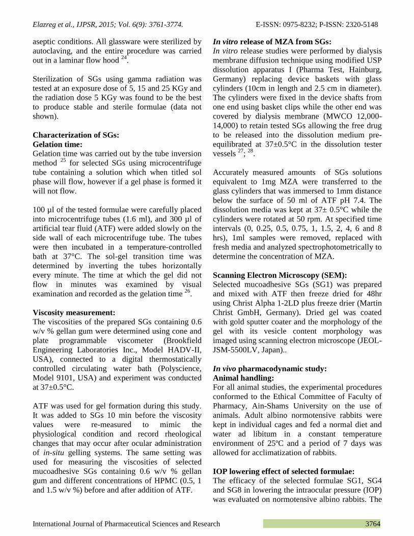
Scanning Electron Microscopy (SEM: SEM imaging was done for SG systems to examine
the effect of mixing MZA-SVs with gellan gum
and HPMC on SVs PS. Since it was not possible to
separate the vesicles from the mucoadhesive ISG
formulations, the only alternative was to freeze
samples then dry them to check the size changes
that may happen during formulation process.
The electron photomicrograph of freeze dried SG1
(Fig.6) shows the spherical vesicles of S1 with
average diameter of 240.12 nm embedded in the
gel matrix of gellan gum and HPMC. SVs of S1
were attached to the surface individually (white
arrow) or in groups. Although some SVs appeared
rounded and spherical, others showed good
spreading while adhering to the gel surface.
SEM image also shows continuous space filling
SVs networks and sometimes patches were noticed
in the gel matrix. The images also revealed the
macroporosity of dried gels with most pores being
in the range of 0.1- 0.5 μm in diameter (black
arrow).
This finding indicates that mixing process of MZA-
SVs with mucoadhesive ISG systems did not
destroy SVs structure and only caused minimal
decrease in PS from (276.26 to 240.12 nm) which
may be due to the effect of freeze drying utilized in
sample preparation.
Elazreg et al., IJPSR, 2015; Vol. 6(9): 3761-3774. E-ISSN: 0975-8232; P-ISSN: 2320-5148
International Journal of Pharmaceutical Sciences and Research 3771
FIG. 6: SCANNING ELECTRON MICROGRAPH OF MZA-
SVs MUCOADHESIVE IN SITU GEL (SG1). SPHERICAL
VESICLES (RED CIRCLES), SVS ATTACHED TO GEL
SURFACE (WHITE ARROW) AND MACROPORES IN GELS
(BLACK ARROW).
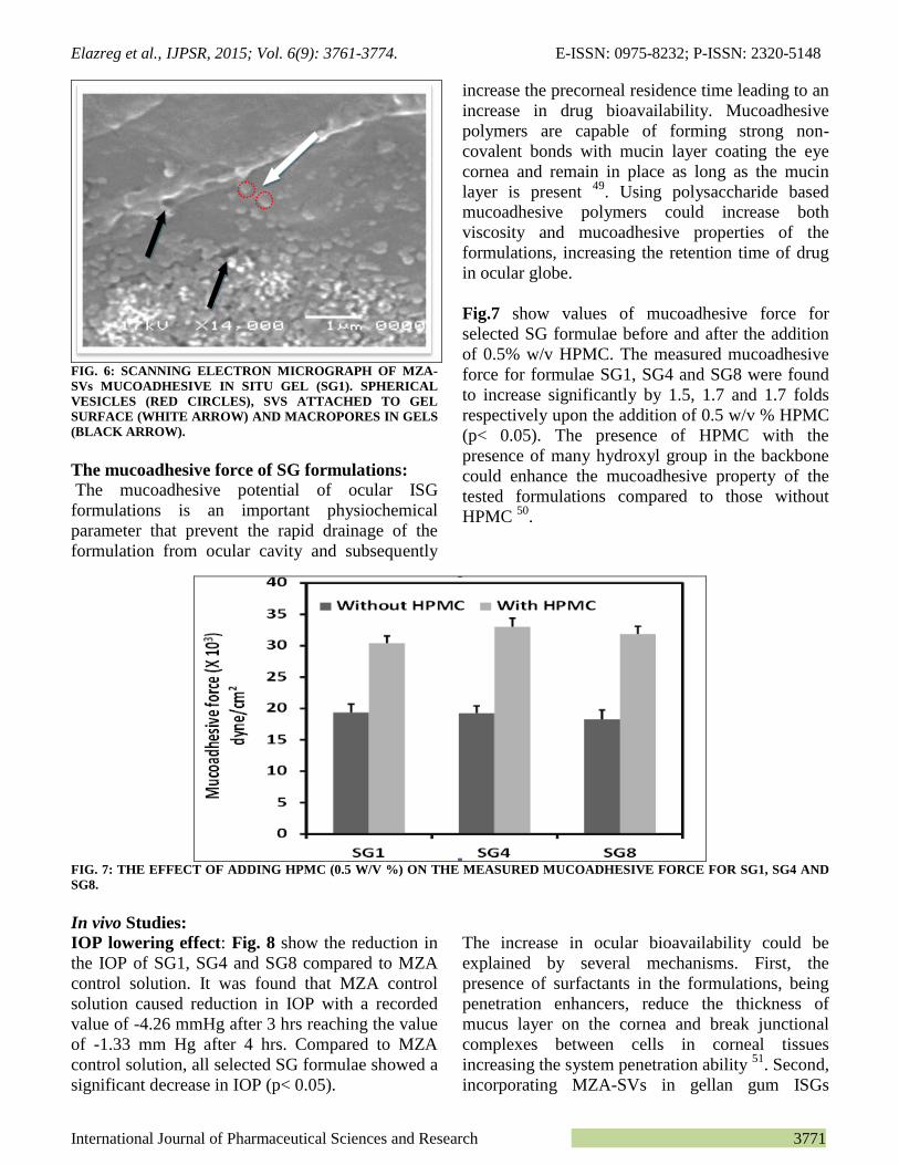
The mucoadhesive force of SG formulations:
The mucoadhesive potential of ocular ISG
formulations is an important physiochemical
parameter that prevent the rapid drainage of the
formulation from ocular cavity and subsequently
increase the precorneal residence time leading to an
increase in drug bioavailability. Mucoadhesive
polymers are capable of forming strong non-
covalent bonds with mucin layer coating the eye
cornea and remain in place as long as the mucin
layer is present 49
. Using polysaccharide based
mucoadhesive polymers could increase both
viscosity and mucoadhesive properties of the
formulations, increasing the retention time of drug
in ocular globe.
Fig.7 show values of mucoadhesive force for
selected SG formulae before and after the addition
of 0.5% w/v HPMC. The measured mucoadhesive
force for formulae SG1, SG4 and SG8 were found
to increase significantly by 1.5, 1.7 and 1.7 folds
respectively upon the addition of 0.5 w/v % HPMC
(p< 0.05). The presence of HPMC with the
presence of many hydroxyl group in the backbone
could enhance the mucoadhesive property of the
tested formulations compared to those without
HPMC 50
.
FIG. 7: THE EFFECT OF ADDING HPMC (0.5 W/V %) ON THE MEASURED MUCOADHESIVE FORCE FOR SG1, SG4 AND
SG8.
In vivo Studies:
IOP lowering effect: Fig. 8 show the reduction in
the IOP of SG1, SG4 and SG8 compared to MZA
control solution. It was found that MZA control
solution caused reduction in IOP with a recorded
value of -4.26 mmHg after 3 hrs reaching the value
of -1.33 mm Hg after 4 hrs. Compared to MZA
control solution, all selected SG formulae showed a
significant decrease in IOP (p< 0.05).
The increase in ocular bioavailability could be
explained by several mechanisms. First, the
presence of surfactants in the formulations, being
penetration enhancers, reduce the thickness of
mucus layer on the cornea and break junctional
complexes between cells in corneal tissues
increasing the system penetration ability 51
. Second,
incorporating MZA-SVs in gellan gum ISGs

Elazreg et al., IJPSR, 2015; Vol. 6(9): 3761-3774. E-ISSN: 0975-8232; P-ISSN: 2320-5148
International Journal of Pharmaceutical Sciences and Research 3772
resulted in the formation of gels upon ocular
administration. The formed gel acted as drug
release controlling matrix that decrease drug
drainage from the eye. The IOP lowering effect of
SG1, SG4 and SG8 reached its peak after 3, 5 and 4
hrs with values of -8.2, -7.3 and -8.0 mm Hg, and
lasted for 12, 10 and 10 hrs respectively. The
fastest onset of action of SG1 might be due to its
small particle size which is expected to increase the
vesicle penetration capability across corneal
membrane and consequently, improve drug
bioavailability. Thus, the particle size represents
the rate limiting step for the process of lowering
IOP 52
.
-12
-10
-8
-6
-4
-2
0
2
4
6
0 5 10 15
Re
du
ctio
n in
IOP
(m
mH
g)
MZAsolution 0.05%
SG1
SG4
SG8
Time(hrs)
FIG. 8: IOP LOWERING EFFECT OF MZA-SVs AFTER ADMINISTRATION OF TOPICAL MZA-LOADED MUCOADHESIVE
ISG FORMULATIONS (SG1, SG4 AND SG8) COMPARED TO MZA SOLUTION.
Assessment of ocular irritancy of mucoadhesive
SGs:
Fig. 9 show the cross sections of control rabbit
corneal tissue, rabbit cornea treated with SG1 and
SG8 twice daily for 10 days respectively. Normal
cornea showing no sign of edema, inflammation or
histological changes. Also no losses were observed
in the epithelial layer of cornea after the
administration of SG1 and SG8.
(a) )b) )c)
FIG 9: HISTOLOGICAL EXAMINATION OF (A) CONTROL RABBIT’S CORNEAL TISSUE AND RABBIT’S CORNEAL
TISSUE TREATED WITH (B)SG1 (C)SG8 WHERE t, s, and d ARE EPITHELIUM, STROMA AND ENDOTHELIUM
RESPECTIVELY.
In the stroma, no keratocyte loss was observed after
the continuous exposure to both formulae for 10
days. Moreover, no neovascularization that may be
associated with the anterior stroma was revealed.
Finally, no endothelial cell losses were noticed in
the eyes of individual treated rabbits. The absence
of any morphological changes after administration
of mucoadhesive SGs is expected due to the lack of

Elazreg et al., IJPSR, 2015; Vol. 6(9): 3761-3774. E-ISSN: 0975-8232; P-ISSN: 2320-5148
International Journal of Pharmaceutical Sciences and Research 3773
any membrane disrupting materials in their content.
The non-ionic nature of span 60, edge activators as
well as the biodegradability of gellan gum and
HPMC would impart a good biocompatibility to
our tested formulation, and could be considered
safe for short and long term treatment. It has been
previously reported that the irritating power of
surfactants is minimum for non-ionic surfactants
when compared to ionic surfactants 53
.
CONCLUSION: MZA was encapsulated successfully
in the elastic SVs using different ratios of Span 60: edge
activators. The best formulae were selected based on
their PS and MZA EE% to be incorporated in
mucoadhesive in-situ gel containing gellan gum/HPMC
mixtures. The prepared formulae showed more
prolonged lowering in IOP when compared to MZA
control solution and were found to be safe and well
tolerated. Thus, this suggests the potential usefulness of
SGs as controlled ocular delivery system to improve
ocular bioavailability of MZA and decrease frequency
of drug administration, superseding MZA control
solution.
ACKNOWLEDGMENTS: Authors wish to
acknowledge the financial grant received from Zawia
University under the Research Grant Scheme from
Libyan government.
REFERENCES:
1. Jarvinen K, Jarvinen T, and Urtti A, Ocular absorption
following topical delivery. Advanced Drug Delivery
Reviews, 1995; 16 (1): 3 – 19.
2. Green K, The effects of preservatives on corneal
permeability of drugs. In: Edman, P. (Ed.),
Biopharmaceutics of Ocular Drug Delivery. CRC Press,
Boca Raton, 1993; 43–49.
3. Merkus F, Schipper N, and Hermeus W, Absorption
enhancers in nasal drug delivery. efficacy and safety,
Journal of Controled Release, 1993; 24: 201–208.
4. Shilpa K, and Kaur IP, Spanlastics—A novel
nanovesicular carrier system for ocular delivery,
International Journal of Pharmaceutics, 2011; 413: 202-
210.
5. El Meshad AN, and Mohsen AM, Enhanced corneal
permeation and antimycotic activity of itraconazole against
Candida albicans via a novel nanosystem vesicle. Drug
Delivery, 2014; 0, 1-9.
6. Kaur IP, Rana C, Singh M, Bhushan S, Singh H, and
Kakkar S, Development and Evaluation of Novel
Surfactant-Based Elastic Vesicular System for Ocular
Delivery of Fluconazole, Journal of Ocular Pharmacology
and Therapeutics, 2012; 28(5): p. 484-496.
7. Williams KA, Irani YD, and Klebe S, Novel Therapeutic
Approaches for Corneal Disease, Discovery Medicine,
2013; 15(84):291-299.
8. Sanzgiri YD, Maschi S, Crescenzi V, Callegaro L, Topp
EM, and Stella VJ, Gellan-based systems for ophthalmic
sustained delivery of methylprednisolone, Journal of
Controled Release, 1993; 26: 195–201.
9. Rozier A, Mazuel C, Grove J, and Plazonnet B,
Functionality testing of gellan gum, a polymeric excipient
material for ophthalmic dosage forms, International
Journal of Pharmaceutics, 1997; 153: 191–198.
10. Ludwig A, The use of mucoadhesive polymers in ocular
drug delivery. Advanced Drug Delivery Reviews, 2005;
57: 1595– 1639.
11. Rahamatullah S, Singh R, Raghu T, James G, David W,
and Ryan D, Mucoadhesive drug delivery systems, 2011;
3(1): 89-100.
12. Maren TH, Haywood JR, Chapman SK, and Zimmerman
TJ, The pharmacology of methazolamide in relation to the
treatment of glaucoma, Invest Ophthalmol Vis Sci, 1977;
16(8): 730–742.
13. Edelhauser HF, and Maren TH, Permeability of human
cornea and sclera to sulfonamide carbonic anhydrase
inhibitors. Arch Ophthalmol, 1988; 106(8): 1110–1115.
14. Fridriksdóttir H, Loftsson T, and Stefánsson E,
Formulation and testing of methazolamide cyclodextrin
eye drop solutions, Journal of Controled Release, 1997;
44(1): 95–99.
15. Chen R, Qian Y, and Li R, Methazolamide calcium
phosphate nanoparticles in an ocular delivery system,
Yakugaku Zasshi, 2010; 130(3): 419–424.
16. Youshia J, Kamel AO, El Shamy A, and Mansour S,
Design of cationic nanostructured heterolipid matrices for
ocular delivery of methazolamide, International Journal of
Nanomedicine, 2012; (7): 2483-2496.
17. Li R, Jiang S, and Liu D, A potential new therapeutic
system for glaucoma: solid lipid nanoparticles containing
methazolamide. Journal of Microencapsulation, 2011;
28(2): 134–141.
18. Kakkar S, and Kaur IP, Spanlastics A novel nanovesicular
carrier system for ocular delivery. International Journal of
Pharmaceutics, 2011; 413: 202– 210.
19. Van den Bergh BAI, Wertz PW, Junginger HE, and
Bouwstra JA, Elasticity of vesicles assessed by electron
spin resonance, electron microscopy and extrusion
measurements, International Journal of Pharmaceutics,
2001; 217: 13–24.
20. El Zaafarany GM, Awad GAS, Holayel SM, and Mortada
ND, Role of edge activators and surface charge in
developing ultradeformable vesicles with enhanced skin
delivery, International Journal of Pharmaceutics, 2010;
397: 164–172.
21. Salama HA, Mahmoud AA, Kamel AO, Abdel Hady M,
and Awad GAS, Phospholipid based colloidal poloxamer–
nanocubic vesicles for brain targeting via the nasal route,
Colloids and Surfaces B: Biointerfaces, 2012; 100: 146-
154.
22. Gan L, Gan Y, Zhu C, Zhang X, and Zhu J, Novel
microemulsion in situ electrolyte-triggered gelling system
for ophthalmic delivery of lipophilic cyclosporine A: in
vitro and in vivo results, International Journal of
Pharmaceutics, 2009; 365(1-2): 143-149.
23. Mansour M, Mansour S, Mortada ND, and Abd Elhady
SS, Ocular poloxamer-based ciprofloxacin hydrochloride
in situ forming gels. Drug Development and Industrial
Pharmacy, 2008; 34(7): 744-752.
24. Guinedi AS, Mortada ND, Mansour S, and Hathout RM,
Preparation and evaluation of reverse-phase evaporation
and multilamellar niosomes as ophthalmic carriers of
acetazolamide. International Journal of Pharmaceutics,
2005; 306(1): 71-82.

Elazreg et al., IJPSR, 2015; Vol. 6(9): 3761-3774. E-ISSN: 0975-8232; P-ISSN: 2320-5148
International Journal of Pharmaceutical Sciences and Research 3774
25. Gupta PN, Mishra V, Rawat A, Dubey P, Mahor S, Jain S,
Chatterji D, and Vyas SP, Non-invasive vaccine delivery
in transfersomes, niosomes and liposomes: a comparative
study. International Journal of Pharmaceutics, 2005;
293(1): 73-82.
26. Kumar R, Jaya K, and Selvadurai M, Formulation and In
vitro Evalution of Gellan Gum/Carbopol and Sodium
Alginate based Solution to Gel Depot of Ketotifen
Fumarate System. Journal of Pharmaceutical Sciences &
Research, 2012; 4: 1973-1977.
27. Nasr M, Mansour S, Mortada ND, and El Shamy AA,
Lipospheres as Carriers for Topical Delivery of
Aceclofenac: Preparation, Characterization and In Vivo
Evaluation. AAPS PharmSciTech, 2008; 9(1): 154–162.
28. Hathout RM, Mansour S, Mortada ND, and Guinedi AS,
Liposomes as an ocular delivery system for acetazolamide:
in vitro and in vivo studies. AAPS Pharm Sci Tech, 2007;
8(1):1.
29. Attama AA, Reichlm S, and Müller- Goymann CC,
Diclofenac sodium delivery to the eye: in vitro evaluation
of novel solid lipid nanoparticle formulation using human
cornea construct. International Journal of Pharmaceutics,
2008; 355(1–2): 307–313.
30. Monem AS, Ali FM, and Ismail MW, Prolonged effect of
liposomes encapsulating pilocarpine HCl in normal and
glaucomatous rabbits. International Journal of
Pharmaceutics, 2000; 198(1): 29–38.
31. Winum J, Casini A, and Mincione F, Carbonic anhydrase
inhibitors: N-(p-sulfamoylphenyl)-alpha-D-
glycopyranosylamines as topically acting antiglaucoma
agents in hypertensive rabbits. Bioorganic & Medicinal
Chemistry, 2004; 14(1): 225–229.
32. Kaur IP, Singh M, and Kanwar M, Formulation and
evaluation of ophthalmic preparation of acetazolamide.
International Journal of Pharmaceutics, 2000; 199: 119–
127.
33. Banchroft JD, Stevens A, and Turner DR, Theory and
practice of histological techniques. Vol. Fourth Ed. 1996.
34. Manosroi A, Wongtrakul P, Manosroi J, Sakai H,
Sugawara F, Yuasa M, and Abe M, Characterization of
vesicles prepared with various non-ionic surfactants mixed
with cholesterol. Colloids and Surfaces B: Biointerfaces,
2003; 30(1): 129-138.
35. Akhilesh D, Bini K, and Kamath J, Review on Span-60
Based Non-Ionic Surfactant vesicles (Niosomes) as Novel
Drug Delivery. International Journal of Research in
Pharmaceutical and Biomedical Sciences, 2012; 3 (1): 6-
12.
36. Yoshioka T, Sternberg B, and Florence A, Preparation and
properties of vesicles (niosomes) of sorbitan monoesters
(Span 20, 40, 60, 80) and sorbitan triesters (Span 85).
International Journal of Pharmaceutics, 1994; 105: 1-6.
37. Lingan M, Sathali A, Kumar M, and Gokila A,
Formulation and evaluation of topical drug delivery
System containing clobetasol propionate niosomes.
Scientific Reviews & Chemical Communications, 2011;
1(1): 7-17.
38. Abdallah M, Sammour O, EL-Ghamry H, and Abu-Selem
M, Preparation and in-vitro evaluation of diclofenac
sodium niosomal formulations. International Journal of
Pharmaceutical Sciences and Research, 2013; 4(5): 1757-
1765.
39. Jacob L, and Anoop K, A review on surfactants as edge
activators in ultradeformable vesicles for enhanced skin
delivery, International Journal of Pharma & Bio Sciences,
2013; 4(3):337-344.
40. Nir S, and Nieva JL, Interactions of peptides with
liposomes: pore formation and fusion, Progress in lipid
research, 2000; 39(2): 181-206.
41. Wilson B, Samanta M, and Santhi K, Poly(n-
butylcyanoacrylate) nanoparticles coated with polysorbate
80 for the targeted delivery of rivastigmine into the brain
to treat Alzheimer’s disease, Brain Res, 2008; 1200: 159–
168.
42. Huang Y, Tsai M, and Wu P, Elastic liposomes as carriers
for oral delivery and the brain distribution of (+)-catechin,
J Drug Target, 2011; 19: 709–718.
43. Girigoswami A, Das S, and De S, Fluorescence and
dynamic light scattering studies of niosomes-membrane
mimetic systems. Spectrochimica Acta Part A: Molecular
and Biomolecular Spectroscopy, 2006; 64(4): 859-866.
44. Tangri P, Khurana S, Basics of ocular drug delivery
systems. International Journal of Research in
Pharmaceutical and Biomedical Sciences, 2011; 2(4):
1541-1552.
45. Ganji F, Abdekhodaie M, and Ramazani S, Gelation time
and degradation rate of chitosan-based injectable hydrogel.
Journal of Sol-Gel Science and Technology, 2007; 42(1):
47-53.
46. Dikstein S, Eyedrops having non-newtonian rheological
properties. 1992; Google Patents.
47. Boddupalli BM, Mohammed ZNK, Nath RA, and Banji D,
Mucoadhesive drug delivery system: An overview, Journal
of Advanced Pharmaceutical Technology & Research.
2010; 1(4): 381-387.
48. Rupenthal ID, Green CR, and Alany RG, Comparison of
ion-activated in situ gelling systems for ocular drug
delivery. Part 1: physicochemical characterisation and in
vitro release. International Journal of Pharmaceutics, 2011.
411(1-2): 69-77.
49. Almeida H, Amaral MH, Lobao P, and Sousa Lobo JM,
Applications of poloxamers in ophthalmic pharmaceutical
formulations: an overview, Expert Opin Drug Deliv, 2013;
10(9): 1223-1237.
50. Harish NM, Prabhu P, Charyulu RN, Gulzar MA, and
Subrahmanyam EVS, Formulation and Evaluation of in
situ Gels Containing Clotrimazole for Oral Candidiasis.
Indian J Pharm Sci., 2009; 71(4): 421–427.
51. Kaur, Garg A, Singla A, and Aggarwel D, Vesicular
systems in ocular drug deliver. International Journal of
Pharmaceutics, 2004; 269: 1-14.
52. Kassem MA, Abdel Rahman AA, Ghorab MM, Ahmed
MB, and Khalil RM, Nanosuspension as an ophthalmic
delivery system for certain glucocorticoid drugs.
International Journal of Pharmaceutics, 2007; 340(1-2):
126-133.
53. Van Abbe N, Eye irritation: studies related to responses in
man and lab animals. Journal of the Society of Cosmetic
Chemists, 1973; 24: 685–687.
All © 2013 are reserved by International Journal of Pharmaceutical Sciences and Research. This Journal licensed under a Creative Commons Attribution-NonCommercial-ShareAlike 3.0 Unported License.
This article can be downloaded to ANDROID OS based mobile. Scan QR Code using Code/Bar Scanner from your mobile. (Scanners are available on Google
Playstore)
How to cite this article:
Elazreg R, Soliman M, Mansour S and El Shamy A: Preparation and Evaluation of Mucoadhesive Gellan Gum In-Situ Gels For the Ocular Delivery of Carbonic Anhydrase Inhibitor Nanovesicles. Int J Pharm Sci Res 2014; 6(9): 3761-74. doi: 10.13040/IJPSR.0975-8232.6(9).3761-74.