visualization of gellan network formation in different
TRANSCRIPT

BMJ Vol 5 Issue 1 ISSN 2519-5972 169
Visualization of Gellan Network Formation in Different
Ionic Environment
Abeda Sultana Shamma1, Nusrat Jahan
2 *
and K. S. Hossain
3
Abstract
Gellan gum is a bacterial polysaccharide with a negatively charged carboxyl ion in its
monomer. It forms thermo-reversible gel due to the formation of a biopolymer network
in an aqueous medium under suitable conditions, and its properties depend on the type
of necessary cations to form a gel. In this study, the Atomic Force Microscopy (AFM)
technique has been used to elucidate cation types effect on gellan network formation.
AFM images were obtained for gellan gel samples prepared without and with adding
salts of monovalent, divalent, and trivalent cations. It was found that gellan gel with
monovalent ion did not show any fiber-like network. Instead, it showed a network
formed by the association of globular objects. While with divalent ion, the presence of
fiber-like objects was seen, and the network was formed due to the crosslinking of these
fibers. The mixture of fibrous and globular structures is realized for the sample with
added trivalent ion; however, the fibers are thicker than the gellan gel sample with
divalent cations.
Keywords: Gellan gum, Polysaccharides, Biopolymer network, Gel, AFM
Introduction
Gellan is an anionic, water-soluble heteropolysaccharide with high molecular weight. It
is composed of repetitive units of tetrasaccharide (β- D-glucose, β- D-glucuronic acid,
β- D-glucose and α-L-rhamnose), and one carboxyl side group per repeating unit
(Jansson, Lindberg and Sanford 1983) (O’Neill, Selvendran and Morris 1983). Gellan
forms a physically cross-linked thermo-reversible gel.
1Department of Physics, University of Louisiana at Lafayette, LA 70504, USA
[email protected] 2Department of Physics, Bangabandhu Sheikh Mujibur Rahman Maritime University,
Dhaka-1216, Bangladesh
[email protected] 3Department of Physics, University of Dhaka, Dhaka-1000, Bangladesh
* Corresponding Author

170 Visualization of Gellan Network Formation in Different Ionic Environment
However, at low concentrations or sufficiently high temperatures, the gellan without
salts does not form a gel as there is a lack of aggregation because of the carboxyl group
(Chandrasekaran, et al. 1988). The carboxyl groups being negatively charged repel each
other, which can be changed by introducing a metal cation in the gellan network
(Moritaka, Fukuba, et al. 1991) (Moritaka, Nishinari, et al. 1992) (E. Ogawa 1996). The
added cations act as the cross-linking agents and hold the helices together, leading to a
continuous 3-dimensional gellan network. However, different cations promote
aggregation differently. (Moritaka, Nishinari, et al. 1992) showed gellan with added K+
ion is more effective in promoting gelation than Na+, and the divalent cation like Ca
2+ is
more effective than any monovalent ion. Moreover, divalent cations can form gel
quickly at far lower concentrations than monovalent cations (Moritaka, Fukuba, et al.
1991) (Miyoshi and Nishinari 1999) and have various applications in tissue engineering
scaffolds (Xu, et al. 2018), self-healing (Lv, et al. 2019) . Therefore, the type, the
valency, and the amount of the cations added to make the solution influence the gel
properties of gellan gum. As most of its application depends on its gel-forming ability,
the gelation mechanism for gellan is widely studied using differential scanning
calorimetry (DSC), Light scattering (Okamoto, Kubota and Kuwahara 1993) ESR
(Tsutsumi, et al. 1993), X-ray small-angle scattering (Yuguchi, et al. 1993 ) osmotic
pressure (E. Ogawa 1993), ultrasonic velocities (Tanaka, Sakurai and Nakamura 1993)
and viscoelastic measurements (Shimazaki and Ogino 1993 ) (Nakamura, Harada and
Tanaka 1993 ).The sample used in these studies was a mixture of several different
cations such as sodium, magnesium, potassium, or calcium. However, (Kirchmajer, et
al. 2014) showed that gelation and mechanical properties of purified gellan are
enhanced compared to commercial samples with a trace of different cations. As a result,
gellan property can be significantly different for a specific cation than others, and its
effect will surely be interesting. Unfortunately, not much research has been done on
purified gellan.
To establish the association behavior of the gellan type bio macromolecules several
models have been proposed using simulations and experimental observations. Most
polysaccharide gelation models have concentrated on the detailed molecular structure of
the junction zones within the gel. However, visualizing the bio macromolecules using
direct observation by microscopic techniques is still at a very early stage. The cations
aggregation of gellan monomer with the carboxyl side group can be directly observed
under Atomic Force Microscopy with higher resolution than transmission electron
microscopy (Gunning et al. 1996). The AFM image with and without added cation was
studied by (Morris, Kirby, and Gunning 1999), and he showed gellan without added
cations form helix without side-by-side aggregation. However, when cation is added
with the potassium-based gellan aqueous solution, side-by-side aggregation of the

BMJ Vol 5 Issue 1 ISSN 2519-5972 171
helical filaments and continuous branched network were created. They concluded from
this study that the coil-helix transition allows the formation of filamentous structures,
which, in the presence of gel-promoting cations, further assemble into branched fibers
formed by the association of these filaments.
On the other hand, researchers found observing AFM images of 1.6% (w/w) gellan
solution with 0.01 M KCl, CsCl, or 0.001 M CaCl2 salt promotes branched rod-like
structures and forms inter helical aggregation (Ikeda et al. 2004). Therefore, Atomic
Force Microscopy (AFM) offers a method for investigating such models for gelation
and visualizing the long-range distributions of macromolecules within the gel network.
Again, most of the study used typically available commercial gellan powder,
predominantly in the potassium salt form. The commercial gellan solution can be made
free of these cations by dialysis against water and the study of network association of
the dialyzed gellan with different types of cation can give an insight into the gellan
gelation properties for specific cation.
In the present study, we aim to investigate the network association of dialyzed gellan
with and without added three different cation using Atomic Force Microscopy (AFM).
Materials and Method
Materials
Gellan powder was obtained from San-ei Gen (Osaka, Japan) and used in this study
without further purification. The metal content present in the sample was analyzed as
Na=0.44%, K =4.78%, Ca= 0.34%, and Mg= 0.012% by a LIBERTY Inductively
Coupled Plasma Optical Emission Spectrometry (ICP-OES) system (Varian Inc., Palo
Alto, CA, USA). Nanopure water was used to prepare the solution.
Sample preparation
Gellan sample without added salt
The powdered sample was mixed with Nanopure water to form a gellan solution. The
mixture was stirred using a magnetic stirrer at 80oC for 1 hour to ensure complete
dissolution. The transparency of the solution recognized the complete dissolution. The
hot solution was transferred to the cylindrical sample holder. A transparent gel was
formed from the uniform solution at ambient temperature. In this way gellan solution of
concentration Cp=0.8 and 1.6 wt. % was prepared.

172 Visualization of Gellan Network Formation in Different Ionic Environment
Dialysis of Gellan Sample
The dialysis was carried out using the Wako dialysis membrane, which was cut, soaked
in nano-pure water, and filled with gellan solution using a funnel. The dialysis
membrane was then put into a beaker with 250 ml nano-pure water and stirred for 3
hours. The dialysis was carried out three times on the sample after a 1-hour interval to
remove any existing metal ions from the gellan solution. The metal ion content after
dialysis was measured by Shimadzu (AA-6800) Flame Atomic Absorption
Spectrometer (FAAS) and the data showed a trace amount of potassium ion still existed
at room temperature, 30 C (Table-1). The prepared solution then dialyzed again, but
this time heat was applied. The metal ion content after dialysis at 50 C was analyzed
yet using FAAS. It showed the existence of the metal contents in the sample are very
low, and the effect of those ions can be considered negligible (Table-1).
Gellan Gel with added KCl, CaCl2, and AlCl3 Salt
Gellan with added salt was prepared by dissolving salt in Nanopure water, which was
then stirred for 30 minutes at 80℃. The concentration of the stock salt solution made
was double than required, and it was then mixed with the same amount of gellan
solution. In this way, the salt and gellan solution's concentration was halved than the
stock solution and five samples of polymer concentration Cp=0.8wt% without dialysis,
Cp=0.8wt% with dialysis and no added salt concentration, Cp=0.8wt% with dialysis and
added salt of concentration Cs=0.1M of KCl, CaCl2, and AlCl3 salt was prepared. The
polymer concentration was chosen at 0.8wt% because, below this concentration, not all
the cations with 0.1M salt concentration could form a gel and this experiment was done
to check the effects of the cation in the gel state of gellan.
Atomic Force Microscopy
A small amount of the liquid solution was spread on the glass substrate and stored at
room temperature 24 hours or more before visualizing the surface with Flex AFM 5
from nanosurf. This method was followed to prepare Cp= 0.8wt% with and without Cs =
0.1M KCl, 0.1M, and 0.1M gellan sample. After making samples according to the
procedure described above, it was ensured that they remain homogenous and fresh
enough to take the measurements with fewer errors. The images of the surface of the
sample were made in contact mode using Nanosurf Easyscan-2 software. Most of the
images obtained in this study contain 256 256 points. For each line, 0.7 seconds was
given to have decently high-resolution images.

BMJ Vol 5 Issue 1 ISSN 2519-5972 173
Result and Discussion
This experiment explores the effect of monovalent, divalent, and trivalent cations on
dialyzed gellan using Atomic Force Microscopy (AFM) images and to analyze the
surfaces' morphology and microstructures. A total of 5 samples of the surface of non-
dialyzed gellan without added salt, dialyzed gellan without added salt, and dialyzed gel
with added monovalent, divalent, and trivalent cations were studied.
For all the samples, a 3μg/ml amount of the solution was deposited on the micro-glass
slide and dried at room temperature for 24 hours. Figure 1 shows the micro-glass slide
used for the sample deposition visualized by AFM. The average roughness of the glass
slides was calculated in the area of 107.8 m2 was = 3.70 nm. In contrast, the native
mica surface's roughness is 0.13 nm in the area of 670 nm2 (Senden and Ducker 1992)
In the previous works on gellan gel, the mica surface was used for sample deposition
(Gunning, et al. 1996; Ikeda, et al. 2004). Still, in this study, the glass surface was
chosen as a substrate as it is rougher than the mica surface, so the solution will adhere
to the surface rather than spreading. SEM and X-ray imaging confirms that the gellan
molecule with added salt is a long chain and does not exist in a single strand but wraps
around and forms a double helix (Yuguchi., Urakawa and Kajiwara 2002). The number
of cross-links increases with increasing gellan concentration. But, at a lower
concentration, this number is not large enough to build a network at ambient
temperature. Hence gellan solution at ambient temperature for concentration less than
1wt% acts like a viscous liquid. Due to the lack of cross-links, all polymer chains clot
together in groups and float in solvent forming a colloid solution. AFM image (Figure
2) revealed that Cp= 0.80 wt% for non- dialyzed samples had no continuous coverage of
polymers rather than sample had discrete aggregates on the glass substrate.
Table-1: Metal ion content in gellan solution after dialysis
Sample Name Na (%) K (%) Ca (%) Mg (%)
0.8 wt. % Gellan Solution
(non-dialyzed)
0.438 4.781 0.336 0.012
Gellan Solution (dialyzed at
30 C)
0.567 3.628 0.258 0.004
Gellan Solution (dialyzed at
50 C)
0.00127 0.0264 0.00696 0.005

174 Visualization of Gellan Network Formation in Different Ionic Environment
Figure 1: Topographical AFM image of glass substrate; (A) color map, (B) 3D
view, and (C) height profile. The scanning size of the image is 10.3μm×10.4μm.
They were randomly oriented in all directions. Without dialysis, a gellan solution can
have different types of metallic ions. Therefore, there may be different types of
aggregation. In Figure 2, from the image of the non-dialyzed gellan film surface, a
significant amount of globules was seen. The average diameter of these globules was
2.28μm. These globules suggest that since the gellan solution was taken at a low
concentration, a lack of aggregation of the carboxyl groups caused gellan to not form
any continuous network. However, the double-helical chains of gellan aggregated
together and formed globules. Figure 3 reveals the surface after dialysis, and it showed
there was no uniform network of branched fibers.
(C)
(A) (B)

BMJ Vol 5 Issue 1 ISSN 2519-5972 175
Figure 2: AFM images of 0.80 wt%
non-dialyzed gellan gum without any
salt. Image size: 10.3μm 10.4μm.
Figure 3: AFM images of 0.80 wt%
dialyzed gellan gum without any salt.
The scanning size of image size:
10.3μm 10.3μm.
Figure 4: AFM images of 0.80 wt%
dialyzed gellan gum with 0.1M KCl.
Image size 10.3μm×10.4μm.
Figure 5(a): AFM images of 0.80 wt%
dialyzed gellan gum with 0.1M CaCl2.
Image size 10.3μm×10.4μm.

176 Visualization of Gellan Network Formation in Different Ionic Environment
Figure 5(b): AFM images of 0.80 wt% dialyzed gellan gum with 0.1M CaCl2. Image
size 831nm×839 nm .
Figure 6: AFM images of 0.80 wt% dialyzed gellan gum with 0.1M AlCl3. Image size
10.3μm×10.4μm.
BMJ Vol 5 Issue 1 ISSN 2519-5972 177
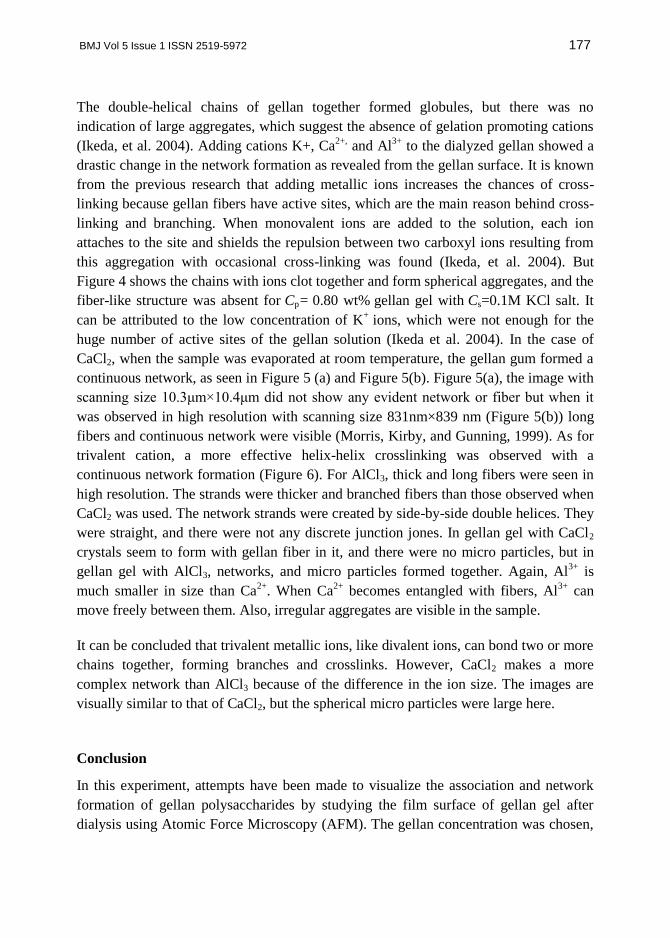
The double-helical chains of gellan together formed globules, but there was no
indication of large aggregates, which suggest the absence of gelation promoting cations
(Ikeda, et al. 2004). Adding cations K+, Ca2+,
and Al3+
to the dialyzed gellan showed a
drastic change in the network formation as revealed from the gellan surface. It is known
from the previous research that adding metallic ions increases the chances of cross-
linking because gellan fibers have active sites, which are the main reason behind cross-
linking and branching. When monovalent ions are added to the solution, each ion
attaches to the site and shields the repulsion between two carboxyl ions resulting from
this aggregation with occasional cross-linking was found (Ikeda, et al. 2004). But
Figure 4 shows the chains with ions clot together and form spherical aggregates, and the
fiber-like structure was absent for Cp= 0.80 wt% gellan gel with Cs=0.1M KCl salt. It
can be attributed to the low concentration of K+
ions, which were not enough for the
huge number of active sites of the gellan solution (Ikeda et al. 2004). In the case of
CaCl2, when the sample was evaporated at room temperature, the gellan gum formed a
continuous network, as seen in Figure 5 (a) and Figure 5(b). Figure 5(a), the image with
scanning size 10.3μm×10.4μm did not show any evident network or fiber but when it
was observed in high resolution with scanning size 831nm×839 nm (Figure 5(b)) long
fibers and continuous network were visible (Morris, Kirby, and Gunning, 1999). As for
trivalent cation, a more effective helix-helix crosslinking was observed with a
continuous network formation (Figure 6). For AlCl3, thick and long fibers were seen in
high resolution. The strands were thicker and branched fibers than those observed when
CaCl2 was used. The network strands were created by side-by-side double helices. They
were straight, and there were not any discrete junction jones. In gellan gel with CaCl2
crystals seem to form with gellan fiber in it, and there were no micro particles, but in
gellan gel with AlCl3, networks, and micro particles formed together. Again, Al3+
is
much smaller in size than Ca2+
. When Ca2+
becomes entangled with fibers, Al3+
can
move freely between them. Also, irregular aggregates are visible in the sample.
It can be concluded that trivalent metallic ions, like divalent ions, can bond two or more
chains together, forming branches and crosslinks. However, CaCl2 makes a more
complex network than AlCl3 because of the difference in the ion size. The images are
visually similar to that of CaCl2, but the spherical micro particles were large here.
Conclusion
In this experiment, attempts have been made to visualize the association and network
formation of gellan polysaccharides by studying the film surface of gellan gel after
dialysis using Atomic Force Microscopy (AFM). The gellan concentration was chosen,

178 Visualization of Gellan Network Formation in Different Ionic Environment
Cp=0.80 wt%, with and without added 0.1M of the monovalent, divalent, and trivalent
cation. Non-dialyzed gellan without added cations show no formation of any networks
because of the low concentration. In the presence of K+ ion, a visible fiber-like network
was not observed but spherical aggregates were observed which suggested that the low
amount of fibers with ions aggregated together to form spherical aggregates. Besides,
the CaCl2 gellan image showed a continuous network with fiber structure and AlCl3
formed a mixture of fibrous and globular structures. The images are consistent with the
fibrous model of gellan gum. Therefore, it can be concluded from the images that two
different types of gellan structures were observed; they were fibrous and globular. The
fiber-like aggregate was found on the surface of divalent and trivalent gellan films,
whereas globular aggregates were found on monovalent gellan film.
Acknowledgments
The authors gratefully acknowledge the financial support from the International Science
Program (ISP), Uppsala University, Sweden, and BSMRMU research grant through
UGC. The author would also thank the Nanophysics and Soft Matter Lab of the
Department of Physics, University of Dhaka for their support in conducting the
experiment.
References
Chandrasekaran, R., L.C. Puigjaner, K.L. Joyce, and S. Arnott. 1988. "Cation
interactions in gellan: an X-ray study of the potassium salt." Carbohydrate Research
181: 23-40.
Gunning, A. P., A. R. Kirby, M. J. Ridout, G. J. Brownsey, and V. J. Morris. 1996. "
Investigation of gellan networks and gels by atomic force microscopy."
Macromolecules 29 ( 6791–6796).
Ikeda, S., Y. Nitta, T. Temsiripong, R. Pongsawatmanit, and K. Nishinari. 2004.
"Atomic force microscopy studies on cation-induced network formation of gellan."
Food Hydrocolloids 727-735.
Jansson, P. E., B. Lindberg, and P. A. Sanford. 1983. "Structural studies of gellan gum,
an extracellular polysaccharide elaborated by Pseudomonas elodea." Carbohydrate
Research 124: 135–139.
Kirchmajer, D.M., B. Steinhoff, H. Warren, R. Clark, and M. Panhuis. 2014. "Enhanced
gelation properties of purified gellan gum." Carbohydrate Research 388 (125-129).

BMJ Vol 5 Issue 1 ISSN 2519-5972 179
Lv, Yukai, Zheng Pan, Cunzheng Song, Yulong Chen*, and Xin Qian*. 2019. "Locust
bean gum/gellan gum double-network hydrogels with superior self-healing and pH-
driven shape-memory properties." Soft Matter.
Miyoshi, E., and K. Nishinari. 1999. "Rheological and thermal properties near the sol–
gel transition of gellan gum aqueous solutions ." Progress in Colloid and Polymer
Science 114 (68–82).
Moritaka, H., H. Fukuba, K. Kumeno, N. Nakahama, and K. Nishinari. 1991. "Effect of
monovalent and divalent cations on the rheological properties of gellan gels ." Food
Hydrocolloids 495-507.
Moritaka, H., K. Nishinari, N. Nakahama, and H. Fukuba. 1992. "Effects of potassium
chloride and sodium chloride on the thermal properties of gellan gum gels." Bioscience,
biotechnology, and biochemistry 595-599.
Morris, V.J., A.R. Kirby, and A.P. Gunning. 1999. "A fibrous model for gellan gels
from atomic force microscopy studies." In Progress in Colloid and Polymer Science, by
K. Nishinari, volume 114. Physical Chemistry and Industrial Application of Gellan
Gum.
Nakamura, K., K. Harada, and Y. Tanaka. 1993 . "Viscoelastic properties of aqueous
gellan solutions: the effects of concentration on gelation ." Food Hydrocolloid 7 (435-
447).
O’Neill, M. A., R. R. Selvendran, and V. J. Morris. 1983. " Structure of the acidic
extracellular gelling polysaccharide produced by Pseudomonas elodea. Carbohydrate
Research, 124, 123–133." Carbohydrate Research 123–133.
Ogawa, E. 1996. "Conformational transition of polysaccharide sodium-gellan gum in
aqueous solutions." Macromolecules 5178-5182.
Ogawa, Etsuyo. 1993. "Osmotic pressure measurements for gellan gum aqueous
solutions." Food Hydrocolloid 7 (397-405).
Okamoto, T., K. Kubota, and N. Kuwahara. 1993. "Light scattering study of gellan
gum." Food Hydrocolloids 7 (363- 371).
Senden, T. J., and W. A. Ducker. 1992. "Surface roughness of plasma-treated mica. ."
Langmuir, 8(2), 733–735.
Shimazaki, T., and K. Ogino. 1993 . "Viscoelastic properties of gellan gum aqueous
solutions ." Food Hydrocolloid 7 (417-426).
Tanaka, Y., M. Sakurai, and K. Nakamura. 1993. "Ultrasonic velocities in aqueous
gellan solutions." Food Hydrocolloid 7 (407-415).

180 Visualization of Gellan Network Formation in Different Ionic Environment
Tsutsumi, A., D. Ya, T. Hiraoki, H. Mochiku, R. Yamaguchi, and N. Takahashi. 1993. "
ESR studies of Mn(II) binding to gellan and carrageenan gels ." Food Hydrocolloids
427-434.
Xu, Zihao, Zhuqing Li, Shan Jiang, and and Kaitlin M. Bratlie. 2018. "Chemically
Modified Gellan Gum Hydrogels with Tunable Properties for Use as Tissue
Engineering Scaffolds." ACS Omega 6998-7007.
Yuguchi, Y., M. Mimura, Kitamura.S., H. Ura kawa, and K. Kajiwara. 1993 .
"Structural characteristics of gellan in aqueous solution ." Food Hydrocolloid 7, 373-
385.
Yuguchi., Y., H. Urakawa, and K. Kajiwara. 2002. "The effect of Potassium salt on the
structural characteristics of gellan gum gel." Food Hydrocolloids Volume 16, Issue 3;
191-195.