gelatin-based composite hydrogels with biomimetic
TRANSCRIPT

Friction 10(2): 232–246 (2022) ISSN 2223-7690 https://doi.org/10.1007/s40544-020-0437-5 CN 10-1237/TH
RESEARCH ARTICLE
Gelatin-based composite hydrogels with biomimetic lubrication and sustained drug release
Kuan ZHANG1,2,†, Jielai YANG3,4,†, Yulong SUN1, Yi WANG1, Jing LIANG4, Jing LUO5, Wenguo CUI4, Lianfu DENG4,
Xiangyang XU3,*, Bo WANG2,*, Hongyu ZHANG1,* 1 State Key Laboratory of Tribology, Department of Mechanical Engineering, Tsinghua University, Beijing 100084, China 2 School of Chemical and Biological Engineering, Shandong University of Science and Technology, Qingdao 266590, China 3 Department of Orthopedics, Ruijin Hospital, Shanghai Jiao Tong University School of Medicine, Shanghai 200025, China 4 Shanghai Key Laboratory for Prevention and Treatment of Bone and Joint Diseases, Shanghai Institute of Traumatology and Orthopaedics,
Ruijin Hospital, Shanghai Jiao Tong University School of Medicine, Shanghai 200025, China 5 Beijing Research Institute of Automation for Machinery Industry Co., Ltd., Beijing 100120, China
Received: 12 June 2020 / Revised: 27 July 2020 / Accepted: 30 July 2020
© The author(s) 2020.
Abstract: The occurrence of osteoarthritis is closely related to progressive and irreversible destruction of the
articular cartilage, which increases the friction significantly and causes further inflammation of the joint. Thus,
a scaffold for articular cartilage defects should be developed via lubrication restoration and drug intervention.
In this study, we successfully synthesized gelatin-based composite hydrogels, namely GelMA–PAM–PMPC, with
the properties of biomimetic lubrication and sustained drug release by photopolymerization of methacrylic
anhydride modified gelatin (GelMA), acrylamide (AM), and 2-methacryloyloxyethyl phosphorylcholine (MPC).
Tribological test showed that the composite hydrogels remarkably enhanced lubrication due to the hydration
lubrication mechanism, where a tenacious hydration shell was formed around the zwitterionic phosphocholine
headgroups. In addition, drug release test indicated that the composite hydrogels efficiently encapsulated
an anti-inflammatory drug (diclofenac sodium) and achieved sustained release. Furthermore, the in vitro test
revealed that the composite hydrogels were biocompatible, and the mRNA expression of both anabolic and
catabolic genes of the articular cartilage was suitably regulated. This indicated that the composite hydrogels
could effectively protect chondrocytes from inflammatory cytokine-induced degeneration. In summary, the
composite hydrogels that provide biomimetic hydration lubrication and sustained local drug release represent
a promising scaffold for cartilage defects in the treatment of osteoarthritis.
Keywords: hydrogel; articular cartilage; zwitterionic polymer; hydration lubrication; drug delivery
1 Introduction
From a biotribological viewpoint, osteoarthritis has
been accepted as a lubrication deficiency-induced
joint disease that is characterized by the breakdown
of articular cartilage and inflammation of the joint.
Therefore, a synergetic therapy integrating both
lubrication and drug intervention is a promising
approach for the treatment of osteoarthritis [1, 2].
However, due to the insufficiency of blood vessels
in articular cartilage, it is very difficult for nutritious
agents/drugs to reach the joint via oral administration
[3, 4]. Thus, the design of a scaffold to simultaneously
achieve enhanced lubrication and sustained drug
† Kuan ZHANG and Jielai YANG contributed equally to this work. * Corresponding authors: Xiangyang XU, E-mail: [email protected]; Bo WANG, E-mail: [email protected]; Hongyu ZHANG,
E-mail: [email protected]

Friction 10(2): 232–246 (2022) 233
www.Springer.com/journal/40544 | Friction
delivery is a good solution for cartilage defects, although
this subject is still facing great challenges.
Hydrogels have been widely studied as an ideal
substitute for articular cartilage and as a drug
carrier. Great effort has been devoted to designing
functional hydrogels with good biocompatibility [5],
high mechanical strength [6], and low coefficient of
friction (COF) [7, 8]. Recently, many hydrogels have
been developed to exhibit excellent physicochemical
properties such as double-network hydrogels [9],
mussel-inspired functional hydrogels [10–12], composite
hydrogels [13], and hydrogen-bonded crosslinked
hydrogels [14]. Gelatin, a single-chain derivative formed
by collagen, is biocompatible and can be biodegraded
in vivo. Additionally, the side chains of gelatin are
rich in reactive groups (–COOH and –NH2), and
thus different gelatin hydrogels (GelMA) have been
synthesized to repair skin, bone, and articular cartilage
[15, 16], in which methacrylic anhydride is introduced
to initiate photopolymerization [17, 18]. However, to
the best of our knowledge, the lubrication properties
of GelMA hydrogels have rarely been investigated.
Many methods have been reported for improving
the mechanical properties of hydrogels, but the impro-
vement in mechanical properties often compromises
lubrication [19–21]. Naturally, articular cartilage has
an ultra-low COF (at a level of 0.001–0.01) based
on hydration lubrication mechanism [22–24], where
polyelectrolyte biomacromolecules (such as hyaluronic
acid, aggrecan, and lubricin) complex with phospha-
tidylcholine (PC) lipids form a lubricating boundary
layer by exposing the hydrated phosphocholine
groups (N+(CH3)3 and PO4−) on the superficial surface
[25]. Poly(2-methacryloyloxyethyl phosphorylcholine)
(PMPC), a biocompatible polymer, has the same
zwitterionic phosphocholine groups as PC lipids, and
it has been widely used to enhance the lubrication
properties of various biomedical materials through
surface modification [26–28].
In this study, bioinspired by the hydration lubrication
mechanism of articular cartilage, we developed gelatin-
based composite hydrogels with biomimetic lubrication
and sustained drug release (GelMA–PAM–PMPC).
Specifically, acrylamide (AM) and MPC were intro-
duced into GelMA to improve the mechanical and
lubrication properties of the composite hydrogels.
Additionally, diclofenac sodium (DS, a typical anti-
inflammatory drug to relieve pain due to osteoarthritis)
was encapsulated while preparing the composite
hydrogels. It is hypothesized that the dual-functional
composite hydrogels developed herein, as a scaffold
for cartilage defects to achieve both lubrication enhan-
cement and local drug delivery, may find applications
in the treatment of osteoarthritis.
2 Materials and methods
2.1 Materials and reagents
2-Methacryloyloxyethyl phosphorylcholine (MPC)
was obtained from Joy-Nature Co. (Nanjing, China).
Gelatin was purchased from Sinopharm Chemical
Reagent Co., Ltd. (Shanghai, China). Methacrylic
anhydride and DS were purchased from J&K Scientific
Ltd. (Beijing, China). Ethyl acetate was purchased
from Modern Oriental Technology Development Co.,
Ltd. (Beijing, China). Dulbecco’s modified eagle’s
medium (DMEM)/nutrient mixture F-12 medium,
bovine serum albumin, fetal bovine serum, and 0.25%
trypsin-ethylenediaminetetraacetic acid (trypsin-EDTA)
were purchased from Gibco Life Technologies Corp.
(CA, USA). Live/dead viability/cytotoxicity kit was
purchased from Invitrogen (Carlsbad, CA, USA). Cell
counting kit-8 (CCK-8) was purchased from Dojindo
Molecular Technologies, Inc. (Kumamoto, Japan).
SYBR® Green PCR Master Mix (DRR420A) and Prime
Script RT reagent kit (DRR037A) were provided by
Takara Biomedical Technology (Beijing) Co., Ltd.
(Tokyo, Japan). Phalloidin was purchased from Sigma-
Aldrich Co. (St. Louis, MO, USA). Recombinant mouse
IL-1β and TNF-α were purchased from PeproTech
(Rocky Hill, NJ, USA).
2.2 Synthesis of composite hydrogels
GelMA was synthesized according to previously
described methods [29, 30]. Gelatin (5 g) was added
to phosphate buffer solution (PBS, 50 mL) and
magnetically stirred at 50 °C until it was completely
dissolved. Then, methacrylic anhydride (5 mL) was
added dropwise to the above solution (0.5 mL/min)
and reacted in a 50 °C isothermal water bath under
magnetic stirring. After 4 h, the reaction solution
was diluted with PBS (200 mL, 50 °C) and stirred to
terminate the reaction. The above solution was placed

234 Friction 10(2): 232–246 (2022)
| https://mc03.manuscriptcentral.com/friction
in a dialysis bag (molecular weight cutoff: 8–14 kDa)
and dialyzed in pure water at 50 °C for 6 d. The
dialyzed solution was poured into a centrifuge tube
for centrifugation (2,500 rpm), and the supernatant
was transferred and lyophilized to obtain the GelMA
product.
The synthesis of the composite hydrogels was
achieved by photopolymerization. Briefly, GelMA (1 g),
MPC (mass ratio to GelMA: 5%, 15%, 30%, and 50%),
crosslinker (bis-acrylamide, 1%), and photoinitiator
(I2959-Tos, 5 mg) were added to the AM solution to
pre-polymerize the monomers at 37 °C. Subsequently,
the pre-polymerized solution was transferred to a
custom-made mold and irradiated using an ultraviolet
spot light source (7.1 mW/cm2, 360–480 nm) for 5 min.
Finally, the hydrogels (GelMA–PAM–PMPC) were
taken out and soaked in PBS at 37 °C for 24 h to
remove the uncrosslinked monomers. Pure GelMA–
PAM and GelMA hydrogels were prepared using
the same method. Unless mentioned otherwise, the
GelMA–PAM–PMPC samples used in the following
tests contained 30% MPC.
2.3 Characterization of composite hydrogels
1H nuclear magnetic resonance (NMR) spectra of
gelatin and GelMA were recorded using an Ascend
400 MHz NMR spectrometer (Bruker, USA) with D2O
as the solvent. Fourier transform infrared (FTIR) spectra
of PMPC, GelMA, and GelMA–PAM–PMPC were
analyzed using a Nexus 670 spectrometer (Nicolet,
USA) at 400–4,000 cm−1. The water content of GelMA
and GelMA–PAM–PMPC was measured via thermo-
gravimetric analysis (TGA) (Q5000IR, TA Instruments,
USA) from 25 to 300 °C at a heating rate of 10 °C /min.
To examine the swelling behavior of the hydrogels,
GelMA and GelMA–PAM–PMPC were incubated in
PBS at 37 °C and sampled at 0, 0.5, 1, 3, and 5 h after
incubation. Then, the hydrogels were freeze-dried, and
the swelling ratio (λw) and the relative volume (λv)
were calculated using Eqs. (1) and (2).
iw
0
W
W (1)
2i i
v 20 0
R H
R H (2)
Here, Wi and W0 are the swollen and dry weights
of the hydrogels, respectively, which were measured
in an equilibrium state. The swelling ratio (λw) was
calculated as the ratio of the weight of swollen
hydrogels to that of dry hydrogels (Eq. (1)). Ri, R0, Hi,
and H0 are the radius and height of the hydrogels
(cylindrical samples) before and after swelling,
respectively. The relative volume (λv) was calculated
as the ratio of the volume of swollen hydrogels to that
of dry hydrogels (Eq. (2)).
The water contact angle of GelMA and GelMA–
PAM–PMPC was obtained using an OCA25 contact
angle goniometer (Dataphysics Instruments, Germany)
by the sessile drop method. Distilled water (3 μL)
was placed on the airside surface of the hydrogels
at room temperature, and the static contact angle was
collected after 10 s. The mean contact angle was cal-
culated from the results of at least three measurements.
To depict the internal microstructures of the
hydrogels, GelMA and GelMA–PAM–PMPC were
freeze-dried to thoroughly remove water. Subsequently,
the hydrogels were coated with Pt/Pd and examined
using a Quanta 200 field emission scanning electron
microscope (SEM, FEI, Eindhoven, Netherlands) under
an accelerating voltage of 5 kV, which was coupled
with energy dispersive spectroscopy (EDS) to enable
elemental composition analysis. Image J software was
used to quantify the pore distribution of the hydrogels
based on a series of SEM images. The pores were
randomly selected on the surface of the hydrogels. The
pore size was nominated as the average pore diameter
on the selected SEM images.
The mechanical properties of GelMA and GelMA–
PAM–PMPC were evaluated using a Zwick Z020
universal testing machine (ZwickRoell, Germany)
with a 0.25 kN load cell. The hydrogels used for the
compressive performance test were cut into cylindrical
shapes (diameter: 15 mm; length: 15 mm). During the
compressive tests, the speed of the crosshead was
maintained at 0.5 mm/min until the hydrogels failed.
A series of five samples were evaluated to ensure the
reproducibility of the data.
2.4 In vitro drug loading and release
Initially, a calibration curve of the drug DS in PBS at
various concentrations (5, 10, 15, 20, and 25 μg/mL)
was obtained by measuring the absorbance value
using a UV-vis spectrophotometer (UV-8000s, Metash

Friction 10(2): 232–246 (2022) 235
www.Springer.com/journal/40544 | Friction
Instruments, China) at 276 nm, as displayed in Fig. S1
in the Electronic Supplementary Material (ESM).
To load the drug, GelMA and GelMA–PAM–PMPC
(100 mg) were added to the DS solution in PBS
(20 mL) and uniformly dispersed by ultrasound. The
mixture was stirred for 12 h, and the hydrogels were
removed and washed with deionized water several
times. The DS remaining in PBS was monitored
according to the calibration curve. Similarly, the DS
released from the hydrogels at various intervals was
evaluated using a UV-vis spectrophotometer. The test
for drug release was performed until the solution
concentration remained nearly constant. The drug
loading capacity (LC), encapsulation efficiency (EE),
and cumulative drug release of the hydrogels were
calculated using Eqs. (3)–(5). Each test was repeated
three times from which the mean value was calculated.
amount of loaded DS
LC(%) 100amount of DS-loaded hydrogels
(3)
amount of loaded DS
EE(%) 100amount of added DS
(4)
t
a
Drug release(%) 100M
M (5)
Here, Mt is the amount of DS released from the
hydrogels at time t, while Ma is the DS encapsulated
in the hydrogels.
2.5 Lubrication properties
The lubrication properties of the hydrogels were
evaluated by a tribological test, which was performed
on a UMT-3 universal material tester (Center for
Tribology Inc., USA) operated in a pin-on-disk
rotating mode (rotation radius: 2 mm). The hydrogels
(GelMA–PAM and GelMA–PAM–PMPC) were fixed
to the platform of the tester with cyanoacrylate glue.
The contact pair was a sphere (bearing steel GCr15)
with a radius of 3 mm. Tribological tests were con-
ducted at 25 °C under various experimental conditions:
the content of MPC (0%, 5%, 15%, 30%, and 50%),
normal load (0.1, 0.2, 0.5, 1, and 2 N), and rotation
frequency (0.5, 1, 2, and 5 Hz). Tribological tests were
performed for 10 min, and sufficient deionized
water was added to the surface of the hydrogels as a
lubricant. The tribological test under each condition
was performed at least three times to obtain reliable
data, and the mean COF was then recorded. After the
tribological test, the surface topology of the hydrogels
and the steel spheres was evaluated using an
optical interferometer (NeXView, ZYGO, USA), and
the surface roughness values (Sa) of the samples were
obtained from at least three measurements at random
positions on the surface [31].
2.6 Primary chondrocytes isolation and culture
Articular cartilage was collected from mice (C57B/L6,
male, 4-7 days old, Ruijin Hospital, School of Medicine,
Shanghai Jiao Tong University). The pieces of articular
cartilage were dissected and separated aseptically
from the underlying bone and connective tissues. The
cartilage was then cut into small pieces, washed with
PBS, and digested using collagenase type II (0.2%)
at 37 °C for 4 h. Afterward, the cartilage tissues were
suspended and seeded into tissue culture plates in
an incubator (37 °C and 5% CO2). The culture medium
was Dulbecco’s modified eagle medium (DMEM)/
nutrient mixture F-12, which was supplemented with
1% penicillin/streptomycin antibiotics and 10% fetal
bovine serum. The cells were passaged using 0.25%
trypsin-EDTA solution when the confluence reached
approximately 80%–90%. Leach solution of hydrogel
materials was used for all the following cell experiments.
2.7 Cell proliferation
Cell cytotoxicity was evaluated using a CCK-8 kit with
reference to the manufacturer’s instructions. Briefly,
primary chondrocytes were cultured in 96-well plates
at 1×10⁴ cells per well. The culture plates were placed
in a humidified atmosphere of 37 °C and 5% CO2
during incubation. After co-culturing with GelMA
and GelMA–PAM–PMPC hydrogels for 1, 3, and
5 days, the cells were washed with PBS, and 10 μL of
the CCK-8 solution was added to each well, before
being further cultured at 37 °C for another 2 h.
Subsequently, the solution absorbance was measured
at 450 nm using a multiskan spectrum microplate
photometer (Thermo Scientific, Finland). The group
treated with untreated chondrocytes was assigned to
the blank group. Three replicates of each group were
evaluated and shown as optical density, which directly
correlated to the number of viable cells.

236 Friction 10(2): 232–246 (2022)
| https://mc03.manuscriptcentral.com/friction
2.8 Cell morphology and viability
The chondrocytes were seeded on glass coverslips
and then co-cultured with the GelMA and GelMA–
PAM–PMPC hydrogels for 1, 3, and 5 days. The
effect of GelMA and GelMA–PAM–PMPC hydrogels
on the morphology and viability of chondrocytes
was evaluated using a live/dead cell kit (Life Tech,
USA). After co-culturing with the hydrogels for 1, 3,
and 5 days, ethidium homodimer-1 (2 μL) and calcein
acetomethoxy (0.5 μL) were mixed with DMEM
(1 mL) for staining the cells in the dark for 30 min.
Following incubation, the constructs were washed
with PBS three times and observed using a fluorescent
microscope (Axio Imager M1, ZEISS, Germany). The
group treated with untreated chondrocytes was
assigned to the blank group.
2.9 Phalloidin staining
The chondrocytes were plated on glass coverslips and
then co-cultured with the GelMA and GelMA–PAM–
PMPC hydrogels for 1, 3, and 5 days following the
procedure described above. The attached cells were
fixed using paraformaldehyde (4%) and permeabilized
with Triton X-100 (0.2%) for 10 min. Afterward, the cells
were stained with 100 nM Alexa Fluor 488-conjugated
phalloidin (A12379, Thermo Fisher) for 30 min in the
dark and then fixed with paraformaldehyde (4%) for
20 min. After washing with PBS, the stained cells were
observed using a laser scanning confocal microscope
(LSM800, ZEISS, Germany). The group treated with
untreated chondrocytes was assigned to the blank group.
2.10 Real-time quantitative polymerase chain
reaction (RT-qPCR) assay
To investigate the protection of the hydrogels for
inflammation-induced chondrocyte degeneration, the
RT-qPCR assay was used to analyze the expression
levels of cartilage-specific genes such as COL2A1,
aggrecan, ADAMTS5, and MMP13. The chondrocytes
were seeded in 6-well plates at 1×10⁵ cells per
well, stimulated with TNF-α or IL-1β (concentration:
TNF-α (5 ng/mL) or IL-1β (10 ng/mL)), and treated
with GelMA and GelMA–PAM–PMPC hydrogels
simultaneously for 24 h. The total RNA of chondrocytes
was extracted from the cells using TRIzol reagent
(Invitrogen, USA). RNA (1 μg) was reverse transcribed
to synthesize complementary DNA (cDNA). For RT-
qPCR, 10 μL of reaction volume was applied, that is,
5 μL of 2X SYBR Master Mix, 4.5 μL of diluted cDNA,
and 0.25 μL of each primer. The parameters of RT-PCR
were set as follows: 95 °C for 10 min, followed by
40 cycles at 95 °C for 15 s and 60 °C for 1 min. The pro-
cedure was conducted using an ABI 7500 Sequencing
Detection System (Applied Biosystems, CA, USA).
The values of cycle threshold (Ct) were obtained and
normalized to β-actin. The relative mRNA level of
each gene was evaluated based on the 2-ΔΔCt method
[33]. The primers were designed with the aid of the
NCBI Primer-Blast Tool, as shown in Table 1. The group
using chondrocytes treated with TNF-α or IL-1β was
assigned to the blank group.
2.11 Statistical analysis
Quantitative data are presented as mean ± standard
deviation. Independent tests were repeated at least
three times to verify the results. One-way analysis of
variance was performed to detect significant differences
between separate groups. Statistical analysis was
conducted using GraphPad Prism software (GraphPad
Software Inc., USA). The level of significance was
displayed as *P < 0.05, **P < 0.01.
Table 1 The primer sequences of the genes used in the study.
Genes Forward sequence Reverse sequence
Aggrecan 5’-TGCAGGACCAGACCGTCAGATAC-3’ 5’-CGAGGCGTGTGGCGAAGAAC-3’
COL2A1 5’-TACTGGAGTGACTGGTCCTAAG-3’ 5’-AACACCTTTGGGACCATCTTTT-3’
MMP13 5’-AACACCTTTGGGACCATCTTTT-3’ 5’-GTCACACTTCTCTGGTGTTTTG-3’
ADAMTS5 5’-GGCAAATGTGTGGACAAAACTA-3’ 5’-GAGGTGCAGGGTTATTACAATG-3’
β-actin 5’-CTACCTCATGAAGATCCTGACC-3’ 5’-CACAGCTTCTCTTTGATGTCAC-3’

Friction 10(2): 232–246 (2022) 237
www.Springer.com/journal/40544 | Friction
3 Results and discussion
3.1 Design of composite hydrogels
We successfully developed gelatin-based composite
hydrogels with the properties of biomimetic lubrication
and sustained local drug release (GelMA–PAM–
PMPC) that could function as a cartilage substitutional
scaffold for treating osteoarthritis. As shown in
Fig. 1(a), the composite hydrogels were synthesized
via photopolymerization of GelMA, AM, and MPC,
using I2959-Tos as the initiator, which was developed
in our previous study [34]. The crosslinked network
of the composite hydrogels included physical (e.g.,
entangled chains and hydrogen bonding in gelatin) and
chemical (e.g., covalent bonds between the monomers)
crosslinking, which enhanced the mechanical properties
of the composite hydrogels. As shown in Fig. 1(b), the
lubrication property of the composite hydrogels was
assessed by a series of tribological tests, which were
performed on a ball-on-disk rotating tribometer. The
zwitterionic phosphocholine groups (N+(CH3)3 and
PO4−) in PMPC, bioinspired by the hydration lubrication
mechanism of articular cartilage, contributed to the
lubrication enhancement. Additionally, the composite
hydrogels were encapsulated with the drug DS during
preparation to endow the hydrogels with sustained
drug release behavior. The chondroprotective potential
of the composite hydrogels was verified by analyzing
the mRNA expression levels of cartilage-specific
genes following co-culturing with the cytokine-treated
chondrocytes.
3.2 Characterization of composite hydrogels
The 1H NMR spectra of gelatin and GelMA are
displayed in Figs. 2(a) and 2(b). The distinct double
peak at 5.36 and 5.64 ppm corresponds to the
CH2=C(CH3)– proton peak. This indicates that the
methacrylic group is successfully grafted onto the
molecular chain of gelatin. The FTIR spectra of PMPC,
GelMA, and GelMA–PAM–PMPC are presented in
Fig. 2(c). The absorption peaks at 1,350 and 1,450 cm−1
are observed for GelMA–PAM–PMPC and are
attributed to the amide groups in PAM. Additionally,
compared with GelMA, new absorption peaks appear
at 950, 1,230, and 1,720 cm−1 for GelMA–PAM–PMPC,
corresponding to the P–O, P=O, and C=O groups in
PMPC, respectively. This indicates that the composite
hydrogels GelMA–PAM–PMPC have been successfully
synthesized by photopolymerization.
The TGA curves of GelMA and GelMA–PAM–PMPC
Fig. 1 Schematic illustration of gelatin-based composite hydrogels for the treatment of osteoarthritis with biomimetic lubrication and sustained drug release. (a) Synthesis of the composite hydrogels through photopolymerization. (b) Lubrication enhancement andchondroprotective potential of the hydrogels.

238 Friction 10(2): 232–246 (2022)
| https://mc03.manuscriptcentral.com/friction
are shown in Fig. 2(d). The water content of GelMA and
GelMA–PAM–PMPC is 61.8% and 73.5%, respectively.
The higher water content of GelMA–PAM–PMPC,
compared with GelMA, is attributed to the introduction
of PMPC, which is regarded as a superhydrophilic
material in the presence of phosphocholine groups
[35]. The relative volume, swelling ratio, and water
contact angle of GelMA and GelMA–PAM–PMPC are
illustrated in Figs. 2(e)–2(h). For the same reason, the
swelling ratio and relative volume of GelMA–PAM–
PMPC are higher than those of GelMA, and the water
contact angle of GelMA–PAM–PMPC (43.7° ± 0.5°) is
much lower than that of GelMA (104.8° ± 1.3°). With
the incubation of the hydrogels in PBS, the swelling
ratio of GelMA and GelMA–PAM–PMPC increases
greatly during the initial 1 h and reaches equilibrium
after 3 h. However, the relative volume of GelMA
and GelMA–PAM–PMPC remains unaffected after
incubation, which is beneficial when the hydrogels
are applied as a scaffold for cartilage defects.
The cross-sectional microstructure, pore size
distribution, and elemental composition of GelMA
and GelMA–PAM–PMPC examined by SEM and EDS
are shown in Figs. 2(i)–2(n). GelMA has a highly
interconnected and loose porous network with a pore
size of approximately 30 μm. Compared with GelMA,
the microstructure of GelMA–PAM–PMPC is denser,
and the pore size reduces dramatically (to ~6 μm),
which is attributed to the increase in the covalent
crosslinking of the hydrogels. Additionally, the
Fig. 2 Characterization of the composite hydrogels. 1H NMR spectrum of (a) gelatin and (b) GelMA. (c) FTIR spectrum of PMPC,GelMA, and GelMA–PAM–PMPC. (d) TGA curve, (e) relative volume, (f) swelling ratio of GelMA and GelMA–PAM–PMPC. Water contact angle, SEM image, pore distribution, EDS spectrum of (g, i, k, m) GelMA and (h, j, l, n) GelMA–PAM–PMPC. (o) Compression stress, and (p) drug release profile of GelMA and GelMA–PAM–PMPC.

Friction 10(2): 232–246 (2022) 239
www.Springer.com/journal/40544 | Friction
detection of P for GelMA–PAM–PMPC further confirms
that the development of composite hydrogels has been
successful.
Mechanical strength is one of the key factors for
cartilage substitutional scaffolds, especially when
the hydrogels should provide sufficient mechanical
support during the early stage of implantation before
adapting to surrounding cartilage tissues. The com-
pressive strength of GelMA and GelMA–PAM–PMPC
is shown in Fig. 2(o). Compressive strength of
GelMA–PAM–PMPC (0.788 MPa at 42% compression
strain) is significantly improved than that of GelMA
(0.084 MPa at 14% compression strain). This result is
attributed to the chemically covalent crosslinking
and the corresponding increase in the density of
the microstructure of the hydrogels. Generally,
gelatin forms physically crosslinked hydrogels by
its own hydrogen bonding, and the introduction of
methacrylic anhydride, AM, and MPC generates
chemically crosslinked hydrogels by UV-induced
photopolymerization.
3.3 In vitro drug loading and release
The in vitro drug loading and release of the hydrogels
was investigated using DS as a nonsteroidal anti-
inflammatory drug for the treatment of osteoarthritis
at 37 °C. The LC and EE of GelMA–PAM–PMPC
(10.9%, 49.2%) are slightly higher than that of GelMA
(9.5%, 42.1%). The drug release profile of DS-loaded
GelMA and GelMA–PAM–PMPC is shown in Fig. 2(p).
Both curves display an initial rapid drug release,
which is followed by a plateau stage. After 10 days,
88.7% of DS is released from GelMA, which is much
higher than that of GelMA–PAM–PMPC (64.5%). DS
released from GelMA–PAM–PMPC is lower at each
time interval, compared with GelMA, which indicates
that the composite hydrogels can achieve a sustained
drug release of DS.
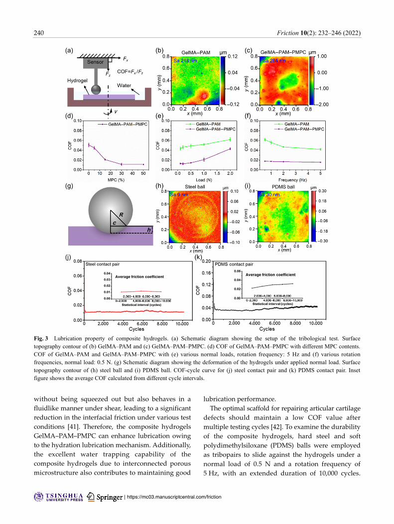
3.4 Lubrication property of composite hydrogels
A series of tribological tests were performed to reveal
the lubrication properties of the composite hydrogels,
as shown in Fig. 3(a). Before the tribological tests, the
surface roughness of GelMA–PAM and GelMA–PAM–
PMPC is measured to be 218 and 284 nm, respectively
(Figs. 3(b) and 3(c)). The COF of GelMA–PAM–PMPC
with various MPC contents is displayed in Fig. 3(d)
(load: 0.5 N; frequency: 2 Hz). The lubrication of the
composite hydrogels is highly dependent on the MPC
content, and with the increase in the MPC content,
the COF value decreases significantly from 0.052 (0%)
to 0.011 (30%). A further increase in the MPC content
to 50% does not improve the lubrication. Consequently,
this setup is applied in the tribological tests under
different conditions.
Hydrogels are viscoelastic and the surface
physicochemical properties can result in a complex
lubrication performance [36, 37]. The lubrication of
hydrogels is related to the applied load and rotation
frequency, which is investigated and illustrated in
Figs. 3(e) and 3(f). The COF values of GelMA–PAM
and GelMA–PAM–PMPC increase greatly from 0.042
to 0.063 and from 0.014 to 0.041 when the normal
load changes from 0.1 to 2 N. This result is attributed
to the effect of the normal load on the contact stress
and deformation of the hydrogels, as schematically
shown in Fig. 3(g). Under a higher normal load, with
the increase in the contact stress and indentation
depth, the lateral friction force will be greatly increased,
thus resulting in a larger COF value. Additionally, the
COF value of GelMA–PAM–PMPC slightly decreases
from 0.018 to 0.011 when the rotation frequency
increases from 0.5 to 5 Hz, and a similar trend is
obtained for GelMA. The short contact time of slip
at a high rotation frequency produces an effective
hydration interface between the tribopairs, which
can result in a reduced COF value [38, 39]. Under each
experimental condition, the COF value of GelMA–
PAM–PMPC is lower than that of GelMA–PAM,
although it has a relatively high surface roughness.
This indicates that the enhanced lubrication of the
composite hydrogels is due to the introduction of
the PMPC.
The zwitterionic phosphocholine groups (N+(CH3)3
and PO4−) in PMPC are the same as that in the PC
lipids, which form a complex with the polyelectrolyte
biomacromolecules and dominate the superlubrication
of articular cartilage based on hydration lubrication.
Phosphocholine groups can attract water molecules
to form a tenacious hydration shell around the
charges as a result of the interaction between the
water dipole and enclosed zwitterionic charges [40].
The hydration shell not only supports high pressures
240 Friction 10(2): 232–246 (2022)
| https://mc03.manuscriptcentral.com/friction
without being squeezed out but also behaves in a
fluidlike manner under shear, leading to a significant
reduction in the interfacial friction under various test
conditions [41]. Therefore, the composite hydrogels
GelMA–PAM–PMPC can enhance lubrication owing
to the hydration lubrication mechanism. Additionally,
the excellent water trapping capability of the
composite hydrogels due to interconnected porous
microstructure also contributes to maintaining good
lubrication performance.
The optimal scaffold for repairing articular cartilage
defects should maintain a low COF value after
multiple testing cycles [42]. To examine the durability
of the composite hydrogels, hard steel and soft
polydimethylsiloxane (PDMS) balls were employed
as tribopairs to slide against the hydrogels under a
normal load of 0.5 N and a rotation frequency of
5 Hz, with an extended duration of 10,000 cycles.
Fig. 3 Lubrication property of composite hydrogels. (a) Schematic diagram showing the setup of the tribological test. Surfacetopography contour of (b) GelMA–PAM and (c) GelMA–PAM–PMPC. (d) COF of GelMA–PAM–PMPC with different MPC contents. COF of GelMA–PAM and GelMA–PAM–PMPC with (e) various normal loads, rotation frequency: 5 Hz and (f) various rotationfrequencies, normal load: 0.5 N. (g) Schematic diagram showing the deformation of the hydrogels under applied normal load. Surface topography contour of (h) steel ball and (i) PDMS ball. COF-cycle curve for (j) steel contact pair and (k) PDMS contact pair. Insetfigure shows the average COF calculated from different cycle intervals.

Friction 10(2): 232–246 (2022) 241
www.Springer.com/journal/40544 | Friction
The surface roughness of the steel ball and PDMS
ball is approximately 9 and 30 nm, respectively, as
shown in Figs. 3(h) and 3(i). It is indicated from
Figs. 3(j) and 3(k) that the COF value remains relatively
unchanged during the test. A lower COF value is
obtained using steel ball as the contact tribopair,
compared with the PDMS ball, although the contact
stress between the steel ball and the hydrogels is
much larger than that between the PDMS ball and
the hydrogels (as the elastic modulus of steel is much
higher than that of PDMS). The larger COF value using
the PDMS ball as the contact tribopair is attributed to
its higher surface roughness. The above results indicate
that the composite hydrogels can sustain low friction
under extended loading cycles, especially when
sliding against a steel ball.
3.5 Cell cytotoxicity and protection for chondrocytes
degeneration
To evaluate the potential clinical application of the
composite hydrogels, we investigated the in vitro
cytotoxicity of GelMA and GelMA–PAM–PMPC on
primary mouse chondrocytes and performed tests to
determine whether GelMA and GelMA–PAM–PMPC
can protect against chondrocyte degeneration. The
hydrogel samples used in the following tests were
encapsulated with DS.
The live/dead assay and CCK-8 test were conducted
to examine the cytotoxicity of the hydrogels on
cell viability and proliferation of primary mouse
chondrocytes. Figures 4(a)–4(c) show the results of the
live/dead assay after co-culturing the chondrocytes
with GelMA and GelMA–PAM–PMPC for 1, 3, and
5 days, where the dead cells are labeled red and living
cells green. Most of the seeded chondrocytes are alive
during culturing, and the density of the cells increases
from day 1 to day 5. The viability of the chondrocytes
co-cultured with the hydrogels is almost the same
as that of the blank group for all incubation times,
indicating that the hydrogels are highly biocompatible
with no detrimental effect on the chondrocytes. Overall,
there are no significant differences in the live/dead cells
among the blank, GelMA, and GelMA–PAM–PMPC
groups at different time intervals.
Fig. 4 In vitro cell viability and proliferation of the chondrocytes incubated with the GelMA and GelMA–PAM–PMPC hydrogels for 1, 3, and 5 days. Blank group: untreated chondrocytes. (a) Representative fluorescence images of the chondrocytes in live/dead cell staining assay. (b, c) Quantitative data of the (b) live and (c) dead cells summarized from the live/dead cell staining assay. (d) Phalloidin staining showing the fibrous actin of the cytoskeleton of the cells. (e) Cell cytotoxicity of the hydrogels examined with CCK-8 assay. The hydrogels are biocompatible with the chondrocytes. NS: no significance.

242 Friction 10(2): 232–246 (2022)
| https://mc03.manuscriptcentral.com/friction
Phalloidin staining was used to observe the fibrous
actin of the cytoskeleton for the chondrocytes, and
the results are shown in Fig. 4(d). The morphology of
the cells in the three groups is complete and normal,
which indicates that the hydrogels have good
biocompatibility. The tetrazolium salt of CCK-8 is
cleaved to a soluble formazan using live cells, and
thus the absorbance is directly related to the number
of viable cells. As displayed in Fig. 4(e), the CCK-8
test indicates that there are no significant differences
between the experimental groups (GelMA and
GelMA–PAM–PMPC) and the blank group at each
time interval, and the number of viable cells is greatly
increased on days 3 and 5 for GelMA and GelMA–
PAM–PMPC. This further indicates that cell prolifera-
tion is not affected by co-culturing the chondrocytes
with the hydrogels. In summary, the live/dead assay,
phalloidin staining, and CCK-8 test all indicate
that the hydrogels are biocompatible and have no
cytotoxicity to chondrocytes.
Multiple factors are involved in the pathogenesis
of osteoarthritis, such as reactive oxygen species,
mechanical loading, and inflammatory cytokines (e.g.,
TNF-α and IL-1β). It is considered that chondrocyte
degeneration, which is accepted to be the most
significant feature of osteoarthritis, is closely
related to the inflammatory cytokines. For example,
inflammatory cytokines play an important role in the
development of osteoarthritis, and the inflammatory
environment contributes to extracellular matrix
degradation and chondrocyte hypertrophy. The
manifestation of osteoarthritis at the cellular level
is due to the increase in catabolic genes and the
degradation of anabolic genes. In this study, IL-1β and
TNF-α were used to treat chondrocytes to mimic the
symptoms of osteoarthritis. The protective effect of the
drug-loaded hydrogels on chondrocyte degeneration
was examined after co-culturing for 24 h. The mRNA
expression levels of anabolic genes (aggrecan and
COL2A1) and catabolic genes (MMP13 and ADAMTS5)
were examined by RT-qPCR analysis. As displayed in
Fig. 5, the mRNA expression of aggrecan and COL2A1
in the hydrogel samples (GelMA and GelMA–PAM–
PMPC) is significantly higher than that of the blank
group, and the mRNA expression of MMP13 and
ADAMTS5 in the hydrogel samples is significantly
lower than that of the blank group. These results
indicate that the DS-loaded hydrogels have chondro-
protective potential for inflammatory cytokine-induced
chondrocytes, and consequently can be an effective
scaffold for cartilage defects in the treatment of
osteoarthritis.
Fig. 5 RT-qPCR analysis showing the mRNA expression levels of anabolic genes (a, e) Aggrecan, (b, f) COL2A1, and catabolic genes (c, g) MMP13, (d, h) ADAMTS5 in IL-1β and TNF-α treated chondrocytes, which are incubated with the GelMA and GelMA–PAM–PMPChydrogels. n = 3, *P < 0.05, **P < 0.01, compared with the blank group.

Friction 10(2): 232–246 (2022) 243
www.Springer.com/journal/40544 | Friction
4 Conclusions
In this study, we successfully synthesized gelatin-based
composite hydrogels, namely GelMA–PAM–PMPC,
with biomimetic hydration lubrication and sustained
drug release via photopolymerization, which could
be used as a scaffold for cartilage defects in the treat-
ment of osteoarthritis. The lubrication test indicated
that the composite hydrogels maintained a relatively
low COF under different experimental conditions
and extended duration, which was attributed to the
hydration lubrication mechanism of the zwitterionic
phosphocholine headgroups. The drug release test
showed that the composite hydrogels efficiently
encapsulated the anti-inflammatory drug of DS and
achieved a sustained release behavior. Additionally,
the in vitro test demonstrated that the composite
hydrogels were biocompatible and protected the
chondrocytes from inflammatory cytokine-induced
degeneration, upregulating the mRNA expression
levels of anabolic genes and downregulating that of
catabolic genes. In summary, the composite hydrogels
prepared herein, with the dual functions of biomimetic
hydration lubrication and sustained drug release,
provide a promising approach for repairing cartilage
defects in the treatment of osteoarthritis.
Acknowledgements
This study was financially supported by the National
Natural Science Foundation of China (Nos. 51675296,
21868011, and 81772372), Shanghai Municipal Science
Foundation (No. SYXF011803), Tsinghua University-
Peking Union Medical College Hospital Initiative
Scientific Research Program (No. 20191080593),
the National Key R&D Program of China (No.
2017YFC1103800), Foshan-Tsinghua Innovation Special
Fund (FTISF), Research Fund of State Key Laboratory
of Tribology, Tsinghua University, China (No. SKLT-
2020C11), and Ng Teng Fong Charitable Foundation
(No. 202-276-132-13).
Electronic Supplementary Material: Supplementary
material (synthesis of photopolymerization initiator,
fabrication of polydimethylsiloxane ball, and calibra-
tion curve of diclofenac sodium) is available in the
online version of this article at https://doi.org/10.1007/
s40544-020-0437-5.
Open Access: This article is licensed under a Creative
Commons Attribution 4.0 International License, which
permits use, sharing, adaptation, distribution and
reproduction in any medium or format, as long as
you give appropriate credit to the original author(s) and
the source, provide a link to the Creative Commons
licence, and indicate if changes were made.
The images or other third party material in this
article are included in the article’s Creative Commons
licence, unless indicated otherwise in a credit line to
the material. If material is not included in the article’s
Creative Commons licence and your intended use is
not permitted by statutory regulation or exceeds the
permitted use, you will need to obtain permission
directly from the copyright holder.
To view a copy of this licence, visit
http://creativecommons.org/licenses/by/4.0/.
References
[1] Ji X, Yan Y, Sun T, Zhang Q, Wang Y, Zhang M, Zhang
H, Zhao X. Glucosamine sulphate-loaded distearoyl
phosphocholine liposomes for osteoarthritis treatment:
combination of sustained drug release and improved
lubrication. Biomater Sci 7: 2716–2728 (2019)
[2] Zheng Y, Yang J, Liang J, Xu X, Cui W, Deng L, Zhang
H. Bioinspired hyaluronic acid/phosphorylcholine
polymer with enhanced lubrication and anti-inflammation.
Biomacromolecules 20: 4135–4142 (2019)
[3] Wan L, Wang Y, Tang X, Sun Y, Luo J, Zhang H.
Biodegradable lubricating mesoporous silica nanoparticles
for osteoarthritis therapy. Friction 10(1): 68–79 (2022)
[4] Tan X, Sun Y, Sun T, Zhang H. Mechanised lubricating silica
nanoparticles for on-command cargo release on simulated
surfaces of joint cavities. Chem Commun 55: 2593–2596
(2019)
[5] Pan Y, Xiao C, Tan H, Yuan G, Li J, Li S, Jia Y, Xiong D,
Hu X, Niu X. Covalently injectable chitosan/chondroitin
sulfate hydrogel integrated gelatin/heparin microspheres
for soft tissue engineering. Int J Polym Mater 70: 149–157
(2021)
[6] Wang M, Chen J, Li W, Zang F, Liu X, Qin S. Paclitaxel-
nanoparticles-loaded double network hydrogel for local
treatment of breast cancer after surgical resection. Mater Sci
Eng C Mater Biol Appl 114: 111046 (2020)

244 Friction 10(2): 232–246 (2022)
| https://mc03.manuscriptcentral.com/friction
[7] Liu Y, Xiong D. Self-healable polyacrylic acid-polyacrylamide-
ferric ion dual-crosslinked hydrogel with good biotribological
performance as a load-bearing surface. J Appl Polym Sci
137: 48499 (2019)
[8] Chen K, Chen G, Wei S, Yang X, Zhang D, Xu L. Preparation
and property of high strength and low friction PVA-HA/PAA
composite hydrogel using annealing treatment. Mater Sci
Eng C Mater Biol Appl 91: 579–588 (2018)
[9] Gong J, Katsuyama Y, Kurokawa T, Osada Y. Double-
network hydrogels with extremely high mechanical strength.
Adv Mater 15: 1155–1158 (2003)
[10] Han L, Liu K, Wang M, Wang K, Fang L, Chen H, Zhou J,
Lu X. Mussel-inspired adhesive and conductive hydrogel
with long-lasting moisture and extreme temperature tolerance.
Adv Funct Mater 28: 1704195 (2018)
[11] Gan D, Xu T, Xing W, Ge X, Fang L, Wang K, Ren F,
Lu X. Mussel-inspired contact-active antibacterial hydrogel
with high cell affinity, toughness, and recoverability. Adv
Funct Mater 29: 1805964 (2019)
[12] Liu K, Han L, Tang P, Yang K, Gan D, Wang X, Wang K,
Ren F, Fang L, Xu X, et al. An anisotropic hydrogel based
on mussel-inspired conductive ferrofluid composed of
electromagnetic nanohybrids. Nano Lett 19: 8343–8356 (2019)
[13] Wang J, Lin L, Cheng Q, Jiang L. A strong bio-inspired
layered pnipam-clay nanocomposite hydrogel. Angew Chem
Int Ed 51: 4676–4680 (2012)
[14] Dai X, Zhang Y, Gao L, Bai T, Wang W, Cui Y, Liu W. A
mechanically strong, highly stable, thermoplastic, and self-
healable supramolecular polymer hydrogel. Adv Mater 27:
3566–3571 (2015)
[15] Wang H, Zhou L, Liao J, Ning C, Tan G. Cell-laden
photocrosslinked gelma-dexma copolymer hydrogels with
tunable mechanical properties for tissue engineering. J Mater
Sci Mater Med 25: 2173–2183 (2014)
[16] Visser J, Gawlitta D, Benders K E, Malda J. Endochondral
bone formation in gelatin methacrylamide hydrogel with
embedded cartilage-derived matrix particles. Biomaterials
37: 174–182 (2015)
[17] Yue K, Santiago G T, Alvarez M M, Tamayol A, Annabi N,
Khademhosseini A. Synthesis, properties, and biomedical
applications of gelatin methacryloyl (gelma) hydrogels.
Biomaterials 73: 254–271 (2015)
[18] Elomaa L, Keshi E, Sauer I M, Weinhart M. Development
of GelMA/PCL and dECM/PCL resins for 3D printing of
acellular in vitro tissue scaffolds by stereolithography. Mater
Sci Eng C Mater Biol Appl 112: 110958 (2020)
[19] Su R, Zihlmann C, Akbari M, Tang X, Khademhosseini A.
Reduced graphene oxide-gelma hybrid hydrogels as scaffolds
for cardiac tissue engineering. Small 12: 3677–3689 (2016)
[20] Yuk H, Zhang T, Lin S, Parada G A, Zhao X. Tough bonding
of hydrogels to diverse non-porous surfaces. Nat Mater 15:
190–196 (2016)
[21] Hu X, Vatankhah-Varnoosfaderani M, Zhou J, Li Q, Sheiko
S S. Weak hydrogen bonding enables hard, strong, tough,
and elastic hydrogels. Adv Mater 27: 6899–6905 (2016)
[22] Jahn S, Seror J, Klein J. Lubrication of articular cartilage.
Annu Rev Biomed Eng 18: 235–258 (2016)
[23] Seror J, Merkher Y, Kampf N, Collinson L, Day A J,
Maroudas A, Klein J. Normal and shear interactions
between hyaluronan-aggrecan complexes mimicking possible
boundary lubricants in articular cartilage in synovial joints.
Biomacromolecules 13: 3823–3832 (2012)
[24] Wang Y, Sun Y, Gu Y, Zhang H. Articular cartilage-inspired
surface functionalization for enhanced lubrication. Adv Mater
Interfaces 6: 1900180 (2019)
[25] Klein J. Hydration lubrication. Friction 1: 1–23 (2013)
[26] Moro T, Takatori Y, Ishihara K, Konno T, Takigawa Y,
Matsushita T, Chung U, Nakamura K, Kawaguchi H. Surface
grafting of artificial joints with a biocompatible polymer for
preventing periprosthetic osteolysis. Nat Mater 3: 829–836
(2004)
[27] Kyomoto M, Moro T, Saiga K, Miyaji F, Kawaguchi H,
Takatori Y, Nakamura K, Ishihara K. Lubricity and stability
of poly(2-methacryloyloxyethyl phosphorylcholine) polymer
layer on Co–Cr–Mo surface for hemi-arthroplasty to prevent
degeneration of articular cartilage. Biomaterials 31: 658–668
(2010)
[28] Kyomoto M, Moro T, Yamane S, Hashimoto M, Takatori
Y, Ishihara K. Poly(ether-ether-ketone) orthopedic bearing
surface modified byself-initiated surface grafting of poly(2-
methacryloyloxyethyl phosphorylcholine). Biomaterials 34:
7829–7839 (2013)
[29] Daniele M A, Adams A A, Naciri J, North S H, Ligler F S.
Interpenetrating networks based on gelatin methacrylamide
and PEG formed using concurrent thiol click chemistries
for hydrogel tissue engineering scaffolds. Biomaterials 35:
1845–1856 (2014)
[30] Liu B, Wang Y, Miao Y, Zhang X, Fan Z, Singh G, Zhang
X, Xu K, Li B, Hu Z, et al. Hydrogen bonds autonomously
powered gelatin methacrylate hydrogels with super-elasticity,
self-heal and underwater self-adhesion for sutureless skin and
stomach surgery and E-skin. Biomaterials 171: 83–96 (2018)
[31] Brown L, Zhang H, Blunt L, Barrans S. Reproduction of
fretting wear at the stem-cement interface in total hip
replacement. Proc Inst Mech Eng Part H-J Eng Med 221:
963–971 (2007)
[32] Zhang H, Zhang S, Luo J, Liu Y, Qian S, Liang F,
Huang Y. Investigation of protein adsorption mechanism
and biotribological properties at simulated stem-cement

Friction 10(2): 232–246 (2022) 245
www.Springer.com/journal/40544 | Friction
interface. J Tribol-Trans ASME 135: 032301 (2013)
[33] Livak K J, Schmittgen T D. Analysis of relative gene
expression data using real-time quantitative PCR and the
2(-Delta Delta C(T)) method. Methods 25: 402–408 (2001)
[34] Yan Y, Sun T, Zhang H, Ji X, Sun Y, Zhao X, Deng L,
Jin Q, Cui W, Santos H, et al. Euryale ferox seed-inspired
superlubricated nanoparticles for treatment of osteoarthritis.
Adv Funct Mater 29: 1807559 (2019)
[35] Lin P, Zhang R, Wang X, Zhou F. Articular cartilage inspired
bilayer tough hydrogel prepared by interfacial modulated
polymerization showing excellent combination of high
load-bearing and low friction performance. ACS Macro Lett
5: 1191–1195 (2016)
[36] Shoaib T, Heintz J, Lopez-Berganza J A, Muro-Barrios R,
Egner S A, Espinosa-Marzal R. Stick-slip friction reveals
hydrogel lubrication mechanisms. Langmuir 34: 756–765
(2018)
[37] Gombert Y, Simic R, Roncoroni F, Dubner M, Geue T,
Spencer N D. Structuring hydrogel surfaces for tribology.
Adv Mater Interfaces 6: 1901320 (2019)
[38] Zhang X, Wang J, Jin H, Wang S, Song W. Bioinspired
supramolecular lubricating hydrogel induced by shear force.
J Am Chem Soc 140: 3186–3189 (2018)
[39] Liu G, Cai M, Zhou F, Liu W. Charged polymer brushes-
grafted hollow silica nanoparticles as a novel promising
material for simultaneous joint lubrication and treatment.
J Phys Chem B 118: 4920–4931 (2014)
[40] Chen H, Sun T, Yan Y, Ji X, Sun Y, Zhao X, Qi J, Cui W,
Deng L, Zhang H. Cartilage matrix-inspired biomimetic
superlubricated nanospheres for treatment of osteoarthritis.
Biomaterials 242: 119931 (2020)
[41] Seror J, Zhu L, Goldberg R, Day A J, Klein J. Supramolecular
synergy in the boundary lubrication of synovial joints. Nat
Commun 6: 6497 (2015)
[42] Ma S, Scaraggi M, Wang D, Zhou F. Stick-slip friction
reveals hydrogel lubrication mechanisms. Adv Func Mater
25: 7366–7374 (2016)
Kuan ZHANG. He received his
B.S. in chemical engineering and
technology in 2016 from Hainan
University. He is currently a joint master student in
Tsinghua University. His research interests include
biotribology and soft materials.
Jielai YANG. He is a Ph.D. student
at Department of Orthopedics &
Shanghai Institute of Traumatology
and Orthopedics, Ruijin Hospital,
Shanghai Jiaotong University School of Medicine.
His research focuses on the novel biomaterials
for osteochondral regeneration and osteoarthritis
management.
Hongyu ZHANG. He received his
B.S. degree from Tianjin University,
China (2005) and Ph.D. degree from
University of Huddersfield, UK
(2009). He is an associate professor
at the State Key Laboratory of
Tribology, Department of Mechanical
Engineering, Tsinghua University, China. His research
interests focus on the development of lubricating
biomaterials such as nanoparticles, coatings, hydrogels,
and electrospun nanofibers, which integrate the
multi-disciplinary knowledge including biotribology,
chemistry, materials science, and medicine to address
clinical issues, e.g., osteoarthritis, anti-tissue/cell/
bacteria adhesion, bone tissue engineering, etc.

246 Friction 10(2): 232–246 (2022)
| https://mc03.manuscriptcentral.com/friction
Xiangyang XU. He is director of foot
and ankle surgery, Ruijin Hospital,
Shanghai Jiao Tong University School
of Medicine. He is deputy director
of foot and ankle surgery, Chinese
Medical Association. He got his Ph.D. degree from
Shanghai Medical University. His research focuses on
diagnosis and treatment of diseases of the foot and
ankle, with expertise in ankle instability and ankle
osteoarthritis.
Bo WANG. He is a professor at
College of Chemical and Biological
Engineering, Shandong University
of Science and Technology, China.
He received his B.S. degree in Hainan University
and Ph.D. degree in Zhejiang University, China. His
research covers green chemistry, bio-catalysis, and
asymmetric synthesis.