photodegradable hydrogels for capture, detection, and...
TRANSCRIPT
Photodegradable Hydrogels Hot PaperDOI: 10.1002/anie.201404323
Photodegradable Hydrogels for Capture, Detection, and Release ofLive Cells**Dong-Sik Shin,* Jungmok You, Ali Rahimian, Tam Vu, Christian Siltanen, Arshia Ehsanipour,Gulnaz Stybayeva, Julie Sutcliffe, and Alexander Revzin*
Abstract: Cells may be captured and released using a photo-degradable hydrogel (photogel) functionalized with antibod-ies. Photogel substrates were used to first isolate human CD4 orCD8 T-cells from a heterogeneous cell suspension and then torelease desired cells or groups of cells by UV-induced photo-degradation. Flow cytometry analysis of the retrieved cellsrevealed approximately 95% purity of CD4 and CD8 T-cells,suggesting that this substrate had excellent specificity. Todemonstrate the possibility of sorting cells according to theirfunction, photogel substrates that were functionalized withanti-CD4 and anti-TNF-a antibodies were prepared. Singlecells captured and stimulated on such substrates were identifiedby the fluorescence “halo” after immunofluorescent stainingand could be retrieved by site-specific exposure to UV lightthrough a microscope objective. Overall, it was demonstratedthat functional photodegradable hydrogels enable the capture,analysis, and sorting of live cells.
The presence of specific cells in bodily fluids and the functionof these cells may serve as diagnostic markers of infections ormalignancies.[1] This is particularly true for immune cellanalysis, as the diagnosis of the acquired immune deficiencysyndrome (AIDS) is based on the enumeration of CD4T-cells, whereas latent tuberculosis (TB) is determined byidentifying cytokine-producing CD4 T-cells.[2] Microengi-neered surfaces and microfluidic devices offer multipleadvantages for the analysis of immune cells in comparisonto standard immunological techniques. These advantages are
related to small volumes, controlled washing conditions,miniature size of the device, and multiplexing capabilities.[3]
However, the ability to capture various immune cells andanalyze the function of these cells needs to be combined withthe possibility of selectively retrieving cells of interest bymaking use of either their surface marker expression orfunction. For example, physicochemical stimulation (e.g.,exposure to EDTA or enzymes or temperature control) canbe applied to retrieve cells from the surfaces.[4] Alternatively,cells can be electrochemically released from electrodes.[5]
However, these methods often threaten cell viability becauseof harsh and physiologically unfavorable conditions, and maybe suboptimal for selective cell retrieval. Light-triggered cellretrieval is particularly promising as it may be used for thehigh-throughput retrieval of small groups of cells or singlecells.[6] Photolabile molecules containing ortho-nitrobenzylmoieties are particularly appealing as photolabile groupsbecause they may be degraded by 365 nm UV light and causeminimal damage to biomolecules or living cells, including T-cells, human umbilical vascular endothelial cells (HUVECs),human leukemic cells, cervical carcinoma cells, and humanmesenchymal stem cells.[7] Our group has previously utilizedsurfaces that contain cell-capture antibodies linked to ortho-nitrobenzyl moieties for the capture and release of cells;[6a]
however, we encountered non-specific binding and foulingissues with this approach. At the same time, we have beenmaking extensive use of poly(ethylene glycol) (PEG) hydro-gels as non-fouling coatings for the printing of antibody arraysand the capture of immune cells.[8] Herein, we describe thedevelopment of an antibody-functionalized photodegradablePEG hydrogel; we hypothesized that such a coating shouldlead to excellent non-fouling properties while additionallyenabling site-specific cell release.
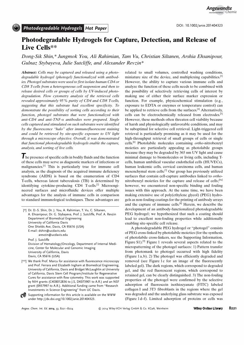
A photodegradable PEG hydrogel or “photogel” consistsof PEG cross-linked by photolabile moieties (for the synthesisof photolabile cross-linkers, see the Supporting Information,Figure S1).[9] Figure 1 reveals several aspects related to themicropatterning of the photogel surfaces: 1) Pattern transferfrom photomask to photogel occurred with high fidelity(Figure 1a, b). 2) The photogel was efficiently degraded andremoved (see Figure 1c for an image of the fluorescentlylabeled gel). The dark regions, which correspond to degradedgel, and the red fluorescent regions, which correspond toretained gel, can be clearly distinguished. 3) The non-foulingproperties of the photogel were confirmed by the selectiveadsorption of fluorescein isothiocyanate (FITC) labeledcollagen I and 3T3 fibroblasts in the regions where the gelwas degraded and the underlying glass substrate was exposed(Figure 1d–f). Limited adsorption of proteins or cells was
[*] Dr. D.-S. Shin, Dr. J. You, A. Rahimian, T. Vu, C. Siltanen,A. Ehsanipour, Dr. G. Stybayeva, Prof. J. Sutcliffe, Prof. A. RevzinDepartment of Biomedical EngineeringUniversity of California DavisOne Shields Ave, Davis, CA 95616 (USA)E-mail: [email protected]
Prof. J. SutcliffeDivision of Hematology/Oncology, Department of Internal Medi-cine, Center for Molecular and Genomic ImagingUniversity of California, DavisDavis, CA 95616 (USA)
[**] We thank Prof. Marcu for assistance with fluorescence microscopyand Prof. Ferrara and Elizabeth Ingham at Biomedical Engineering,University of California, Davis and Bridget McLaughlin at Universityof California, Davis Stem Cell Program/Institute for RegenerativeCures for assistance with flow cytometry. This work was supportedby NIH grants (C4EB012836 to J.S, DK073901 to A.R.) and an NSFgrant (0937997 to A.R.). Additional funding came from “ResearchInvestments in Science Engineering” from UC Davis.
Supporting information for this article is available on the WWWunder http://dx.doi.org/10.1002/anie.201404323.
AngewandteChemie
8221Angew. Chem. Int. Ed. 2014, 53, 8221 –8224 � 2014 Wiley-VCH Verlag GmbH & Co. KGaA, Weinheim
seen on the photogel. Through optimization experiments thatare described in detail in the Supporting Information andFigure S2, we arrived at the following procedure for gelformation and gel degradation. The photogel was grafted onacrylated glass surfaces by radical polymerization initiated byammonium persulfate (AP) and tetramethylethylenediamine(TEMED). Regions of the photogel on the glass surface werethen exposed to 365 nm UV light from a fluorescence micro-scope for various exposure times. The photodegradationefficiency was determined by measuring the degraded area asa function of exposure time. The photodegradation amountedto more than 95 % after 20 seconds of exposure(600 mWcm�2). Importantly, a simple adjustment of micro-scope aperture allowed modulating the diameter of theexposure area from 50 to 200 mm, offering the possibility toaddress small groups of cells or single cells (Figure S3).
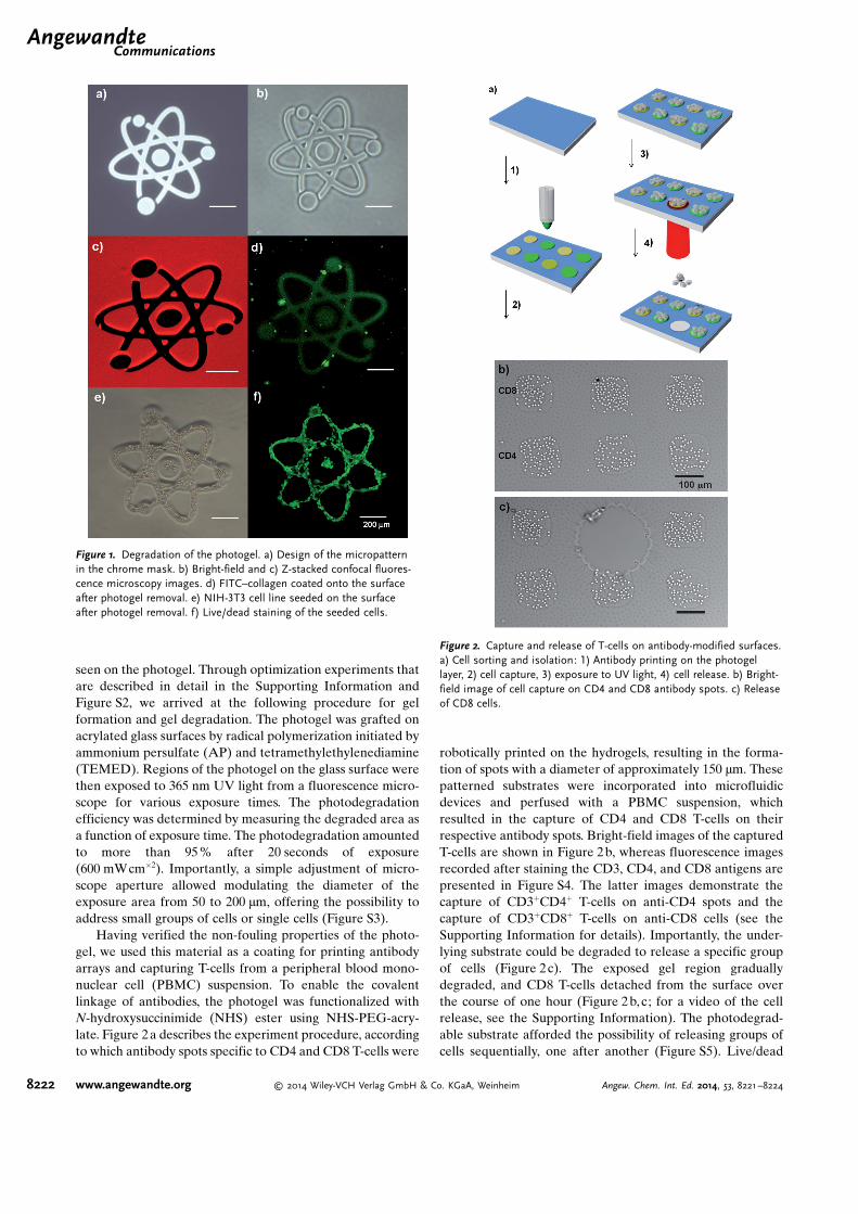
Having verified the non-fouling properties of the photo-gel, we used this material as a coating for printing antibodyarrays and capturing T-cells from a peripheral blood mono-nuclear cell (PBMC) suspension. To enable the covalentlinkage of antibodies, the photogel was functionalized withN-hydroxysuccinimide (NHS) ester using NHS-PEG-acry-late. Figure 2a describes the experiment procedure, accordingto which antibody spots specific to CD4 and CD8 T-cells were
robotically printed on the hydrogels, resulting in the forma-tion of spots with a diameter of approximately 150 mm. Thesepatterned substrates were incorporated into microfluidicdevices and perfused with a PBMC suspension, whichresulted in the capture of CD4 and CD8 T-cells on theirrespective antibody spots. Bright-field images of the capturedT-cells are shown in Figure 2b, whereas fluorescence imagesrecorded after staining the CD3, CD4, and CD8 antigens arepresented in Figure S4. The latter images demonstrate thecapture of CD3+CD4+ T-cells on anti-CD4 spots and thecapture of CD3+CD8+ T-cells on anti-CD8 cells (see theSupporting Information for details). Importantly, the under-lying substrate could be degraded to release a specific groupof cells (Figure 2c). The exposed gel region graduallydegraded, and CD8 T-cells detached from the surface overthe course of one hour (Figure 2 b, c; for a video of the cellrelease, see the Supporting Information). The photodegrad-able substrate afforded the possibility of releasing groups ofcells sequentially, one after another (Figure S5). Live/dead
Figure 2. Capture and release of T-cells on antibody-modified surfaces.a) Cell sorting and isolation: 1) Antibody printing on the photogellayer, 2) cell capture, 3) exposure to UV light, 4) cell release. b) Bright-field image of cell capture on CD4 and CD8 antibody spots. c) Releaseof CD8 cells.
Figure 1. Degradation of the photogel. a) Design of the micropatternin the chrome mask. b) Bright-field and c) Z-stacked confocal fluores-cence microscopy images. d) FITC–collagen coated onto the surfaceafter photogel removal. e) NIH-3T3 cell line seeded on the surfaceafter photogel removal. f) Live/dead staining of the seeded cells.
.AngewandteCommunications
8222 www.angewandte.org � 2014 Wiley-VCH Verlag GmbH & Co. KGaA, Weinheim Angew. Chem. Int. Ed. 2014, 53, 8221 –8224
staining of retrieved cells showed over 97% viability,suggesting that photodegradation of the gel did not havedeleterious effects on the cells (Figure S6).
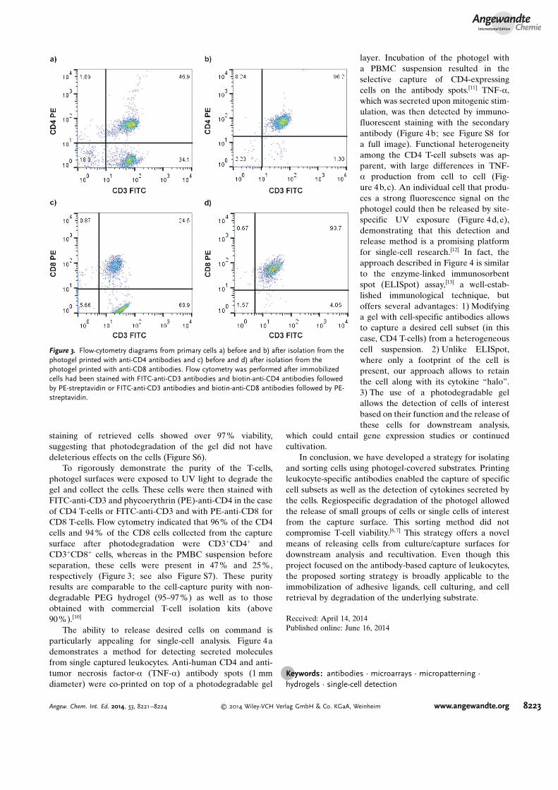
To rigorously demonstrate the purity of the T-cells,photogel surfaces were exposed to UV light to degrade thegel and collect the cells. These cells were then stained withFITC-anti-CD3 and phycoerythrin (PE)-anti-CD4 in the caseof CD4 T-cells or FITC-anti-CD3 and with PE-anti-CD8 forCD8 T-cells. Flow cytometry indicated that 96% of the CD4cells and 94 % of the CD8 cells collected from the capturesurface after photodegradation were CD3+CD4+ andCD3+CD8+ cells, whereas in the PMBC suspension beforeseparation, these cells were present in 47% and 25%,respectively (Figure 3; see also Figure S7). These purityresults are comparable to the cell-capture purity with non-degradable PEG hydrogel (95–97 %) as well as to thoseobtained with commercial T-cell isolation kits (above90%).[10]
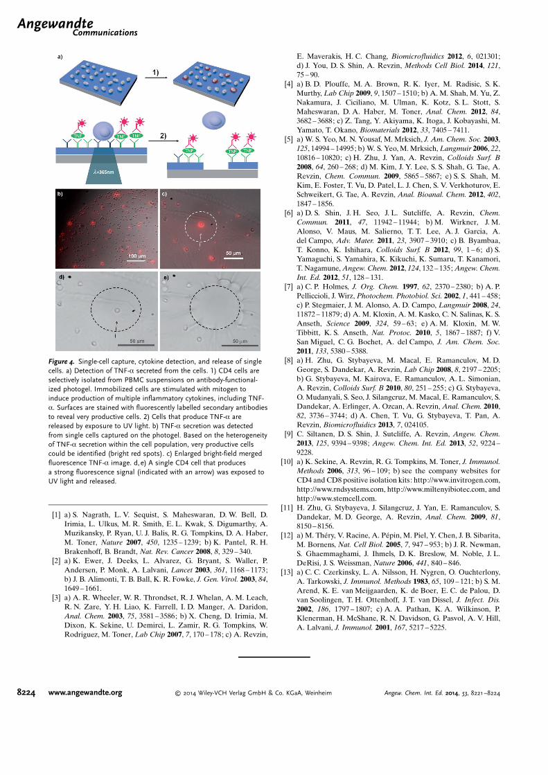
The ability to release desired cells on command isparticularly appealing for single-cell analysis. Figure 4ademonstrates a method for detecting secreted moleculesfrom single captured leukocytes. Anti-human CD4 and anti-tumor necrosis factor-a (TNF-a) antibody spots (1 mmdiameter) were co-printed on top of a photodegradable gel
layer. Incubation of the photogel witha PBMC suspension resulted in theselective capture of CD4-expressingcells on the antibody spots.[11] TNF-a,which was secreted upon mitogenic stim-ulation, was then detected by immuno-fluorescent staining with the secondaryantibody (Figure 4b; see Figure S8 fora full image). Functional heterogeneityamong the CD4 T-cell subsets was ap-parent, with large differences in TNF-a production from cell to cell (Fig-ure 4b,c). An individual cell that produ-ces a strong fluorescence signal on thephotogel could then be released by site-specific UV exposure (Figure 4d,e),demonstrating that this detection andrelease method is a promising platformfor single-cell research.[12] In fact, theapproach described in Figure 4 is similarto the enzyme-linked immunosorbentspot (ELISpot) assay,[13] a well-estab-lished immunological technique, butoffers several advantages: 1) Modifyinga gel with cell-specific antibodies allowsto capture a desired cell subset (in thiscase, CD4 T-cells) from a heterogeneouscell suspension. 2) Unlike ELISpot,where only a footprint of the cell ispresent, our approach allows to retainthe cell along with its cytokine “halo”.3) The use of a photodegradable gelallows the detection of cells of interestbased on their function and the release ofthese cells for downstream analysis,
which could entail gene expression studies or continuedcultivation.
In conclusion, we have developed a strategy for isolatingand sorting cells using photogel-covered substrates. Printingleukocyte-specific antibodies enabled the capture of specificcell subsets as well as the detection of cytokines secreted bythe cells. Regiospecific degradation of the photogel allowedthe release of small groups of cells or single cells of interestfrom the capture surface. This sorting method did notcompromise T-cell viability.[6,7] This strategy offers a novelmeans of releasing cells from culture/capture surfaces fordownstream analysis and recultivation. Even though thisproject focused on the antibody-based capture of leukocytes,the proposed sorting strategy is broadly applicable to theimmobilization of adhesive ligands, cell culturing, and cellretrieval by degradation of the underlying substrate.
Received: April 14, 2014Published online: June 16, 2014
.Keywords: antibodies · microarrays · micropatterning ·hydrogels · single-cell detection
Figure 3. Flow-cytometry diagrams from primary cells a) before and b) after isolation from thephotogel printed with anti-CD4 antibodies and c) before and d) after isolation from thephotogel printed with anti-CD8 antibodies. Flow cytometry was performed after immobilizedcells had been stained with FITC-anti-CD3 antibodies and biotin-anti-CD4 antibodies followedby PE-streptavidin or FITC-anti-CD3 antibodies and biotin-anti-CD8 antibodies followed by PE-streptavidin.
AngewandteChemie
8223Angew. Chem. Int. Ed. 2014, 53, 8221 –8224 � 2014 Wiley-VCH Verlag GmbH & Co. KGaA, Weinheim www.angewandte.org
[1] a) S. Nagrath, L. V. Sequist, S. Maheswaran, D. W. Bell, D.Irimia, L. Ulkus, M. R. Smith, E. L. Kwak, S. Digumarthy, A.Muzikansky, P. Ryan, U. J. Balis, R. G. Tompkins, D. A. Haber,M. Toner, Nature 2007, 450, 1235 – 1239; b) K. Pantel, R. H.Brakenhoff, B. Brandt, Nat. Rev. Cancer 2008, 8, 329 – 340.
[2] a) K. Ewer, J. Deeks, L. Alvarez, G. Bryant, S. Waller, P.Andersen, P. Monk, A. Lalvani, Lancet 2003, 361, 1168 – 1173;b) J. B. Alimonti, T. B. Ball, K. R. Fowke, J. Gen. Virol. 2003, 84,1649 – 1661.
[3] a) A. R. Wheeler, W. R. Throndset, R. J. Whelan, A. M. Leach,R. N. Zare, Y. H. Liao, K. Farrell, I. D. Manger, A. Daridon,Anal. Chem. 2003, 75, 3581 – 3586; b) X. Cheng, D. Irimia, M.Dixon, K. Sekine, U. Demirci, L. Zamir, R. G. Tompkins, W.Rodriguez, M. Toner, Lab Chip 2007, 7, 170 – 178; c) A. Revzin,
E. Maverakis, H. C. Chang, Biomicrofluidics 2012, 6, 021301;d) J. You, D. S. Shin, A. Revzin, Methods Cell Biol. 2014, 121,75 – 90.
[4] a) B. D. Plouffe, M. A. Brown, R. K. Iyer, M. Radisic, S. K.Murthy, Lab Chip 2009, 9, 1507 – 1510; b) A. M. Shah, M. Yu, Z.Nakamura, J. Ciciliano, M. Ulman, K. Kotz, S. L. Stott, S.Maheswaran, D. A. Haber, M. Toner, Anal. Chem. 2012, 84,3682 – 3688; c) Z. Tang, Y. Akiyama, K. Itoga, J. Kobayashi, M.Yamato, T. Okano, Biomaterials 2012, 33, 7405 – 7411.
[5] a) W. S. Yeo, M. N. Yousaf, M. Mrksich, J. Am. Chem. Soc. 2003,125, 14994 – 14995; b) W. S. Yeo, M. Mrksich, Langmuir 2006, 22,10816 – 10820; c) H. Zhu, J. Yan, A. Revzin, Colloids Surf. B2008, 64, 260 – 268; d) M. Kim, J. Y. Lee, S. S. Shah, G. Tae, A.Revzin, Chem. Commun. 2009, 5865 – 5867; e) S. S. Shah, M.Kim, E. Foster, T. Vu, D. Patel, L. J. Chen, S. V. Verkhoturov, E.Schweikert, G. Tae, A. Revzin, Anal. Bioanal. Chem. 2012, 402,1847 – 1856.
[6] a) D. S. Shin, J. H. Seo, J. L. Sutcliffe, A. Revzin, Chem.Commun. 2011, 47, 11942 – 11944; b) M. Wirkner, J. M.Alonso, V. Maus, M. Salierno, T. T. Lee, A. J. Garcia, A.del Campo, Adv. Mater. 2011, 23, 3907 – 3910; c) B. Byambaa,T. Konno, K. Ishihara, Colloids Surf. B 2012, 99, 1 – 6; d) S.Yamaguchi, S. Yamahira, K. Kikuchi, K. Sumaru, T. Kanamori,T. Nagamune, Angew. Chem. 2012, 124, 132 – 135; Angew. Chem.Int. Ed. 2012, 51, 128 – 131.
[7] a) C. P. Holmes, J. Org. Chem. 1997, 62, 2370 – 2380; b) A. P.Pelliccioli, J. Wirz, Photochem. Photobiol. Sci. 2002, 1, 441 – 458;c) P. Stegmaier, J. M. Alonso, A. D. Campo, Langmuir 2008, 24,11872 – 11879; d) A. M. Kloxin, A. M. Kasko, C. N. Salinas, K. S.Anseth, Science 2009, 324, 59 – 63; e) A. M. Kloxin, M. W.Tibbitt, K. S. Anseth, Nat. Protoc. 2010, 5, 1867 – 1887; f) V.San Miguel, C. G. Bochet, A. del Campo, J. Am. Chem. Soc.2011, 133, 5380 – 5388.
[8] a) H. Zhu, G. Stybayeva, M. Macal, E. Ramanculov, M. D.George, S. Dandekar, A. Revzin, Lab Chip 2008, 8, 2197 – 2205;b) G. Stybayeva, M. Kairova, E. Ramanculov, A. L. Simonian,A. Revzin, Colloids Surf. B 2010, 80, 251 – 255; c) G. Stybayeva,O. Mudanyali, S. Seo, J. Silangcruz, M. Macal, E. Ramanculov, S.Dandekar, A. Erlinger, A. Ozcan, A. Revzin, Anal. Chem. 2010,82, 3736 – 3744; d) A. Chen, T. Vu, G. Stybayeva, T. Pan, A.Revzin, Biomicrofluidics 2013, 7, 024105.
[9] C. Siltanen, D. S. Shin, J. Sutcliffe, A. Revzin, Angew. Chem.2013, 125, 9394 – 9398; Angew. Chem. Int. Ed. 2013, 52, 9224 –9228.
[10] a) K. Sekine, A. Revzin, R. G. Tompkins, M. Toner, J. Immunol.Methods 2006, 313, 96 – 109; b) see the company websites forCD4 and CD8 positive isolation kits: http://www.invitrogen.com,http://www.rndsystems.com, http://www.miltenyibiotec.com, andhttp://www.stemcell.com.
[11] H. Zhu, G. Stybayeva, J. Silangcruz, J. Yan, E. Ramanculov, S.Dandekar, M. D. George, A. Revzin, Anal. Chem. 2009, 81,8150 – 8156.
[12] a) M. Th�ry, V. Racine, A. P�pin, M. Piel, Y. Chen, J. B. Sibarita,M. Bornens, Nat. Cell Biol. 2005, 7, 947 – 953; b) J. R. Newman,S. Ghaemmaghami, J. Ihmels, D. K. Breslow, M. Noble, J. L.DeRisi, J. S. Weissman, Nature 2006, 441, 840 – 846.
[13] a) C. C. Czerkinsky, L. A. Nilsson, H. Nygren, O. Ouchterlony,A. Tarkowski, J. Immunol. Methods 1983, 65, 109 – 121; b) S. M.Arend, K. E. van Meijgaarden, K. de Boer, E. C. de Palou, D.van Soolingen, T. H. Ottenhoff, J. T. van Dissel, J. Infect. Dis.2002, 186, 1797 – 1807; c) A. A. Pathan, K. A. Wilkinson, P.Klenerman, H. McShane, R. N. Davidson, G. Pasvol, A. V. Hill,A. Lalvani, J. Immunol. 2001, 167, 5217 – 5225.
Figure 4. Single-cell capture, cytokine detection, and release of singlecells. a) Detection of TNF-a secreted from the cells. 1) CD4 cells areselectively isolated from PBMC suspensions on antibody-functional-ized photogel. Immobilized cells are stimulated with mitogen toinduce production of multiple inflammatory cytokines, including TNF-a. Surfaces are stained with fluorescently labelled secondary antibodiesto reveal very productive cells. 2) Cells that produce TNF-a arereleased by exposure to UV light. b) TNF-a secretion was detectedfrom single cells captured on the photogel. Based on the heterogeneityof TNF-a secretion within the cell population, very productive cellscould be identified (bright red spots). c) Enlarged bright-field mergedfluorescence TNF-a image. d, e) A single CD4 cell that producesa strong fluorescence signal (indicated with an arrow) was exposed toUV light and released.
.AngewandteCommunications
8224 www.angewandte.org � 2014 Wiley-VCH Verlag GmbH & Co. KGaA, Weinheim Angew. Chem. Int. Ed. 2014, 53, 8221 –8224