biodegradable hydrogels from silk sericin & gelatin...
TRANSCRIPT
BIODEGRADABLE HYDROGELS FROM SILK
SERICIN & GELATIN: DEVELOPMENT AND
CHARACTERIZATION FOR MEDICAL
APPLICATIONS
A DISSERTATION SUBMITTED
BY
DEEPTHI M. NAIR
IN PARTIAL FULFILLMENT OF THE REQUIREMENTS
FOR THE DEGREE OF
MASTER OF PHILOSOPHY - 2015
SREE CHITRA TIRUNAL INSTITUTE FOR MEDICAL SCIENCES
AND
TECHNOLOGY
TRIVANDRUM-695011
2 P a g e
DECLARATION
I, Deepthi. M. Nair, hereby declare that I had personally carried out the work depicted
in the dissertation entitled „Biodegradable hydrogels from silk sericin: Development
and characterization for medical applications’ under the direct supervision of Dr.
Roy Joseph, Scientist F, Polymer Processing Laboratory, Biomedical Technology
Wing, Sree Chitra Tirunal Institute for Medical Sciences and Technology, Trivandrum,
Kerala, India. External help sought are acknowledged.
DEEPTHI. M. NAIR
3 P a g e
SREE CHITRA TIRUNAL INSTITUTE FOR MEDICAL SCIENCES &
TECHNOLOGY
TRIVANDRUM-695011,INDIA
(An Institute of National Importance under Govt. of India with the status of University
By an act of Parliament in1980)
CERTIFICATE
This is to certify that the dissertation entitled “Biodegradable hydrogels from silk
sericin: Development and characterization for medical applications” submitted
by Deepthi. M. Nair has been carried out in partial fulfilment for the Degree of
Master of Philosophy in Biomedical Technology to be awarded by this Institute.
The entire work was done by her under my supervision and guidance at Polymer
Processing Laboratory, Biomedical Technology Wing, Sree Chitra Tirunal Institute
for Medical Sciences and Technology (SCTIMST), Trivandrum-695012.
Thiruvanathapuram
(Dr. Roy Joseph)
Date: (Research Supervisor)
4 P a g e
The Dissertation
Entitled
BIODEGRADABLE HYDROGELS FROM SILK SERICIN &
GELATIN: DEVELOPMENT AND CHARACTERIZATION
FOR MEDICAL APPLICATIONS
Submitted
By
DEEPTHI M. NAIR
For
MASTER OF PHILOSOPHY
Of
SREE CHITRA TIRUNAL INSTITUTE FOR MEDICAL SCIENCES AND
TECHNOLOGY
TRIVANDRUM-695011
Evaluated and approved
By
Signature Signature
Name of Supervisor Examiner’s Name and Designation
5 P a g e
ACKNOWLEDGEMENT
I bow before "GOD Almighty'', without whose help and blessings, this work would not
have been a success.
I wish to thank Director, Head, Associate Head and Biomedical Technology Wing,
SCTIMST, for giving me this opportunity to carry out my MPhil research project and
for providing all the facilities.
I sincerely express my gratitude and respect to Dr. Roy Joseph, Scientist F, Polymer
Processing Laboratory, SCTIMST, for his inspiring guidance, scholarly supervision
and providing all facilities to complete my M.Phil dissertation.
I am also obliged to Deputy Registrar, for the academic support bestowed on me to
complete this M.Phil Programme.
I extend my sincere gratitude to Dr. Annie John, Scientist F, SIC, TEM Lab for giving
me the opportunity to do cell culture works. I am extremely grateful to Dr. Lissy
Krishnan, SIC, Thrombosis Research Unit, for providing me human dermal fibroblasts
for my cell culture works.
I am also grateful to Dr. V. Kalliyanakrishnan, SIC , Dental Products Laboratory, for
providing FTIR & Micro CT facilities to carry out my work. With great pleasure I
extend my heartfelt thanks to Dr. Anilkumar P. R ,TIC , for providing
spectroflourimetry facility and for his help & valuable advices.
I would like to thank Dr. H.K Varma, SIC, BCL and Mr.Nishad for providing SEM
facility. I would also like to thank Dr. K. Sreenivasan, SIC ,LPA , Dr. Radhakumary ,
for providing TGA facility. I wish to thank Dr. Anugya Bhatt, Ms.Priyanka , TRU for
Hemolysis, Ms.Renu Ramesh, Ms.Subha, for their help and support.
6 P a g e
I would like to express my profound sense of gratitude to Ms.Resmi. R, for teaching me
cell culture works and also for her sincere support and guidance towards the
completion of my project work.
My heartfelt thanks to Ms. Vibha, Dr.Renjith, Ms. Rethikala, Ms.Lakshmy for their
help and support rendered on me.
I would like to mention my special thanks to Ms. Parvathy, Ms.Mayuri, Ms. Remya,
Ms.Jincy, Mr. Susanth, Mr. Sudhin Thampi, Ms. Sreelakshmi, Ms. Nayana, ,Ms.
Anupama, Ms. Dhanya, for their whole hearted co-operation for my work & for the
services rendered towards my thesis preparation. They all deserve special thanks for
their immense support & friendship.
I extend my sincere thanks to all my friends of the M.Phil 2014-2015 batch and all my
classmates during the course work and for their support, joyful times and for the ever
memorable days in this campus.
Last but not the least, my parents and sister deserve special mention for their prayers,
affection and encouragement which has been an inspiring, driving and motivating
force in my life.
7 P a g e
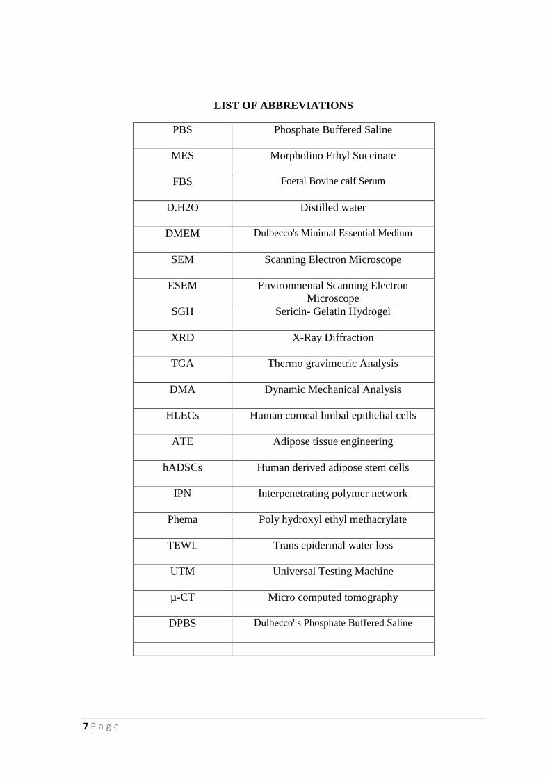
LIST OF ABBREVIATIONS
PBS
Phosphate Buffered Saline
MES
Morpholino Ethyl Succinate
FBS
Foetal Bovine calf Serum
D.H2O
Distilled water
DMEM
Dulbecco's Minimal Essential Medium
SEM
Scanning Electron Microscope
ESEM
Environmental Scanning Electron
Microscope
SGH
Sericin- Gelatin Hydrogel
XRD
X-Ray Diffraction
TGA
Thermo gravimetric Analysis
DMA
Dynamic Mechanical Analysis
HLECs
Human corneal limbal epithelial cells
ATE
Adipose tissue engineering
hADSCs
Human derived adipose stem cells
IPN
Interpenetrating polymer network
Phema
Poly hydroxyl ethyl methacrylate
TEWL
Trans epidermal water loss
UTM
Universal Testing Machine
µ-CT
Micro computed tomography
DPBS
Dulbecco' s Phosphate Buffered Saline
8 P a g e
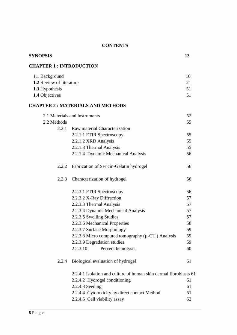
CONTENTS
SYNOPSIS 13
CHAPTER 1 : INTRODUCTION
1.1 Background 16
1.2 Review of literature 21
1.3 Hypothesis 51
1.4 Objectives 51
CHAPTER 2 : MATERIALS AND METHODS
2.1 Materials and instruments 52
2.2 Methods 55
2.2.1 Raw material Characterization
2.2.1.1 FTIR Spectroscopy 55
2.2.1.2 XRD Analysis 55
2.2.1.3 Thermal Analysis 55
2.2.1.4 Dynamic Mechanical Analysis 56
2.2.2 Fabrication of Sericin-Gelatin hydrogel 56
2.2.3 Characterization of hydrogel 56
2.2.3.1 FTIR Spectroscopy 56
2.2.3.2 X-Ray Diffraction 57
2.2.3.3 Thermal Analysis 57
2.2.3.4 Dynamic Mechanical Analysis 57
2.2.3.5 Swelling Studies 57
2.2.3.6 Mechanical Properties 58
2.2.3.7 Surface Morphology 59
2.2.3.8 Micro computed tomography (µ-CT ) Analysis 59
2.2.3.9 Degradation studies 59
2.2.3.10 Percent hemolysis 60
2.2.4 Biological evaluation of hydrogel 61
2.2.4.1 Isolation and culture of human skin dermal fibroblasts 61
2.2.4.2 Hydrogel conditioning 61
2.2.4.3 Seeding 61
2.2.4.4 Cytotoxicity by direct contact Method 61
2.2.4.5 Cell viability assay 62
9 P a g e
2.2.4.6 Pico green assay 62
2.2.4.7 Two dimensional cell attachment 63
2.2.4.8 Actin/Hoechst Staining 63
CHAPTER 3: RESULTS AND DISCUSSION 64
3.1 Raw material characterization
3.1.1 FTIR Spectroscopy 64
3.1.1 X-Ray Diffraction 66
3.1.2 Thermal Analysis 67
3.1.3 Dynamic Mechanical Analysis 69
3.2 Fabrication Sericin-Gelatin Hydrogel 69
3.2.1 Process optimisation Fabrication
3.3 Characterization of Hydrogel
3.3.1 FTIR Spectroscopy 71
3.3.2 XRD Analysis 72
3.3.3 Thermal Analysis 74
3.3.4 Dynamic Mechanical Analysis 76
3.3.5 Swelling studies 76
3.3.6 Mechanical Properties 78
3.3.7 Surface morphology 81
3.3.8 Micro computed tomography (µ-CT ) Analysis 82
3.3.9 Degradation study 84
3.3.10 Percent hemolysis 84
3.4 Biological evaluation of hydrogels
3.4.1 Cytocompatability of hydrogel 85
3.4.2 Cell Viability Assay 85
3.4.3 Biochemical evaluation for Cell Proliferation- Pico green 87
3.4.4 Two dimensional Cell Attachment 88
3.4.5 Cell attachment assay 88
CHAPTER 4: SUMMARY AND CONCLUSION 90
REFERENCES 93
APPENDIX
101
10 P a g e
LIST OF FIGURES
Fig:
No
Caption Page
no:
1 Sericin and fibroin 22
2 Properties of sericin 25
3 Applications of sericin 30
4 Tensile testing of hydrogel materials 58
5 FTIR Spectrum of Gelatin 64
6 FTIR Spectrum of Sericin 65
7 XRD spectrum of gelatin 66
8 XRD Spectrum of Sericin 66
9 A) TGA curve of Sericin B) TGA curve of Gelatin 67,68
10 Temperature dependence of storage modulus and tan delta of
Gelatin 69
11 Hydrogel fabricated in different shapes 70
12 (A) Reaction mixture before gelation, (B) After gelation
(B) 71
13 FTIR spectrum of sericin, gelatin , sericin –gelatin blend (SGB)
and hydrogel (SGH) 73
14 XRD pattern of sericin, gelatin SGB and SGH 74
15 TGA curves of gelatin, hydrogel and sericin 75
16 Temperature dependence of E‟ and tan δ for SGH 76
17 Swelling behaviour of hydrogel in different media 77
18 Effect of medium on percentage swelling 78
19 Tensile strength of hydrogels immersed in different media
(D.H2O, PBS and MES) 79
20 Youngs modulus of hydrogels immersed in different media
(D.H2O, PBS and MES) 79
21 Elongation at break % of hydrogels immersed in different
media (D.H2O, PBS and MES) 80
22 Stress- strain curves of hydrogels immersed in different media:
(a) PBS (b) MES (c) Distilled water 80
23 -A) : SEM images lyophilized hydrogel after swelling in water 81
24 Frequency distribution of pore diameter 82
25 A) 3D morphology image B) Pore size distribution image, C)
Thickness distribution image
83
26 Pore size distribution of SGH washed in PBS 83
27 Weight loss (%) pattern of hydrogel in PBS, MES and D.H2O
due to degradation over a period of 3 weeks 84
28 Percentage hemolysis of SGH with control 85
29 Phase contrast microscopy images showing cytocompatibility 86
30 Graphical representation of trypan blue exclusion assay 87
31 Quantitative data on cell proliferation after 3 days & 6 days 87
32 ESEM images of cell seeded scaffold 88
11 P a g e
LIST OF TABLES
Table
No
Caption Page No:
1 List of Materials used
52
2 List of Equipments used
53
3 Process parameters used for the preparation of
hydrogel
70
4 Peak assignments of hydrogel along with the
gelatine, sericin and their blend.
73
5 Mechanical properties of dried SGH sample
80
6 Porosity volume percentage of SGH sample
83

12 P a g e
SYNOPSIS
Silk protein, sericin has a long history of being discarded as a waste material in the
textile industry by a process called degumming. But nowadays, sericin has
become a hot topic in the area of tissue engineering and regenerative medicine. It
is a hydrophilic protein with large amount of OH groups. Exploration of the
potential applications of sericin has just begun. Sericin has got wide applications
in various industrial sectors. In the cosmetic industry, it is being used as a
moisturizer since it resembles natural moisturizing factor (NMR). In addition, it
has the property of inhibiting tyrosinase enzyme, which is responsible for melanin
biosynthesis. Also, it is a natural ingredient for the food industry because its
consumption enhances the bioavailability of Zinc, Iron, Magnesium and Calcium.
Sericin plays an important role in wound healing, by promoting collagen
synthesis. It possesses antimicrobial property and accelerates mammalian cell
growth during serum deprivation. Moreover, studies are going on the basis of
transforming sericin into a biomaterial by cross-linking with natural as well as
synthetic polymers.
Without a doubt, gelatin has an important role in the area of biomaterial science. It
has being widely used in a variety of forms since it is a biodegradable,
biocompatible natural polymer. Hydrogels are three dimensional polymeric
networks with high water holding capacity. They can be cross-linked either by
physical or chemical methods. They are extremely useful for a variety of
applications in pharmaceutical as well as medical industry. They are capable of
retaining large amounts of water, possess soft and rubbery consistency and
13 P a g e
provide a moist environment. These features make them closely resemble with
living tissues. Since it has a low interfacial tension, its adsorption with proteins in
the body fluid is negligible. Also, the cross linked network contributes high
porosity, which is required for cell proliferation and attachment. An effort is put
forth in the present study to develop a hydrogel from silk protein sericin and
gelatin without any chemical cross-linkers. Owing to this background information,
it can be hypothesized that:
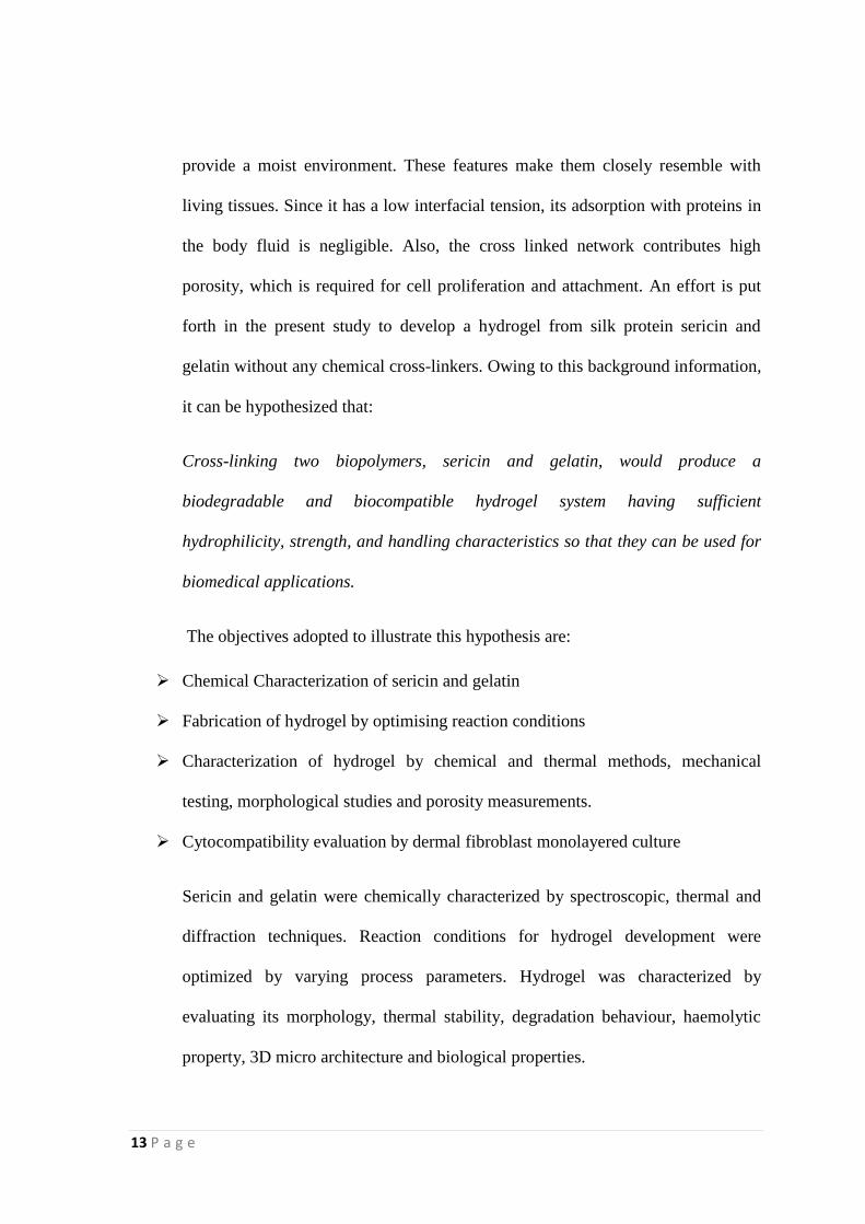
Cross-linking two biopolymers, sericin and gelatin, would produce a
biodegradable and biocompatible hydrogel system having sufficient
hydrophilicity, strength, and handling characteristics so that they can be used for
biomedical applications.
The objectives adopted to illustrate this hypothesis are:
Chemical Characterization of sericin and gelatin
Fabrication of hydrogel by optimising reaction conditions
Characterization of hydrogel by chemical and thermal methods, mechanical
testing, morphological studies and porosity measurements.
Cytocompatibility evaluation by dermal fibroblast monolayered culture
Sericin and gelatin were chemically characterized by spectroscopic, thermal and
diffraction techniques. Reaction conditions for hydrogel development were
optimized by varying process parameters. Hydrogel was characterized by
evaluating its morphology, thermal stability, degradation behaviour, haemolytic
property, 3D micro architecture and biological properties.
14 P a g e
Surface morphology and 3D micro architecture was evaluated by using scanning
electron microscopy and Micro Computer Tomography techniques. The hydrogel
was found to possess interconnected porous network with an average pore
diameter of 23.5µm. Mechanical testing of hydrogel showed sufficient strength of
the material. Biological characterization of the hydrogel revealed its
cytocompatibility with limited ability for cell attachment.
15 P a g e
CHAPTER 1
INTRODUCTION
1.1 BACKGROUND
Hydrogels are three dimensional materials with the ability to absorb large amount
of water, retaining its structural integrity. The history of hydrogels began in 1960,
when Wichterle and Lim introduced the use of hydrophilic networks of cross-
linked poly(2-hydroxyethyl methacrylate) (pHEMA,) as soft contact lens material
(Wichterle, 1960). Hydrogels have become a hot topic during the last few decades,
as indicated by the increasing number of papers on hydrogel-based materials
published from 1995 onwards. Among the large variety of definitions, the most
acceptable one for hydrogel is referred to as hydrogels are water-swollen, cross-
linked polymeric structures containing: (1) covalent bonds produced by the
reaction of one or more co monomers, (2) physical cross-links due to chain
entanglements, (3) association bonds including hydrogen bonds or strong van der
Waals interactions between chains, or (4) crystallites bringing together two or
more macromolecular chains (Peppas, 2000).
Silk worm, Bombyx Mori is gaining a much important space in the area of
biomedical technology because of its two powerful protein members Sericin and
Fibroin. Fibroin is widely used in textiles, industrial and medical applications
(Panilaitis, 2003). Sericin is a glue-like protein that holds fibroin fibres together.
For centuries, it has been discarded as a waste material by a process called
degumming in textile industry. Exposing this unutilised sericin to environment
poses serious threat due to high oxygen demand for its degradation (Aramwit,
16 P a g e
2012). Polymers of natural origin always hold a better position for use as a
biomaterial. Since they possess compatibility with the living tissues, favourable
cell-material interaction, non toxic degradation products, minimal immunological
responses and adverse reactions to a greater extent, biomaterial from bio-
polymers, particularly proteins, is highly relevant. Current study focuses on the
fabrication of a hydrogel by cross-linking sericin with gelatin.
Sericin is a polypeptide composed of 18 amino acids with high content of serine
and aspartate (Takasu, 2002). The molecular weight of sericin depends on the
method of its extraction from silkworm cocoons. Ethanol precipitation of sericin
yields a molecular weight of 800 Da (Yoko, 2002). It is a hydrophilic
macromolecule with high content of hydroxyl and carboxyl groups (Patel, 2011).
Structural analysis of sericin gene revealed two closely related mRNAs of length
11.0 and 9.6 kilobases. A strong homologous region in the sericin and
fibroingenes at their corresponding 5‟ flanking sequences was identified.
(Harumasa et al. 1982). Sericin is soluble in hot water and insoluble in cold water;
however, it is easily hydrolyzed. In this process, the long protein breaks down into
smaller molecules which are easy to solubilise in hot water (Gulrajani, 1988).
Sericin is highly advantageous because of its special properties like oxidation
resistance, antibacterial and UV resistance. It absorbs and release moisture easily
(Mondal, 2007). Reports are available on the diverse biological activities of
sericin, such as anti-oxidation (Kato, 1998), anti-bacterium (Sasaki,2000) anti-
coagulation (Takeuchi, 2005) and its ability to promote cell growth and
differentiation (Miyazaki,2004). Sericin is reported to be composed of three
polypeptides, sericin A, M and P with amino acids like serine, aspartic acid,
17 P a g e
glutamic acid and glycine in large proportions (Yoko, 2014). In the field of
regenerative medicine, owing to its biodegradability, easy availability, and
hydrophilicity with many polar side groups, sericin is mostly copolymerized,
cross-linked, or blended with other polymers to form various scaffolds in order to
help obtain improved properties for relevant biomedical applications, such as skin
regeneration (Wang, 2014).
Sericin is widely utilised in cosmetic industry. Excessive Trans epidermal water
loss (TEWL) is one of the causes of dry skin and skin moisturizers have been used
to overcome it. The silk sericin has resemblance with the natural moisturizing
factor (NMR) (Patel, 2011). Moreover, it is being regarded as a valuable natural
ingredient for food industry, since it increases the bioavailability of Zn, Fe, Mg
and Ca (Masahiro et al, 2000).
Acidic or basic hydrolysis of collagen yields a biodegradable protein, Gelatin,
involving breakage of the collagen's triple helix structure into random coils
(Bouhadir, 2001). Gelatin is composed of a variety of amino acids forming a
linear polymer with molecular weight varying between 15,000-250,000 Daltons.
The sol state of gelatin transforms into the gel state upon cooling of aqueous
solution of gelatin, which involves a partial rearrangement of the gelatin structure
from random coil into triple helix (Bigi, 2004). Gelatin is widely used in
biomedical applications, for example in tissue engineering, wound dressing, gene
therapy, and drug delivery due to its high biocompatibility and biodegradability.
Gelatin will easily form hydrogel in water at room temperature, but is fragile.
18 P a g e
Gelatin is tremendously utilised in the field of biomaterials by cross-linking with
synthetic or natural polymers. Sufficient mechanical properties may be attained by
modifying primary amino acid residues present in gelatin like lysine,
hydroxylysine, histidine, and arginine. There are two types of gelatin available:
Type A and B. Type A gelatin is obtained from porcine skin by acidic treatment
while Type B gelatin from bovine skin by alkaline treatment. Both types of gelatin
are rich in glycine followed by proline and arginine. Gelatin is able to form
physical and chemical gels. Physical gel is formed by merely cooling gelatine
solution to room temperature. Chemical gels are formed with the help of a cross-
linker. A cross-linker forms covalent links C-N with the amine groups of gelatin
coils (lysine, hydroxylysine, histidine).
The main goal of the study is to fabricate sericin- gelatin cross-linked hydrogel.
High serine content in sericin makes it highly hydrophilic. Also the richness of
acidic amino acids in sericin and basic amino acids in gelatin is being targeted
here. If the carboxyl group in sericin is made available to crosslink with the amino
groups in gelatin, it can form a covalently cross linked network. Reports are
available on this by using chemical crosslinkers like formaldehyde,
glutaraldehydes (Jayakrishnan and Jameela 1996), glyceraldehyde, hydrogen
peroxide, benzene, sulphonic acid, guanidine hydrochloride and genipin (Huang et
al. 1999). But here, the crosslinking is aimed to achieve without the involvement
of any chemical cross linkers, thus eliminating the possibility any kind of
toxicities.

19 P a g e
Hydrogels are widely accepted as a scaffold which supports effective cell growth
and proliferation because of their high porosity that leads to high permeability of
oxygen, nutrients, and metabolites. At the same time hydrogels can protect the
cells from the host immune system and high molecular weight complexes like
immunoglobulin (Li, 2006; Auslander, 2012). Furthermore, the softness and
flexibility rendered by hydrogels reduces the mechanical stress and friction on
cells and also on adjacent tissue upon transplantation (Wilson, 2013). Not only
that, the highly hydrophilic nature of hydrogels lead to high water content,
providing a tissue like environment to the attached cells (Billiet, 2012). On the
basis of these advantages, it is expected that the hydrogel resulting from the cross
linking of sericin and gelatin can offer possibilities for future studies in tissue
engineering. Indeed, gelatin and sericin are already known as a favourable
material for tissue engineering and regenerative medicine. However, the
combination of both these proteins without any cross-linkers is not yet studied. In
the present work, we attempt to fabricate hydrogel from sericin and gelatin by
optimising the reaction conditions.
20 P a g e
1.2. REVIEW OF LITERATURE
As designed by the mother nature, the silk spinnerets of Bombyx mori produce a
delicate twin thread of silk and surround it with a protective cover of sericin
during the spinning procedure. This principle is now being imitated and perfected
to provide an innovative active ingredient for the pharma industry. For centuries
ago, silk has been a scientific curiosity. Silk fibers have been used in the form of
sutures. Recently silk solutions have become an ideal candidate in the field of
biomaterials such as gels, sponges and films, for medical applications. Surface
properties of silk can be tuned through chemical modification of amino acid side
chains so as to immobilize cellular growth factors. Silks with precise features can
be developed through molecular engineering of silk sequences: for example cell
recognition or mineralization. The mode of processing and the subsequent content
of β sheet crystallinity will in turn relate to the degradability of silk biomaterials.
Different silk biomaterials have been a successful substratum for the growth of
several primary cells and cell lines so as to demonstrate a range of biological
outcomes. Also they proved to be biocompatible in vitro and in vivo. Silk
scaffolds have been a flourishing nominee in wound healing and in tissue
engineering of bone, cartilage, tendon and ligament tissues (Vepari, 2007).
1.2.1 Silk protein sericin
Silk is mainly composed of two types of proteins, silk fibroin and sericin. Sericin
contributes about 20-30 per cent of total cocoon weight. It is a biopolymer with
high serine content (32%) and 18 amino acids, including essential amino acids.
Isolation of sericin from silk thread can be done by different modes. Sericin‟s
21 P a g e
characteristic features like solubility, molecular weight, and gelling properties are
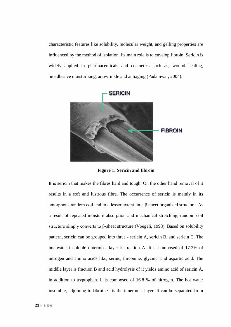
influenced by the method of isolation. Its main role is to envelop fibroin. Sericin is
widely applied in pharmaceuticals and cosmetics such as, wound healing,
bioadhesive moisturizing, antiwrinkle and antiaging (Padamwar, 2004).
Figure 1: Sericin and fibroin
It is sericin that makes the fibres hard and tough. On the other hand removal of it
results in a soft and lustrous fibre. The occurrence of sericin is mainly in its
amorphous random coil and to a lesser extent, in a β-sheet organized structure. As
a result of repeated moisture absorption and mechanical stretching, random coil
structure simply converts to β-sheet structure (Voegeli, 1993). Based on solubility
pattern, sericin can be grouped into three - sericin A, sericin B, and sericin C. The
hot water insoluble outermost layer is fraction A. It is composed of 17.2% of
nitrogen and amino acids like, serine, threonine, glycine, and aspartic acid. The
middle layer is fraction B and acid hydrolysis of it yields amino acid of sericin A,
in addition to tryptophan. It is composed of 16.8 % of nitrogen. The hot water
insoluble, adjoining to fibroin C is the innermost layer. It can be separated from
22 P a g e
fibroin by treating with hot dilute acid or alkali. Its acid hydrolysis yields proline
besides amino acids of sericin B. It also contains sulphur and 16.6 % of nitrogen
(Shaw, 1951).
Sericin has been classified into various species derived from its relative
solubilities. Many other researchers have also nominated various fractions of
sericin with respect to their dissolution behaviour like sericin A and B, or sericin I,
II, III, and IV, or S1, S2, S3, S4, and S5, and as α, β, and γ modification
(Komatsu,1996). Soluble form of sericin always attains a random coil molecular
conformation. But the β-sheet structure is more difficult to dissolve. The more
crystalline structure resulting from repeated moisture absorption contributes to
reduced solubility. Moisture absorption increases crystallinity by making
molecular aggregation structure denser. Three layer structure of sericin was
reported by -ray study. The outer layer contained some fibre direction filaments,
middle layer exhibits cross-fibre direction filaments, and the inner layer shows
longitudinal filaments (Wang, 1985). Casting temperature also influences the
structure of sericin. As the casting temperature lowers, sericin molecules attain
more β-sheet structure rather than random coil.
1.2.2 Availability
The cultivated silkworm, Bombyx mori Linn., is a lepidopteran molecular model.
Now a days, it is being considered as an important economic insect that can serve
as an ideal molecular genetic resource so as to resolve a broad range of biological
troubles. B. Mori, in its final stage of larval development, produces a bulk amount
23 P a g e
of silk proteins. The middle silk gland is the storehouse of these proteins. At the
end of the fifth instar, these are expelled through the anterior duct and spinneret.
Sericulture is now in the path of technological development as silkworm serve as
a bio factory for the production of useful protein using the silk gland. Owing to
this background, silkworm can be classified as a value added biomaterial for
medical application, silk protein fibroin and sericin as a biomaterial and other seri
-by products (Mondal, 2007).
1.2.3 Properties
Sericin is insoluble in cold water, however, it is easily hydrolyzed. In this process,
the long protein breaks down into smaller molecules which are easy to solubilise
in hot water (Gulrajani, 1988). Sericin is highly advantageous because of its
special properties like oxidation resistant, antibacterial, UV resistant and absorbs
and release moisture easily, inhibitory activity of tyrosine and kinase, etc.
(Mondal, 2007).
24 P a g e
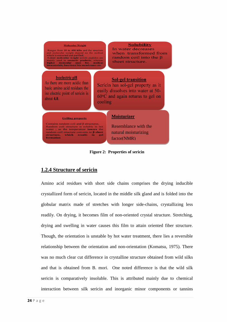
Figure 2: Properties of sericin
1.2.4 Structure of sericin
Amino acid residues with short side chains comprises the drying inducible
crystallized form of sericin, located in the middle silk gland and is folded into the
globular matrix made of stretches with longer side-chains, crystallizing less
readily. On drying, it becomes film of non-oriented crystal structure. Stretching,
drying and swelling in water causes this film to attain oriented fiber structure.
Though, the orientation is unstable by hot water treatment, there lies a reversible
relationship between the orientation and non-orientation (Komatsu, 1975). There
was no much clear cut difference in crystalline structure obtained from wild silks
and that is obtained from B. mori. One noted difference is that the wild silk
sericin is comparatively insoluble. This is attributed mainly due to chemical
interaction between silk sericin and inorganic minor components or tannins
Moisturizer
Resemblance with the
natural moisturizing
factor(NMR)
25 P a g e
present within wild silk (Tsukada, 1983). Sericin with random coil structure was
found to be extracted from the liquid silk and fresh cocoon shells of a silkworm
hope with 5- 10 % b-structure and no α-helix (Lizuka,1969).
Also, in aqueous solution, sericin exhibits both random coil and β- structure, but
lacks α-helix (Komatsu, 1975). The relationship between solubility and molecular
conformation is confirmed by studying sericin from both the liquid silk in the silk
gland and the cocoon filament (Komatsu, 1982). Till now, it is evident that
occurrence of sericin is mainly in its random coil or β-structure. Also it is believed
that β structure is intrinsic to liquid silk. Later, a circular dichroism spectrum
showed that sericin extracted from liquid silk for 45 minutes with water contains a
small fraction (10%) of α-helix. During the dissolution of the liquid silk in water,
part of the sericin IV become a white suspension due to the coexisting cocoon
yarn wax but does not coagulate and the β structure was originally present in the
liquid silk. β -structure sericin is more insoluble than random coil sericin
(Komatsu 1980). Though, these reports are not in conformity with reports of
Tsukada (1983).
The hygroscopic conditions favour the molecular transition of sericin from its
random coil to β - structure (Komatsu, 1980). Ishikawa and Hirabayashi (1968)
reported that sericins have a cross β structure in a water solution of sericin
containing 50% dioxane (Iizuka, 1969) or methanol (Ishikawa et al., 1987)
Komatsu (1975) believed that the sericin fraction closest to fibroin in the cocoon
filament has molecular chains also arranged in cross β structure, rather than
having the main axis of crystallites oriented at right angle to the fiber axis. The
intra- molecular bonds of sericin having random coils are broken by the absorption
26 P a g e
of water molecules and the folded structure becomes unfolded into an extended
structure and is transformed into a β structure. β Structure is more stable regarding
energy. Part of the sericin thus transformed into β structure is fixed by its new
intermolecular hydrogen bonds and remains crystallized even when the water
molecules are removed by drying. In a new cycle of water absorption, the
crystalline structure already formed, remains unaffected, whereas, a further
fraction of random coils state crystallizes into a β structure thereby increasing the
portion of β structure and promotes crystallization. Heating during moisture
absorption activates the thermal motions of segments and accelerates
transformation. Thus the sericin gets modified in the direction of difficult
solubility due to repeated moisture absorption and loss.
According to scientific data, sericin of cocoon shell fall under two proportions:
(1) α-sericin and (2) β-sericin. Outer layer of shell is composed of α - Sericin and
β -sericin in the inner layer. α -sericin contains lesser C and H and somewhat more
N and O than the β -sericin (Bose et al., 1989). α -sericin is more soluble in the
boiling water than β - sericin. Sadov et al., (1987) have drawn a fact that native
sericin is mixture of two substances, Sericin A and Sericin B. Degumming of silk
results in the isolation of sericin in aqueous solution. It is not a single chemical
substance rather a mixture of at least two substances. There are scientific
descriptions that the sericin layers are formed from the outside to inside in the
order of I, II, III to cover the fibroin on the cocoon thread. These three fractions of
sericin were found to have different solubilities and named them sericin fractions
I, II, and III. The order was based on the ease of hot water dissolution and their
27 P a g e
ratios were approximately 40:40:20. Sericin I was found to be amorphous in
nature whereas the other fractions as crystalline. Komatsu (1975) observed a
fourth fraction of sericin which was much harder to dissolve as sericin IV. This
fraction was reported to have higher specific gravity and crystallinity.
Robson (1985) reported that sericin may be separated into sericin I, II, III and IV
by their different solubilities in hot water and assessing the degree of solubility by
UV absorption. The greatest sericin content is present in the outer layer of cocoon
whereas the least sericin proportion is present in the innermost layer of a cocoon.
1.2.5 Amino acid composition
The total amount of hydroxy amino acids in sericin is 45.8 per cent. There are 42.3
per cent of polar amino acid and 12.2 per cent of nonpolar amino acid residues
(Padamwar, 2004). Sericin of mulberry wild silkworm, Bombyx morimandarina,
is characterized by the same kind of amino acids as the domesticated silkworm, B.
mori (Yamada, 1978). On the other hand, the wild silkworm sericin is composed
of serine, proline, methoinine, glucosamine, galactosamine and histidine in lower
amount and throeonine, glutamic acid, cystine and phenyl amine in higher amount.
The inner layer of cocoons was found to be rich in threonine, galactosamine and
glucosamine than the outer layer. Moreover, the sericin extracted from the floss
showed high contents of serine, glycine, valine and tyrosine but low contents in
threonine, aspartic acid, alanine, cystine, leucine, glucosamine, galactosamine,
lysine and histidine as compared with the sericin of cocoon layer.
28 P a g e
More non polar amino acid and less polar amino acids were observed in the wild
silkworm sericin when compared to that of the domesticated silkworm sericin. It
was showed that the amino acid composition of sericin extracted from the cocoon
is species specific. Structural analysis of sericin gene revealed two closely related
mRNAs of length 11.0 and 9.6 kilobases. A strong homologous region in the
sericin and fibroingenes at their corresponding 5‟ flanking sequences was
identified (Harumasa et al., 1982). The co-efficient of pattern-similarity in amino
acid composition was high between the sericin of wild silkworm and domesticated
silkworms, while sericin of the wild silkworm such as Antheraea or Phylosamia
showed significantly low similarity.
1.2.6 Applications in the market
Sericin is an excellent member in the cosmetic area that possesses moisture
absorption and preservative ability. The hydrophilicity of sericin renders it to
absorb water 50 times high than that of glycerine. Also it was shown that sericin
has a capability to inhibit tyrosine kinase activity, which is involved in the
production of skin melanin. Applying it on the skin will result in softness and
smoothness and makes the hair soft and flexible. It is also an ideal candidate in
shaping of hair. Sericin is a less irritant moisturizer and is gentle on a delicate
skin. Sericin cream is available in the market with wound healing properties, since
it is essential in collagen synthesis. Dermal and corneal wound healing have been
tried by many researchers using sericin.
29 P a g e
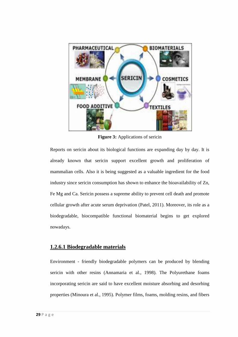
Figure 3: Applications of sericin
Reports on sericin about its biological functions are expanding day by day. It is
already known that sericin support excellent growth and proliferation of
mammalian cells. Also it is being suggested as a valuable ingredient for the food
industry since sericin consumption has shown to enhance the bioavailability of Zn,
Fe Mg and Ca. Sericin possess a supreme ability to prevent cell death and promote
cellular growth after acute serum deprivation (Patel, 2011). Moreover, its role as a
biodegradable, biocompatible functional biomaterial begins to get explored
nowadays.
1.2.6.1 Biodegradable materials
Environment - friendly biodegradable polymers can be produced by blending
sericin with other resins (Annamaria et al., 1998). The Polyurethane foams
incorporating sericin are said to have excellent moisture absorbing and desorbing
properties (Minoura et al., 1995). Polymer films, foams, molding resins, and fibers
30 P a g e
containing sericin (0.01-50% w/w) can be produce by reacting a composition
comprising a polyol, tolylene, di- isocyanet, di-butyltin di-laurate (catalyst) and
trichloromonofluoromethane (a blowing agent) in the presence of sericin. The
moister absorption/desorption rate of the sericin containing polyurethane form was
found to be two-to five fold greater than that of control. Other procedures have
also been reported for producing sericin-containing polyurethane with excellent
mechanical and thermal properties (Hatakeyama, 1996).
1.2.6.2 Membrane materials
Membrane based separations (e.g., reverse osmosis, dialysis, ultra filtration and
micro filtration) are used in process such as desalination of water, production of
extremely pure water, the bioprocessing industry and some chemical processes.
Pure sericin not easily made into membranes, but membranes of sericin cross-
linked, blended, or copolymerized with other substance are made readily, because
sericin contains large amount of amino acid with neutral polar functional groups.
Sericin and fibroin can be used to make membranes for use in separation
processes. The in solubilized silk fibroin membrane could be used to separate the
mixture of water and alcohol (Chisti, 1998). Mizoguchi et al. (1991) describe a
cross- linked thin film made of sericin for use as a separating membrane for water
and ethanol. Sericin containing membranes are quite hydrophilic. Acrylonitrile
used in making certain synthetic polymers can be copolymerized with sericin to
prepare a protein containing synthetic polymer film for separating water from
organics (Yamada and Fuwa 1994; Yamada et al., 1993).
31 P a g e
1.2.6.3 Functional biomaterials
Nakajima (1994) has found that sericin film located on lay of liquid crystal can
uniformly orient the liquid crystal molecules to provide distortion-free high-
quality crystal displays. Sericin- coated film is used on the surface of refrigeration
equipment because of its anti frosting action (Tanaka and Mizuno, 2001). Use of
the coated sericin film is an effective anti frosting method that can be widely
applied to refrigerators, deep freezers and refrigerated trucks and ships. Moreover
use of the coated film on roads and roof can prevent frost damage. Sericin protein
can be coated on surfaces of various durable materials to enhance functionality
(Li, 1996). Sericin can be used in preparation of art pigments and for surface
protection of articles. The material coated the sericin have excellent
weatherability, good permeability and do not warp on drying. Sericin blends with
water-soluble polymers, especially with polyvinyl alcohol (PVA). A blended
hydrogel made of sericin and fibroin and PVA is said to have excellent moisture
absorbing and desorbing properties and elasticity (Yoshii et al., 2000). The
hydrogel can be used as a soil conditioner and in medical materials and wound
dressing.
1.2.6.4 Medical biomaterials
Silkworm silk fibers have been the primary silk- like material used in biomedical
applications particularly as sutures. During decades of use, silk fibres have proven
to be effective in many clinical applications. At the same time, some biological
responses to the protein have raised questions about biocompatibility. Tasubouchi
(1999a) developed a silk fibroin- based wound dressing that could accelerate
32 P a g e
healing and could be peeled off without damaging the newly formed skin. The
non-crystalline fibroin film of the wound dressing had a water content of 3-16%
and a thickness of 10-100 μm. Subsequently, the wound dressing was made with a
mixture of both fibroin and sericin (Tsubouchi, 1999b). A membrane composed of
sericin and fibroin is an effective substrate for the proliferation of adherent animal
cells and can be used as a substitute for collagen. Minoura et al., (1995) and
Tsukada et al., (1999) investigated the attachment and growth of animal cells on
films made of sericin and fibroin. Cell attachment and growth were dependent on
maintaining a minimum of around 90% sericin in the composite membrane. Film
made of sericin and fibroin has excellent oxygen permeability and is similar to
human cornea in its functional properties. It hoped that the sericin- fibroin blended
film could be used to form article corneas (Murase, 1994). A novel mucoadhesive
polymer has been prepared by template polymerization of acrylic acid in the
presence of silk sericin (Ahn et al., 2001). Silk protein can be made into a
biomaterial with anticoagulant properties, by a sulfonation treatment of sericin and
fibroin (Tamada, 1997).
Kato et al. (1998) provided the first evidence of antioxidant action of the silk
protein by showing that sericin suppressed in vitro lipid peroxidation.
Furthermore, sericin also found to inhibit tyrosinase activity. These results
suggested that sericin is the valuable natural ingredient for food and cosmetics.
The biopolymer sericin has a strong affinity to keratin. Excessive transepidermal
water loss (TEWL) is one of the causes of dry skin and skin moisturizers have
been used to overcome it. The silk sericin has resemblance with the natural
33 P a g e
moisturizing factor (NMR). Sericin gel is prepared by using sericin solution with
pluronic and carbopol as a stabilizer to prevent water loss from the upper layer of
the skin. It forms a moisturizing, semi-occlusive, protective, antiwrinkle film on
the skin surface imparting an immediate, long lasting, smooth, silky feeling
(Padamwar et al., 2005). The configuration of sericin is very close to the one of
human beings. That is why sericin can naturally saturate into skin and revitalize
cells. It is discovered that sericin can restrain the functions of active-oxygen
(major factor of aging), which brings wrinkles and dark spots.
The use of oxygen-permeable membranes from silk fibroin and silk sericin,
containing about 60% water for contact lens, artificial skin, etc. The other uses of
sericin includes, as a soil conditioner, coagulant for purification of waste waters,
hygroscopic moisture-releasing polyurethane foams and their manufacture for
furniture and interior materials, as additives for health foods to prevent colon
cancers, medical composites of sericin, additives to rice cooking, fabric care
compositions, light and sunscreen compositions, foam-forming aerosol shaving
gels, sericin-coated powders for cosmetics, as dermatitis inhibitor, as wound
protection film, nail cosmetics, and chewing gums (Gulrajani, 2005). Fibroin has
been explored as a biomedicine for various applications.
Sericin and fibroin have been recently explored in the field of drug delivery
Systems. Silk protein sericin, suppress DMBA-TPA induced mouse skin tumor
genesis by reducing oxidative stress, inflammatory responses and endogenous
tumor promoter TNF-alpha (Zhaorigetu et al., 2003).
34 P a g e
1.2.7 HYDROGEL
Hydrogels are, physically or chemically cross linked three dimensional polymer
networks with an interesting ability to absorb aqueous solutions. The
hydrophilicity of the material is the underlying reason for this capability. These
products are a hot topic in the field of biomaterial research. They can be of
different types based on many criteria. Since hydrogels possess low interfacial
tension, they show negligible tendency to adsorb proteins from body fluids
(Gupta, 2002). Literature on this particular area is expanding day by day.
1.2.7.1 Preparation
Hydrogel is defined as polymer networks having hydrophilic properties. Mostly
these are prepared from hydrophilic monomers. However, occasionally, hydrogels
are prepared from hydrophobic monomers in order to regulate the properties for
specific applications. Generally, hydrogels can be made from synthetic or natural
polymers. When compared to natural polymers, synthetic are hydrophobic and
chemically stronger. They possess a slow degradation rate owing to their
mechanical strength and provide the durability as well. Through an optimal
design, these two opposite features can be balanced (Tabata, 2009). When the
natural polymers have suitable functional groups or have been functionalized with
radically polymerizable groups, it is applicable for the preparation of natural
polymer hydrogels also (Shantha, 2002).
In the most precise sense, a hydrogel is regarded as a hydrophilic polymeric
network cross-linked in some fashion resulting in an elastic structure. In this way,
any method that results in the formation of a cross-linked polymer can be used to
35 P a g e
produce a hydrogel. Some common methods are copolymerization / cross-linking
free-radical polymerization by reacting hydrophilic monomers with
multifunctional cross-linkers. Similarly, water-soluble linear polymers of both
natural and synthetic origin are cross-linked to form hydrogels in a number of
ways:
1) Linking polymer chains via chemical reaction.
2) Using ionizing radiation to generate main-chain free radicals which can
recombine as cross-link junctions.
3) Physical interactions such as entanglements, electrostatics, and crystallite
formation.
In most cases, the three integral parts of the hydrogel preparation are monomer,
initiator, and cross-linker. Diluents like water or other aqueous solutions can be
applied to control the heat of polymerization and the final hydrogel properties.
Then, the hydrogel mass needs to be washed to remove impurities left from the
preparation process. These include non-reacted monomer, initiators, cross-linkers,
and unwanted products produced via side reactions. Usually polar monomers
serve as the starting material for hydrogels. They can be classified based on the
starting material into natural polymer hydrogels, synthetic polymer hydrogels, and
combination of the two classes. Many preparative methods are there like, graft
polymerization, cross-linking polymerization, networks formation of water-
soluble polymer, and radiation cross-linking, etc.. The polymerization techniques
include bulk polymerisation, suspension polymerisation/cross-linking, suspension
36 P a g e
polymerisation/inverse suspension polymerisation, grafting to a support,
polymerisation by irradiation (Enas, 2013).
1.2.7.2 Classification of hydrogels
Classification based on source
Hydrogels can be classified into two groups based on their natural or synthetic
origins (Wen, 2013).
Classification according to polymeric composition
The method of preparation leads to formations of some important classes of
hydrogels. These can be exemplified by the following:
a) Homopolymeric hydrogels are referred to polymer network derived from a
single species of monomer, which is a basic structural unit comprising of any
polymer network (Takashi, 2007). Homopolymers may have cross-linked
skeletal structure depending on the nature of the monomer and polymerization
technique.
(b) Copolymeric hydrogels are comprised of two or more different monomer
species with at least one hydrophilic component, arranged in a random, block or
alternating configuration along the chain of the polymer network (Yang, 2002)
(c) Multipolymer Interpenetrating polymeric hydrogel (IPN), an important class of
hydrogels, is made of two independent cross-linked synthetic and/or natural
polymer component, contained in a network form. In semi-IPN hydrogel, one
component is a cross-linked polymer and other component is a non-cross-linked
polymer (Maolin, 2000; Hacker, 2011).
Classification based on configuration
37 P a g e
The classification of hydrogels depends on their physical structure and chemical
composition can be classified as follows:
(a) Amorphous (non-crystalline).
(b) Semicrystalline: A complex mixture of amorphous and crystalline phases.
(c) Crystalline.
Classification based on type of cross-linking
Hydrogels can be divided into two categories based on the chemical or physical
nature of the cross-link junctions. Chemically cross-linked networks have
permanent junctions, while physical networks have transient junctions that arise
from either polymer chain entanglements or physical interactions such as ionic
interactions, hydrogen bonds, or hydrophobic interactions (Hacker, 2011).
Classification based on physical appearance
Hydrogels appearance as matrix, film, or microsphere depends on the technique of
polymerization involved in the preparation process.
Classification according to network electrical charge
Hydrogels may be categorized into four groups on the basis of presence or
absence of electrical charge located on the cross-linked chains:
(a) Nonionic (neutral).
(b) Ionic (including anionic or cationic).
(c) Amphoteric electrolyte (ampholytic) containing both acidic and basic groups.
(d) Zwitterionic (polybetaines) containing both anionic and cationic groups in
each structural repeating unit.
38 P a g e
Hydrogel-forming natural polymers include proteins such as collagen and gelatine
and polysaccharides such as starch, alginate, and agarose. Synthetic polymers that
form hydrogels are traditionally prepared using chemical polymerization methods.
Hydrogel product sensitive to environmental conditions
As mentioned, hydrogels as three-dimensional cross-linked hydrophilic polymer
networks are capable of swelling or de-swelling reversibly in water and retaining
large volume of liquid in swollen state. Hydrogels can be designed with
controllable responses as to shrink or expand with changes in external
environmental conditions. They may perform dramatic volume transition in
response to a variety of physical and chemical stimuli, where the physical stimuli
include temperature, electric or magnetic field, light, pressure, and sound, while
the chemical stimuli include pH, solvent composition, ionic strength, and
molecular species.
The extent of swelling or de-swelling in response to the changes in the external
environment of the hydrogel could be so drastic that the phenomenon is referred to
as volume collapse or phase transition (Jinsub, 2010). Synthetic hydrogels have
been a field of extensive research for the past four decades, and it still remains a
very active area of research today.
1.2.8 Natural polymer hydrogels
Hydrogels may be of natural polymer based; for example polysaccharides and
proteins. These natural macromolecules are repetition of glycosidic and amino
acid monomers. Since hydrogels are characterized of its softness and rubbery
consistence, they resemble closely with the living tissues. Even if they shows
minimal tendency to adsorb serum proteins, natural polymers like collagen,
39 P a g e
gelatine and their derivatives often possess affinity towards serum fibronectin.
Interestingly, studies on this fact provides the potential of these hydrogels to
function as ideal scaffolds that support cell growth for tissue engineering purposes
and as dug delivery vehicles (van Vlierberghe, 2011).
Hydrogels of natural polymers, especially polysaccharides, are in general, non-
toxic and biodegradable. Considerable research and technical work have been
reported. Hyaluronic acid, chitosan, chondroitin sulphate, cellulose derivatives,
alginate are the excellent members under this category. The chemical modification
of starch or modified starch via vinyl graft copolymerization constitutes the most
important fields for improving the properties of starch and enlarging the range of
its utilization. The starch graft-copolymer such as starch-g-polystyrene, starch-g-
polyvinyl alcohol, starch-g-methacrylonitrile, and starch-g-acrylonitrile have been
produced by generating free radicals on the surface of the starch granules followed
by copolymerization of these free radicals with the respective vinyl monomers.
These copolymers have also limited biodegradability because of the presence of a
non-biodegradable part of the polymer (Doyle, 2006).
It has been reported that the synthesis of hydrogels by modification of natural
polymers (for example, biocatalytic) has been used for preparation of sugar-
containing poly(acrylate) hydrogels. These authors found that by the introduction
of small quantities of agar, they were able to eliminate the relative brittleness of
the polyacrylamide hydrogels and reduce the formation of undesirable fine
particles during wet milling. Raju et al. (2001) grafted acrylonitrile onto cassava
starch. These authors investigated the effects of the reactant concentrations and
duration of the polymerization.
40 P a g e
1.2.8.1 Protein hydrogels
Protein is the natural macromolecule with amino acid monomers. In the area of
biopolymer based hydrogels, protein is an ideal candidate. Collagen, gelatin,
elastin, fibroin and sericin are the prominent members under this category.
Hydrogels based on these are excellent tools in the field of tissue engineering. To
date, hydrogels from these are useful for bone tissue engineering, skin tissue
engineering, blood vessel regeneration and ocular tissue engineering purposes.
Porous collagen scaffolds have been made by freeze drying and stereolithographic
methods. They can be used for bone application by mixing with calcium
phosphates. Also, it can be blended with synthetic polymers like, poly(lactic acid)
(Gong, 2007; Yang, 2009) poly(glycolic acid), (Wen, 2007; Kawazoe, 2009) or
poly(caprolactone) as well as (modified) glycosaminoglycans (Ohyabu, 2009;
Haugh, 2009; Tierney, 2009) such as (photocross- linkable) hyaluronic acid
forming semi-IPNs.
Gelatin, a hydrolysed product of collagen is a crucial member in this field. It can
be chemically / enzymatically cross-linked so as to get a stable hydrogel product.
Porous scaffolds from this can be made by freeze drying and phase separation
methods. It can also be blended with poly(L-lactic acid), (Lazzeri, 2007)
polyurethanes, (Wei, 2008) and PCL (Rim, 2009).
Elastin constitutes the greater part of elastic, thus mechanically active tissues
including tendon, blood vessels, and elastic cartilage (Krishna, 2010). Matriderm
41 P a g e
is a commercially available dermal substitute composed of elastin and collagen
described frequently in literature (Keck, 2009; Kolokythas, 2008).
Silk fibroin synthesized by the silkworm Bombyx mori, is a natural protein (Cao,
2009). Its primary structure mainly consists of glycine, alanine, and serine
(Sundar, 2010). The protein is an ideal candidate of biomaterial science and drug
delivery from which, films, (Lawrence, 2010; Nogueira, 2010) nanofibers, (Wang,
2009; Wang et al., 2009; Jin, 2004; Meinel, 2009) scaffolds, (She, 2009)
membranes (Ghassemifar, 2010) gels, (Gong, 2010; Yucel, 2010) and powders
(Yucel, 2010; Tao, 2010) can be developed (Sundar, 2010). Fan et al. have
developed porous gelatin-based hybrid scaffolds for ligament tissue engineering.
In addition to composite scaffolds with other proteins (e.g., collagen, fibroin has
also been combined with GAGs including hyaluronan (Ren, 2009; Garcia, 2009).
The scaffolds developed were suitable to support mesenchymal stem cell adhesion
(Garcia, 2009). Porous fibroin scaffolds can be made by freeze drying and
leaching out porogens. In addition to freeze-drying, (Mandal, 2009; Mauney,
2007) leaching out of porogens (Makaya, 2009).
1.2.9 SERICIN AS A BIOMATERIAL
Tissue engineering provides combinations of cells and biomaterials, and/or drugs,
genes & gene products that may be designed and fabricated in the form of
biological substitutes. An important element of this field is biomaterial design.
This involves incorporation of physical, chemical and biological cues in order to
lead cells into functional tissues through the process of migration, adhesion and
differentiation. Mostly, biomaterials should possess a degradation rate matching
42 P a g e
with the new tissue formation so that cells will be capable to deposit new
extracellular matrix (ECM) and regenerate functional tissue. These biomaterials
must be able to support mechanically, in accordance with the level of functional
tissue development as well. On the whole, for a material to be called as a
biomaterial, it must be biocompatible and elicit little to no host immune response.
In this context, silk have been investigated as biomaterial due to the successful use
of silk fibers from B. mori as suture material for centuries (Moy, 1991). Silks
obtained from different species and within a species shows some functional
differences as a result of the changes in primary amino acid sequence, processing,
and the impact of environmental factors (Vollrath, 2001). Silks are a unique
family of structural proteins having biocompatibility, degradability, mechanical
superiority. This ultra class of proteins offer a wide range of properties, open to
aqueous or organic solvent processing and can undergo chemical modification in
order to go well with a wide range of biomedical applications.
For the treatment of superficial wounds, genipin cross-linked sericin/poly(vinyl
alcohol) (PVA) films were proved to be an efficient 2 dimensional wound dressing
agent. The property of silk sericin in enhancing collagen production and resultant
epithelialisation in wounds is applied in this system. Moreover excellent
attachment of fibroblast and keratinocytes on sericin also contribute to the
excellence in using sericin as a wound dressing material. Complete investigation
of the physical and biological properties of the system revealed increased surface
density, tensile strength, and percentage of elongation, but decreased percentage
43 P a g e
of light transmission, water vapor transmission rate, and water swelling, compared
to the non-cross-linked films, thus promoting the formation of a more rigid
molecular structure by minimising the mobility of molecular chain. The release of
sericin from film was in a sustained manner. Desirable percentage of viability
could be shown by L929 mouse fibroblast or HaCat keratinocyte cells when
cultured on the silk sericin / PVA films cross-linked with 0.075 and 0.1%w/v
genipin. Safety test conducted in this study renders the system a completely
biocompatible one (Siritientong, 2013).
A 3D tissue engineered sericin construct has been developed as an alternative to
autografts, allografts and xenografts. In the fabricated matrix co-culture of
fibroblast and keratinocytes were used. Keratinocytes were grown on the upper
surface and fibroblasts on the lower surface. Freeze drying was employed in the
fabrication method and genipin was the crosslinker resulting in a porous matrix.
The characterisation of the matrix showed a good growth of co-cultured
fibroblasts and keratinocytes in vitro on 3D sericin matrices. This construct was
aimed to deliver at the target wound site where it could promote the wound
closure and gradually accelerate healing mechanism. This 3D tissue-engineered
skin model is a potential source of living skin equivalents for grafting in vivo
(Sunita, 2013).
Sericin is a highly hydrophilic protein family acting as the glue in Bombyx mori
silk. In order to apply sericin as a wound dressing, a novel sericin film named gel
film was prepared by a simple process without using any chemical modifications.
Sericin solution was gelled with ethanol into a sheet shape and then dried. Infrared
44 P a g e
analysis revealed that the sericin gel film contained water-stable beta-sheet
networks formed in the gelation step. This structural feature rendered the gel film
morphologically stable against swelling and gave it good handling properties in
the wet state. The sericin gel film rapidly absorbed water, equilibrating at a water
content of about 80%, and exhibited elastic deformation up to a strain of about
25% in the wet state. A culture of mouse fibroblasts on the gel film indicated that
it had low cell adhesion properties and no cytotoxicity. These characteristics of
sericin gel film suggest its potential as a wound dressing (Teramoto et al., 2008).
A study was conducted by Theodora (2014) to investigate the synergetic effect of
racemic flavanone Naringenin (NRG) and the protein sericin as TNF-α blockers.
Sericin micro particles were fabricated by using spray-drying method, which was
loaded with naringenin. Characterization in terms of morphology and particle size
distribution, and encapsulation efficiency was done, which were correlated well
with the motto of the study. This particular study provides the proof of concept
that sericin-based microspheres loaded with TNF-α-blockers could contribute to
the down regulation of the cytokine and represents the starting point for the
development of new topical formulations for the treatment of middle-stage
psoriasis. It is to be drawn from this work that sericin may improve skin barrier
function, thus preventing water loss from the upper layer of the skin (Theodora,
2014).
Silk fibers have been the primary silk- like material used in biomedical
applications particularly as sutures. During decades of use, silk fibres have proven
45 P a g e
to be effective in many clinical applications. Subsequently, the wound dressing
was made with a mixture of both fibroin and sericin (Tsubouchi, 1999b). A
membrane composed of sericin and fibroin is an effective substrate for the
proliferation of adherent animal cells and can be used as a substitute for collagen.
Minoura et al., (1995) and Tsukada et al., (1999) investigated the attachment and
growth of animal cells on films made of sericin and fibroin. Cell attachment and
growth were dependent on maintaining a minimum of around 90% sericin in the
composite membrane.
In the area of cartilage tissue engineering, bio hybrids consisting of biodegradable
scaffolds with cultured cells is a recent development. In a study aimed at
developing a 3D porous scaffold based on natural compounds, collagen-sericin
scaffold supporting human adipose –derived stem cells (ASCs) was fabricated and
characterized. It was already proved that Hyaluronic acid and chondroitin sulphate
are the most potent prochondrogenic factors involved in the biomaterial design for
cartilage tissue engineering application. The fabricated scaffolds were of good
porosity, high water absorption capacity; non-denatured triple helical structure of
collagen. These features make them perfect for cartilage tissue engineering.
Cytotoxic assays also were found to be positive (Sorina, 2013).
Role of sericin in ocular tissue engineering is in its path of exploration. In a study
conducted under this field, investigated attachment and growth in vitro of human
corneal limbal epithelial cells (HLECs) on sericin-based membranes. Primary cell
cultures from two donors were established in serum supplemented media in the
presence of murine feeder cells. Membranes made of sericin and sericin-fibroin
blends were assessed in terms of cell growth and attachment. The results
46 P a g e
demonstrated that B. mori sericin-based materials were mechanically weak
compared to sericin-fibroin blend. However, the attachment of human corneal
limbal epithelial cells to sericin or sericin-rich blends was far superior to that to
fibroin or fibroin-rich blends, with no evidence of any cytopathologic response.
This is a strong recommendation that sericin is an apt material that could serve as
a scaffold in ocular surface regeneration (Traian, 2013).
The role of sericin as an active participant in tissue engineering field becomes a
hot topic. Fabrication of a novel 3D sericin / gelatine scaffold and 2D films
supports this fact. The fabricated matrices were characterized and optimized
biophysically and biologically. The results were in accordance with the aim of the
study. It was showed that the matrices were cytocompatible and biocompatible.
They showed excellent characteristics like uniform pore distribution, improved
compressive strength, high swell ability and overall high porosity, the critical
parameters for tissue engineering and biomedical applications. This work also
provides a new dimension to silk-protein sericin in the form of novel blended 3-D
scaffolds for efficient and effective use as biomaterial (Biman, 2009).
Limitations in the current clinical strategies for adipose tissue engineering (ATE)
strongly recommend the application of a 3D cell-scaffold construct. In this
respect, a 3D scaffold made of 60% collagen and 40% collagen preceded with
human adipose derived stem cells (hADSCs). These scaffolds were well
characterized and optimized for the specific application. Interestingly, it was
shown that incorporation of the sticky protein sericin along with collagen
47 P a g e
enhanced the adhesion and proliferation rate of seeded cells, making it a
biocompatible marix. Sericin showed an astonishing ability of stimulating
PPARγ2 over expression, triggering a consequent up regulated expression profile
of FAS, aP2 and perilipin adipogenic markers. In addition to this, sericin is well
known by its property to stimulate collagen synthesis. Moreover, higher rate of
adipogenesis was correlated with the presence of sericin in the cellular
environment. These remarkable features makes the fabricated matrix out of it as a
strong candidate for soft tissue reconstruction and wound healing applications.
However, in vivo implantation of these constructs should be addressed in further
work during ATE applications in order to confirm the reproducibility of these data
and to validate safety claims (Sorina, 2013).
Antibacterial property of sericin was examined by growing Micrococcus leutus
bacteria by modified agar diffusion method. A zone of inhibition of (9-12) mm in
diameter for 10 & 20 mg/ml concentration was observed. Also, the antibacterial
efficiency was found to be increased with increasing concentration of sericin. This
study provides an insight to use sericin for medical applications after isolation and
identification of some pathogenic bacteria like Pseudomonas aeruginosa,
Staphyllococcus aureous Escherchia coli to produce medical bandages, mouth
wash, antibacterial soaps & tooth paste. In this context, more research has to be
explored (Khalid, 2010).
Sericin based nano biomaterials was reported. Nano sized sericin was obtained
using urea treatment by a simple and cost effective method. Nano sericin obtained
48 P a g e
was tested for many applications like hydrogel, biopolymers, biomaterials, and
antioxidant and for other antimicrobial applications. Moisture absorbing and
releasing property, inherent gelling property rapid gelation on addition of cross-
linkers make it an ideal candidate for wound dressing application and as soil
conditioner. The developed nano sericin is suitable for various applications like
moisturizer in cosmetics, as scaffold, replacing binding material in perfume sticks
and in the production of antimicrobial products (Pushpa, 2013).
1.2.9.1 Sericin based hydrogel
Silk sericin is a precious protein resource obtained from silk worm, the queen of
textiles. Usually, it is considered as a waste product and discarded by a process
called degumming in the textile industry. Studies have proved that sericin is a
valuable biomaterial that can be made into gels, membrane, foam, fiber and other
materials. Hydrogels based on sericin is a remarkable output of such studies.
These sericin hydrogels are ideal for drug delivery applications and tissue
engineering. Hydrogel matrix produced from sericin shows excellent cell homing
ability, which makes them suitable for wound healing application.
In situ semi interpenetrating on-mulberry tropical tasar silk sericin/polyacrylamide
hydrophilic network showed cell homing ability and rapid gelation. They
successfully entrapped sericin physically within the 3D hydrophilic structure of
polyacrylamide. The injectable composite material developed through such a
relatively simple technique possessed favourable properties required for in situ
cell scaffolding and tissue regeneration. The characteristics of the matrix like the
gelation time, hydrogel micro-architecture, swelling, and equilibrium water
49 P a g e
content, compressive strength and in vitro degradation were found to be
influenced by sericin concentration. Another remarkable achievement of this work
is that no chemical cross linkers are involved in this (Kundu, 2012).
Sericin hydrogels were also used as drug vehicle. Interpenetrating polymer
networks (IPNs) composed of silk sericin (SS) and poly (N-isopropylacrylamide)
(PNIPAAm) were prepared by the simultaneous-IPN method. The miscibility of
the two was confirmed by the single Tg found in thermograms. The interaction
between two members which favoured miscibility was deduced from FTIR. The
release profile of a model drug BSA was studied. It showed a burst release within
first 12 hours. This IPN based on sericin is being modulated as an ideal drug
carrier (Zuo, 2006).
A photo luminescent, injectable hydrogel had developed from sericin via covalent
cross linking method with glutaraldehyde for delivery of cells and drugs. The
fabricated hydrogel system was showed to acquire marvellous cell adhesive
capability, guiding efficient cell attachment, proliferation and long term survival
of various types of cells. Moreover, the system was able to release bioactive
agents in a sustained manner. In addition to that, the sericin hydrogel could exhibit
good elasticity, high porosity and pH dependent degradation dynamics which
make it suitable to function as a delivery vehicle for cells and therapeutics. Since
the system is injectable, it allows minimal invasive approaches for implantation.
The photoluminescence property enables bioimaging and in vivo tracking. Among
the three types of cell types tried, HEK293, C2C12, HaCaT, HEK293 exhibited
50 P a g e
cluster type growth on hydrogel surface, whereas the other cell types did not show
this particular pattern. This demonstrates that sericin hydrogel influences spatial
distribution of cells (Wang, 2014).
1.3 HYPOTHESIS
Cross-linking two biopolymers, sericin and gelatin, would produce a
biodegradable and biocompatible hydrogel system having sufficient
hydrophilicity, strength, and handling characteristics so that they can be used for
biomedical applications.
1.4 OBJECTIVES
Fabrication of hydrogel by optimising reaction conditions
To carry out physico-chemical Characterization of Hydrogel
To perform biological evaluation of Hydrogel
51 P a g e
CHAPTER 2
MATERIALS AND METHODS
2.1 MATERIALS AND EQUIPMENTS
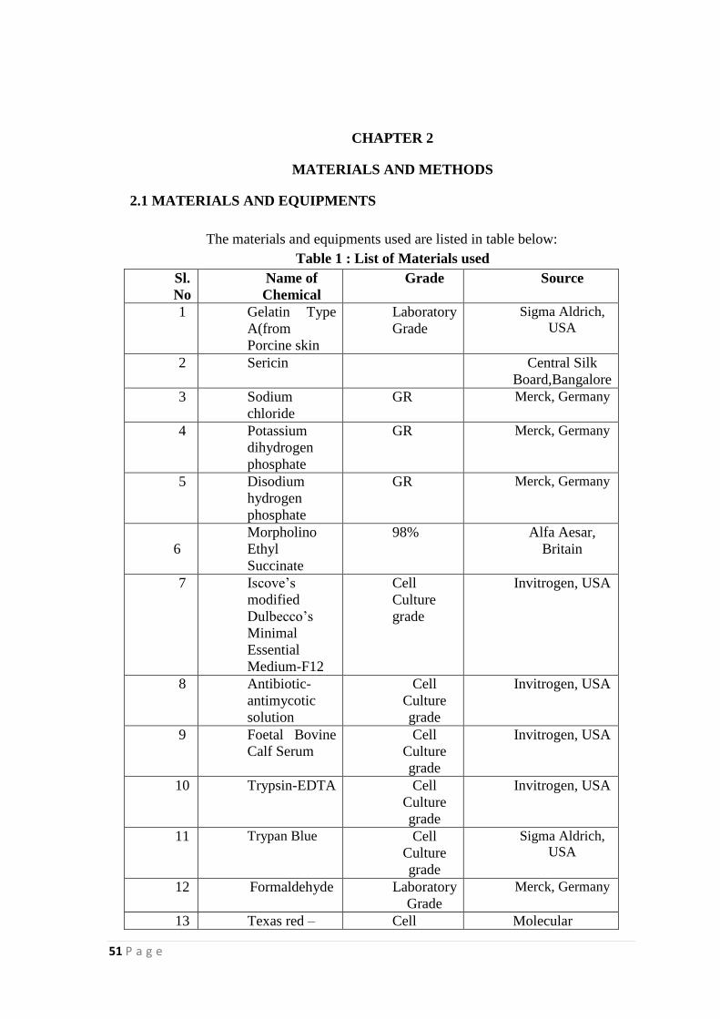
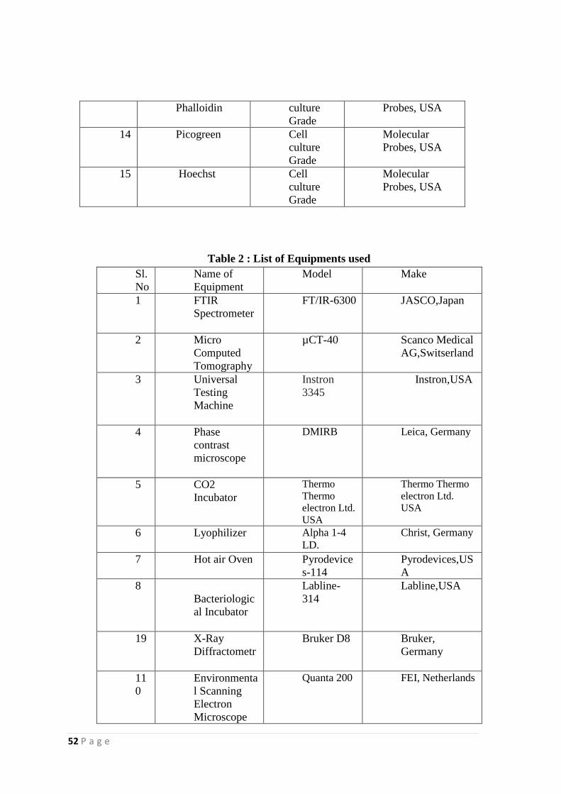
The materials and equipments used are listed in table below:
Table 1 : List of Materials used
Sl.
No
Name of
Chemical
Grade Source
1 Gelatin Type
A(from
Porcine skin
Laboratory
Grade
Sigma Aldrich,
USA
2 Sericin Central Silk
Board,Bangalore
3 Sodium
chloride
GR Merck, Germany
4 Potassium
dihydrogen
phosphate
GR Merck, Germany
5 Disodium
hydrogen
phosphate
GR Merck, Germany
6
Morpholino
Ethyl
Succinate
98% Alfa Aesar,
Britain
7 Iscove‟s
modified
Dulbecco‟s
Minimal
Essential
Medium-F12
Cell
Culture
grade
Invitrogen, USA
8 Antibiotic-
antimycotic
solution
Cell
Culture
grade
Invitrogen, USA
9 Foetal Bovine
Calf Serum
Cell
Culture
grade
Invitrogen, USA
10 Trypsin-EDTA Cell
Culture
grade
Invitrogen, USA
11 Trypan Blue
Cell
Culture
grade
Sigma Aldrich,
USA
12 Formaldehyde Laboratory
Grade
Merck, Germany
13 Texas red – Cell Molecular
52 P a g e
Phalloidin culture
Grade
Probes, USA
14 Picogreen Cell
culture
Grade
Molecular
Probes, USA
15 Hoechst Cell
culture
Grade
Molecular
Probes, USA
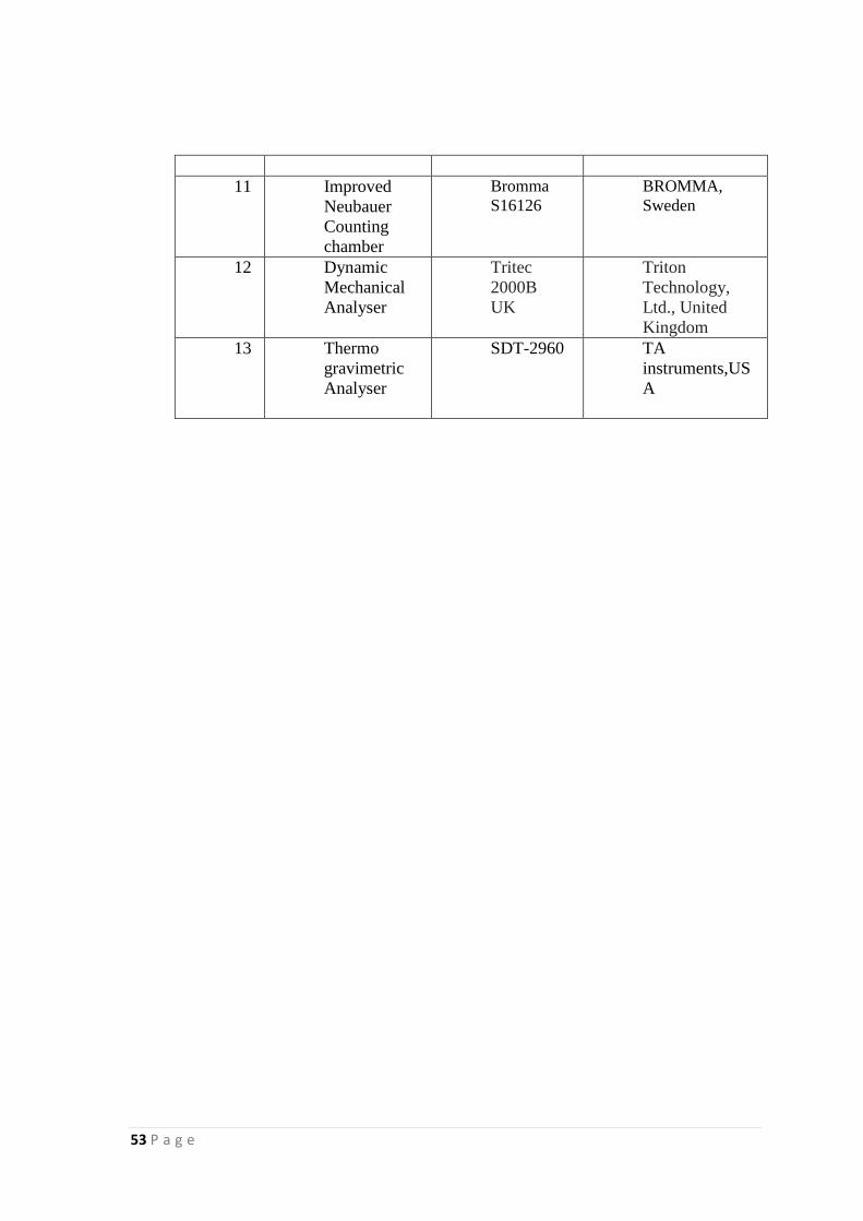
Table 2 : List of Equipments used
Sl.
No
Name of
Equipment
Model Make
1 FTIR
Spectrometer
FT/IR-6300 JASCO,Japan
2 Micro
Computed
Tomography
µCT-40 Scanco Medical
AG,Switserland
3 Universal
Testing
Machine
Instron
3345
Instron,USA
4 Phase
contrast
microscope
DMIRB Leica, Germany
5 CO2
Incubator
Thermo
Thermo
electron Ltd.
USA
Thermo Thermo
electron Ltd.
USA
6 Lyophilizer
Alpha 1-4
LD. Christ, Germany
7 Hot air Oven
Pyrodevice
s-114
Pyrodevices,US
A
8
Bacteriologic
al Incubator
Labline-
314
Labline,USA
19 X-Ray
Diffractometr
Bruker D8 Bruker,
Germany
11
0
Environmenta
l Scanning
Electron
Microscope
Quanta 200 FEI, Netherlands
53 P a g e
11 Improved
Neubauer
Counting
chamber
Bromma
S16126
BROMMA,
Sweden
12 Dynamic
Mechanical
Analyser
Tritec
2000B
UK
Triton
Technology,
Ltd., United
Kingdom
13 Thermo
gravimetric
Analyser
SDT-2960 TA
instruments,US
A
54 P a g e
2.2 METHODS
2.2.1 RAW MATERIAL CHARACTERIZATION
2.2.1.1 FTIR Spectroscopy:
Fourier Transform Infrared (FTIR) spectra of raw materials (sericin and gelatin)
were recorded using a FTIR spectrophotometer (model 6300, Jasco, Japan).
Samples in the powder form was mixed with KBr and made into thin transparent
disks using a hydraulic hand press by applying a pressure of about 10-ton for
about 3 minutes. KBr without the samples was used as the control.
After warming up the instrument for 20 minutes, background measurements were
made with control KBr pellet. CO2 and moisture peak removal was made effective
after this recording. The sample disks were monitored after background
measurements using the “Spectra manager” software. Spectra were collected in
the range 400-4000 cm-1
using at least 16 scans at a resolution of 4 cm-1
. The
obtained FTIR spectra were processed for baseline corrections, smoothening and
labelling of peaks using the software.
2.2.1.2 X-Ray Diffraction analysis
X-Ray Diffraction (XRD) data of the powdered samples were analyzed with a
Bruker D8 Advance diffractometer with Cu Kα radiation at a scan speed of 40/min
over 10-900.
2.2.1.3 Thermal Analysis
Thermal stability of the sample was studied using thermogravimetry (TGA) based
on the method given in ASTME-1131-19 by using a SDT-2960 model
thremogravimetric analyser (M/s. TA instruments, USA). An amount of 50 mg of
55 P a g e
powdered sample was heated at a rate of 100C/min in an aluminum crucible from
30 to 500 0C in a dynamic inert nitrogen atmosphere. Continuous recording of
sample temperature, sample weight and heat flow were made.
2.2.1.4 Dynamic Mechanical Analysis
DMA was done in tensile mode with a Tritec 2000B DMA instrument (Triton
Technology, Ltd., United Kingdom) on a rectangular gelatin sample of 4-mm
width, 20-mm length and 0.2-mm thickness. A frequency of 1 Hz with an
amplitude of dynamic deformation of 50µm was applied in the temperature-ramp
experiments. A heating rate of 10C/min was used over a temperature range from -
1000C to 90
0C
2.2.2 FABRICATION OF SERICIN –GELATIN HYDROGEL
Hydrogels of sericin and gelatin (SGH) were fabricated by mixing solutions of
both gelatin and sericin at approximate temperatures and later drying in an
incubator. Silk protein, sericin was dissolved in hot water at 600C and gelatin in
distilled water at 400C. Then the two solutions were mixed at different reaction
conditions. Reaction mixture was then incubated at 370C for 10-60 minutes for
gelation. The process of fabrication of hydrogel was optimized by varying the
parameters such as solution concentration, temperature, reaction time and
incubation.
2.2 .3 CHARACTERIZATION OF HYDROGEL
2.2.3.1 FTIR Spectroscopy
FTIR characterization of the hydrogel was done using the method mentioned
above.
56 P a g e
2.2.3.2 XRD Analysis
XRD data of the powdered sample was done using the method reported above.
2.2.3.3 Thermal Analysis
Thermal stability of the hydrogel was analysed by using TGA and DSC.
2.2.3.4 Dynamic Mechanical Analysis
Dynamic Mechanical Analysis (DMA) of the materials were performed in tensile
mode with a Tritec 2000B DMA instrument (Triton Technology, Ltd., United
Kingdom) on a rectangular sample of 4 mm width, 20 mm length and 0.2 mm
thickness. A frequency of 1 Hz with an amplitude of dynamic deformation of
50µm was applied in the temperature-ramp experiments. A heating rate of
10C/min was used over a temperature range from -100
0C to 90
0C.
2.2.3.5 Swelling studies
The dynamic swelling of the hydrogel was analysed in alkaline (PBS), acidic
(MES) and neutral (distilled water) media. Dried circular disk samples (2cm
diameter × 1 mm thickness) were pre-weighed and immersed in three media at
37°C for 24 h. At different time points, hydrogels were wiped with Whatmann
No.1 filter paper to remove surface water and swollen weight was determined
using an analytical balance.
Degree of swelling (Q) was calculated by the formula
Q = (Ws-Wd)/Wd
Percentage Swelling was calculated by the formula
Percentage Swelling = [(Ws-Wd)/Wd] × 100
Equilibrium Water content was calculated by the formula
EWC = [(Ws-Wd)/Ws] × 100
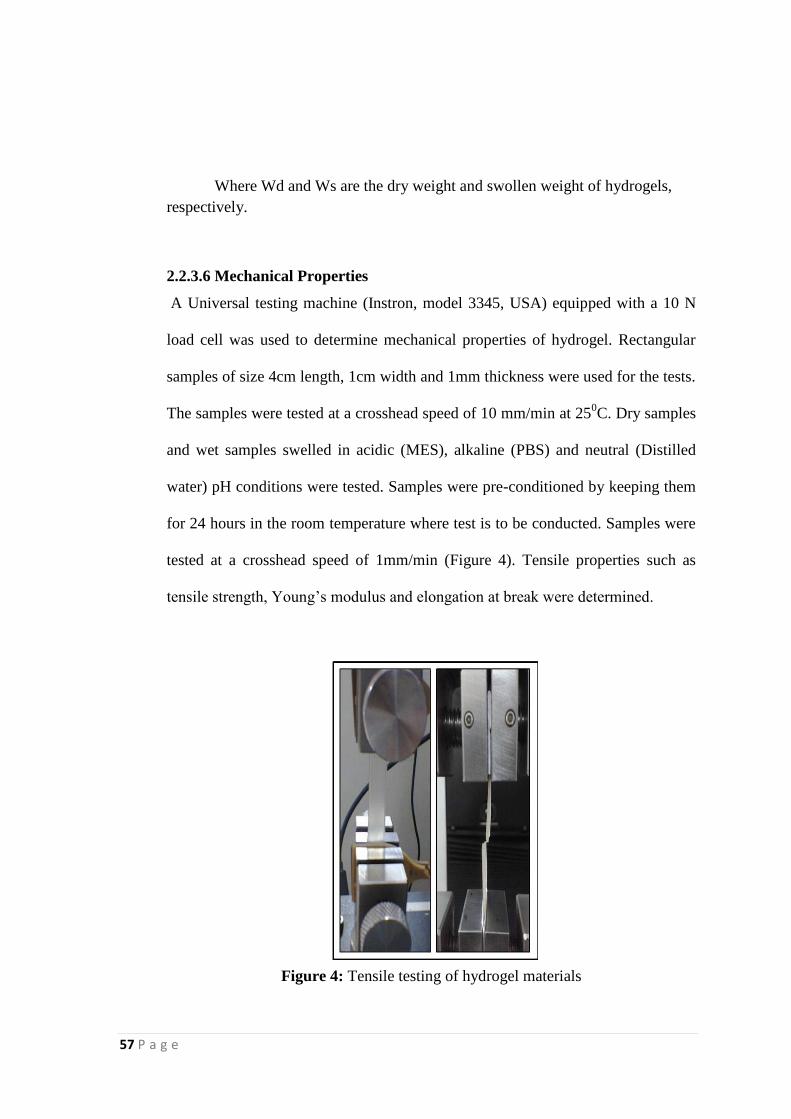
57 P a g e
Where Wd and Ws are the dry weight and swollen weight of hydrogels,
respectively.
2.2.3.6 Mechanical Properties
A Universal testing machine (Instron, model 3345, USA) equipped with a 10 N
load cell was used to determine mechanical properties of hydrogel. Rectangular
samples of size 4cm length, 1cm width and 1mm thickness were used for the tests.
The samples were tested at a crosshead speed of 10 mm/min at 250C. Dry samples
and wet samples swelled in acidic (MES), alkaline (PBS) and neutral (Distilled
water) pH conditions were tested. Samples were pre-conditioned by keeping them
for 24 hours in the room temperature where test is to be conducted. Samples were
tested at a crosshead speed of 1mm/min (Figure 4). Tensile properties such as
tensile strength, Young‟s modulus and elongation at break were determined.
Figure 4: Tensile testing of hydrogel materials
58 P a g e
2.2.3.7 Surface Morphology
The microstructure of the hydrogel sample was investigated using a scanning
electron microscope (SEM; Hitachi S2400, Japan). Briefly, after fabrication the
hydrogels were immersed in de-ionized water and washed thrice to remove the un-
reacted components, if any. The resultant gels were lyophilized and dried. The
samples were gold coated and examined under vacuum under SEM with a
working voltage of 25 KV. The images were taken at different magnifications.
The pore sizes within the hydrogels were measured using Image J 1.40 software.
2.2.3.8 Micro Computed Tomography Analysis
Lyophilised hydrogel samples were subjected to micro computed tomography
(µCT). The samples were placed in the sample holder (PMMA tube) for detecting
the X-Ray attenuation and scanning was performed with 45KV X-ray energy,
177µA intensity and 10µm resolution (2D slice thickness). Two-dimensional
reconstructions was done using Cone beam algorithm. Region of interest in the 2D
slices were contoured and 3D evaluation was performed by setting appropriate
threshold value of X-ray attenuation to actual 3D image of the sample.
2.2.3.9 Degradation studies
Hydrogel samples were fabricated as described previously. Degradation of
hydrogel in alkaline (PBS), acidic (MES) and neutral (distilled water) media was
studied for 3 weeks. Pre-weighed hydrogel samples in the form of circular disk (2
cm diameter × 1mm thickness) of average weight 0.1g were immersed in 5ml
PBS, MES and distilled water along with 0.1% Sodium azide solution and
incubated at 370C. The weight loss of the sample was determined using an
59 P a g e
analytical balance in seven days interval, which indicates hydrolytic degradation
of the samples.
2.2.3.10 Hemolysis
Hemocompatibility test of hydrogel systems was carried out broadly on the basis
of ISO 10993-4:2002 (E) „Selection of tests for interaction of materials with
blood‟. The test is mainly aimed at finding the extent of hemolysis caused by the
sample. Blood from healthy human volunteer was collected into the anticoagulant,
citrate-phosphate-dextrose-adenine [CPD-A]. Samples kept in PBS were taken out
and placed in polystyrene plates. Blood (2 mL) was added to each sample; 1 mL
blood was taken immediately for initial analysis and another 1 mL blood was
incubated with the samples for 30 min under agitation at 70±5 rpm using an
Environ shaker (Labline Instruments Inc., Melrose Park, USA) thermostated at
35±2°C. Four empty polystyrene culture dishes were exposed with blood as
reference. The total hemoglobin in the whole blood samples was measured using
automatic hematology analyzer (Sysmex-K 4500, Sysmex Corporation, Japan).
The free hemoglobin liberated into the plasma after exposure to materials was
measured using diode array spectrophotometer (HP 8453; Hewlett-Packard
GmbH, Germany) and the hemolysis (%) was calculated using the formula
60 P a g e
2.2.4 BIOLOGICAL EVALUATION OF HYDROGEL
2.2.4.1 Isolation and culture of human skin dermal fibroblasts
Human skin dermal fibroblast was received from Thrombosis Research Unit,
SCTIMST, Trivandrum.
2.2.4.2 Hydrogel conditioning
Lyophilised washed hydrogels were rehydrated for 24 hours in PBS and conditioned
in serum free medium for 6 days. Medium was changed on alternative days.
2.2.4.3 Seeding
Scaffolds were analyzed for their cell attachment and proliferation. Primary
fibroblasts after second passage were used for hydrogel wound dressing seeding.
Scaffolds were statically seeded with a cell density of 5000 cells/cm2 and allowed
to attach. Unattached cells were removed after 12 hours and replenished with fresh
medium.
2.2.4.4 Cytotoxicity by direct contact Method
Primary fibroblasts were seeded on to tissue culture treated polystyrene surfaces
and allowed to form a complete monolayer. Circular hydrogel discs were placed
on top of the cell monolayer and fed with complete medium. Efforts were taken to
avoid the floating of the hydrogel. Medium was replenished on every alternative
day. After 48 hours of culture, scaffolds were removed and cell monolayer
integrity was observed through phase contrast microscope.
61 P a g e
2.2.4.5 Cell viability Assay
Trypan blue exclusion assay was used to quantify the cell viability in the presence
of hydrogel. The cells cultured in the presence of hydrogel for 72 hrs as mentioned
above were harvested by trypsinization after removing the gel from the dish. The
cell pellet was then re-suspended in lmL sterile PBS. Trypan Blue stain was
diluted into 1:9 ratio with DPBS and mixed 90µL stain to 10µL cell suspension.
About 20µL was taken into the Neubauer counting chamber and counted using 10
X magnification of microscope. Cells seeded in culture wells served as control.
2.2.4.6 Biochemical evaluation for Cell Proliferation- Pico green
The proliferation of human dermal fibroblasts on day 3 and day 6 days in culture
was determined using Picogreen® dsDNA Quantitation reagent (Molecular
probes). For this, the cell-seeded materials (n=6) were washed with PBS twice and
kept in -80°C until analysis. Frozen cell samples were thawed for 20 min on ice
and lysed with 1% Triton X-1 00 (300 µl ) for 20 min with sonication for 10 min.
The lysate (10 µl) then mixed with Picogreen in Tris-EDTA buffer (190 µl) for 5
min and the intensity of fluorescence was measured with a multifunction
microplate reader (HIDEX Chameleon) at an excitation and emission wavelength
of 485/ 535 nm. Relative fluorescence units were correlated with cell number
using a calibration line constructed from cell suspensions with increasing
concentrations of cell numbers.

62 P a g e
2.2.4.7 Two dimensional cell attachment
Scaffolds were statistically seeded with a cell density of 5000 cells/cm2
and
allowed to attach for 72 hours. The scaffold was then observed under
Environmental scanning electron microscope (ESEM). Material alone served as
the control.
2.2.4.8 Cell attachment assay
Scaffolds were statically seeded with a cell density of 5000 cells /cm2 and allowed
to attach for 48 hours. The scaffolds were then fixed with 3. 7% formaldehyde for
20 minutes and then permeabilised with 0.2% triton X 100. Texas red conjugated
antibodies against actin cytoskeleton were added and kept at 4 0 C for overnight.
Scaffolds were counter stained with Hoechst. Washed thoroughly and observed
through an inverted fluorescent microscopy.
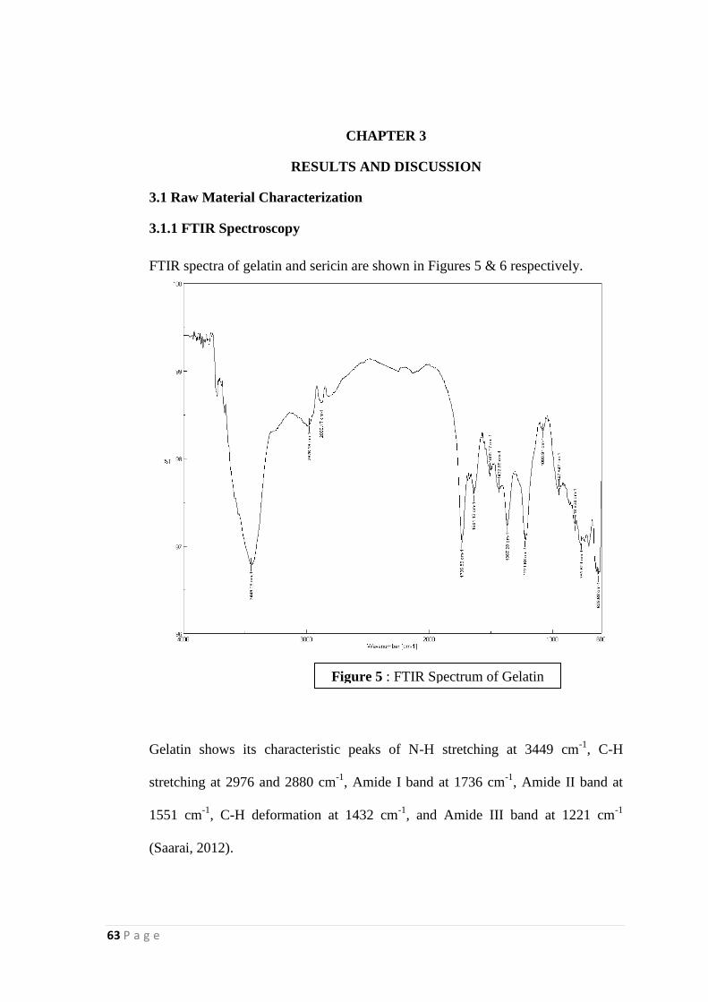
63 P a g e
CHAPTER 3
RESULTS AND DISCUSSION
3.1 Raw Material Characterization
3.1.1 FTIR Spectroscopy
FTIR spectra of gelatin and sericin are shown in Figures 5 & 6 respectively.
Gelatin shows its characteristic peaks of N-H stretching at 3449 cm-1
, C-H
stretching at 2976 and 2880 cm-1
, Amide I band at 1736 cm-1
, Amide II band at
1551 cm-1
, C-H deformation at 1432 cm-1
, and Amide III band at 1221 cm-1
(Saarai, 2012).
Figure 5 : FTIR Spectrum of Gelatin
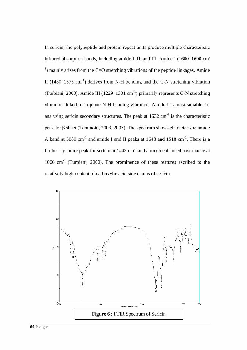
64 P a g e
In sericin, the polypeptide and protein repeat units produce multiple characteristic
infrared absorption bands, including amide I, II, and III. Amide I (1600–1690 cm-
1) mainly arises from the C=O stretching vibrations of the peptide linkages. Amide
II (1480–1575 cm-1
) derives from N-H bending and the C-N stretching vibration
(Turbiani, 2000). Amide III (1229–1301 cm-1
) primarily represents C-N stretching
vibration linked to in-plane N-H bending vibration. Amide I is most suitable for
analysing sericin secondary structures. The peak at 1632 cm-1
is the characteristic
peak for β sheet (Teramoto, 2003, 2005). The spectrum shows characteristic amide
A band at 3080 cm-1
and amide I and II peaks at 1648 and 1518 cm-1
. There is a
further signature peak for sericin at 1443 cm-1
and a much enhanced absorbance at
1066 cm-1
(Turbiani, 2000). The prominence of these features ascribed to the
relatively high content of carboxylic acid side chains of sericin.
Figure 6 : FTIR Spectrum of Sericin
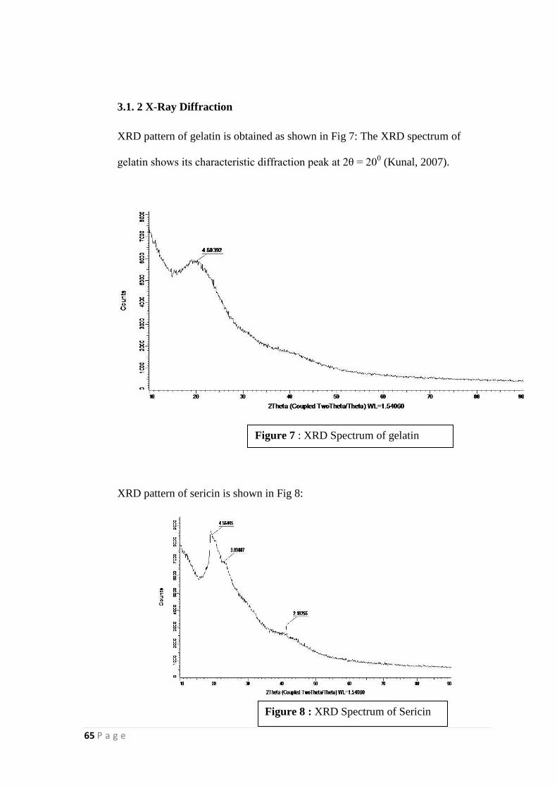
65 P a g e
3.1. 2 X-Ray Diffraction
XRD pattern of gelatin is obtained as shown in Fig 7: The XRD spectrum of
gelatin shows its characteristic diffraction peak at 2θ = 200 (Kunal, 2007).
XRD pattern of sericin is shown in Fig 8:
Figure 7 : XRD Spectrum of gelatin
Figure 8 : XRD Spectrum of Sericin
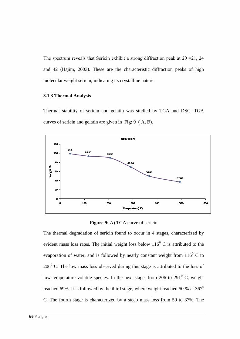
66 P a g e
The spectrum reveals that Sericin exhibit a strong diffraction peak at 2θ =21, 24
and 42 (Hajim, 2003). These are the characteristic diffraction peaks of high
molecular weight sericin, indicating its crystalline nature.
3.1.3 Thermal Analysis
Thermal stability of sericin and gelatin was studied by TGA and DSC. TGA
curves of sericin and gelatin are given in Fig: 9 ( A, B).
Figure 9: A) TGA curve of sericin
The thermal degradation of sericin found to occur in 4 stages, characterized by
evident mass loss rates. The initial weight loss below 1160 C is attributed to the
evaporation of water, and is followed by nearly constant weight from 1160 C to
2060 C. The low mass loss observed during this stage is attributed to the loss of
low temperature volatile species. In the next stage, from 206 to 2910 C, weight
reached 69%. It is followed by the third stage, where weight reached 50 % at 3670
C. The fourth stage is characterized by a steep mass loss from 50 to 37%. The
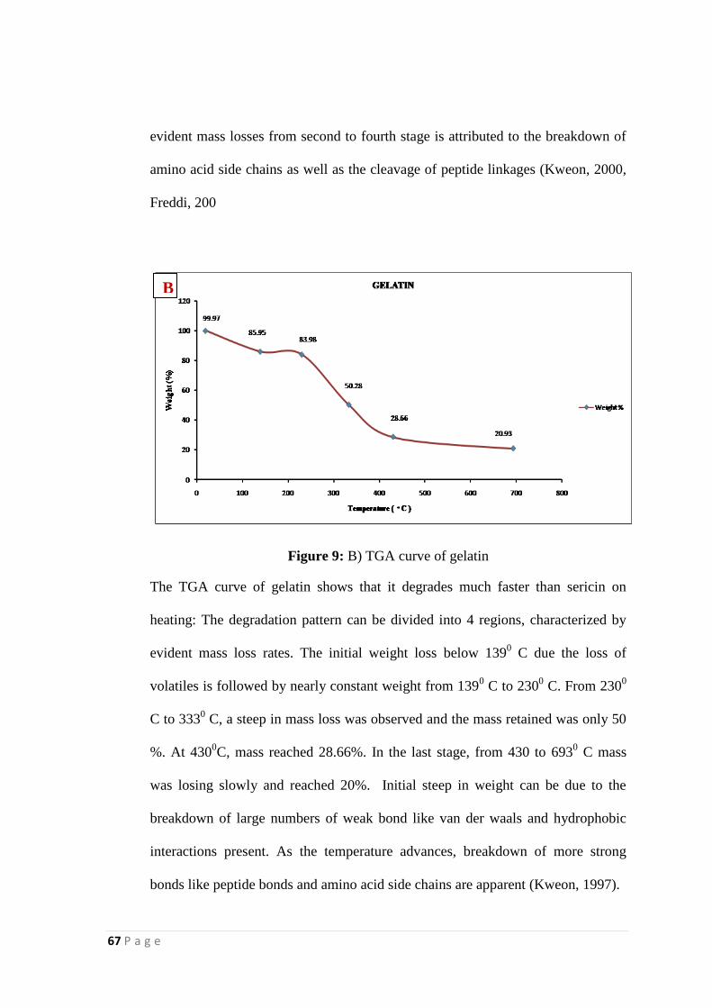
67 P a g e
evident mass losses from second to fourth stage is attributed to the breakdown of
amino acid side chains as well as the cleavage of peptide linkages (Kweon, 2000,
Freddi, 200
Figure 9: B) TGA curve of gelatin
The TGA curve of gelatin shows that it degrades much faster than sericin on
heating: The degradation pattern can be divided into 4 regions, characterized by
evident mass loss rates. The initial weight loss below 1390 C due the loss of
volatiles is followed by nearly constant weight from 1390 C to 230
0 C. From 230
0
C to 3330 C, a steep in mass loss was observed and the mass retained was only 50
%. At 4300C, mass reached 28.66%. In the last stage, from 430 to 693
0 C mass
was losing slowly and reached 20%. Initial steep in weight can be due to the
breakdown of large numbers of weak bond like van der waals and hydrophobic
interactions present. As the temperature advances, breakdown of more strong
bonds like peptide bonds and amino acid side chains are apparent (Kweon, 1997).
B
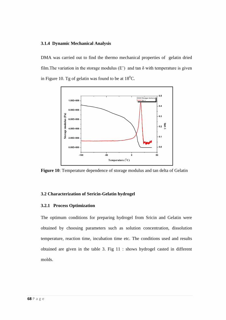
68 P a g e
3.1.4 Dynamic Mechanical Analysis
DMA was carried out to find the thermo mechanical properties of gelatin dried
film.The variation in the storage modulus (E‟) and tan δ with temperature is given
in Figure 10. Tg of gelatin was found to be at 180C.
Figure 10: Temperature dependence of storage modulus and tan delta of Gelatin
3.2 Characterization of Sericin-Gelatin hydrogel
3.2.1 Process Optimization
The optimum conditions for preparing hydrogel from Sricin and Gelatin were
obtained by choosing parameters such as solution concentration, dissolution
temperature, reaction time, incubation time etc. The conditions used and results
obtained are given in the table 3. Fig 11 : shows hydrogel casted in different
molds.
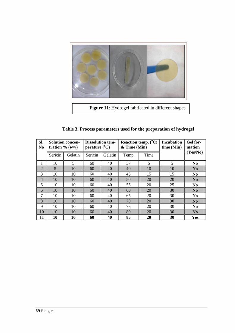
69 P a g e
Table 3. Process parameters used for the preparation of hydrogel
Sl.
No
Solution concen-
tration % (w/v)
Dissolution tem-
perature (oC)
Reaction temp. (0C)
& Time (Min)
Incubation
time (Min)
Gel for-
mation
(Yes/No) Sericin Gelatin Sericin Gelatin Temp Time
1 10 5 60 40 37 5 5 No
2 5 10 60 40 40 10 10 No
3 10 10 60 40 45 15 15 No
4 10 10 60 40 50 20 20 No
5 10 10 60 40 55 20 25 No
6 10 10 60 40 60 20 30 No
7 10 10 60 40 65 20 30 No
8 10 10 60 40 70 20 30 No
9 10 10 60 40 75 20 30 No
10 10 10 60 40 80 20 30 No
11 10 10 60 40 85 20 30 Yes
Figure 11: Hydrogel fabricated in different shapes
shapes
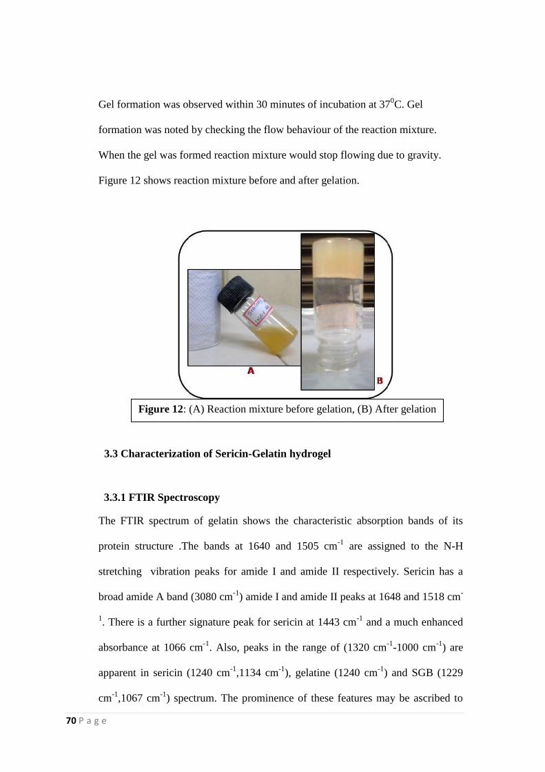
70 P a g e
Gel formation was observed within 30 minutes of incubation at 370C. Gel
formation was noted by checking the flow behaviour of the reaction mixture.
When the gel was formed reaction mixture would stop flowing due to gravity.
Figure 12 shows reaction mixture before and after gelation.
3.3 Characterization of Sericin-Gelatin hydrogel
3.3.1 FTIR Spectroscopy
The FTIR spectrum of gelatin shows the characteristic absorption bands of its
protein structure .The bands at 1640 and 1505 cm-1
are assigned to the N-H
stretching vibration peaks for amide I and amide II respectively. Sericin has a
broad amide A band (3080 cm-1
) amide I and amide II peaks at 1648 and 1518 cm-
1. There is a further signature peak for sericin at 1443 cm
-1 and a much enhanced
absorbance at 1066 cm-1
. Also, peaks in the range of (1320 cm-1
-1000 cm-1
) are
apparent in sericin (1240 cm-1
,1134 cm-1
), gelatine (1240 cm-1
) and SGB (1229
cm-1
,1067 cm-1
) spectrum. The prominence of these features may be ascribed to
Figure 12: (A) Reaction mixture before gelation, (B) After gelation
71 P a g e
the relatively high content of carboxylic acid and alkyl hydroxyl containing amino
acid side chain in sericin. In the case of hydrogel, all those signature peaks of
sericin and gelatin in the fingerprint region was found to be significantly reduced
(1230 cm-1
only), indicating the unavailability of these functional groups. But in
the spectrum of sericin-gelatin blend, fingerprint regions of sericin and gelatin
remained as such to an extent. N-H bend was observed at 1649 cm-1
in sericin and
1641 cm-1
in gelatin. In SGH, a peak was observed at 1659 cm-1
, inferring a shift
in N-H bend as a result of crosslinking. The peak at 1518 cm-1
in sericin reveals
Amide II band. Gelatin also exhibits Amide II at 1505 cm-1
. In SGH, a shift was
observed at this peak towards 1482 cm-1
. Moreover, such a peak is absent in the
case of SGB. The significant reduction of peaks corresponding to C-O stretch of
acids and N-H bend of amines in SGH suggest the unavailability these functional
groups. Owing to these observations, it can be said that a crosslinked hydrogel
network was formed as a result of the interactions of reactive groups in sericin and
gelatin. Table 4 shows the peaks obtained for Sericin, gelatin, hydrogel and a
blend of gelatin and hydrogel.
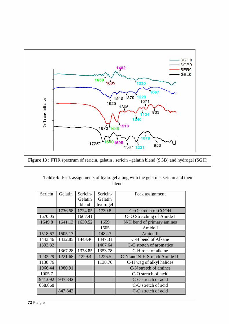
72 P a g e
.
Table 4: Peak assignments of hydrogel along with the gelatine, sericin and their
blend.
Sericin Gelatin Sericin-
Gelatin
blend
Sericin-
Gelatin
hydrogel
Peak assignment
1736.58 1724.05 1730.8 C=O stretch of COOH
1670.05 1667.41 C=O Stretching of Amide I
1649.8 1641.13 1630.52 1659 N-H bend of primary amines
1605 Amide I
1518.67 1505.17 1482.7 Amide II
1443.46 1432.85 1443.46 1447.31 C-H bend of Alkane
1393.32 1407.64 C-C stretch of aromatics
1367.28 1378.85 1353.78 C-H rock of alkane
1232.29 1221.68 1229.4 1226.5 C-N and N-H Stretch Amide III
1138.76 1138.76 C-H wag of alkyl halides
1066.44 1080.91 C-N stretch of amines
1005.7 C-O stretch of acid
941.092 947.842 C-O stretch of acid
858.868 C-O stretch of acid
847.842 C-O stretch of acid
Figure 13 : FTIR spectrum of sericin, gelatin , sericin –gelatin blend (SGB) and hydrogel (SGH)
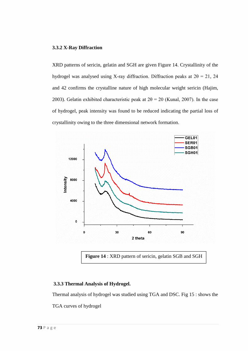
73 P a g e
3.3.2 X-Ray Diffraction
XRD patterns of sericin, gelatin and SGH are given Figure 14. Crystallinity of the
hydrogel was analysed using X-ray diffraction. Diffraction peaks at 2θ = 21, 24
and 42 confirms the crystalline nature of high molecular weight sericin (Hajim,
2003). Gelatin exhibited characteristic peak at 2θ = 20 (Kunal, 2007). In the case
of hydrogel, peak intensity was found to be reduced indicating the partial loss of
crystallinity owing to the three dimensional network formation.
3.3.3 Thermal Analysis of Hydrogel.
Thermal analysis of hydrogel was studied using TGA and DSC. Fig 15 : shows the
TGA curves of hydrogel
Figure 14 : XRD pattern of sericin, gelatin SGB and SGH
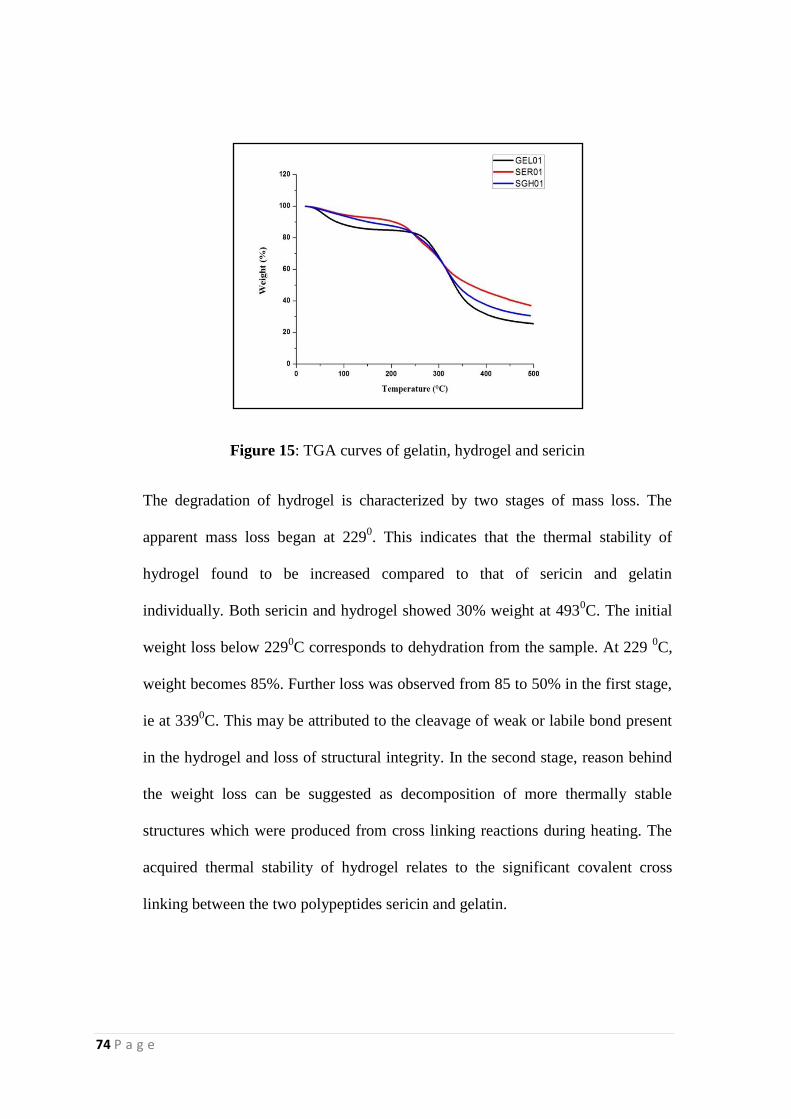
74 P a g e
Figure 15: TGA curves of gelatin, hydrogel and sericin
The degradation of hydrogel is characterized by two stages of mass loss. The
apparent mass loss began at 2290. This indicates that the thermal stability of
hydrogel found to be increased compared to that of sericin and gelatin
individually. Both sericin and hydrogel showed 30% weight at 4930C. The initial
weight loss below 2290C corresponds to dehydration from the sample. At 229
0C,
weight becomes 85%. Further loss was observed from 85 to 50% in the first stage,
ie at 3390C. This may be attributed to the cleavage of weak or labile bond present
in the hydrogel and loss of structural integrity. In the second stage, reason behind
the weight loss can be suggested as decomposition of more thermally stable
structures which were produced from cross linking reactions during heating. The
acquired thermal stability of hydrogel relates to the significant covalent cross
linking between the two polypeptides sericin and gelatin.
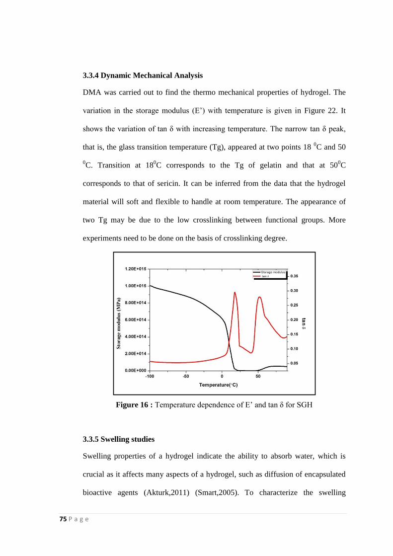
75 P a g e
3.3.4 Dynamic Mechanical Analysis
DMA was carried out to find the thermo mechanical properties of hydrogel. The
variation in the storage modulus (E‟) with temperature is given in Figure 22. It
shows the variation of tan δ with increasing temperature. The narrow tan δ peak,
that is, the glass transition temperature (Tg), appeared at two points 18 0C and 50
0C. Transition at 18
0C corresponds to the Tg of gelatin and that at 50
0C
corresponds to that of sericin. It can be inferred from the data that the hydrogel
material will soft and flexible to handle at room temperature. The appearance of
two Tg may be due to the low crosslinking between functional groups. More
experiments need to be done on the basis of crosslinking degree.
Figure 16 : Temperature dependence of E‟ and tan δ for SGH
3.3.5 Swelling studies
Swelling properties of a hydrogel indicate the ability to absorb water, which is
crucial as it affects many aspects of a hydrogel, such as diffusion of encapsulated
bioactive agents (Akturk,2011) (Smart,2005). To characterize the swelling
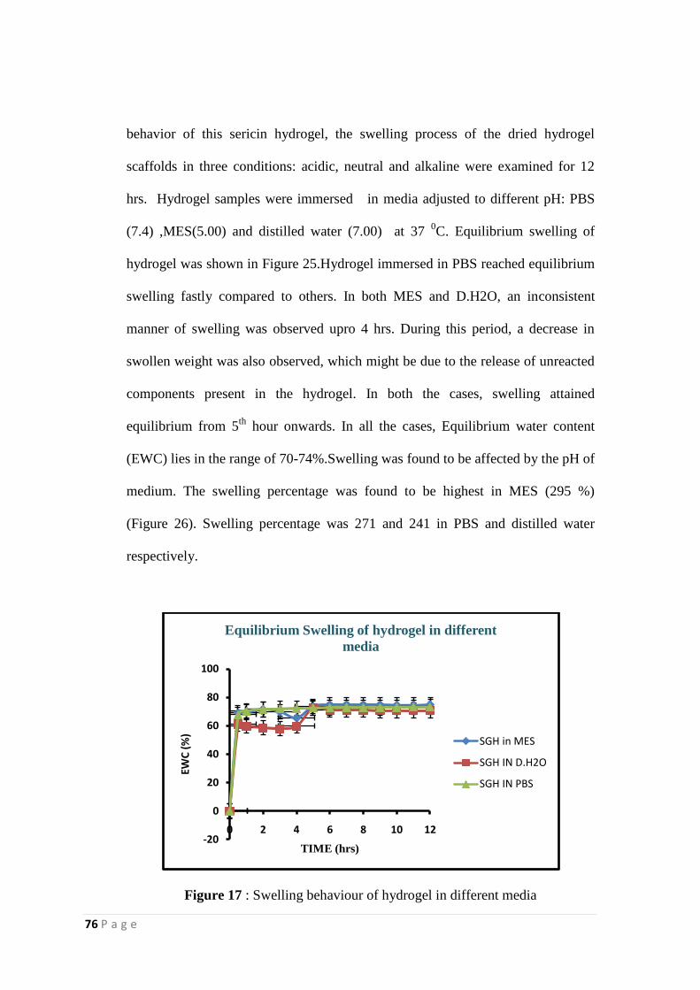
76 P a g e
behavior of this sericin hydrogel, the swelling process of the dried hydrogel
scaffolds in three conditions: acidic, neutral and alkaline were examined for 12
hrs. Hydrogel samples were immersed in media adjusted to different pH: PBS
(7.4) ,MES(5.00) and distilled water (7.00) at 37 0C. Equilibrium swelling of
hydrogel was shown in Figure 25.Hydrogel immersed in PBS reached equilibrium
swelling fastly compared to others. In both MES and D.H2O, an inconsistent
manner of swelling was observed upro 4 hrs. During this period, a decrease in
swollen weight was also observed, which might be due to the release of unreacted
components present in the hydrogel. In both the cases, swelling attained
equilibrium from 5th
hour onwards. In all the cases, Equilibrium water content
(EWC) lies in the range of 70-74%.Swelling was found to be affected by the pH of
medium. The swelling percentage was found to be highest in MES (295 %)
(Figure 26). Swelling percentage was 271 and 241 in PBS and distilled water
respectively.
Figure 17 : Swelling behaviour of hydrogel in different media
-20
0
20
40
60
80
100
0 2 4 6 8 10 12
EWC
(%
)
TIME (hrs)
Equilibrium Swelling of hydrogel in different
media
SGH in MES
SGH IN D.H2O
SGH IN PBS
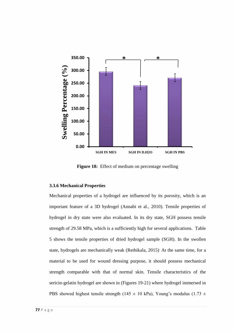
77 P a g e
Figure 18: Effect of medium on percentage swelling
3.3.6 Mechanical Properties
Mechanical properties of a hydrogel are influenced by its porosity, which is an
important feature of a 3D hydrogel (Annabi et al., 2010). Tensile properties of
hydrogel in dry state were also evaluated. In its dry state, SGH possess tensile
strength of 29.58 MPa, which is a sufficiently high for several applications. Table
5 shows the tensile properties of dried hydrogel sample (SGH). In the swollen
state, hydrogels are mechanically weak (Rethikala, 2015). At the same time, for a
material to be used for wound dressing purpose, it should possess mechanical
strength comparable with that of normal skin. Tensile characteristics of the
sericin-gelatin hydrogel are shown in (Figures 19-21) where hydrogel immersed in
PBS showed highest tensile strength (145 ± 10 kPa), Young‟s modulus (1.73 ±
0.00
50.00
100.00
150.00
200.00
250.00
300.00
350.00
SGH IN MES SGH IN D.H2O SGH IN PBS
Sw
elli
ng P
erc
en
tage
(%)
* *
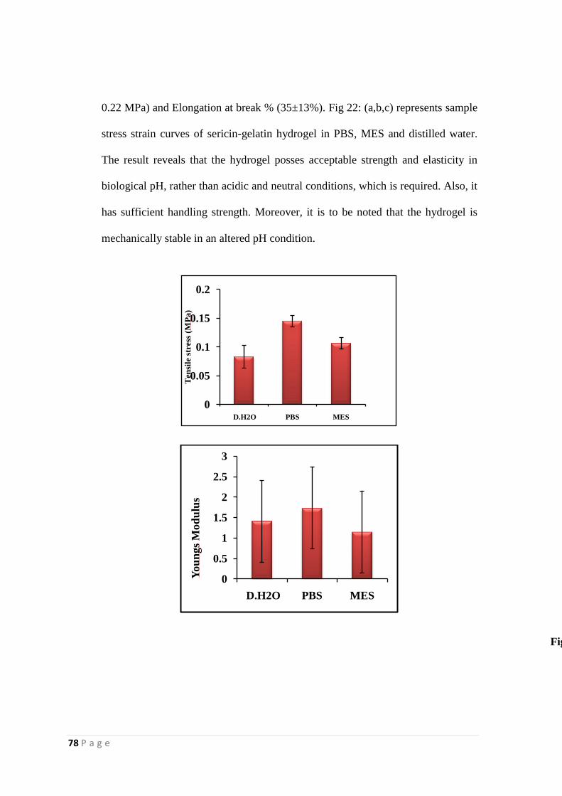
78 P a g e
0.22 MPa) and Elongation at break % (35±13%). Fig 22: (a,b,c) represents sample
stress strain curves of sericin-gelatin hydrogel in PBS, MES and distilled water.
The result reveals that the hydrogel posses acceptable strength and elasticity in
biological pH, rather than acidic and neutral conditions, which is required. Also, it
has sufficient handling strength. Moreover, it is to be noted that the hydrogel is
mechanically stable in an altered pH condition.
Figure 20 : Tensile strength of hydrogels immersed in different media (D.H2O,PBS and MES) Data represents Mean±SD (n=5)
Figure 21 :Youngs Modulus of hydrogels immersed in different media (D.H2O,PBS and MES) Data represents Mean±SD (n=
0
0.5
1
1.5
2
2.5
3
D.H2O PBS MES
You
ngs
Mod
ulu
s
0
0.05
0.1
0.15
0.2
D.H2O PBS MES
Ten
sile
str
ess
(MP
a)
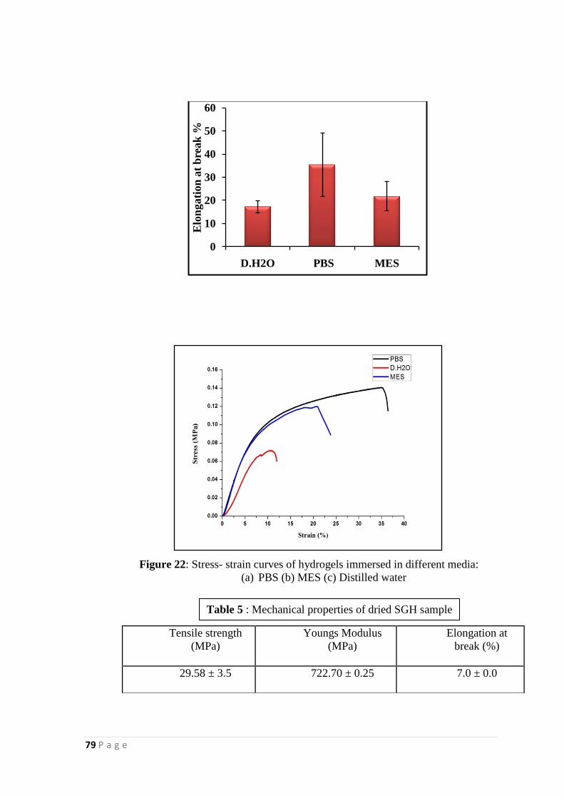
79 P a g e
Figure 22 : Elonation at break (%) of hydrogel immersed in different media (D.H2O, PBS and MES) Mean±SD (n=5)
Figure 22: Stress- strain curves of hydrogels immersed in different media:
(a) PBS (b) MES (c) Distilled water
0
10
20
30
40
50
60
D.H2O PBS MES
Elo
ngati
on
at
bre
ak
%
Tensile strength
(MPa)
Youngs Modulus
(MPa)
Elongation at
break (%)
29.58 ± 3.5
722.70 ± 0.25 7.0 ± 0.0
Table 5 : Mechanical properties of dried SGH sample
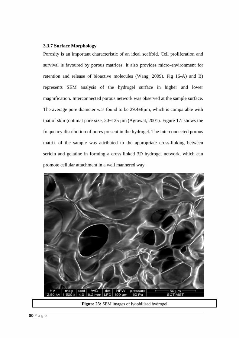
80 P a g e
3.3.7 Surface Morphology
Porosity is an important characteristic of an ideal scaffold. Cell proliferation and
survival is favoured by porous matrices. It also provides micro-environment for
retention and release of bioactive molecules (Wang, 2009). Fig 16-A) and B)
represents SEM analysis of the hydrogel surface in higher and lower
magnification. Interconnected porous network was observed at the sample surface.
The average pore diameter was found to be 29.4±8µm, which is comparable with
that of skin (optimal pore size, 20~125 µm (Agrawal, 2001). Figure 17: shows the
frequency distribution of pores present in the hydrogel. The interconnected porous
matrix of the sample was attributed to the appropriate cross-linking between
sericin and gelatine in forming a cross-linked 3D hydrogel network, which can
promote cellular attachment in a well mannered way.
Figure 23: SEM images of lyophilised hydrogel
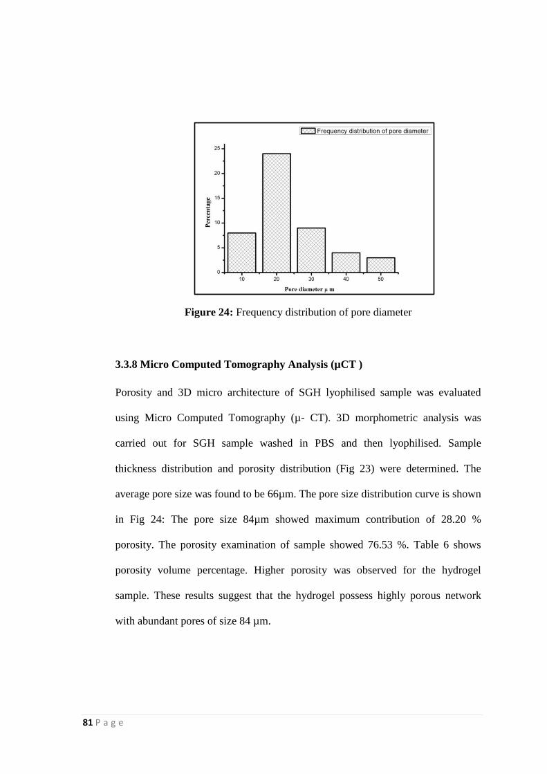
81 P a g e
Figure 24: Frequency distribution of pore diameter
3.3.8 Micro Computed Tomography Analysis (µCT )
Porosity and 3D micro architecture of SGH lyophilised sample was evaluated
using Micro Computed Tomography (µ- CT). 3D morphometric analysis was
carried out for SGH sample washed in PBS and then lyophilised. Sample
thickness distribution and porosity distribution (Fig 23) were determined. The
average pore size was found to be 66µm. The pore size distribution curve is shown
in Fig 24: The pore size 84µm showed maximum contribution of 28.20 %
porosity. The porosity examination of sample showed 76.53 %. Table 6 shows
porosity volume percentage. Higher porosity was observed for the hydrogel
sample. These results suggest that the hydrogel possess highly porous network
with abundant pores of size 84 µm.

82 P a g e
Figure 25 : A) 3D morphology image B) Pore size distribution image, C)
Thickness distribution image
Figure 26 : Pore size distribution of SGH washed in PBS
Table 6: Porosity volume percentage of SGH sample
Micro CT parameter
SGH sample
Total volume (mm3)
0.2598
Scaffold volume (mm3)
0.0610
Scaffold volume/Total volume (mm3)
0.2347
Porosity volume (%) 76.53
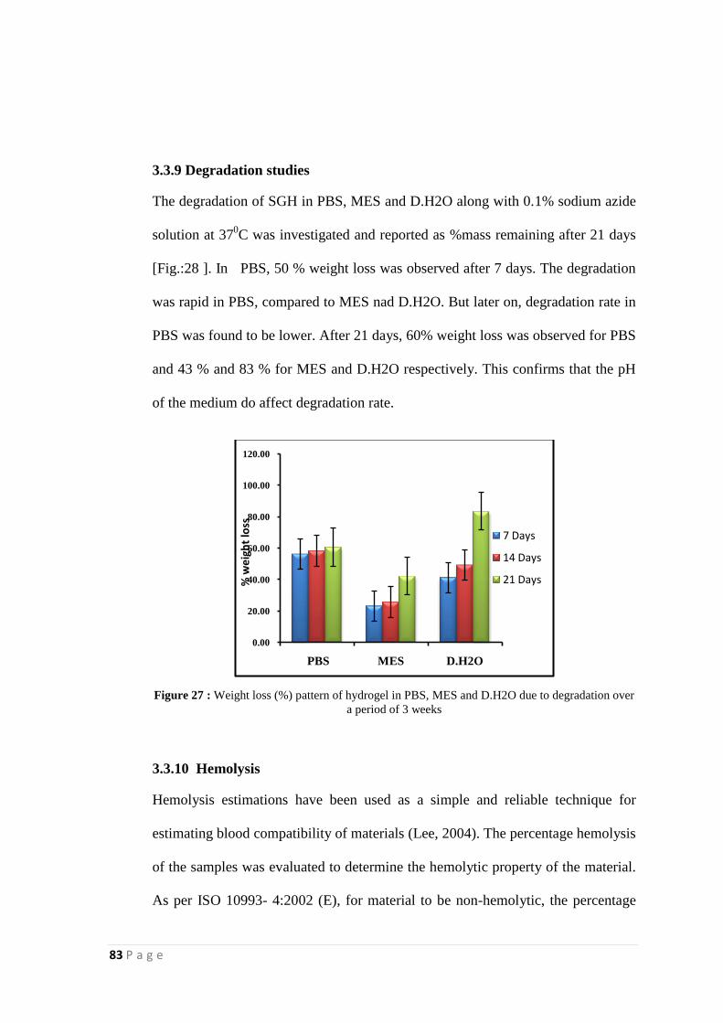
83 P a g e
3.3.9 Degradation studies
The degradation of SGH in PBS, MES and D.H2O along with 0.1% sodium azide
solution at 370C was investigated and reported as %mass remaining after 21 days
[Fig.:28 ]. In PBS, 50 % weight loss was observed after 7 days. The degradation
was rapid in PBS, compared to MES nad D.H2O. But later on, degradation rate in
PBS was found to be lower. After 21 days, 60% weight loss was observed for PBS
and 43 % and 83 % for MES and D.H2O respectively. This confirms that the pH
of the medium do affect degradation rate.
Figure 27 : Weight loss (%) pattern of hydrogel in PBS, MES and D.H2O due to degradation over
a period of 3 weeks
3.3.10 Hemolysis
Hemolysis estimations have been used as a simple and reliable technique for
estimating blood compatibility of materials (Lee, 2004). The percentage hemolysis
of the samples was evaluated to determine the hemolytic property of the material.
As per ISO 10993- 4:2002 (E), for material to be non-hemolytic, the percentage
0.00
20.00
40.00
60.00
80.00
100.00
120.00
PBS MES D.H2O
% w
eig
ht
loss
7 Days
14 Days
21 Days
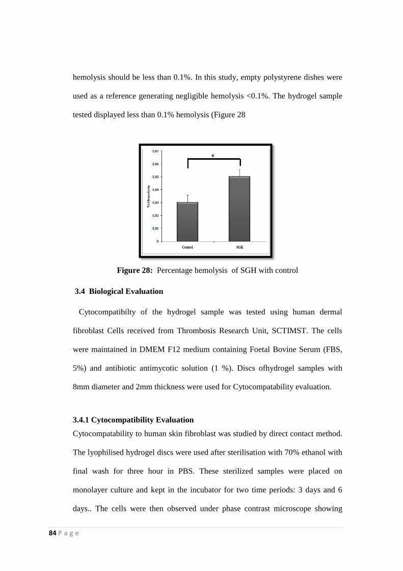
84 P a g e
hemolysis should be less than 0.1%. In this study, empty polystyrene dishes were
used as a reference generating negligible hemolysis <0.1%. The hydrogel sample
tested displayed less than 0.1% hemolysis (Figure 28
Figure 28: Percentage hemolysis of SGH with control
3.4 Biological Evaluation
Cytocompatibilty of the hydrogel sample was tested using human dermal
fibroblast Cells received from Thrombosis Research Unit, SCTIMST. The cells
were maintained in DMEM F12 medium containing Foetal Bovine Serum (FBS,
5%) and antibiotic antimycotic solution (1 %). Discs ofhydrogel samples with
8mm diameter and 2mm thickness were used for Cytocompatability evaluation.
3.4.1 Cytocompatibility Evaluation
Cytocompatability to human skin fibroblast was studied by direct contact method.
The lyophilised hydrogel discs were used after sterilisation with 70% ethanol with
final wash for three hour in PBS. These sterilized samples were placed on
monolayer culture and kept in the incubator for two time periods: 3 days and 6
days.. The cells were then observed under phase contrast microscope showing
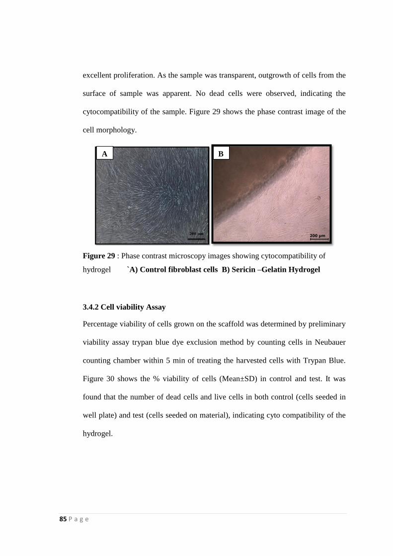
85 P a g e
excellent proliferation. As the sample was transparent, outgrowth of cells from the
surface of sample was apparent. No dead cells were observed, indicating the
cytocompatibility of the sample. Figure 29 shows the phase contrast image of the
cell morphology.
Figure 29 : Phase contrast microscopy images showing cytocompatibility of
hydrogel `A) Control fibroblast cells B) Sericin –Gelatin Hydrogel
3.4.2 Cell viability Assay
Percentage viability of cells grown on the scaffold was determined by preliminary
viability assay trypan blue dye exclusion method by counting cells in Neubauer
counting chamber within 5 min of treating the harvested cells with Trypan Blue.
Figure 30 shows the % viability of cells (Mean±SD) in control and test. It was
found that the number of dead cells and live cells in both control (cells seeded in
well plate) and test (cells seeded on material), indicating cyto compatibility of the
hydrogel.
A B
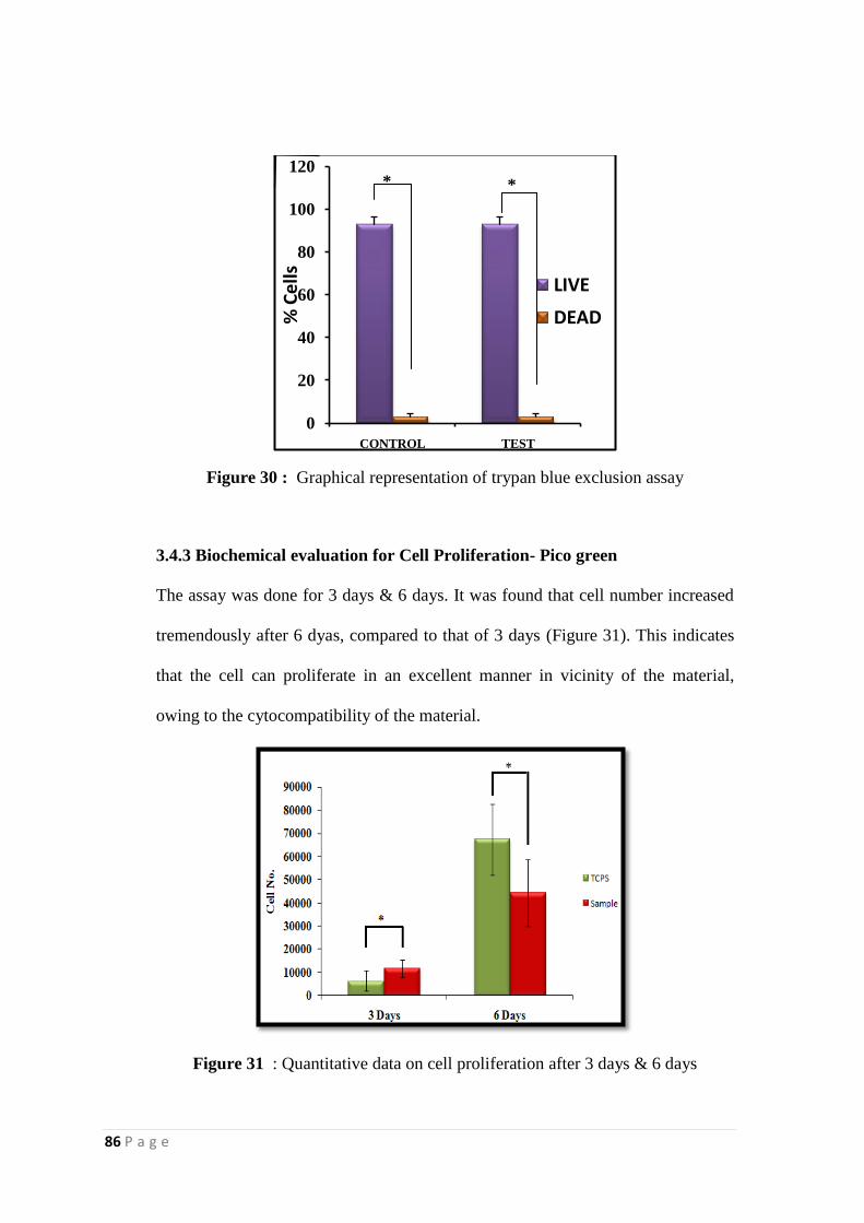
86 P a g e
Figure 30 : Graphical representation of trypan blue exclusion assay
3.4.3 Biochemical evaluation for Cell Proliferation- Pico green
The assay was done for 3 days & 6 days. It was found that cell number increased
tremendously after 6 dyas, compared to that of 3 days (Figure 31). This indicates
that the cell can proliferate in an excellent manner in vicinity of the material,
owing to the cytocompatibility of the material.
Figure 31 : Quantitative data on cell proliferation after 3 days & 6 days
0
20
40
60
80
100
120
CONTROL TEST
% C
ells LIVE
DEAD
**
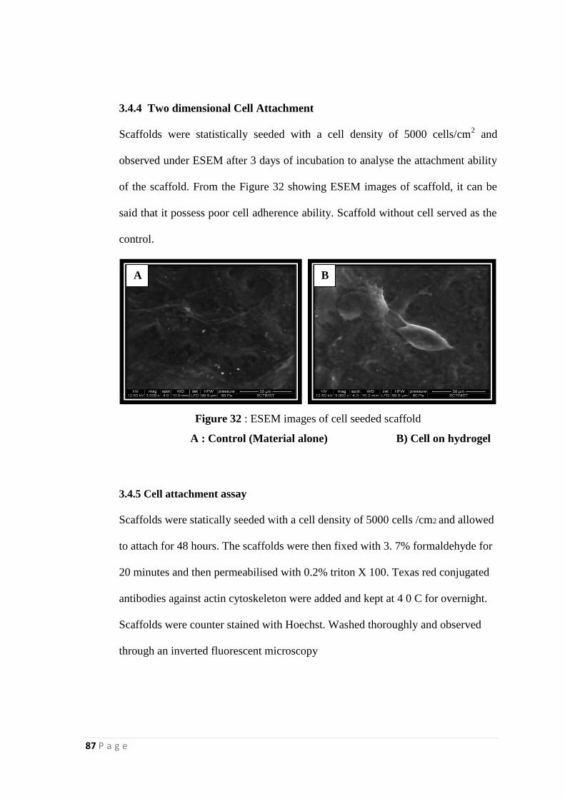
87 P a g e
3.4.4 Two dimensional Cell Attachment
Scaffolds were statistically seeded with a cell density of 5000 cells/cm2 and
observed under ESEM after 3 days of incubation to analyse the attachment ability
of the scaffold. From the Figure 32 showing ESEM images of scaffold, it can be
said that it possess poor cell adherence ability. Scaffold without cell served as the
control.
Figure 32 : ESEM images of cell seeded scaffold
A : Control (Material alone) B) Cell on hydrogel
3.4.5 Cell attachment assay
Scaffolds were statically seeded with a cell density of 5000 cells /cm2 and allowed
to attach for 48 hours. The scaffolds were then fixed with 3. 7% formaldehyde for
20 minutes and then permeabilised with 0.2% triton X 100. Texas red conjugated
antibodies against actin cytoskeleton were added and kept at 4 0 C for overnight.
Scaffolds were counter stained with Hoechst. Washed thoroughly and observed
through an inverted fluorescent microscopy
A B
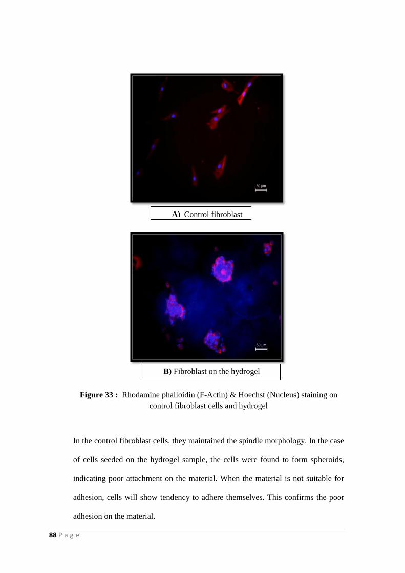
88 P a g e
Figure 33 : Rhodamine phalloidin (F-Actin) & Hoechst (Nucleus) staining on
control fibroblast cells and hydrogel
In the control fibroblast cells, they maintained the spindle morphology. In the case
of cells seeded on the hydrogel sample, the cells were found to form spheroids,
indicating poor attachment on the material. When the material is not suitable for
adhesion, cells will show tendency to adhere themselves. This confirms the poor
adhesion on the material.
A) Control fibroblast
B) Fibroblast on the hydrogel
89 P a g e
CHAPTER 4
SUMMARY AND CONCLUSION
Biopolymers are a versatile class of materials with widespread applications in the
area of tissue engineering and regenerative medicine. Members under this class
were found to have thermo-responsive solubility behaviour. This opens
perspectives in developing gels with water retaining capacity, defined as hydrogel.
Hydrogels serve as an important class of biomaterials. Silk protein sericin is an
emerging candidate in the biomedical field. It has a long history of being
discarded as a waste material. Natural polymers are always given a prominence
for use as a biomaterial, owing to its biocompatibility. Gelatin, a product derived
from collagen is being explored widely in this area.
In the current study, an effort was put forth to develop a hydrogel from silk protein
sericin and gelatin. The sericin-gelatin hydrogel in the present investigation uses a
simple and cost effective fabrication technique without any chemical cross-linkers.
The fabricated hydrogel was characterized chemically, mechanically and
biologically. The raw materials were characterized by spectroscopic, thermal and
mechanical methods.
The parameters for hydrogel formation were optimized. The fabricated hydrogel
was also characterized spectroscopically and compared with that of raw materials.
Thermal stability of the hydrogel was also done, which was found to be increased
90 P a g e
when compared to raw materials. Swelling, degradation and mechanical studies
were done in hydrogel immersed in different media PBS (slightly alkaline), MES
(acidic) and distilled water (neutral). Swelling and degradation was found to be
affected by the pH of the medium. Tensile properties of hydrogel were the highest
when it was swelled in PBS. Dynamic mechanical analysis showed two glass
transitions temperatures corresponding to sericin and gelatin.
Two dimensional surface morphology of the hydrogel was revealed by Scanning
Electron microscope, with a porous network with an average pore diameter
comparable with that of human skin. Poor adhesion was confirmed by Rhodamine
phalloiidn & Hoechst staining. Further, three dimensional micro architecture was
done by Micro CT, showing a continuous and uniform pore size distribution with
a porosity volume of 76.53.
Hemocompatibility of the hydrogel (0.05%) was revealed by percent hemolysis
method. Biological evaluation was carried out by cytocompatibility studies using
human dermal fibroblasts. Phase contrast images of the scaffold seeded with cells
revealed no cytotoxicity and excellent proliferation. ESEM images of scaffold
with cells showed poor cell adhesion. Preliminary evaluation of cytotoxicity was
quantified by trypan blue exclusion method, which revealed 90 % viability of cells
seeded on scaffold.
91 P a g e
From the results obtained, it can be concluded that the sericin –gelatin hydrogel
possess good swelling, mechanical and thermal stability, adequate porosity and
cytocompatibility with poor cell adhesion, to use as a wound dressing material.
The present study can be considered as a pioneer work, which has to be explored
further in terms of biological evaluation for use as a potential wound dressing
material. This study involves a simple and novel method of developing hydrogel
from two bio polymers without any chemical cross-linkers. Incorporation of
growth factors and /or drugs and the release studies could be done in the future. As
literature supports the fact that sericin possess anti microbial activity, studies
could be extended in terms of sericin release from hydrogel and subsequent anti
microbial activity. Moreover, the present study is an attempt to explore the utility
and value of Sericin, a waste material as green material.

92 P a g e
REFERENCES
1. Agrawal Pushpa*, B.V. Goutham Vishnu and Thippa Reddy K.S, Preparation of
nano silk sericin based hydrogels from silk industry waste, Journal of Environmental
Research And Development , 2013, Vol. 8 No. 2.
2. Agrawal, C. M.&Ray, R. B. Biodegradable polymeric scaffolds for
musculoskeletal tissue engineering. J Biomed Mater Res 55, 141–150 (2001).
3. Ahn, J.S., Choi, H.K., Lee, K.H., Nahm, J.H. and Cho, S. (2001) Novel
mucoadhesive polymer prepared by template polymerization of acrylic acid in the
presence of silk sericin. J. Appl. Polym. Sci. 80, 274–280. application. Japan patent
06-080741A.
4. Akturk, O. et al. Evaluation of sericin/collagen membranes as prospective wound
5. Annabi, N. et al. Controlling the porosity and microarchitecture of hydrogels for
tissue engineering. Tissue Eng Part B Rev 16, 371–383 (2010).
6. Aramwit, P., Siritientong, T. & Srichana, T, Potential applications of silk
sericin, a natural protein from textile industry by-products. Waste Manage Res
,2012,30, 217–224.
7. Ausl¨ander S., M. Wieland and M. Fussenegger, Metab. Eng., 2012, 14, 252–260.
8. Baba, T.,Hanada, K. &Hashimoto, I. The study of ultraviolet b-induced apoptosis
9. Banani Kundu, Subhas C. Kundu, Silk sericin/polyacrylamide in situ forming
hydrogels for dermal reconstruction, Biomaterials,2012,Vol 33 7456-7467.
10. Bapi Sarker,a Dimitrios G. Papageorgiou,b Raquel Silva,a Tobias Zehnder,a
Farhana Gul-E-Noor,†c Marko Bertmer,c Joachim Kaschta,d Konstantinos
Chrissafis,b Rainer Detscha and Aldo R. Boccaccini*, Fabrication of alginate–
gelatin crosslinked hydrogel microcapsules and evaluation of the microstructure
and physico-chemical properties. J. Mater. Chem. B, 2014, 2, 1470–1482.
11. Bapi Sarker,a Dimitrios G. Papageorgiou,b Raquel Silva,a Tobias Zehnder,a
12. Bigi. A., S. Panzavolta and K. Rubini, Biomaterials, 2004, 25, 5675–5680
13. Billiet T., M. Vandenhaute, J. Schelout, S. Van Vlierberghe and P. Dubruel,
Biomaterials, 2012, 33, 6020–6041.
14. Biman B. Mandal, Anjana S. Priya, S.C. Kundu *, Novel silk sericin/gelatin 3-D
scaffolds and 2-D films: Fabrication and characterization for potential tissue
engineering applications, Acta Biomaterialia. 2009, 5, 3007–3020.
15. Bouhadir K. H, K. Y. Lee, E. Alsberg, K. L. Damm, K. W. Anderson and D. J.
Mooney, Biotechnol. Prog, 2001,17, 945–950.
by coating with silk sericin, Trans Mater Res Soc Jpn ,2004 29, 4.
16. Cao, Y.; Wang, B. C. Biodegradation of silk biomaterials. Int. J. Mol. Sci. 2009,
10, 1514–1524.
17. Charu Vepari and David L. Kaplan, Silk as a Biomaterial, Prog Polym Sci. 2007 ;
32(8-9): 991–1007.
18. Chisti, Y. (1998) Strategies in downstream processing; in Bioseparation and
bioprocesssing: a handbook. G. Subramanian (ed). New York: Wiley-VCH. pp. 3–
30.
colon carcinogenesis induced by 1,2-dimethylhydrazine in mice. Oncol Rep,2000,
7, 1049–1052.
93 P a g e
19. Doyle, J. P.; Giannouli, P.; Martin, E. J.; Brooks, M.; Morris,E. R. Effect of sugars,
galactose content, and chainlength on freeze-thaw gelation of galactomannans.
Carbohydr. Polym. 2006, 64, 391–401.
dressing biomaterial. J Biosci Bioeng,2011,112, 279–288.
20. Enas M. Ahmed, Hydrogel: Preparation, characterization, and applications.
http://dx.doi.org/10.1016/j.jare.2013.07.006.
21. Farhana Gul-E-Noor,†c Marko Bertmer,c Joachim Kaschta,d Konstantinos
Chrissafis,b Rainer Detscha and Aldo R. Boccaccini, Fabrication of alginate–
gelatin crosslinked hydrogel microcapsules and evaluation of the microstructure
and physico-chemical properties, J. Mater. Chem. B,2014, 2, 1470.
22. Freddi G, Monti P, Nagura M, Gotoh Y & Tsukada M. Structure and Molecular
Conformation of Tussah Silk Fibroin Films: Effect of Heat Treatment. Journal of
Polymer Science Part B: Polymer Physics ,1997; 35(5): 841–847.
23. Garcia-Fuentes, M.; Meinel, A. J.; Hilbe, M.; Meinel, L.; Merkle, H. P. Silk
fibroin/hyaluronan scaffolds for human mesenchymal stem cell culture in tissue
engineering. Biomaterials, 2009, 30, 5068– 5076
24. Ghassemifar, R.; Redmond, S.; Zainuddin; Chirila, T. V.Advancing towards a
tissue-engineered tympanic membrane: silk fibroin as a substratum for growing
human eardrum keratinocytes. J. Biomater. Appl. 2010, 24, 591–606.
25. Gong, Y. H.; Zhu, Y. B.; Liu, Y. X.; Ma, Z. W.; Gao, C. Y.; Shen, J. C. Layer-by-
layer assembly of chondroitin sulfate and collagen on aminolyzed poly(L-lactic
acid) porous scaffolds to enhance their chondrogenesis. Acta Biomater. 2007, 3,
677–685.
26. Gong, Z. G.; Yang, Y. H.; Huang, L.; Chen, X.; Shao, Z. Z. Formation kinetics and
fractal characteristics of regenerated silk fibroin alcogel developed from
nanofibrillar network. Soft Matter 2010,6, 1217–1223
27. Gregory H. Altman,Frank Diaz,Caroline Jakuba,Tara Calabro,Rebecca L. Horan,
28. Gulrajani M.L. (2005) Sericin: A Bio-molecule of value. Souveni 20th congress of
the international sericultural commission, Bangalore, India 15-18th December
2005.pp. 21-29.
29. Gupta, P.; Vermani, K.; Garg, S. Hydrogels: from controlled release to pH-
responsive drug delivery. Drug Discovery Today 2002, 7, 569–579.
30. Hacker MC, Mikos AG. Synthetic polymers, principles of regenerative medicine.
2nd ed.; 2011. p. 587–622.
31. Hajim Miyake, Hiroyuki Wakisaka, Yoshitaka Yamashia and Masanobu Nagura.
Moisture characterestics nd properties of high moleculr weight sericin films,
Polymer journal,2003,Vol.35,No.8,pp 683-687.
32. Harumasa Okamotoa, Etsuko Ishikawa, and YoshiaSkui zukig ,Structural Analysis
of Sericin Genes homologies with fibroin gene in the 5‟ flanking nucleotide
sequences*,1982.
33. Haugh, M. G.; Jaasma, M. J.; O‟Brien, F. J. The effect of dehydrothermal
treatment on the mechanical and structural properties of collagen-GAG scaffolds.
J. Biomed. Mater. Res., Part A 2009, 89A, 363–369.
34. Hidetoshi Teramoto, T Sunenori Kameda and Yasushi Tameda , Preparation of gel
film from Bombyx mori and its characterization as a wound dressing
,Biosci.Biotechnol.Biochem,2008, Vol 72, 12, 3189-3196.
94 P a g e
in cultured mouse keratinocytes and in mouse skin. J Dermatol Sci ,1996,12, 18–
23.
35. Jayakrishnan, A. and S. R. Jameela, Glutaraldehyde as a fixative in bioprostheses
and drug delivery matrices,Biomaterials,1996 17(5): 471-484.
36. Jin, H. J.; Chen, J. S.; Karageorgiou, V.; Altman, G. H.; Kaplan, D. L. Human
bone marrow stromal cell responses on electrospun silk fibroin mats. Biomaterials
2004, 25, 1039–1047.
Jingsong Chen,Helen Lu,John Richmond, David L. Kaplan ,Silk-based
biomaterials, Biomaterials, 2003, 24, 401–416.
37. Jinsub. Shin, Paul.V. Braun, Wonmok. Lee Fast response photonic crystal pH
sensor based on templated photo-polymerized hydrogel inverse opal Sens Actuat
B: Chem, 150 (1) (2010), pp. 183–190
38. K.L. Shantha, D.R.K. Harding Synthesis and evaluation of sucrose-containing
polymeric hydrogels for oral drug delivery, J Appl Polym Sci, 84 (2002), p. 2597
39. K.M. Raju, M.P. Raju Synthesis of novel superabsorbing copolymers for
agricultural and horticultural applications Polym Int, 50 (2001), pp. 946–951
40. Kato, N. et al. Silk protein, sericin, inhibits lipid peroxidation and tyrosinase
activity. Biosci Biotechnol Biochem ,1998,62, 145–147.
41. Kato, N., Sato, S., Yamanaka, A., Yamadam,H., Fuwam, N. and Nomura, M.
(1998) Silk protein, sericin, inhibits lipid peroxidation and tyrosinase activity.
Biosci. Biotechnol. Biochem. 62, 145–147.
42. Kawazoe, N.; Lin, X. T.; Tateishi, T.; Chen, G. P. Threedimensional cultures of rat
pancreatic RIN-5F cells in porous PLGAcollagen hybrid scaffolds. J. Bioact.
Compat. Polym. 2009, 24, 25–42.
43. Keck, M.; Haluza, D.; Burjak, S.; Eisenbock, B.; Kamolz, L. P.; Frey, M.
Cultivation of keratinocytes and preadipocytes on a collagenelastin scaffold
(Matriderm): first results of an in vitro study. Eur. J. Surg. 2009, 41, 189–193.
44. Khalid Nassif Jassim (1) Omar Jaffar Al-Saree, Study of the antimicrobial activity
of silk sericin from silkworm bombyx mori , Iraqi J. Comm. Med.,2010, Vol -23, 2.
45. Kolokythas, P.; Aust, M. C.; Vogt, P. M.; Paulsen, F. Dermal substitute with the
collagen-elastin matrix Matriderm in burn injuries: a comprehensive review.
Handchirurgie Mikrochirurgie Plastische Chirurgie 2008, 40, 367–371.
46. Komatsu K, Silk (its formation, structure, character, and utilization), the polymeric
materials encyclopedia, (CRC Press) 1996
47. Komatsu, K. (1975) Studies on dissolution behaviors and structural characteristic
of silk Sericin. Bull. Sericult. Exp. Sta. 26, 135- 256.
48. Komatsu, K. (1982) Silk III. Sericin physical structure. Sericologia. 22, 14-23.
49. Komatsu, K., 1980, Recent advances in sericin research. J. Sericult. Sci. Japan. 69,
457-465.
50. Krishna, O. D.; Kiick, K. L. Protein- and peptide-modified synthetic polymeric
biomaterials. Biopolymers ,2010, 94, 32–48.
51. Kunal Pal,1 Ajit K. Banthia,1 and Dipak K. Majumda,Preparation and
Characterization of Polyvinyl Alcohol–Gelatin Hydrogel Membranes for
Biomedical Applications ,Materials Science Centre, 2007,Indian Institute of
Technology, Kharagpur.
52. Kweon H & Park YH. Dissolution and Characterization of Regenerated Antheraea
pernyi Silk Fibroin. Journal of Applied Polymer Science ,2001; 82: 750–758.
95 P a g e
53. Kweon HY, Um IC & Park YH. Thermal behavior of regenerated Antheraea
pernyi silk fibroin film treated with aqueous methanol. Polymer ,2000; 41: 7361–
7367.
54. L. Takashi, T. Hatsumi, M. Makoto, I. Takashi, G. Takehiko, S. Shuji Synthesis of
porous poly(N-isopropylacrylamide) gel beads by sedimentation polymerization
and their morphology, J Appl Polym Sci, 104 (2) (2007), p. 842
55. L. Yang, J.S. Chu, J.A. Fix Colon-specific drug delivery: new approaches and in
vitro/in vivo evaluation Int J Pharm, 235 (2002), pp. 1–15
56. Lawrence, B. D.; Wharram, S.; Kluge, J. A.; Leisk, G. G.;Omenetto, F. G.;
Rosenblatt, M. I.; Kaplan, D. L. Effect of hydration on silk film material
properties. Macromol. Biosci. 2010, 10, 393–403.
57. Lazzeri, L.; Cascone, M. G.; Danti, S.; Serino, L. P.; Moscato,S.; Bernardini, N.
Gelatine/PLLA sponge-like scaffolds: morphological and biological
characterization. J. Mater. Sci.: Mater. Med. 2007,18, 1399–1405.
58. Lee DW, Powers K and Baney R. Physicochemical properties and blood
compatibility of acylated chitosan nanoparticles. Carbohydr Polym 2004; 58: 371–
377.
59. Li H.-B., H. Jiang, C.-Y. Wang, C.-M. Duan, Y. Ye, X.-P. Su, Q.-X. Kong, J.-F.
Wu and X.-M. Guo, Biomed. Mater., 2006, 1, 42–47.
60. Li, X. (1996) Usages of sericin in durable material. China patent.1116227A.
61. M N Padamwar and A P Pawar*, Silk sericin and its applications: A review,
Journal of Scientific & Industrial Research , 2004 ,Vol. 63,323-329.
62. M. Friedman, J. Agric. Food Chem., 2004, 52, 385–406.
63. M. Mondal*, K. Trivedy and S. Nirmal Kumar, The silk proteins, sericin and
fibroin in silkworm, Bombyx mori Linn., - a review. Caspian J. Env. Sci. 2007,
Vol. 5 No. 2 pp. 63~76 .
64. Magoshi J, Biospinning (silk fibre formation), the polymeric materials
encyclopedia (CRC Press) 1996.
65. Makaya, K.; Terada, S.; Ohgo, K.; Asakura, T. Comparative study of silk fibroin
porous scaffolds derived from salt/water and sucrose/hexafluoroisopropanol in
cartilage formation. J. Biosci. Bioeng. 2009, 108, 68–75.
66. Mandal, B. B.; Kundu, S. C. Cell proliferation and migration in silk fibroin 3D
scaffolds. Biomaterials ,2009, 30, 2956–2965.
67. Mauney, J. R.; Nguyen, T.; Gillen, K.; Kirker-Head, C.; Gimble, J. M.; Kaplan, D.
L. Engineering adipose-like tissue in vitro and in vivo utilizing human bone
marrow and adipose-derived mesenchymal stem cells with silk fibroin 3D
scaffolds. Biomaterials 2007,28, 5280–5290.
68. Meinel, A. J.; Kubow, K. E.; Klotzsch, E.; Garcia-Fuentes, M.; Smith, M. L.;
Vogel, V.; Merkle, H. P.; Meinel, L. Optimization strategies for electrospun silk
fibroin tissue engineering scaffolds. Biomaterials, 2009, 30, 3058–3067.
69. Mei-po Ho1, Hao Wang1 and Kin-tak Lau1,2*,Thermal properties and structure
conformation on silkworm silk fibre, Centre of Excellence in Engineered Fibre
Composites, Faculty of Engineering and Surveying, University of Southern
Queensland, Toowoomba, Queensland Australia 2 Department of Mechanical
Engineering, The Hong Kong Polytechnic University, Kowloon, Hong Kong,
SAR, China.
96 P a g e
70. Minoura, N., Aiba, S., Gotoh, Y., Tsukada, M.and Imai, T. (1995) Attachment and
growth of cultured fibroblast cells on silk protein matrices. J. Biomed. Mat. 29,
1215–1221
71. Miyazaki, T. et al. Control of bioresorption of porous alpha-tricalcium phosphate
72. Mizoguchi, K., Iwatsubo, T. and Aisaka, N (1991) Separating membrane made of
cross-linked thin film of sericin and production thereof. Japan Patent. 03-
284337A.
73. Moy RL, Lee A, Zalka A. Commonly used suture materials in skin surgery. Am
Fam Physician 1991;44(6):2123–2128. [PubMed: 1746393]
74. Murase, M. (1994) Method for solubilising and molding cocoon silk, artificial
organ made of cocoon silk, and medical element made of cocoon silk. Japan
Patent 06-166850A.
75. Nakajima, Y. (1994) Liquid crystal element. Japan Patent 06-018892.
76. Nogueira, G. M.; Rodas, A. C. D.; Weska, R. F.; Aimoli, C. G.; Higa, O. Z.;
Maizato, M.; Leiner, A. A.; Pitombo, R. N. M.; Polakiewicz, B.; Beppu, M. M.
Bovine pericardium coated with biopolymeric films as an alternative to prevent
calcification: In vitro calcification and cytotoxicity results. Mater. Sci. Eng., C
2010, 30, 575–582
77. Ohyabu, Y.; Adegawa, T.; Yoshioka, T.; Ikoma, T.; Shinozaki,K.; Uemura, T.;
Tanaka, J. A collagen sponge incorporating a hydroxyapatite/chondroitinsulfate
composite as a scaffold for cartilage tissue engineering. J. Biomater. Sci., Polym.
Ed. 2009, 20, 1861–1874
78. Panilaitis, B. et al. Macrophage responses to silk. Biomaterials,2003 24, 3079–
3085.
79. Peppas, N. A.; Bures, P.; Leobandung, W.; Ichikawa, H. Hydrogelsin
pharmaceutical formulations. Eur. J. Pharm. Biopharm. 2000, 50 (1), 27–46.
80. Ren, Y. J.; Zhou, Z. Y.; Liu, B. F.; Xu, Q. Y.; Cui, F. Z. Preparation and
characterization of fibroin/hyaluronic acid composite scaffold. Int. J. Biol.
Macromol. 2009, 44, 372–378.
81. Rethikala PK and Krishnan V Kalliyana, Photopolymerized poly(2- hydroxyethyl
methacrylate)/ poly(ε-caprolactone)/ poly(ethylene glycol) system as a potential
wound dressing material . A. Saarai, T. Sedlacek, V. Kasparkova, T. Kitano and P.
Saha, J. Appl. Polym. Sci., 2012, 126, E79–E88 .
82. Rim, N. G.; Lee, J. H.; Jeong, S. I.; Lee, B. K.; Kim, C. H.; Shin, H. Modulation of
osteogenic differentiation of human mesenchymal stem cells by poly[(L-lactide)-
co-(epsilon-caprolactone)]/gelatin nanofibers. Macromol. Biosci. 2009, 9, 795–
804.
83. RJ. Patel*and M. K. Modasiya, Sericin: Pharmaceutical Applications,
International journal of research in pharmaceutical and biomedical science,
2011,Vol. 2 (3).
84. S. Van Vlierberghe, P. Dubruel, and E. Schacht , Biopolymer-Based Hydrogels As
Scaffolds for Tissue Engineering Applications: A Review. Biomacromolecules
2011, 12, 1387–1408.
85. Sadov, F., Korchagin, M. and Matetsky, A. (1987) Chemical technology of fibrous
materials. Mir Publication, Moscow, pp. 306-307
86. Sasaki, M., Kato, N.,Watanabe, H. & Yamada, H. Silk protein, sericin, suppresses
87. Shaw J T B & Smith S G, Amino acid of silk sericin, Nature, 4278 (1951) 745.
97 P a g e
88. She, Z. D.; Liu, W. Q.; Feng, Q. L. Self-assembly model, hepatocytes attachment
and inflammatory response for silk fibroin/chitosan scaffolds. Biomed. Mater.
2009, 4, 045013.
89. Smart, J. D. The basics and underlying mechanisms of mucoadhesion. Adv Drug
Deliv Rev,2005,57, 1556–1568.
90. Sorina Dinescu 1,†, Bianca Galateanu 1,†, Madalina Albu 2, Anisoara Cimpean
1,Anca Dinischiotu 1 and Marieta Costache 1,*Sericin Enhances the
Bioperformance of Collagen-Based Matrices Preseeded with Human-Adipose
Derived Stem Cells (hADSCs), Int. J. Mol. Sci. 2013, 14, 1870-1889.
91. Sorina Dinescu,1 Bianca Gslsueanu,1 Msdslina Albu,2 Adriana Lungu,3 Eugen
Radu,4 Anca Hermenean,5,6 andMarieta Costache1, Biocompatibility Assessment
of Novel Collagen-Sericin Scaffolds Improved with Hyaluronic Acid and
Chondroitin ulfate for Cartilage Regeneration, BioMed Research International,
2013,11 pages
Structural effect of silk sericin. J R Soc Interface,2005, 2, 373–378.
92. Sundar, S.; Kundu, J.; Kundu, S. C. Biopolymeric nanoparticles. Sci. Technol. Adv.
Mater. 2010, 11, 014104.
93. Sung, H. W., D. M. Huang, et al.,Evaluation of gelatin hydrogel crosslinked with
various crosslinking agents as bioadhesives: in vitro study, J Biomed Mater Res46
,1999(4): 520-530.
94. Sunita Nayak, Sancharika Dey, Subhas C. Kundu*, Skin Equivalent Tissue-
Engineered Construct: Co-Cultured Fibroblasts/ Keratinocytes on 3D Matrices of
Sericin Hope Cocoons, Plos one,2.13, Vol 8, 9.
95. Takasu, Y., Yamada, H. & Tsubouchi, K. Isolation of three main sericin
components from the cocoon of the silkworm, bombyx mori. Biosci Biotechnol
Biochem,2012, 66, 2715–2718.
96. Takeuchi, A. et al. Heterogeneous nucleation of hydroxyapatite on protein:
97. Tanaka, K. amd Mizuno, S. (2001) Homologues of fibroin L-chain and P25 of
Bombyx mori are present in Dendrolimus spectabilis and Papilio xuthus but not
detectable in Antheraea yamamai. Insect Biochem. Mol Biol. 31, 665-677.
98. Tao, Y. Z.; Yan, Y.; Xu, W. L. Physical characteristics and properties of
waterborne polyurethane materials reinforced with silk fibroin powder. J. Polym.
Sci., Part B: Polym. Phys. 2010, 48, 940–950.
99. Thirupathamma D1* Savithri G 1, and Kavya Sudha K, Silk for biomedical
applications. Research Journal of Pharmaceutical, Biological and Chemical
Sciences, 2013,Vol 4 No.2,657-663.
100. Tierney, C. M.; Jaasma, M. J.; O‟Brien, F. J. Osteoblast activity on collagen-GAG
scaffolds is affected by collagen and GAG concentrations.J. Biomed. Mater. Res.,
Part A 2009, 91A, 92–101.
101. Tippawan Siritientong, Juthamas Ratanavaraporn, Teerapol Srichana, and
Pornanong Aramwit, Preliminary Characterization of Genipin-Cross-Linked Silk
Sericin/Poly(vinyl alcohol) Films as Two-Dimensional Wound Dressings for the
Healing of Superficial Wounds, Biomed Res Int. 2013; 2013: 904314.
102. Traian V Chirila1,2,3,4,5*, Shuko Suzuki1, Laura J Bray1,6,7, Nigel L
Barnett1,6,8 and Damien G Harkin1,6,9, Evaluation of silk sericin as a biomaterial:
in vitro growth of human corneal limbal epithelial cells on Bombyx mori sericin
membranes, Progress in Biomaterials, 2013, 2:14
98 P a g e
103. Tsubouchi, K. (1999a) Wound covering material. US patent 5951506.
104. Tsubouchi, K. (1999b) Occlusive dressing consisting essentially of silk fibroin
and silk sericin and its production. Japan Patent 11-070160A.
105. Tsukada, M. (1983) Structure of silk sericins removed from wild silk by boiling in
water. J. Sericult. Sci. Japan. 52, 296-299.
106. Tsukada, M., Hayasaka, S., Inoue, K.,Nishikawa, S. and Yamamoto, S. (1999)
Cell culture bed substrate for proliferation of animal cell and its preparation. Japan
Patent 11-243948A.
107. Turbiani .R.B *, Jose Tomadon Jr., Fernanda Lini Seixas, Marcelino Luis
Gimenes, Properties and Structure of Sericin Films: Effect of the Crosslinking
Degree Franciele Department of Chemical Engineering. State University of
Maringá Av. Colombo. 5790. CEP: 87020-900. Maringá. PR. BraziL
108. Van Vlierberghe S, P. Dubruel, and E. Schacht*, Biopolymer-Based Hydrogels
As Scaffolds for Tissue Engineering Applications: A Review 2011 ,
Biomacromolecules,2011, 12, 1387–1408
109. Vasconcelos, A. C. Gomes and A. Cavaco-Paulo, Acta Biomater, 2012, 8, 3049–
3060.
110. Voegeli R, Meier J & Blust R, Sericin silk protein: unique structure and
properties,Cosmet Toilet, 108 (1993) 101-108.
111. Vollrath F, Knight DP. Liquid crystalline spinning of spider silk. Nature
2001;410(6828):541–548. [PubMed: 11279484]
112. Wang T, Wang J & Zhou J, γ- Ray study on the sericin structure of cocoon silk,
Fangzhi Xuebao, 6(3) (1985) 133-134
113. Wang, S. D.; Zhang, Y. Z.; Wang, H. W.; Yin, G. B.; Dong,Z. H. Fabrication and
properties of the electrospun polylactide/silk fibroin-gelatin composite tubular
scaffold. Biomacromolecules, 2009,10, 2240–2244.
114. Wang, S. D.; Zhang, Y. Z.; Yin, G. B.; Wang, H. W.; Dong, Z. H. Electrospun
polylactide/silk fibroin-gelatin composite tubular scaffolds for small-diameter
tissue engineering blood vessels. J. Appl. Polym. Sci. 2009, 113, 2675–2682.
115. Wang, S. D.; Zhang, Y. Z.; Yin, G. B.; Wang, H. W.; Dong, Z. H. Electrospun
polylactide/silk fibroin-gelatin composite tubular scaffolds for small-diameter
tissue engineering blood vessels. J. Appl. Polym. Sci., 2009, 113, 2675–2682
116. Wei, X.; Xiaohong, W.; Yongnian, Y.; Renji, Z. A polyurethane-gelatin hybrid
construct for manufacturing implantable bioartificial livers. J. Bioact. Compat.
Polym. 2008, 23, 409–422.
117. Wen, F.; Chang, S.; Toh, Y. C.; Teoh, S. H.; Yu, H. Development of poly(lactic-
co-glycolic acid)-collagen scaffolds for tissue engineering. Mater. Sci. Eng., C
2007, 27, 285–292.
118. Wichterle, O.; Lim, D. Hydrophilic gels for biological use. Nature, 1960, 185,
117–118.
119. Wilson J. L, T. C. McDevitt, Biotechnol. Bioeng., 2013,110, 667–682.
120. Y. Tabata Biomaterial technology for tissue engineering applications J R Soc
Interf, 6 (2009), pp. S311–S324
121. Yamada, H. and Fuwa, N. (1994) Protein containing high molecular material and
its
122. Yamada, H., Fuwa, N. and Nomura, M. (1993) Synthetic fiber having improved
hygroscopicity. Japan patent 05-339878A.
99 P a g e
123. Yamada, M. (1978) Amino acid composition of the sericin extracted from cocoon
of the mulberry wild silkworm, Bombyx mori and its species specificity. J.
Sericult. Sci. Japan. 47, 108-112.
124. Yang, Y.; Zhu, X. L.; Cui, W. G.; Li, X. H.; Jin, Y. Electrospun composite mats
of poly[(D,L-lactide)-co-glycolide] and collagen with high porosity as potential
scaffolds for skin tissue engineering. Macromol. Mater. Eng. 2009, 294, 611–619.
125. Yoko Takasua, Hiromi Yamadaa & Kozo Tsubouchia, Isolation of Three Main
Sericin Components from the Cocoon of the Silkworm, Bombyx mori, National
Institute of Agrobiological Sciences Owashi,2014.
126. Yoshii, F., Kume, N., Makuuchi, K. and Sato, F. (2000) Hydrogel composition
containing silk protein. Japan Patent 06-017373A.
127. Yucel, T.; Kojic, N.; Leisk, G. G.; Lo, T. J.; Kaplan, D. L. Nonequilibrium silk
fibroin adhesives. J. Struct. Biol. 2010, 170, 406–412.Jang, E. S.; Park, J. W.;
Kweon, H.; Lee, K. G.; Kang, S. W.;Baek, D. H.; Choi, J. Y.; Kim, S. G.
Restoration of peri-implant defects in immediate implant installations by
Choukroun platelet-rich fibrin and silk fibroin powder combination graft. Oral
Surg. Oral Med. Oral Pathol. Oral Radiol. Endodontol. 2010, 109, 831–836.
128. Z. Maolin, L. Jun, Y. Min, H. Hongfei The swelling behaviour of radiation
prepared semi-interpenetrating polymer networks composed of polyNIPAAm and
hydrophilic polymers, Radiat Phys Chem, 58 (2000), pp. 397–400
129. Zhao Wen, Jin Xing, Cong Yang, Liu Yuying, Fu Jun Degradable natural polymer
hydrogels for articular cartilage tissue engineering, J Chem Technol Biotechnol, 88
(3) (2013), pp. 327–339
130. Zhaorigetu, S.N., Sasakim M., Watanbe, H.and Kato, N. (2003) Silk protein,
sericin,suppresses DMBA-TPA induced mouse skin tumorigenesis by reducing
oxidative stress, inflammatory responses and endogenous tumor promoter TNF-
alpha. Oncol Rep. 10, 537-543.
131. Zheng Wang1,2*, Yeshun Zhang1*, Jinxiang Zhang2, Lei Huang1, Jia Liu1,
Yongkui Li1, Guozheng Zhang4,Subhas C. Kundu5 & Lin Wang1,3, Exploring
natural silk protein sericin for regenerative medicine: an injectable,
photoluminescent, cell-adhesive 3D hydrogel,Nature,2014,Vol 4,7064.
100 P a g e
APPENDIX
REAGENTS USED:
1. Dulbecco's Phosphate buffered saline (DPBS):
NaCl- 8g
KC1-0.2g
KH2P04- 0.2g
Na2HP04- 1.7g
DI Water-1000 mL
pH -7.4
2. Fibroblast culture medium (50mL):
DMEM-F12- 45mL
Foetal Bovine Calf Serum (FBS)- 5mL
Antibiotic-antimycotic solution - 500µL
3. 3.7% Formaldehyde (l0 mL):
Formaldehyde (37%)- lmL
DPBS-9mL