gastrointestinal disorders and malabsorption
TRANSCRIPT

Gastrointestinal disorders
and
malabsorption
Elodie Hanon
5th March 2015

Content
1) Introduction
2) The stomach
3) The stomach: gastrin secretion regulation
4) Disorders and investigations of gastric function
5) The pancreas
6) Disorders of the exocrine pancreas
7) Pancreatic cancer
8) The small intestine
9) Colon
10)Digestion and absorption
11)Malabsorption
12)Coeliac disease
13)Inflammatory bowel diseases
14)Clinical consequences of malabsorption
15)Cases

Source: http://www.austincc.edu/apreview/PhysText/Digestive.html
Introduction

The stomach
Divided in 3 major zones: cardiac zone, body and pyloric zone (antrum, pyloric
canal and sphincter)
Mix of food with acidic gastric juice
Approx. 2.5L per 24h

The stomach
HCl → acidic pH necessary
for pepsin (from
pepsinogens) to begin
digestion of proteins and
stimulates bile secretion
Mucus → protection of the
mucosal surface
Intrinsic factor → binds to
vit B12, allows its
absorption in the terminal
ileum

The stomach
Three phases for regulation of gastric secretion:
Cephalic: neuronal (vagus nerve)
Gastric: local response to food in stomach, hormonal (gastrin)
Intestinal: inhibitory phase, response to entry of gastric contents into
duodenum and jejunum, hormonal and neuronal

The stomach: gastrin secretion regulation
Production by G cells of antrum
Stimulated by antral distention (meals), protein digestion in stomach and vagal
stimulation
Transported by blood through liver to the parietal cells of the fundus of the stomach
Stimulation of gastric acid secretion
Gastrin also stimulates:
Secretion of gastric pepsinogens, intrinsic factor
Secretion of secretin (small intestin)
Secretion of bicarbonate, enzymes (pancreas)
Secretion of hepatic bile
Motility, mucosal growth, blood flow to stomach
Inhibition of gastrin by the action of acid on the G cells

Disorders and investigations of gastric function
Limited use of biochemical tests
Use of endoscopy or contract radiography
Peptic ulceration: most cases due to colonisation by Helicobacter pylori
Diagnosis by serology
Other cause: hypergastrinaemia
Disorder Gastric secretion Gastrin response to secretin
Zollinger-Ellison syndrome Greatly ↑ ↑
Hypersecretion of gastrin
by antral G-cells
Greatly ↑
None or ↓
Pernicious anaemia ↓ ↓
Post vagotomy ↓ ↓
CKD Variable ↓

Disorders and investigations of gastric function
Intrinsic factor
Intrinsic factor: glycoprotein, essential for absorption of vitamin B12 (cyancobalamin) in
terminal ileum
Pernicious anaemia
Autoimmune destruction of parietal cells in stomach
Resulting in achlohydria and anaemia
Macrocytic, megaloblastic, heamolytic anaemia
Diagnosis: low serum B12, positive parietal cell autoantibodies, high plasma gastrin
B12 absorption restored by administration of intrinsic factor

The pancreas
Endocrine function
• Secretion in the blood stream
• Islets of Langerhans
• Production of insulin, glucagon, somatostatin,
pancreatic polypeptide
Exocrine function
• Secretion out of the body (gut lumen)
• Alkaline, bicarbonate-rich solution (pH 8.0)
• Approx. 1.5 L per day
• Proenzyme forms of proteases, trypsin,
chymotrypsin, carboxypeptidase
• Lipase, colipase, amylase
Secretion under the control of 2 hormones secreted by the small intestine:
Secretin: stimulation of secretion of an alkaline fluid
Cholecystokinin (CCK): stimulation of secretion of pancreatic enzymes and
contraction of the gallbladder
Secretin and CCK secretion in response to presence of acid, AA, partly digested proteins in
the duodenum

Disorders of the exocrine pancreas
Major disorders of exocrine pancreas
Acute pancreatitis: biochemistry essential in diagnosis and management
Chronic pancreatitis: biochemistry of limited used
Pancreatic cancer: biochemistry of little use
Cystic fibrosis: inherited metabolic disease
Acute pancreatitis
Acute abdomen with severe pain
Most frequent causes: excessive alcohol ingestion, gallstones
Less common causes: infection, hypertriglycaemia, hypercalcaemia
Severe acute pancreatitis → mortality up to 25%
Biochemistry
High serum amylase activity (>10 ULN)
Serum lipase activity: more specific, but
less used in UK
Plasma may be lipaemic
Mild increase bilirubin and ALP
Early increase AST: characteristic of
pancreatitis caused by gallstones
Causes of increased plasma amylase
>10 ULN: acute pancreatitis
>5 ULN: perforated duodenal ulcer, intestinal
obstruction, other acute abdominal disorders,
acute oliguric renal failure, DKA, ruptured
Fallopian tube
Usually <5 ULN: salivary gland disorders, CKD,
macroamylasaemia

Disorders of the exocrine pancreas
Pancreatic insufficiency
Reduced exocrine function: fat malabsorption, clinically significant when 90% of
function lost
Symptoms: weight loss, malaise, diarrhoea, steatorrhoea
Causes
Chronic pancreatitis
Pancreatic surgery
Acute pancreatitis
Cystic fibrosis
Diabetes
Assessing pancreatic function
Gold standard: faecal elastase, single stool sample
Pancreatic specific
Not degraded in the intestine
Normally high concentration in faeces
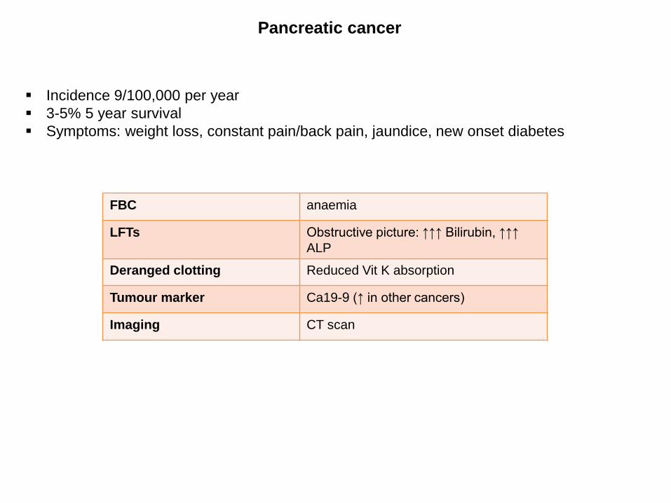
Pancreatic cancer
Incidence 9/100,000 per year
3-5% 5 year survival
Symptoms: weight loss, constant pain/back pain, jaundice, new onset diabetes
FBC anaemia
LFTs Obstructive picture: ↑↑↑ Bilirubin, ↑↑↑
ALP
Deranged clotting Reduced Vit K absorption
Tumour marker Ca19-9 (↑ in other cancers)
Imaging CT scan

The small intestine
Divided into 3 sections: duodenum, jejunum and ileum
Approx. 7m long (adult)
Intestinal villus: increased surface area

Colon
Approx. 1.5 m (adult) from the
ileum to anus
Divided into: cecum, appendix,
colon, rectum and anal canal

Digestion and absorption

Digestion and absorption
Most of it in duodenum and jejunum
B12 and bile salts in the terminal ileum
Reabsorption of Na and water
Fluid, vitamins and minerals Location
Fat soluble vitamins (A, D, E, K)
Iron, zinc, calcium
Folate
Jejunum
Vit B12
Magnesium, calcium
Fluids and electrolytes
Ileum
Fluids and electrolytes Colon

Digestion and absorption
Peptides
Pancreatic enzymes, e.g. trypsinogen, activated in the small intestine
Absorbed as amino acids, dipeptides and tripeptides
Hydrolysed in enterocytes to free amino acids
Lipids
Emulsification with bile and bile acids
Pancreatic lipase and co-lipase
Triglycerides hydrolysed on brush border to monoglycerides and fatty acids
Re-esterified in enterocytes to triglycerides
Incorporated into chylomicrons with apo-lipoprotein B48
Transported via lymph to thoracic duct and superior vena cava (in contrast to amino acids
and sugars – transported via portal vein to liver)

Digestion and absorption
Carbohydrates
Diet: polysaccharides (starch and glycogen), disaccharides and monosaccharides
Salivary and pancreatic amylase for hydrolysation
In brush border: disaccharideases → glucose, galactose, fructose
Disaccharidease deficiency:
• Fermentation in colon (flatulence and diarrhoea)
• Often acquired following gut infection
• Lactase activity lost after weaning (abdominal discomfort and explosive diarrhoea)
Others
Calcium: active transport, controlled by 1,25-dihydroxyvitamin D
Iron: rate of absorption determined by saturation of circulating transferrin
When saturated, iron is not absorbed

Malabsorption
Malabsorption: impaired absorption of products of digestion
Mucosal defects
Biochemical or genetic abnormalities
Coeliac disease (gluten-sensitive enteropathy)
Other protein sensitivities: cows’ mil (infants), soya protein
Disaccharidase deficiencies: inherited (lactase) or acquired (post-infection)
Cystinuria, etc…
Inadequate absorptive surface
Small intestinal resection
Small bowel fistulae
Inflammatory, infiltrative or infective disorders
Crohn’s disease
Tropical and non-tropical sprue
Infective enteritis
Parasites, etc…
Others
Malnutrition: mucosal/gut atrophy
Lymphatic obstruction

Malabsorption
Defective luminal digestion
Pancreatic insufficiency
Chronic pancreatitis
Pancreatic carcinoma
Cystic fibrosis
Pancreatectomy
Biliary insufficiency
Liver disease
Bacterial overgrowth
Terminal ileal disease/resection
Drugs
Others
Postgastrectomy steatorrhoea
Endocrine disease

Coeliac disease
Autoimmune mediated jejunal mucosal inflammation
Proximal small bowel affected
Symptoms: diarrhoea, weight loss, failure to thrive, stomatitis, bloating and pain
Patient history: trigger by food
Investigations: tissue transglutaminase antibodies
duodenal biopsy → gold standard

Inflammatory bowel diseases
Crohn’s disease
Affects any part of the GI tract
Skip lesions
Usually small bowel
Transmural inflammation
Crypt abscesses
Ulcerative colitis
Proximal from rectum
Continuous involvement
Always rectum
Inflammed mucosa
Superficial inflammation
No crypt abscesses

Inflammatory bowel disease
Investigations
Colonoscopy
Faecal calprotectin: distinguish between IBD and non-inflammatory bowel conditions
(IBS), to monitor IBD activity
IBD and IBS can cause similar symptoms
Released into the intestines in excess when inflammation
NICE diagnostics guidance DG11

Clinical consequences of malabsorption
Generalised malnutrition and weight loss (failure to thrive in children)
Fat and carbohydrates calorie malabsorption
Decreased absorption/reabsorption of fluid and electrolytes
Fermentation of unabsorbed carbohydrates (and fat) in colon
Gross fluid loss: hypotension and pre-renal failure
Vitamin and mineral deficiencies
Vitamin and mineral Consequences
Iron, B12, folate Anaemia
Vit D, Ca Rickets - osteomalacia
Vit K Bruising and bleeding
Iron, B12, B vitamins Glossitis and stomatitis
K Muscular weakness
Ca and Mg Tetany, paraesthesiae

Cases

Case 1
Man, 53
Presents with severe abdominal pain. Radiates through to the back
No previous H/O of GI disease
Heavy alcohol intake over many years
Examination: tender epigastric region, midly shocked
Test result Reference range
Urea 10 mmol/L 3.3-6.7 mmol/L
Creatinine 90 µmol/L 60-120 µmol/L
Calcium 2.10 mmol/L 2.2-2.6 mmol/L
Albumin 30 g/L 35-50 g/L
Glucose 12 mmol/L 2.8-6.0 mmol/L (fasting)
Amylase 5000 U/L <300 U/L
↑↑ amylase = suggestive of acute
pancreatitis
Slightly ↑ urea + N creatinine = renal
hypoperfusion due to shock
Hypocalcaemia: protein-rich exudate in
the peritoneal cavity, formation of
insoluble calcium salts of fatty acids

Case 2
3 year old baby boy
Failure to thrive: below the 3rd centile for height, 10th centile for weight
Frequent diarrhoea
Did not enjoy his food
Examination: anaemia, abdominal distension, wasting of the muscles of the limbs, buttocks
and shoulder girdle
Test result Reference range
Albumin 30 g/L 34-42 g/L
Haemoglobin 97 g/L 105-127 g/L
Tissue transglutaminase antibody
Strongly positive
Duodenal biopsy Total villous atrophy
History and findings on examination
suggestive of GI disorder
Positive serology for tissue transglutaminase
antibody suggestive of coeliac disease
Supported by biopsy appearance