gastrin-releasing peptide receptor silencing suppresses ... · gastrin-releasing peptide receptor...
TRANSCRIPT

Gastrin-releasing peptide receptor silencingsuppresses the tumorigenesis and metastaticpotential of neuroblastomaJingbo Qiao*, JungHee Kang*, Titilope A. Ishola*, Piotr G. Rychahou*, B. Mark Evers*†, and Dai H. Chung*†‡
*Department of Surgery and †Sealy Center for Cancer Cell Biology, University of Texas Medical Branch, Galveston,TX 77555-0353
Edited by Patricia K. Donahoe, Massachusetts General Hospital, Boston, MA, and approved July 9, 2008 (received for review December 19, 2007)
Neuroblastoma accounts for nearly 15% of all pediatric cancer-related deaths. We have previously shown that gastrin-releasingpeptide (GRP) stimulates neuroblastoma growth, and that its cellsurface receptor, GRP-R, is overexpressed in advanced-stage hu-man neuroblastomas; however, the effects of GRP/GRP-R on tu-morigenesis and metastasis in vivo are not clearly elucidated. In thepresent study, we found that GRP-R knockdown in the aggressivecell line BE(2)-C induced cell morphology changes, reduced cell size,decreased cell proliferation, and inhibited DNA synthesis, corre-sponding to cell cycle arrest at G2/M phase. Activated Akt, a crucialregulator of cell survival and metastasis, was down-regulated byGRP-R silencing. In addition, expression of p-p70S6K and its down-stream target molecule S6, key regulators of protein synthesis andcell metabolism, were also significantly decreased by GRP-R silenc-ing. GRP-R knockdown also up-regulated the expression of tumorsuppressor PTEN, the inhibitor of the PI3K/Akt pathway. Further-more, silencing GRP-R as well as GRP in BE(2)-C cells suppressedanchorage-independent growth in vitro. Conversely, overexpres-sion of GRP-R in less aggressive SK-N-SH neuroblastoma cellsresulted in soft agar colony formation, which was inhibited by aGRP-blocking antibody. Moreover, GRP-R deficiency significantlydelayed tumor growth and diminished liver metastases in vivo. Ourfindings demonstrate that GRP and GRP-R have important onco-genic properties beyond their established mitogenic functions.Therefore, GRP-R may be an ideal therapeutic target for thetreatment of aggressive neuroblastomas.
Akt � RNAi � anchorage independence � metastasis
Advanced-stage neuroblastoma in children remains highlylethal with mortality rates exceeding 50% (1). Because of
their neuroendocrine lineage, neuroblastomas can produce avariety of peptides that contribute to the classic clinical symp-toms and are related to tumor prognosis, including gastrin-releasing peptide (GRP) (2). GRP and its receptor, GRP-R, areknown to be up-regulated in various cancers, including undif-ferentiated neuroblastoma (3), small cell lung carcinoma (4), andprostate cancer (5). We have previously shown that GRP issecreted by neuroblastoma cells and acts as an autocrine growthfactor to promote proliferation (3). We have also found thatstable transfection of GRP-R, a member of the G proteincoupled receptor family, causes an increase in the bindingcapacity for its ligand GRP to stimulate a constitutive cellulargrowth rate in SK-N-SH neuroblastoma cells (6).
GRP/GRP-R signaling functionally correlates with aberra-tions in neuroblastoma behavior including cell cycle progressionand angiogenesis. We have found that GRP treatment inducesG1-S phase progression (7). We have also shown that bombesin,the amphibian equivalent of GRP, increases the vascularizationof neuroblastoma xenografts in vivo by the up-regulation ofvascular endothelial growth factor (8). These processes wereshown to be mediated in part by the phosphatidylinositol 3-kinase (PI3K)/Akt survival pathway (7, 8). Correspondingly,GRP-R overexpression up-regulates Akt activation in neuro-blastoma cells and we also found that the ratio of Akt, in
comparison to its negative regulator PTEN, was increased inhuman malignant neuroblastomas (6). This finding is especiallyrelevant because a recent study has shown that Akt activationcorrelates with poor prognosis in primary neuroblastoma (9).
The mitogenic actions of GRP in tumor cells have beenwell-established; however, another less known property of GRP/GRP-R is its morphogenic capability (10). Morphogenesis is animportant step for cell motility during the development of theinvasive nature of various cancers, including breast and colon(11, 12). In addition to its growth factor functions, we have alsonoted morphological alterations in neuroblastoma cells thatoverexpress GRP-R (6). Therefore, GRP/GRP-R may be in-volved in regulating multiple steps of tumorigenesis. The mo-lecular mechanisms responsible for GRP-mediated tumor ag-gressiveness and metastatic potential are not clearly defined. Thepurpose of our current investigation was to elucidate, in broaderdetail, the oncogenic effects of GRP-R in relation to neuroblas-toma survival, invasive potential, and metastasis development.
In this study, we report that down-regulation of GRP-Rreversed the aggressive phenotype of human neuroblastoma cellline BE(2)-C, decreased cell proliferation, inhibited DNA syn-thesis, and induced cell cycle arrest at G2/M phase in vitro.GRP-R silencing also significantly blocked neuroblastoma tu-morigenicity by reducing colony formation in vitro, and inhibitingxenograft growth and liver metastasis in vivo. We also observed,at the molecular level, that cell survival mediator Akt and itsdownstream targets p70S6K and S6 are regulated by GRP-R inhuman neuroblastoma. Our results further demonstrate thatGRP-R is a clinically relevant therapeutic target in humanneuroblastomas.
ResultsStable Knockdown of GRP-R Inhibits Neuroblastoma Cell Growth andDown-Regulates the PI3-K/Akt Pathway. We have shown that GRPacts as an autocrine growth factor for neuroblastoma cells (3);this effect appears to depend on ligand binding to GRP-R (3, 8).We wanted to confirm the importance of GRP-R in mediatingneuroblastoma growth; therefore, we measured the effect ofGRP-R silencing on the cell proliferative capacity. We used shorthairpin RNA (shRNA) vectors to establish stable knockdown ofGRP-R in BE(2)-C cells and confirmed the knockdown withreverse transcription-PCR (RT-PCR) and Western blot analysis(Fig. 1E). Over a time course, the BE(2)-C cells expressing
Author contributions: J.Q. and D.H.C. designed research; J.Q., J.H.K., T.A.I., and P.G.R.performed research; J.Q., J.H.K., T.A.I., P.G.R., B.M.E., and D.H.C. analyzed data; and J.Q.,T.A.I., B.M.E., and D.H.C. wrote the paper.
The authors declare no conflict of interest.
This article is a PNAS Direct Submission.
‡To whom correspondence should be addressed at: University of Texas Medical Branch, 301University Blvd., Galveston, TX 77555-0353. E-mail: [email protected].
This article contains supporting information online at www.pnas.org/cgi/content/full/0711861105/DCSupplemental.
© 2008 by The National Academy of Sciences of the USA
www.pnas.org�cgi�doi�10.1073�pnas.0711861105 PNAS � September 2, 2008 � vol. 105 � no. 35 � 12891–12896
CELL
BIO
LOG
Y
GRP-R shRNA (shGRP-R) displayed stagnant growth andsignificantly reduced proliferation in comparison with controlshRNA (shCON) cells, whose growth rate steadily increasedbetween each time point (Fig. 1 A). In addition, we measured theamount of BrdU incorporation in shGRP-R cells and found thatDNA synthesis was decreased to 57% compared with controlcells (Fig. 1B). This reduction corresponded with a 37% decreaseof shGRP-R cells in S phase of the cell cycle and an increase ofcells arrested in G2/M phase, in comparison to shCON cells (Fig.1C). We confirmed that the transfected control cells did notchange the endogenous protein levels of GRP-R as well as theproliferative capacity when compared to native BE(2)-C cells[supporting information (SI) Fig. S1]. These data show thatGRP-R plays a vital role in maintaining neuroblastoma cellularproliferation.
Our previous studies found that GRP/GRP-R could activatethe PI3K/Akt survival pathway (7, 8). We also found that GRP-Roverexpression enhanced the levels of phosphorylated (p-)Akt,and decreased the levels of PTEN, a tumor suppressor thatnegatively regulates the PI3K/Akt pathway (6). Furthermore, wedetermined that the ratio of Akt/PTEN was increased in poorlydifferentiated human neuroblastoma tissue samples (6). SinceAkt activation regulates numerous oncogenic processes andcorrelates with poor prognosis in primary human neuroblasto-mas (9); we next assessed the role of GRP-R knockdown onp-Akt and PTEN expression. shGRP-R significantly decreasedp-Akt (Ser-473) expression without notably affecting the levelsof total Akt (Fig. 1D). Additionally, PTEN was up-regulated incells expressing shGRP-R. Therefore, PI3K pathway activationappears to be regulated by GRP-R; on the other hand, activatedERK1/2 protein levels remained relatively unchanged withshGRP-R expression (Fig. 1D).
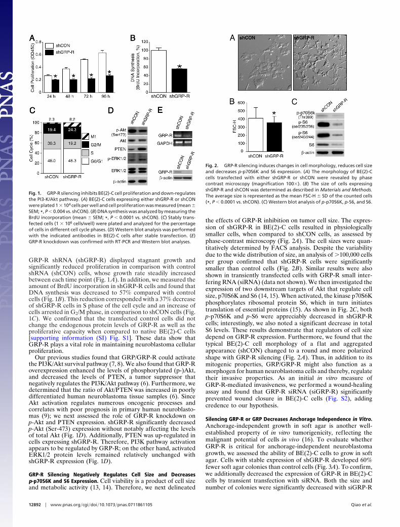
GRP-R Silencing Negatively Regulates Cell Size and Decreasesp-p70S6K and S6 Expression. Cell viability is a product of cell sizeand metabolic activity (13, 14). Therefore, we next delineated
the effects of GRP-R inhibition on tumor cell size. The expres-sion of shGRP-R in BE(2)-C cells resulted in physiologicallysmaller cells, when compared to shCON cells, as assessed byphase-contrast microscopy (Fig. 2A). The cell sizes were quan-titatively determined by FACS analysis. Despite the variabilitydue to the wide distribution of size, an analysis of �100,000 cellsper group confirmed that shGRP-R cells were significantlysmaller than control cells (Fig. 2B). Similar results were alsoshown in transiently transfected cells with GRP-R small inter-fering RNA (siRNA) (data not shown). We then investigated theexpression of two downstream targets of Akt that regulate cellsize, p70S6K and S6 (14, 15). When activated, the kinase p70S6Kphosphorylates ribosomal protein S6, which in turn initiatestranslation of essential proteins (15). As shown in Fig. 2C, bothp-p70S6K and p-S6 were appreciably decreased in shGRP-Rcells; interestingly, we also noted a significant decrease in totalS6 levels. These results demonstrate that regulators of cell sizedepend on GRP-R expression. Furthermore, we found that thetypical BE(2)-C cell morphology of a flat and aggregatedappearance (shCON) changed to a round and more polarizedshape with GRP-R silencing (Fig. 2 A). Thus, in addition to itsmitogenic properties, GRP/GRP-R might also function as amorphogen for human neuroblastoma cells and thereby, regulatetheir invasive properties. As an initial in vitro measure ofGRP-R-mediated invasiveness, we performed a wound-healingassay and found that GRP-R siRNA (siGRP-R) significantlyprevented wound closure in BE(2)-C cells (Fig. S2), addingcredence to our hypothesis.
Silencing GRP-R or GRP Decreases Anchorage Independence in Vitro.Anchorage-independent growth in soft agar is another well-established property of in vitro tumorigenicity, reflecting themalignant potential of cells in vivo (16). To evaluate whetherGRP-R is critical for anchorage-independent neuroblastomagrowth, we assessed the ability of BE(2)-C cells to grow in softagar. Cells with stable expression of shGRP-R developed 60%fewer soft agar colonies than control cells (Fig. 3A). To confirm,we additionally decreased the expression of GRP-R in BE(2)-Ccells by transient transfection with siRNA. Both the size andnumber of colonies were significantly decreased with siGRP-R
Fig. 1. GRP-R silencing inhibits BE(2)-C cell proliferation and down-regulatesthe PI3-K/Akt pathway. (A) BE(2)-C cells expressing either shGRP-R or shCONwere plated 1 � 104 cells per well and cell proliferation was measured (mean �SEM; *, P � 0.004 vs. shCON). (B) DNA synthesis was analyzed by measuring theBrdU incorporation (mean � SEM; *, P � 0.0001 vs. shCON). (C) Stably trans-fected cells (1 � 106 cells/well) were plated and analyzed for the percentageof cells in different cell cycle phases. (D) Western blot analysis was performedwith the indicated antibodies in BE(2)-C cells after stable transfection. (E)GRP-R knockdown was confirmed with RT-PCR and Western blot analyses.
Fig. 2. GRP-R silencing induces changes in cell morphology, reduces cell sizeand decreases p-p70S6K and S6 expression. (A) The morphology of BE(2)-Ccells transfected with either shGRP-R or shCON were revealed by phasecontrast microscopy (magnification 100�). (B) The size of cells expressingshGRP-R and shCON was determined as described in Materials and Methods.The average size is represented as the mean FSC-H � SD of the counted cells(*, P � 0.0001 vs. shCON). (C) Western blot analysis of p-p70S6K, p-S6, and S6.
12892 � www.pnas.org�cgi�doi�10.1073�pnas.0711861105 Qiao et al.

(47% of control; Fig. 3B Left and Center). In addition, coloniesalso formed more slowly than the control colonies transfectedwith a nontargeting control (siNTC). The efficiency of trans-fection was confirmed by Western blotting (Fig. 3B Right). Todetermine whether these effects were mediated by the binding ofGRP ligand to GRP-R, and not just a result of receptormanipulation, we transfected BE(2)-C cells with GRP siRNA(siGRP), before soft agar analysis. Transfection with siGRPblocked the colony growth of BE(2)-C cells by 54% whencompared to cells transfected with siNTC (Fig. 3C Left andCenter). Effective GRP knockdown was confirmed by RT-PCR(Fig. 3C Right). Therefore, GRP binding to its cell surfacereceptor is an important step in the process of GRP-R-inducedcolony formation. Of note, Akt silencing produced similarinhibitory effects on colony growth as GRP-R knockdown (Fig.S3).
GRP-R Expression Level Correlates with Malignant Potential in HumanNeuroblastoma Cells. To examine whether endogenous GRP-Rexpression correlates with anchorage independence in neuro-blastoma cells and to eliminate cell-line specific effects, wecompared the colony formation of BE(2)-C to another humanneuroblastoma cell line, SK-N-SH. We found that BE(2)-C cells,which behave aggressively in an in vivo xenograft model (data notshown), exhibited significantly increased malignant potential byformation of soft agar colonies when compared to SK-N-SH cells(Fig. 4A Left). Furthermore, BE(2)-C cells expressed higherlevels of GRP-R than SK-N-SH cells by Western blotting (Fig.4A Right). This finding is consistent with our previous studies, inwhich more abundant GRP-R expression was noted in poorlydifferentiated, more aggressive neuroblastomas (3).
To further examine the role of GRP-R on neuroblastomamalignant potential, we stably transfected SK-N-SH cells witheither pEGFP or pEGFP-GRP-R plasmid, and then incubatedthe cells in soft agar for 4 weeks. GRP-R overexpressingSK-N-SH neuroblastoma cells demonstrated a significantly in-creased number of colonies in soft agar, indicating stimulation ofanchorage-independent cell growth by increased GRP-R expres-sion (Fig. 4B Left). In contrast, cells transfected with controlvector, pEGFP, showed only a few smaller, isolated soft agarcolonies. The number of colonies induced by GRP-R overex-pression was markedly increased by nearly 8-fold (Fig. 4BCenter). The expression of GFP and GFP-tagged GRP-R wasconfirmed by fluorescent microscopy, where intense green flu-orescence was noted in GRP-R overexpressing SK-N-SH cells(Fig. 4B Right). Similar results were obtained by transienttransfection of SK-N-SH cells with a pEGFP-GRP-R plasmid
(data not shown). Our findings suggest that the cell surfacereceptor, GRP-R, is tumorigenic in human neuroblastoma SK-N-SH cells. To further confirm the role of the ligand GRP inneuroblastoma colony formation, we added a specific GRP-neutralizing antibody to the culture media. The presence of theGRP-specific antibody significantly inhibited colony formationin SK-N-SH cells overexpressing GRP-R (Fig. 4C Left). Thenumber of colonies was significantly decreased to 25% of control(Fig. 4C Right). These results further demonstrate that GRP,secreted by neuroblastoma cells, binds to GRP-R to act as a
Fig. 3. Knockdown of GRP-R or GRP inhibits soft agar colony formation. (A) BE(2)-C cells expressing either shGRP-R or shCON were plated in soft agar (2.5 �103 cells per well) for 3 weeks, and colony formation was quantitatively assessed (*, P � 0.0001 vs. shCON). (B) BE(2)-C cells were transfected with siGRP-R or siNTCfor 48 h and then plated in soft agar (2.5 � 103 cells/well) for 3 weeks (live cells, magnification 40�; Left). Bar graph represents the quantitative assessment(*, P � 0.05 vs. siNTC; Center). Western blotting confirmed inhibition of GRP-R protein levels by siRNA (Right). (C) BE(2)-C cells were transfected with siGRP orsiNTC for 48 h, incubated in soft agar at 2.5 � 103 cells/well for 3 weeks, and then photographed after staining (Left). Bar graph represents the quantitativeassessment of colony growth (*, P � 0.05 vs. siNTC; Center). The knockdown of GRP mRNA by siRNA was confirmed with RT-PCR (Right).
Fig. 4. Constitutive expressions of GRP-R correlate to anchorage-independent growth in human neuroblastoma cells. (A) BE(2)-C and SK-N-SHcells were incubated in soft agar at 2.5 � 103 cells/well in a six-well plate for3 weeks and 5 � 103 cells per well for 4 weeks, respectively, and thenphotographed after staining (Left). Western blot analysis of endogenouslevels of GRP-R in BE(2)-C and SK-N-SH cells (Right). (B) Soft agar analysis ofSK-N-SH cells stably transfected to overexpress GFP or GFP-tagged GRP-R;arrows indicate colonies (live cells, magnification 40�; Left). Quantitativeanalysis of soft agar assay (*, P � 0.05 vs. GFP control cells; Center). GFP andGFP-tagged GRP-R were expressed in SK-N-SH cells stably transfected withpEGFP and pEGFP-GRP-R plasmids (Right). (C) GRP-R stably transfected SK-N-SH cells (5 � 103 cells per well) were incubated in soft agar for 4 weeks.GRP-neutralizing antibody (1 ng/ml) was applied to the top of the soft agarand colonies were photographed after staining (Left). Quantitative analysis ofsoft agar assay (*, P � 0.05 vs. without antibody; Right).
Qiao et al. PNAS � September 2, 2008 � vol. 105 � no. 35 � 12893
CELL
BIO
LOG
Y

growth factor in soft agar assay, suggesting that the mitogenicactions of GRP may be an important mechanism in enhancinganchorage-independent growth.
Decreased Expression of GRP-R Inhibits Tumor Growth and Metastasisin Vivo. Based on the above in vitro findings, both GRP andGRP-R appear to be important in the anchorage-independentgrowth of neuroblastoma cells. To investigate whether theeffects of GRP-R knockdown on neuroblastoma growth inhibi-tion are sustained in vivo, we next established shGRP-R stablytransfected BE(2)-C xenografts in athymic nude mice. Strikingly,as depicted in Fig. 5A, mice injected with shGRP-R cells failedto establish any noticeable tumors, whereas the control micewere significantly affected by tumor burden by day 23. In fact, thenude mice xenografts with shGRP-R expression did not developtumors up to 43 days after subcutaneous (s.c.) injection, whereascontrol mice developed tumors within 10 days (Fig. 5B). Inanother experiment, for the purpose of same-day comparison,tumor volumes and weights were assessed 19 days after injection.The tumor volumes of mice bearing shCON xenografts weresignificant by day 9, in comparison to day 1 (Fig. 5C Left). Asexpected, because of the minute size of shGRP-R xenografts, itwas not possible to measure the tumor volumes; however, therewere constant GFP signals observed with the bioluminescentimaging system throughout the duration of the experiment.Correspondingly, the tumor weights (Fig. 5C Right) of miceinjected with shGRP-R cells were drastically lower than micebearing shCON xenografts (n � 5 per group).
Metastatic disease is common in neuroblastoma, and becausewe observed anchorage-independence in vitro, we next examinedthe effect of GRP-R silencing on tumor metastasis in vivo. Weused a spleen-liver metastasis model using BE(2)-C cells trans-fected with pEGFP/shGRP-R or pEGFP/shCON. Before death,strong green fluorescence signals were detected in the abdom-inal region of control mice, whereas fluorescence was barelydetectable in the shGRP-R group. However after death, weobserved that shGRP-R cells formed primary tumors withdemonstrable GFP signal in the spleen (Fig. 6A, arrowheads), incontrast to control tumors which formed not only at primarysites, but also aggressively metastasized to the livers (Fig. 6A,arrows). The livers of mice with shGRP-R tumors appeared
fairly normal, whereas the control mice livers had multiplemetastatic lesions (Fig. 6 A and B). In addition, the average liverweight (relative to mouse body weight) of the shGRP-R groupwas 34% of the control group (Fig. 6B). Furthermore, H&Estaining of the livers demonstrated numerous, densely packedneuroblastoma metastases in the shCON group, whereas arelatively low number of micrometastases were noted on theperiphery of the livers in the shGRP-R group (Fig. 6C). Takentogether, these results demonstrate that GRP-R knockdown isan advantageous strategy for in vivo neuroblastoma growthinhibition and metastasis suppression.
DiscussionWe previously demonstrated the growth-stimulatory function ofGRP in neuroblastoma cells and its correlation to PI3K/Aktpathway activation (3, 6, 7). In the present study, we show thatGRP-R overexpression led to anchorage-independent growth insoft agar, which is considered a criterion for cell transformationand malignant potential in vivo. Conversely, GRP-R knockdownwith siRNA and shRNA inhibited colony growth in soft agar.Furthermore, GRP-R silencing resulted in the inhibition oftumor growth and loss of cell metastatic potential in vivo.Previously uncharacterized, these results show that GRP/GRP-Rplays a role in the oncogenic process of anchorage-independenceand is critical for tumor growth and metastasis in humanneuroblastoma. Our findings further corroborate the multiplemitogenic functions of GRP, which are known to be associatedwith various malignant and aggressive human tumors (4, 5, 17,18).
GRP and its equivalent bombesin act as autocrine and/orparacrine growth factors to promote cell proliferation in cancercells (19, 20). In accordance with this, overexpression of thebombesin peptide receptor has been shown to promote cellproliferation in Rat-1 fibroblasts (21). Similarly, we have previ-ously shown that GRP-R overexpression in SK-N-SH neuroblas-toma cells increased the proliferative capacity of the cells (6). Inaddition, we have found that exogenous bombesin promotesneuroblastoma growth and that its antagonist suppresses tumorprogression in vivo (8). We now report that GRP-R overexpres-sion induces anchorage-independent growth that requires theGRP ligand, because a neutralizing antibody reversed the ef-
A
C
*** **
shCON shGRP-R
†
†
†
†
Tum
or V
olum
e (m
m3 )
Tum
or W
eigh
t (g)
Days1 5 9 12 15 19
0
100
200
300
400
500
600
700
0
0.05
0.10
0.15
0.20
0.25
0.30
B
Days
shCON
* **
shGRP-R
shC
ON
shG
RP
-R†
†
†
1 10 14 17 21 23 27 30 34 37 4143 47 50 54 61
Tum
or V
olum
e (m
m3 )
0
500
1000
1500
2000
2500
shCONshGRP-R
Fig. 5. GRP-R silencing blocks tumorigenesis of BE(2)-C cells in vivo. (A) BE(2)-C cells stably transduced with shGRP-R or shCON were grown in subscapular regionof athymic nude mice. Pictures were taken at 23 and 61 days postinjection for shCON group (Upper) and shGRP-R group (Lower), respectively. (B) Tumor volumesof transduced BE(2)-C xenografts of mice killed on day 23 (shCON) or 61 (shGRP-R) postinjection (n � 3 per group (*, P � 0.05 vs. shCON; †, P � 0.05 vs. shCON,day 1). (C) Tumor volumes (Left) of transduced xenografts of mice killed on day 19 (n � 5 per group; *, P � 0.05 vs. shCON; †, P � 0.05 vs. shCON, day 1). Tumorweights (Right) of mice killed on day 19 (n � 5 per group; *, P � 0.05 vs. shCON).
12894 � www.pnas.org�cgi�doi�10.1073�pnas.0711861105 Qiao et al.

fects. We also determined that cell proliferation, DNA synthesis,and cell cycle progression are intricately related to GRP-Rexpression, as silencing GRP-R inhibited each process. GRP hasalso been thought to be a morphogen, because it is capable ofaltering cell morphology in colon cancer cells (22). In this study,GRP-R silencing induced a round, polarized shape in BE(2)-Ccells, which normally exist in flatter, densely packed formations.Dynamic cytoskeletal modifications are a function of cell mo-tility and, thus, characteristic of invasive cells (23). This isconsistent with our results because GRP-R knockdown inhibitedmetastatic potential in vitro and in vivo, correlating to the loss ofthe anchorage-independent phenotype. Consequently, xenograftdevelopment was significantly delayed in mice injected withshGRP-R cells. Moreover, GRP-R-silenced neuroblastomaxenografts in the spleen lacked the metastatic aggressiveness ofcontrol tumors, suggesting that in addition to proliferation,GRP-R signaling is also an effective regulator of malignanttransformation in neuroblastoma cells.
PI3K/Akt pathway activation is frequently observed in humancancers; specifically in neuroblastoma, activation of Akt predictspoor outcome (9). Akt contributes to diverse cellular roles, whichinclude cell survival, growth, proliferation, angiogenesis, metab-olism, and migration (24). One of the best-conserved functionsof Akt is its role in promoting cell growth and increasing cellularmass by regulating nutrient uptake and metabolism (25). TheAkt-regulated mediators of this process include mTOR and itstarget S6K (14, 15, 26). In this study, we found that GRP-R
silencing down-regulated the phosphorylation of Akt and itsdownstream effectors p70S6K and S6, in addition to reducingcell size. Interestingly, we did not see significant changes inmTOR (data not shown), suggesting that Akt may additionallyregulate p70S6K and S6 independent of mTOR. This is consis-tent with many studies in which Akt functions in both mTOR-dependent and -independent pathways (24). Akt also plays a rolein the regulation of cell cycle G2/M phase transition (27) andcorrespondingly, our results demonstrated that GRP-R knock-down resulted in a decrease in activated Akt and further causeda decrease in DNA synthesis and cell cycle arrest at G2/M phase.GRP-R knockdown also appears to indirectly inhibit Akt acti-vation through PTEN, the endogenous inhibitor of PI3K/Akt,whose expression was increased with shGRP-R. Therefore,down-regulation of Akt activity is most likely an important factorcoordinating tumor growth inhibition subsequent to GRP-Rsilencing.
In conclusion, our study demonstrates that GRP/GRP-R is acrucial regulator of neuroblastoma cell growth, transformation,and metastasis and that Akt may be an important downstreameffector of GRP/GRP-R-mediated oncogenic properties. Thesein vitro and in vivo results are consistent with previous histolog-ical findings in which GRP and GRP-R expression were in-creased in poorly differentiated, aggressive human tumor sam-ples (3). In a recent review (10), the biological importance ofGRP and GRP-R in relation to various cancers, was discussed.However, it also emphasized the need for further investigationin regards to cancer-specific strategies. This study clearly dem-onstrates the roles of GRP-R in neuroblastoma tumorigenesisand secondary metastasis formation. These findings are clini-cally relevant because advanced-stage neuroblastomas are re-fractory to current treatment modalities; hence, understandingGRP/GRP-R regulation of tumor metastatic potential couldprovide a novel therapeutic adjunct for aggressive, undifferen-tiated neuroblastomas.
Materials and MethodsAntibodies and Reagents. Primary antibodies used include GRP-R (Abcam),p-ERK1/2 (Promega), and PTEN, ERK, p-Akt (Ser-473), Akt, p-p70S6K (Thr-389),p-S6 (Ser 235/236 and Ser-240/244), and S6 from Cell Signaling. Rabbit anti-GRP IgG (GRP-neutralizing antibody) was from Bachem. Agarose was fromCambrex Bio Science. �-actin antibody and all other reagents were fromSigma-Aldrich.
Cell Culture and Transfections. SK-N-SH and BE(2)-C cells were transfected withplasmids or siRNA pool (Dharmacon) as described (6, 7). All stably transfected cellswere selected with G418 (Cellgro) at 300 �g/ml and/or zeocin at 50 �g/ml for 2weeks. For GRP-R overexpression and silencing, pEGFP-GRP-R and pENTR/H1/TO(Invitrogen) were used, respectively. The sequence targeting GRP-R (NM�005314)is underlined in the following shRNA sequence: 5�-CACCGTAACGTGTGCTCCAGT-GGACGAATCCACTGGAGCACACGTTA-3�, the nonspecific control shCON was:5�-CACCGGGCGCGCTTTGTAGGATTCGCCGAAGCGAATCCTACAAAGCGCGCC-3�.
RT-PCR and Western Blot Analyses. Total RNA was isolated using RNAqueouskit (Ambion) according to the manufacturer’s instructions. Isolated RNA wasused to synthesize cDNA using the High-Capacity cDNA Reverse TranscriptionKit (Applied Biosystems). Primers designed to amplify a 151-bp GRP fragment(NM�002091): forward primer 5�-GCTGGGTCTCATAGAAGCAAAG-3�; reverseprimer 5�-TGGAGCAGAGAGTCTACCAAC-3�. GRP-R and GAPDH-specific oligo-nucleotide primers were the same as described (28). Amplification was per-formed for 40 cycles (30 cycles for GAPDH) of 2 min at 94°C, 30 sec at 55°C, and40 sec at 72°C. GAPDH was used as a control. Western blot analysis wasperformed using �-actin as a loading control as described (6).
Cell Proliferation and DNA Synthesis Analyses. Cells were seeded onto 96-wellplates at a density of 1 � 104 cells per well in RPMI culture medium with 10%FBS and cell number was assessed using Cell Counting Kit-8 (Dojindo Molec-ular Technologies) for cell proliferation. For DNA synthesis, BrdU was added tothe cell culture for 4 h before detection by Cell Proliferation ELISA (Roche)according to the manufacturer’s instructions.
Fig. 6. Knockdown of GRP-R inhibits tumor cell metastasis in vivo. (A)Representative GFP and gross images of primary tumor (arrowheads) in spleenand secondary liver metastases (arrows) from mice injected with BE(2)-C/GFP/shCON (Upper) and BE(2)-C/GFP/shGRP-R (Lower) cells. (B) Representativeimages of livers from mice injected with BE(2)-C/GFP/shGRP-R and BE(2)-C/GFP/shCON cells (Upper). Quantitative analysis of liver weight relative to bodyweight (*, P � 0.05 vs. shCON; Lower). (C) Representative H&E-stained sectionsof livers from mice injected with BE(2)-C/GFP/shCON (Upper) and BE(2)-C/GFP/shGRP-R (Lower) cells.
Qiao et al. PNAS � September 2, 2008 � vol. 105 � no. 35 � 12895
CELL
BIO
LOG
Y

Flow Cytometry Analyses. Cell cycle analyses were assessed as described (7). Forcell size determination, the average cell size in 100,000-cell samples wasassessed using CellQuest v3.3 software (BD), according to a previous report(29). The resulting parameter, mean forward scatter height (FSC-H), is ameasure of relative cell size.
Soft Agar Colony Formation Assay. Cells were trypsinized and resuspended inRPMI medium 1640 containing 0.4% agarose and 7.5% FBS. SK-N-SH andBE(2)-C cells were overlaid onto a bottom layer of solidified 0.8% agarosein RPMI medium 1640 containing 5% FBS, at cell concentrations of 5 � 103
cells per well and 2.5 � 103 cells per well of a six-well plate, and incubatedfor 4 and 3 weeks, respectively. Colonies were stained with 0.05% crystalviolet, photographed, and quantified.
Xenograft Tumor Growth. Male athymic nude mice (4–6 weeks old) weremaintained as described (8). All studies were approved by the InstitutionalAnimal Care and Use Committee at the University of Texas Medical Branch andwere conducted in accordance with guidelines issued by the National Insti-tutes of Health. BE(2)-C cells transfected with shGRP-R or shCON alone orcotransfected with pEGFP and xenografts were established as previouslydescribed (8). Briefly, 1 � 106 cells/100 �l of HBSS were injected s.c. into thebilateral flanks using a 26-gauge needle (n � 3–5 per group). Tumor growthwas assessed biweekly by measuring the two greatest perpendicular tumordimensions with vernier calipers (Mitutoyo), and body weights were recorded
weekly. At 23 days postinjection, tumors were excised, weighed, and fixed informalin.
Liver Metastasis Model. BE(2)-C cells were stably transfected with pEGFP(N3)vector (Clontech) and shGRP-R or shCON. Mice were anesthetized with isoflu-orane, and a small left flank incision was made to isolate and exteriorize thespleen; viable pEGFP/shCON or pEGFP/shGRP-R cells (4 � 106 cells/100 �l ofHBSS) were injected into the splenic capsule using a 27-gauge needle. Thespleen was returned, and the abdominal wall was closed with metal woundclips. Tumor growth was observed biweekly with Illumatool TLS (LightoolsResearch). At death, livers were excised, weighed and fixed in formalin forfurther assessment.
Statistical Analysis. For in vitro experiments, conditions were compared byusing Student’s paired t test. One-way ANOVA on the ranks for repeatedmeasures was performed for multiple comparisons. In vivo experiments wereanalyzed as described previously (8). Body weight was analyzed using ANOVAfor a two-factor experiment with repeated measures on time. The two factorswere treatment group (control and target knockdown) and day. For allexperiments, P � 0.05 was considered significant.
ACKNOWLEDGMENTS. We thank Karen Martin for manuscript preparationand Tatsuo Uchida for statistical analysis. This work was supported by GrantsRO1 DK61470, RO1 DK48498, RO1 CA104748, PO1 DK35608, and F31DK079422 from the National Institutes of Health.
1. Kushner BH, et al. (2004) Reduction from seven to five cycles of intensive inductionchemotherapy in children with high-risk neuroblastoma. J Clin Oncol 22:4888–4892.
2. Gustafson WC, De Berry BB, Evers BM, Chung DH (2005) Role of gastrointestinalhormones in neuroblastoma. World J Surg 29:281–286.
3. Kim S, et al. (2002) Gastrin-releasing peptide is a growth factor for human neuroblas-tomas. Ann Surg 235:621–629; discussion 629–630.
4. Moody TW, Carney DN, Cuttitta F, Quattrocchi K, Minna JD (1985) High affinityreceptors for bombesin/GRP-like peptides on human small cell lung cancer. Life Sci37:105–113.
5. Markwalder R, Reubi JC (1999) Gastrin-releasing peptide receptors in the humanprostate: relation to neoplastic transformation. Cancer Res 59:1152–1159.
6. Qiao J, Kang J, Cree J, Evers BM, Chung DH (2005) Gastrin-releasing peptide-induceddown-regulation of tumor suppressor protein PTEN (phosphatase and tensin homologdeleted on chromosome ten) in neuroblastomas. Ann Surg 241:684–691; discussion691–692.
7. Ishola TA, Kang J, Qiao J, Evers BM, Chung DH (2007) Phosphatidylinositol 3-kinaseregulation of gastrin-releasing peptide-induced cell cycle progression in neuroblas-toma cells. Biochim Biophys Acta 1770:927–932.
8. Kang J, et al. (2007) Bombesin induces angiogenesis and neuroblastoma growth.Cancer Lett 253:273–281.
9. Opel D, Poremba C, Simon T, Debatin KM, Fulda S (2007) Activation of Akt predicts pooroutcome in neuroblastoma. Cancer Res 67:735–745.
10. Patel O, Shulkes A, Baldwin GS (2006) Gastrin-releasing peptide and cancer. BiochimBiophys Acta 1766:23–41.
11. Paszek MJ, Weaver VM (2004) The tension mounts: Mechanics meets morphogenesisand malignancy. J Mamm Gland Biol Neoplasia 9:325–342.
12. van den Brink GR, Offerhaus GJ (2007) The morphogenetic code and colon cancerdevelopment. Cancer Cell 11:109–117.
13. Jorgensen P, Tyers M (2004) How cells coordinate growth and division. Curr Biol14:R1014–R1027.
14. Fingar DC, Blenis J (2004) Target of rapamycin (TOR): An integrator of nutrient andgrowth factor signals and coordinator of cell growth and cell cycle progression.Oncogene 23:3151–3171.
15. Fingar DC, Salama S, Tsou C, Harlow E, Blenis J (2002) Mammalian cell size is controlledby mTOR and its downstream targets S6K1 and 4EBP1/eIF4E. Genes Dev 16:1472–1487.
16. Ponten J (1971) Spontaneous and virus induced transformation in cell culture. VirologyMonographs, eds Herausgegeben V, Gard S, Hallauer C, Meyer KF (Springer, NewYork), vol 8, pp 4–19.
17. Ferris HA, Carroll RE, Rasenick MM, Benya RV (1997) Constitutive activation of thegastrin-releasing peptide receptor expressed by the nonmalignant human colon epi-thelial cell line NCM460. J Clin Invest 100:2530–2537.
18. Lui VW, et al. (2003) Mitogenic effects of gastrin-releasing peptide in head and necksquamous cancer cells are mediated by activation of the epidermal growth factorreceptor. Oncogene 22:6183–6193.
19. Alexander RW, et al. (1988) Effects of bombesin on growth of human small cell lungcarcinoma in vivo. Cancer Res 48:1439–1441.
20. Rozengurt E, Sinnett-Smith J (1983) Bombesin stimulation of DNA synthesis and celldivision in cultures of Swiss 3T3 cells. Proc Natl Acad Sci USA 80:2936–2940.
21. Charlesworth A, Broad S, Rozengurt E (1996) The bombesin/GRP receptor transfectedinto Rat-1 fibroblasts couples to phospholipase C activation, tyrosine phosphorylationof p125FAK and paxillin and cell proliferation. Oncogene 12:1337–1345.
22. Carroll RE, Matkowskyj KA, Tretiakova MS, Battey JF, Benya RV (2000) Gastrin-releasingpeptide is a mitogen and a morphogen in murine colon cancer. Cell Growth Differ11:385–393.
23. Yamaguchi H, Wyckoff J, Condeelis J (2005) Cell migration in tumors. Curr Opin CellBiol 17:559–564.
24. Manning BD, Cantley LC (2007) AKT/PKB signaling: navigating downstream. Cell129:1261–1274.
25. Gottlob K, et al. (2001) Inhibition of early apoptotic events by Akt/PKB is dependent onthe first committed step of glycolysis and mitochondrial hexokinase. Genes Dev15:1406–1418.
26. Ohanna M, et al. (2005) Atrophy of S6K1(-/-) skeletal muscle cells reveals distinct mTOReffectors for cell cycle and size control. Nat Cell Biol 7:286–294.
27. Kandel ES, et al. (2002) Activation of Akt/protein kinase B overcomes a G(2)/m cell cyclecheckpoint induced by DNA damage. Mol Cell Biol 22:7831–7841.
28. Chatzistamou I, Schally AV, Sun B, Armatis P, Szepeshazi K (2000) Inhibition of growthof OV-1063 human epithelial ovarian cancers and c-jun and c-fos oncogene expressionby bombesin antagonists. Br J Cancer 83:906–913.
29. Ruvinsky I, et al. (2005) Ribosomal protein S6 phosphorylation is a determinant of cellsize and glucose homeostasis. Genes Dev 19:2199–2211.
12896 � www.pnas.org�cgi�doi�10.1073�pnas.0711861105 Qiao et al.