gastrin release - jci
TRANSCRIPT

Disturbed Islet-Cell Function Related to Endogenous
Gastrin Release
STUDIES ONINSULIN SECRETIONANDGLUCOSETOLERANCE
IN PERNICIOUS ANEMIA
JENS F. RiEHuDFrom the Department of Clinical Chemistry, Bispebjerg Hospital,DK-2400 Copenhagen NV, Denmark
A B S T R A C T The insulin and gastrin response to oralglucose, intravenous glucose, or a protein-rich meal weremeasured in 44 nondiabetic patients with perniciousanemia (PA) and in 44 control subjects. 36 of the PA-patients had hypergastrinemia, while serum gastrin con-centrations in the remaining eight patients were belownormal. Three hypergastrinemic PA-patients were inaddition studied during an oral glucose loading withsynchronous intravenous infusion of gastrin-17.
During both oral and intravenous glucose tests bloodglucose concentrations were similar in patients and incontrols. After ingestion of protein blood glucose con-centrations in PA-patients with hypergastrinemia wereabove those of the controls (P <0.05). Parenteral in-fusion of gastrin-17 during oral glucose loading alsoincreased blood glucose concentrations above the levelsobserved after glucose alone. In PA-patients with hyper-gastrinemia the insulin response was augmented in alltests. In patients with hypogastrinemia serum insulinconcentrations were lower than normal in the fastingstate and during stimulation with glucose intravenously(P < 0.01). In hypergastrinemic patients serum gastrinconcentrations decreased after oral as well as intravenousglucose administration. The decrease was larger duringthe oral test. In hypogastrinemia oral glucose induced,as in controls, a small initial rise followed by a slow fallin serum gastrin concentrations. No variations wereseen in these patients during the intravenous glucoseinfusion. Gel filtration of serum from hypergastrinemicpatients disclosed a decrease in the concentrations of all
Dr. Rehfeld's present address is Institute of Medical Bio-chemistry, University of Aarhus, DK-8000 Aarhus C, Den-mark.
Received for publication 1 December 1975 and in rezHsedform 16 February 1976.
four main components of gastrin during the glucoseloadings.
Taken together with earlier studies on the effect ofexogenous gastrin the results suggest that endogenoushypergastrinemia induces hyperglycemia and potentiatesinsulin secretion. In contrast hypogastrinemia is associ-ated with hypoinsulinism.
INTRODUCTIONExogenous gastrin in form of the heptadecapeptideamide (gastrin-17)1 and its C-terminal tetrapeptideamide can stimulate insulin release in man (1-3). Butdose-response studies with exogenous gastrin-17 cor-related to endogenous gastrin concentrations in serumduring meals point to a limited role of gastrin in theregulation of insulin secretion in normal human sub-jects (4). However, since gastrin-17 in high doses re-leases large amounts of insulin and moreover increasesblood glucose concentrations (4), endogenous hyper-gastrinemia might also influence insulin secretion andglucose metabolism.
Endogenous hypergastrinemia is most often found inpatients with achlorhydria, among which those withpernicious anemia (PA) constitute a well-defined group.Approximately 75% of PA-patients have increased con-centrations of gastrin in serum (5). A relationship be-tween PA and impaired glucose tolerance and insulinsecretion was suggested already in 1910 (6), and it isnow well-known that the frequency of diabetes mellitusin PA is increased (7-9). The pathogenetic link betweenPA and diabetes of juvenile-onset type is possibly of
'Abbreviations used in this paper: gastrin-17, heptadeca-peptide; PA, pernicious anemia.
The Journal of Clinical Investigation Volume 58 July 1976* 41-49 41

autoimmune nature (10-14). But diabetes mellitus ofmaturity-onset type in PA might be due, at least in partto hypergastrinemia.
The present study is an attempt to evaluate whetherthe disturbed gastrin release in PA influences insulinsecretion and glucose homeostasis.
METHODS
Patients and control subjects44 patients with treated PA and 44 healthy control sub-
jects participated in the study. All patients and controlswere within 10% of their desirable weight (15). The PA-diagnosis was established on the basis of analysis of vitaminB,2 concentration in serum, bone marrow examination,Schilling test without and with oral hog intrinsic factor,and an augmented histamine test. None of the patients haddiseases other than PA, except five with vitiligo and twohaving rheumatoid arthritis. Neither diabetes mellitus northyroid diseases were known in the patients, nor had anybeen submitted to gastrointestinal surgery before the in-vestigation.
The PA-patients were divided into three groups: Group Icomprised 27 patients with gastrin concentrations in serumin the fasting state ranging from 120 to 1,614 pmol -1'. Theirage ranged from 30 to 83 yr, with a mean of 64 yr. 21 ofthese patients were females. Group II comprised eightpatients with gastrin concentrations in serum below theupper limit of the normal reference group in the fastingstate, which in this department is 50 pmol 1-1 (mean+2SD). Their age ranged from 55 to 80 yr, with the meanbeing 68 yr. Five were females. Group III comprised ninepatients with enhanced gastrin concentration in serum inthe fasting state, range: 167-2,000 pmol 1-1. Their ageranged from 32 to 79 yr and the mean was 60 yr. Eightwere females.
Age (+2 years) and sex matched subjects without PA,known gastrointestinal disorders or family history of dia-betes mellitus, and with gastrin concentrations in serumbelow 50 pmol ' in the fasting state served as controls.Informed consent was obtained from all patients andcontrols.
Both patients and controls were on a diet containing atleast 250 g carbohydrates per day 3 days before each in-vestigation. After an overnight fast the examination beganbetween 8:00 and 9:00 a.m. Blood samples were drawnfrom an antecubital vein. Serum was stored at - 200C untilassay.
Experimental proceduresOral glucose loading. Groups I and II and corresponding
controls were given 50 g glucose as a 25% solution flavouredwith lemon. Blood samples were drawn 10 and 5 minbefore glucose loading and 5, 10, 15, 20, 30, 40, 50, 60, 90,120, 150, and 180 min after.
Intravenous glucose infusion. 1 wk after the oral testeach patient in groups I and II and the correspondingcontrol subjects was submitted to an intravenous glucoseinfusion test designed to imitate the changes in blood glu-cose concentrations measured during the oral glucose test:16.7 g glucose in concentrations from 33 to 50%o was givenintravenously at a constant infusion rate. Termination of theinfusion was aimed to coincide with the peak blood glucoseconcentration reached during the oral test in the same in-
dividual. Blood samples were drawn from the contralateralarm at the same intervals as those in the oral glucose test.The simple infusion test described here has been evaluatedin detail elsewhere (16). It is based on the observationthat approximately one third of the glucose given orallyescapes hepatic extraction in subjects with a normal glucosetolerance (17).
Protein-rich meal. Group III of the PA-patients andthe respective controls were given an appetizing meal com-posed of beefsteak, sauce, vegetables, and a glass of water.The meal was finished within 20 min. Blood samples weredrawn 15, 10, and 5 min before, and 5, 10, 20, 30, 45, 60,and 90 min after the onset of the meal.
Intravenous gastrin infusion. In three PA-patients fromgroup III, who previously were submitted to a 50-g oralglucose load, pure human nonsulfated gastrin-17, 2 pug/kg/h(a generous gift from Professor R. A. Gregory and Dr.H. J. Tracy, Liverpool, England) was infused intravenously30 min before and 90 min after administration of 50 gglucose per os. Blood samples were drawn from the contra-lateral arm 30, 20, and 10 min before, and 5, 10, 20, 30,40, 50, 60, 90, 105, 120, 150, and 180 min after the onsetof the glucose load.
Laboratory methodsBlood glucose. Concentrations were measured with a
glucose oxidase method on Auto Analyzer (Technicon In-struments Corp., Tarrytown, N. Y.).
Serum insulin. Concentrations were measured radioim-munochemically. Reliability criteria of the actual assay havebeen given in detail previously (4). The antiserum employedbinds monocomponent human insulin and human proinsulinwith equimolar potency.
Serum gastrin. Concentrations were measured radioim-munochemically. Reagents, procedure, and reliability param-eters for the assay have been described in detail previously(5, 18, 19). In the present study antiserum no. 2604-g wasemployed (18). Gastrin components ' in PA-serum werestudied on Sephadex G-50 superfine columns (25 X 2,000mm) eluted with 0.25 moll1' ammonium bicarbonate, pH8.2 at 4°C. The columns were calibrated with pure humangastrin-34, -17, and -14 (generous gifts from Professor R. A.Gregory and Dr. H. J. Tracy [Liverpool, England]) andwith 'SI-human albumin and 'NaCl (Amersham, England)for indication of void volume and total volume. The relativeaffinity of the large gastrins in PA-serum, components I andII, to antiserum 2604-8 was measured as shown in Fig. 1.It appears that the affinity of components I, II (gastrin-34-like), and III (gastrin-17-like) to antiserum 2604-8 are
'There are four main components of gastrin in humanserum (20, 21). They have been named I-IV according totheir molecular size (22). From tumour and antral tissueGregory and Tracy have purified three pairs of gastrinwith a known sequence of 34, 17, and 14 amino acids, re-spectively (23-25). Walsh et al. (26) have proposed thatthey should be named gastrin-34, gastrin-17, and gastrin-14.The tissue gastrins have roughly the same molecular sizeand charge as Components II, III, and IV, respectively, butthere are also indications that serum components and tissuegastrins are not quite identical (27). Until the serum com-ponents are characterized better and possible identity withtissue gastrin is proven the flexible component nomenclatureshould be used for circulating gastrins, whereas the rigidamino acid-number nomenclature can be maintained forthe well-characterized tissue gastrins.
42 1. F. Rehfeld

Before tryptie cleivage
100 ~
50
L
20 40
11 0
II50 100
pmot eq human gastrin-17/1After tryptic cleavage
pmol human gastrin-13/1
III
100
IV -100 F
50 50
I I I
0 50 100 0 50 100
IMMUNOREACTIVEGASTRINpmol human gastrin-17/1
FIGURE 1 Comparison of immunoreactivity of the maincomponents of gastrins, I-IV, in human serum against thestandard gastrin of the assay (synthetic human gastrin-17[nonsulphated]), employing antiserum 2604-8 and mono-iodinated gastrin-17. Components I, II, and III from seraof hypergastrinemic patients with PA were separated by gelfiltration, pooled, and concentrated by lyophilization. Com-ponents I and II (diagrams I and II, respectively) wereeach divided in two equal volumes, of which one was incu-bated with trypsin. This converts these large components tothe smaller component III. After termination of trypticcleavage by boiling the immunoreactivity of each volumewas quantitated in six different dilutions. Component III(diagram III) was compared with the standard gastrin-17in five different dilutions. Component IV was only presentin PA-sera in very low amounts. Instead pure, naturalgastrin-13 (nonsulphated) (diagram IV) in known con-
centrations was compared with standard gastrin-17.
similar on a molar basis. Separate experiments with gas-trin-14 disclosed a binding of 60% to antiserum 2604-8 com-
pared with gastrin-17 (Fig. 1). However, since the gastrin-14-like component IV constitutes only a few per cent ofthe total concentration of gastrin in serum, the concentrationmeasured with antiserum 2604-8 in molar terms closely re-
flects the total amount of gastrin components present.
CalculationsThe integrated glucose stimulation and insulin response
for each experiment were computed by planimetry of thearea under the blood glucose and serum insulin curves inthe time intervals indicated using the lowest levels observedas base line. The significance of differences between means
was tested by Student's t test. The concentrations are givenas mean+SEM.
RESULTS
Response to oral glucose loadingGroup I (Fig. 2 and Table I). The blood glucose
curves and the total glucose stimulation in patients weresimilar to that of controls. Serum insulin concentrationsin the patients rose from 62±10 to 560+69 pmol-after 60 min. Controls showed an increase from 60±+10to 371+45 pmol- l' in the same period. The mean peakconcentration in PA was above normal (P < 0.01). Soalso were the insulin responses in the 0.5-2 h intervaland the total insulin response (P < 0.05). Mean serumgastrin concentration in PA fell rapidly (P < 0.01).The controls showed a small rise initially but otherwisedisplayed no variations in serum gastrin levels.
Group II (Fig. 3 and Table I). Blood glucose con-centrations rose faster than normal to a peak after 30
LU
Uf)0
0IO) 0D-J _
D-5, Eo E0
co
8
6
4.50
z
v) _ 300_
=
E
Xn 150
0
500
z
en 104 =
0 - 2500
X E
(n
00 2 3
150
3to
100:loo 3
60
40 =3
20
1000
.0
500 3
0
HOURS
FIGURE 2 Blood glucose, serum insulin, and serum gastrinconcentrations in 27 hypergastrinemic PA-patients (groupI: * *) and in 27 matched controls (A A) duringa 50 g oral glucose tolerance test. Mean±+SEM of concen-trations.
Insulin and Gastrin Secretion in Pernicious Anemia 43
Befeore tryptic cleavage
20 I
ze<lx L
I
I

150 tions rose in 30 min in PA's from 65±11 to 228±248
pmol. 1-'. This peak concentration is above the 30-min3 concentration in normals of 142±16 pmol P-1 (P <
100 0.01). The insulin response was increased in the pa-3 tients, most pronounced in the first 30 min after onset of
the glucose infusion (P <0.05). The intravenous glu-cose produced a slight decrease in serum gastrin concen-trations in PA-patients, whereas serum gastrin concen-trations in controls remained constant.
60 Group II (Fig. 5 and Table I). Blood glucose curvesand concentrations in patients and controls were simi-lar. Serum insulin concentrations in the patients were
L0 -l decreased both in the fasting state (P <0.01), and in3 response to intravenous glucose being 38% of the normal
response (P < 0.01). Serum gastrin concentrations in20 patients were lower than normal throughout the test
(P < 0.01) and showed no significant variations.0 Response to a protein-rich meal
0 2 3
HOURS
100 Group III (Fig. 6). Blood glucose concentrations inin PA-patients rose from 4.85±0.20 to 6.49±0.36 mmol-
50Ie3
wUn0
,0 _
oE8E0
FIGuRE 3 Blood glucose, serum insulin, and serum gastrinconcentrations in eight hypogastrinemic PA-patients (groupII: * *) and in eight matched controls (A A)during a 50 g oral glucose tolerance test. Mean+SEM ofconcentrations.
min. The controls reached peak concentration after 50min from the same basal level. Serum insulin concentra-tion in the patients in the fasting state was 32±14 pmol-1P1, which is below that of controls, 77+7 pmol 11 (P <0.02). The rise of insulin concentration in patients afteroral glucose was higher than normal (P < 0.05). After1 h the insulin concentrations fell below those of thecontrols (P < 0.05), so that the entire insulin responseto oral glucose in patients was similar to that of normalsubjects (Table I). The gastrin concentrations in thefasting PA-patients were lower than normal (P < 0.05).The gastrin concentrations varied as in controls duringglucose ingestion, but at a lower level.
Response to intravenous glucose infusionGroup I (Fig. 4 and Table I). Blood glucose curves
were almost similar in patients and in controls. Thetotal glucose stimulation in PA was normal althoughthe patients had insignificantly higher glucose concen-trations throughout the test. Serum insulin concentra-
8
6
z 300
Z *_- _z2 ° 150
an
0
500
z
E250Lnw.
L
w
0
HOURS
FIGURE 4 Blood glucose, serum insulin, and serum gastrinconcentrations in 27 hypergastrinemic PA-patients (groupI: 0-O) and in 27 matched controls (A A) duringan intravenous infusion of 16.7 g glucose. Mean±+SEM ofconcentrations.
44 J. F. Rehfeld
w#A0 uU.
0-a E0 E0
6 -
i450
300 Fz
U'%
wnU'=
150 1
0
F0[
20 I
z
,0'.9X Ewla"
0
150
3i-100 o0
3
50
20 3
0
ID
o
2 3I
I I

TABLE I
Integrated Glucose Stimulation (G, in mmol/liter X min) and Insulin Response (I, in nmol/liter X min) during Oral and IntravenousGlucose Administration in Two Groups of Patients with Pernicious Anemia and in Control Subjects (mean ±SEM)
Oral glucose tolerance test, 50 g Intravenous glucose infusion test, 16.7 g
Interval, minutes ......... 0-30 30-60 60-120 120-180 0-180 0-30 30-60 60-120 120-180 0-180
PA-group I* (G) 88±5 139410 217426 115415 559439 97±6 126±6 167416 85±8 475428(I) 0.72±0.08 1.5440.17? 2.66±33$ 1.23±0.16? 6.12±0.581 0.51±0.07? 0.6540.07 0.9340.15 0.40±0.04 2.50±0.30
Control group I (G) 93±6 145±7 211±29 116+22 565±46 9748 131±6 166±16 103±16 497465(I) 0.71±0.10 1.10+0.12 1.82±0.23 0.90±0.15 4.53±0.35 0.38±0.05 0.510.09 0.72±0.09 0.46±0.13 2.07±0.26
PA-group IIi (G) 114±44 13149 177423 105±18 527±39 102±9 129413 165±17 108±14 504±40(I) 0.7440.08 1.44±0.20$ 1.5940.30 0.3640.08? 4.13±0.55 0.18±0.02? 0.2640.04: 0.2840.05t 0.1440.03? 0.86±0.12t
Control group II (G) 9049 136412 195±25 100±17 521±49 101±10 144412 167±27 109±27 520±65(I) 0.63 40.05 0.98±-0.07 1.73 ±0.2 1 0.87 ±0.20 4.21±0.45 0.40±-0.04 0.58±-0.22 0.76±40.10 0.44 ±0.08 2.19±-0.23
* PA patients with hypergastrinemia.? Significant difference between PA patients.§ PA patients without hypergastrinemia.
1'. The peak concentrations were reached after 45 min.In control subj ects blood glucose concentration de-creased initially from 4.90±0.15 to 4.72±0.18 mmol* 1',after which a rise to 5.85±0.39 mmol' 1' was observedafter 45 min. Blood glucose concentrations in PA-pa-
tients were above normal throughout the 90-min periodafter onset of the meal (P < 0.05). Serum insulin con-centrations rose in PA-patients from 77±9 to 468±137pmol' P-1 within 45 min, after which the concentrationswere reduced to half the peak value. In control subjects
2 3
CA 70150 n -
J ._
5
3 m
100 g
3
LS0
50 z
Un *
oz =
a:zc
-
3
_ L0
- 20
J 0
100
z
m -,,.
In,50
0 ~ x
HOURS
FIGURE 5 Blood glucose, serum insulin, and serum gastrinconcentrations in eight hypogastrinemic PA-patients (groupIII: 0 O) and in eight matched controls (A A)during an intravenous infusion of 16.7 g glucose. Mean±SEMof concentrations.
300
150
0
1000
750
100
0
1303
100 3
60
40-rc
20
0
2000
1500 3
200
00 0.5 1.0 1.5
HOURS
FIGURE 6 Blood glucose, serum insulin, and serum gastrinconcentrations in nine hypergastrinemic PA-patients (0--*) and in nine matched control subjects (A A)after a protein-rich meal. Mean±+SEM of concentrations.
Insulin and Gastrin Secretion in Pernicious Anemia
8
6
An0,U0V-
a0Co EJ
300
z
J
z =
- :z
:X OE tS0 _
w
tn
O _
L0z
wO..n0V _
= E20 j
00
x
i
45

the increase was from 82±6 to 234±40 pmol 11, whichis below the peak concentration (P < 0.02). Serum gas-trin concentrations in the patients rose within 10 min,and remained above basal concentrations throughout thetest period. Control subjects responded rapidly to thefood.
Response to oral glucose loading combined withintravenous gastrin infusions (Fig. 7)
Blood glucose concentrations were slightly enhancedafter gastrin infusion, both in the basal state and duringglucose ingestion in comparison to the levels found dur-ing the usual oral glucose tolerance test. Basal serum in-sulin concentrations rose immediately after onset of thegastrin infusion, and remained above control levelsthroughout the infusion period. Serum gastrin concen-trations were doubled during the infusion period.
un0
.
n cc'ao E
coc5
IJ 70
450.r
300 1
150 F
GASTRIN-17 I.V.0
1000
r
in 0*t -
m Ew C
500
0
HOURS
FIGURE 7 Blood glucose, serum insulin, and serum gastrinconcentrations in three hypergastrinemic PA-patients duringa 50-g oral glucose tolerance test (A*A) and in thesame three PA-patients during a 50-g oral glucose toler-ance test combined with intravenous infusion of 2 ,g purenatural gastrin-17 (nonsulfated) per kg per hour (--0). The infusion of gastrin began 0.5 h before glucoseingestion and continued 1.5 h after. Mean±SEM of con-centrations.
Effect of oral and intravenous glucose adminis-tration on gastrin component pattern in serum(Table II)Comparison of components in the fasting state and at
nadir showed unchanged patterns after intravenous glu-cose infusion. After oral glucose the concentration of allcomponents was reduced.
DISCUSSION
The present study shows that both hyper- and hypogas-trinemia are associated with abnormalities in insulin se-cretion. Moreover it shows that protein-rich food, whichmaintains enhanced gastrin levels in hypergastrinemiapatients, increases blood glucose concentrations abovenormal in spite of hyperinsulinemia. Combination ofthese results with the dose-response study in normalsubjects (4), and the results of parenteral gastrin-infu-sion in PA suggest that gastrin may be important inregulation of islet-cell secretion in certain diseases.
Selection of patients with hypergastrinemia. In con-trast to other diseases with increased serum gastrin lev-els achlorhydria is, in the form found in PA, useful forstudies on the effect of endogenous hypergastrinemia onislet-cell function because: 1) gastrin in PA, as innormal subjects, is produced mainly by the antrum (28).2) All evidence available so far indicates that circulatinggastrins in normal subjects and PA-patients are quali-tatively similar (20, 21, 27). 3) The increased gastrinrelease in PA is probably caused only by lack of inhibi-tion from gastric acid, and the gastrins in serum aremetabolized at a normal rate (29). 4) Zollinger-Ellisonpatients are unsuited for studies like the present inthat gastrinomas often are associated with subclinicalneoplasias of alpha- and beta cells (30, 31). Insulin andglucagon secretion in the Zollinger-Ellison syndromemay consequently deviate a priori, and thereby invali-date studies on the insulinogenic effect of gastrin basedon gastrinoma patients (32, 33). Anyway, Zollinger-Ellison patients display increased insulin response toglucose and diabetic glucose tolerance (34). 5) Gastrinsecretion in duodenal ulcer patients is also abnormallyincreased, at least during meals (35, 36). Serum levelsin these patients and those of food-stimulated normalsubjects (37) are, however, placed so much to the lefton the dose-response curve (4) that it is difficult todemarcate insulinogenic effects of endogenous gastrinsin these conditions. 6) Patients with severe kidney dam-age are hypergastrinemic, but their general metabolicderangement, including abnormal levels of betacyto-trophic hormones other than gastrin, makes them unfitfor studies like the present.
Little is known about secretion of hormones withbetacytotrophic actions in PA other than gastrin. Se-cretin levels are apparently normal in PA (29), but the
46 1. F. Rehfeld
Z
D '.4Aad-=
i-2 0:3ECK CLw(A
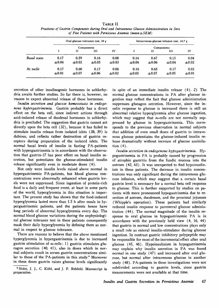
TABLE IIFractions of Gastrin Components during Oral and Intravenous Glucose Administration in Sera
of Five Patients with Pernicious Anemia (mean±SEM)
Oral glucose tolerance test, 50 g Intravenous glucose infusion test, 16.7 g
Components ComponentsI II III IV I II III IV
Basal state 0.17 0.59 0.16 0.08 0.14 0.67 0.15 0.0440.04 410.02 :1:0.05 4-0.03 :1:0.04 :1:0.06 :1:0.04 :1:0.02
At nadir 0.17 0.60 0.17 0.06 0.16 0.65 0.17 0.024:0.01 ±0.07 410.06 :1:0.02 40.05 40.07 40.05 40.01
secretion of other insulinogenic hormones in achlorhy-dria awaits further studies. So far there is, however, noreason to expect abnormal release of these hormones.
Insulin secretion and glucose homeostasis in endoge-nous hypergastrinemia. Gastrin probably has a directeffect on the beta cell, since indirect actions throughacid-induced release of duodenal hormones in achlorhy-dria is precluded. The suggestion that gastrin cannot actdirectly upon the beta cell (32), because it has failed tostimulate insulin release from isolated islets (38, 39) isdubious, and reflects rather destruction of gastrin re-ceptors during preparation of the isolated islets. Thenormal basal levels of insulin in fasting PA-patientswith hypergastrinemia is in accordance with the observa-tion that gastrin-17 has poor effect on basal insulin se-cretion, but potentiates the glucose-stimulated insulinrelease significantly even in moderate doses (4).
Not only were insulin levels raised above normal inhypergastrinemic PA-patients, but blood glucose con-centrations were abnormally enhanced when gastrin lev-els were not suppressed. Since ingestion of protein-richfood is a daily and frequent event, at least in some partsof tne world, hyperglycemia in this situation is impor-tant. The present study has shown that the food-inducedhyperglycemia lasted more than 1.5 h after meals in hy-pergastrinemic patients, and the patients hence havelong periods of abnormal hyperglycemia every day. Thenormal blood glucose variations during the unphysiologi-cal glucose tolerance test in these patients consequentlymask their daily hyperglycemia by defining them as nor-mal in respect to glucose tolerance.
There are reasons to believe that the above mentionedhyperglycemia in hypergastrinemia is due to increasedgastrin stimulation of a2-cells: 1) gastrin stimulates glu-cagon secretion (40, 41), also in doses which in nor-mal subjects result in serum gastrin concentrations simi-lar to those of the PA-patients in this study.8 Moreoverin these doses gastrin raises glucose levels significantly
5Holst, J. J., C. Kiihl, and J. F. Rehfeld. Manuscript inpreparation.
in spite of an immediate insulin release (4). 2) Thenormal glucose concentrations in PA after glucose in-gestion may reflect the fact that glucose administrationsuppresses glucagon secretion. However, since the in-sulin response to glucose is increased there is still anabnormal relative hyperglycemia after glucose ingestion,which may suggest that a2-cells are not normally sup-pressed by glucose in hypergastrinemia. This corre-sponds to the previous observation in normal subjectsthat addition of even small doses of gastrin to intrave-nous glucose potentiates the glucose-induced insulin re-lease dramatically without increase of glucose assimila-tion (4).
Insulin secretion in endogenous hypogastrinemia. Hy-pogastrinemia in PA is probably caused by progressionof atrophic gastritis from the fundic mucosa into theantrum (42, 43). It was unexpected to find hypoinsulin-ism in these patients. The decrease in insulin concen-trations was only significant during the intravenous glu-cose infusion, which may suggest that a certain basalgastrin level is necessary for a normal beta cell responseto glucose. This is further supported by studies on pa-tients with more pronounced hypogastrinemia due to re-section of antrum, duodenum, and the proximal jejunum(Whipple's operation). These patients had similarlyreduced insulin response to parenteral glucose adminis-tration (44). The normal magnitude of the insulin re-sponse to oral glucose in hypogastrinemic PA is inaccordance with the previously held view (4, 16, 44)that gastrin in normal and low concentrations plays onlya small role as enteral insulin-stimulator during glucoseingestion. In contrast gastric inhibitory polypeptide maybe responsible for most of the incremental effect after oralglucose (45, 46). Hypoinsulinism in hypogastrinemiamay explain why insulin secretion in PA was foundnormal in one study (47) and increased after oral glu-cose, but normal after intravenous glucose in anotherstudy (48). PA-patients in these investigations were notsubdivided according to gastrin levels, since gastrinmeasurements were not available at that time.
Insulin and Gastrin Secretion in Pernicious Anemia 47
The effect of glucose on serum gastrin components inendogenous hypergastrinemia. Endogenous gastrin cir-culates in four main components both in normal subjectsand hypergastrinemic patients (20, 22, 49). The gastrinsare stimulated by protein-rich food (29, 49, 50); but, asshown in hypergastrinemic PA-patients, the concentra-tion of the components is effectively depressed by oralglucose. The suppression is probably to a small extentdue to direct inhibition of gastrin release by glucoseper se, because intravenous glucose administration low-ers the gastrin concentrations slightly (Figs. 4 and 5).The mechanism behind the remaining fall in gastrin con-centrations after oral glucose is unknown. A possibilityis that gut hormones released by enteral glucose likeentero-glucagon and GIP inhibits the secretion ofgastrin.
The present study has not disclosed which of thegastrin components is most active and important in theregulation of insulin and glucagon secretion. Furtherdose-response studies are now needed to decide whetherthe various gastrins stimulate the endocrine pancreas ina molar ratio similar to their stimulation of gastric acid(26, 51).
ACKNOWLEDGMENTS
Ninna Haack, Annegrethe Pedersen, and Ulla Soegaard aregratefully acknowledged for skillful technical assistance.The study was supported by grants from the Danish Medi-cal Research Council (n.no. 512-1048, 512-1323, 512-2540),The Medical Research Foundation for Greater Copenhagen,Faroe Islands & Greenland, and ithe King Chr. X Foun-dation.
REFERENCES1. Dupre, J., J. D. Curtis, R. H. Unger, R. W. Waddell,
and J. Beck. 1969. Effects of secretin, pancreozymin, orgastrin on the response of the endocrine pancreas toadministration of glucose or arginine in man. J. Clin.Invest. 48: 745-757.
2. Ohgawara, H., Y. Mizuno, Y. Tasaka, and K. Kosaka.1969. Effect of the C-terminal tetrapeptide amide of gas-trin on insulin secretion in man. J. Clin. Endocrinol.Metab. 29: 1261-1262.
3. Rehfeld, J. F. 1971. Effect of gastrin and its C-terminaltetrapeptide on insulin secretion in man. Acta Endo-crinol. (Copenhagen). 66: 169-176.
4. Rehfeld, J. F., and F. Stadil. 1973. The effect of gastrinon basal- and glucose-stimulated insulin secretion inman. J. Clin. Invest. 52: 1415-1426.
5. Stadil, F., and J. F. Rehfeld. 1973. Determination ofgastrin in serum. An evaluation of the reliability of aradioimmunoassay. Scand. J. Gastroenterol. 8: 101-112.
6. Parkinson, J. 1910. A case of pernicious anaemia ter-minating in acute diabetes. Lancet. II: 416-418.
7. Root, H. F. 1931. Diabetes and pernicious anemia. J.Am. Med. Assoc. 96: 928-933.
8. Sundberg, A., and A. Gronberg. 1960. Diabetes mellitusand pernicious anaemia. Acta Med. Scand. .166: 147-150.
9. Munichoodappa, C., and G. P. Kozak. 1970. Diabetesmellitus and pernicious anemia. Diabetes. 19: 719-723.
10. Moore, J. M., and J. McE. Neilson. 1963. Antibodies togastric mucosa and thyroid in diabetes mellitus. Lancet.II: 645-647.
11. Ungar, B., A. E. Stocks, F. I. R. Martin, S. Whitting-ham, and I. R. Mackay. 1968. Intrinsic-factor antibody,parietal-cell antibody, and latent pernicious anaemia indiabetes mellitus. Lancet. II: 416-418.
12. Irvine, W. J., B. F. Clarke, L. Scarth, D. R. Cullen, andL. J. P. Duncan. 1970. Thyroid and gastric autoimmunityin patients with diabetes mellitus. Lancet. II: 163-168.
13. Nerup, J., 0. 0. Andersen, G. Bendixen, J. Egeberg, andJ. E. Poulsen. 1971. Antipancreatic cellular hypersensi-tivity in diabetes mellitus. Diabetes. 20: 424-427.
14. MacCuish, A. C., E. W. Barnes, W. J. Irvine, andL. P. J. Duncan. 1974. Antibodies to pancreatic isletcells in insulin-dependent diabetics with coexistent auto-immune disease. Lancet. II: 1529-1531.
15. Natvig, H. 1956. New height-weight tabels for Nor-wegian women and men. Landsforeningen for kostholdog helse. Oslo.
16. Rehfeld, J. F., and F. Stadil. 1975. The glucose-inducedgastrointestinal stimulation of insulin secretion in man:relation to age and to gastrin release. Eur. J. Clin. In-vest. 5: 273-283.
17. Perley, M. J., and D. M. Kipnis. 1967. Plasma insulinresponses to oral and intravenous glucose: studies innormal and diabetic subjects. J. Clin. Invest. 46: 1954-1962.
18. Rehfeld, J. F., F. Stadil, and B. Rubin. 1972. Produc-tion and evaluation of antibodies for the radioimmuno-assay of gastrin. Scand. J. Clin. Lab. Invest. 30: 221-232.
19. Stadil, F., and J. F. Rehfeld. 1972. Preparation of 'MI-labeled synthetic human gastrin for radioimmunonalysis.Scand. J. CliG. Lab. Invest. 30: 361-368.
20. Rehfeld, J. F. 1972. Three components of gastrin inhuman serum. Gel filtration studies on the molecularsize of immunoreactive serum gastrin. Biochim. Biophys.Acta. 285: 364-372.
21. Rehfeld, J. F., F. Stadil, and J. Vikelsode. 1974. Immuno-reactive gastrin components in human serum. Gut. 15:102-111.
22. Rehfeld, J. F. 1973. Gastrins in serum. A review of gas-trin radioimmunoanalysis and the discovery of gastrinheterogeneity in serum. Scand. J. Gastroenterol. 8: 577-583.
23. Gregory, R. A., and H. J. Tracy. 1964. The constitutionand properties of two gastrins extracted from hog an-tral mucosa. Gutt. 5: 103-115.
24. Gregory, R. A., and H. J. Tracy. 1974. Isolation of twominigastrins from Zollinger-Ellison tumour tissue. Gut.15: 683-685.
25. Gregory, R. A., and H. J. Tracy. 1975. Chemistry of thegastrins: some recent advances. In Gastrointestinal Hor-mones. J. C. Thompson, editor. University of TexasPress, Austin. 1: 13-24.
26. Walsh, J. H., H. T. Debas, and M. I. Grossman. 1974.Pure human big gastrin. Immunochemical properties,disappearance half time, and acid-stimulating action indogs. J. Clin. Invest. 54: 477-485.
27. Rehfeld, J. F., F. Stadil, J. Malmstrom, and M. Miyata.1975. Gastrin heterogeneity in serum and tissue. Aprogress report. In Gastrointestinal Hormones. J. C.Thompson, editor. University of Texas Press. Austin andLondon. 1: 43-58.
48 1. F. Rehfeld

28. Malmstr6m, J., F. Stadil, and J. F. Rehfeld. 1976. Gas-trins in tissue: Concentration and component pattern ingastric, duodenal, and jejunal mucosa of normal humansubjects and patients with duodenal ulcer. Gastroeniterol-ogy. In press.
29. Fahrenkrug, J., 0. B. Schaffalitzky de Muckadell, I.Hornum, and J. F. Rehfeld. 1976. The mechanism ofhypergastrinemia in achlorhydria: Effect of food, acid,and calcitonin on serum gastrin concentrations andcomponent pattern in pernicious anemia-with correla-tion to endogenous secretin concentrations in plasma.Gastroenterology. In press.
30. Larsson, L. I., 0. Ljungberg, F. Sundler, R. Hakanson,S. 0. Svensson, J. F. Rehfeld, F. Stadil, and J. Holst.1973. Antropyloric gastrinoma associated with pancre-atic nesidioblastosis and proliferation of islets. VirchowsArchiv. fur Pathologie. Abteilung A. 360: 305-314.
31. Larsson, L.-I., L. Grimelius, R. Hakanson, J. F. Reh-feld, F. Stadil, J. Holst, L. Angervall, and F. Sundler.1975. Mixed endocrine pancreatic tumors producing sev-eral peptide hormones. Am. J. Pathol. 79: 271-284.
32. Hayes, J. R., J. Ardill, and K. D. Buchanan. 1975.Gastrin and insulin release. Diabetologia. 11: 89-92.
33. Track, N. S., R. Arnold, and W. Creutzfeldt. 1974. En-dogenous gastrin and insulin release in man. Diabeto-logia. 10: 389. (Abstr.)
34. Rehfeld, J. F., K. B. Lauritsen, and F. Stadil. 1976.Insulin secretion in the Zollinger-Ellison syndrome.Scand. J. Gastroenterol. 37: (Suppl.) 63-66.
35. McGuigan, J. E., and W. L. Trudeau. 1973. Differencesin rates of gastrin release in normal persons and pa-tients with duodenal-ulcer disease. N. Engl. J. Med.288: 64-66.
36. Fordtran, J. S., and J. H. Walsh. 1973. Gastric acidsecretion rate. and buffer content of the stomach aftereating: results in normal subjects and in patients withduodenal ulcer. J. Clin. Invest. 52: 645-657.
37. Budillon, G., G. Mazzacca, and G. Squame. 1973. Failureof endogenous gastrin release to affect serum insulin.Digestion. 8: 201-207.
38. Buchanan, K. D., J. E. Vance, and R. H. Williams. 1969.Insulin and glucagon release from isolated islets ofLangerhans. Effect of enteric factors. Diabetes. 18: 381-386.
39. Hinz, M., N. Katsilambros, B. Schweitzer, S. Raptis,and E. F. Pfeiffer. 1971. The role of the exocrine pan-creas in the stimulation of insulin secretion by intestinalhormones. I. The effect of pancreozymin, secretin, gas-trin-pentapeptide and of glucagon upon insulin secretionon isolated islets of rat pancreas. Diabetologia. 7: 1-5.
40. Iversen, J. 1971. Secretion of glucagon from the isolatedperfused canine pancreas. J. Clin. Invest. 50: 2123-2136.
41. Kaneto, A., Y. Mizuno, Y. Tasaka, and K. Kosaka. 1970.Stimulation of glucagon secretion by tetragastrin. En-docrinology. 86: 1175-1180.
42. Howitz, J., and J. F. Rehfeld. 1974. Serum gastrin invitiligo. Lancet. I: 831-833.
43. Howitz, J., and J. F. Rehfeld. 1974. Serum-gastrin,vitiligo, and achlorhydric atrophic gastritis. Lancet. II:1399-1400.
44. Rehfeld, J. F., F. Stadil, H. Baden, and K. Fischermann.1975. The enteral insulin-stimulation after Whipple'soperation. Diabetologia. 11: 207-210.
45. Dupre, J., S. A. Ross, D. Watson, and J. C. Brown.1973. Stimulation of insulin secretion by gastric inhibi-tory polypeptide in man. J. Clin. Endocrinol. Metab.37: 826-828.
46. Cataland, S., S. E. Crockett, J. C. Brown, and E. L.Mazzaferri. 1974. Gastric inhibitory polypeptide (GIP)stimulation by oral glucose in man. J. Clin. Endocrinol.Metab. 39: 223-228.
47. Kipnis, D. M. 1970. Insulin secretion in normal and dia-betic individuals. Adv. Intern. Med. 16: 103-134.
48. Rehfeld, J. F., and E. Hippe. 1970. Serum insulin re-sponse to oral glucose in pernicious anemia. Scand. J.Gastroenterol. 5: 713-717.
49. Yalow, R. S., and S. A. Berson. 1971. Further studieson the nature of immunoreactive gastrin in humanplasma. Gastroenterology. 60: 203-214.
50. Stadil, F., J. F. Rehfeld, L. A. Christiansen, and J.Malmstr6m. 1975. Patterns of gastrin components inserum during feeding in normal subjects and duodenalulcer patients. Scand. J. Gastroenterol. 10: 863-868.
51. Walsh, J. H. 1975. Biological activity and disappearancerates of big, little, and minigastrins in dog and man. InGastrointestinal Hormones. J. C. Thompson, editor. Uini-versity of Texas Press. Austin and London. 1: 75-84.
Insulin and Gastrin Secretion in Pernicious Anemia 49