facilitation of the inhibitory transmission by gastrin...
TRANSCRIPT

RESEARCH Open Access
Facilitation of the inhibitory transmission bygastrin-releasing peptide in the anteriorcingulate cortexXiaoYan Cao1, Valentina Mercaldo1, Pingyang Li1, Long-Jun Wu1, Min Zhuo1,2*
Abstract
Gastrin-releasing peptide (GRP) has been proposed as a peptidergic molecule for behavioral fear and itching.Immunohistochemistry and in situ hybridization studies have shown that GRP and GRP receptor are widely distrib-uted in forebrain areas. Less information is available for the functional action for GRP in the prefrontal cortexincluding the anterior cingulate cortex (ACC). Here we used whole-cell patch-clamp recording technique to studythe modulation of synaptic transmission by GRP in the ACC. We found that GRP increased the frequency of sIPSCsrecorded while had no significant effect on sEPSCs in ACC pyramidal neurons. The facilitatory effect of GRP onsIPSCs was blocked by the GRP receptor antagonist, RC3095. In the presence of TTX, however, GRP had no effecton the mIPSCs. Therefore, activation of GRP receptor may facilitate the excitation of the interneurons andenhanced spontaneous GABAergic, but not glutamatergic neurotransmission. Similar results on GRP modulation ofGABAergic transmission were observed in the insular cortex and amygdala, suggesting a general possible effect ofGRP on cortical inhibitory transmission. Our results suggest that GRP receptor is an important regulator of inhibi-tory circuits in forebrain areas.
IntroductionGastrin-releasing peptide (GRP) is a mammalian analo-gue of bombesin (BB), a 14 amino acid-containing pep-tide first isolated from the skin of the frog Bombinabombina [1,2]. Anatomic studies have shown that GRPand its receptors are widely distributed in the centralnervous system, in addition to the gastrointestinal (GI)tract [2-10]. GRP has been implicated in many physiolo-gical and pathological conditions such as the regulationof the circadian rhythm, exocrine and endocrine secre-tions, smooth muscle contraction, inflammation, feeding,fear and behavioral itch [11-17].Recent studies on GRP in sensory systems have trig-
gered new interests on GRP. At the level of the spinalcord, it has been reported that GRP may serve as aselective signaling transmitters for itching sensation[14,15]. In the amygdala, it has been reported that GRPmay contribute to regulation of neuronal excitability,and contribute to behavioral fear [18]. Although it has
been known that GRP is distributed in cortical areas,less is known about the possible modulatory effects ofGRP on cortical circuits. The anterior cingulate cortex(ACC),a key structure of the prefrontal cortex, plays anestablished role in learning and memory, drug addiction,and chronic pain [19-22]. In the present study, we haveinvestigated the effects of GRP on both excitatory andinhibitory transmission in the ACC. Our results showthat the GRP selectively facilitate GABAergic but notglutamatergic neurotransmission. The facilitation mayresult from the GRP-induced inward current and firingof GABAergic interneurons in the ACC.
MethodsAnimalsAdult male C57BL/6 mice were purchased from CharlesRiver (6-10 weeks old). All mice were maintained on a12 h light/dark cycle with food and water provided adlibitum. All protocols used were approved by The Ani-mal Care and use Committee at the University of Tor-onto and conform to NIH guidelines.* Correspondence: [email protected]
1Department of Physiology, Faculty of Medicine, University of Toronto, 1King’s College Circle, Toronto, Ontario M5S 1A8, CanadaFull list of author information is available at the end of the article
Cao et al. Molecular Pain 2010, 6:52http://www.molecularpain.com/content/6/1/52 MOLECULAR PAIN
© 2010 Cao et al; licensee BioMed Central Ltd. This is an Open Access article distributed under the terms of the Creative CommonsAttribution License (http://creativecommons.org/licenses/by/2.0), which permits unrestricted use, distribution, and reproduction inany medium, provided the original work is properly cited.
Whole-cell Patch Clamp RecordingsAdult male mice were anesthetized with 1-2% halothaneand decapitated. Coronal slices (300 μm) containing theACC, amygdala or insular cortex will be prepared usingroutine methods used in our laboratory [23,24]. Sliceswere then transferred to a submerged recovery chamberwith oxygenated (95% O2 and 5% CO2) ACSF at roomtemperature. After a one-hour recovery period, sliceswere placed in a recording chamber on the stage of anAxioskop 2FS microscope (Zeiss) equipped with infraredDIC optics for visually-guided whole cell patch clamprecordings. Pyramidal neurons or interneurons in LayerII-III in the ACC were recorded with an Axon 200Bamplifier (Molecular device, Union city, CA). Recordingelectrodes (2-5 M) contained an internal solution com-posed of (in mM): Kgluconate, 120; NaCl, 5; MgCl2 1;EGTA, 0.5; Mg-ATP, 2; Na3GTP, 0.1; HEPES, 10; pH 7.2;280-300 mOsmol. The membrane potentials were held at-70 mV throughout all experiments. When recordingGABAA receptor-mediated currents, K-gluconate wasreplaced by Cs-MeSO3 and a holding potential of 10 mV.Spontaneous EPSCs were recorded in the presence ofGABAA receptor antagonist, picrotoxin (100 μM) andspontaneous IPSCs were recorded in the presence of aNMDA receptor antagonist, AP5 (100 μM) and a non-NMDA receptor antagonist, CNQX (20 μM). GRP and
its receptor antagonist RC3095 were purchased fromSigma. To examine the mIPSCs, TTX (1 μM) was bath-applied in the perfusion solution. The sIPSCs/mIPSCswere analyzed with the Mini Analysis Program v5.2.4(Synaptosoft Inc., Decatur, GA). Access resistance was15-30MO and monitored throughout the experiments.Data were discarded if access resistance changed > 15%during an experiment. Signals were filtered at 1 kHz,digitized at 10 kHz.
Passive and Active Membrane PropertiesOff-line analysis was performed using Clampfit version 9(Axon Instruments). Resting membrane potential (RMP)was the low-pass readout of the electrode amplifier andwas not corrected for liquid junction potential (~12 mV)after terminating the recording. The membrane potentialwas measured immediately after establishing the whole-cell configuration. Only neurons that had a resting mem-brane potential more negative than -60 mV were furtherinvestigated. Conductance was determined from the lin-ear slope (between -60 mV to -80 mV) of the current-voltage (I-V; Vhold = -70 mV) relationships. Actionpotentials (APs) were detected in response to suprathres-hold current injections from a holding potential around-70 mV. Depolarizing currents of 5~200 pA (400-msduration) were delivered in increments of 5 pA until an
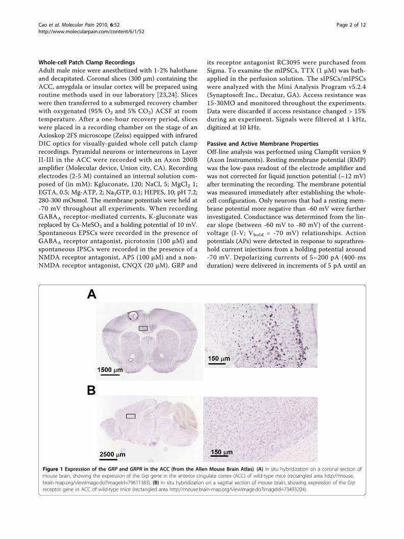
Figure 1 Expression of the GRP and GRPR in the ACC (from the Allen Mouse Brain Atlas). (A) In situ hybridization on a coronal section ofmouse brain, showing the expression of the Grp gene in the anterior cingulate cortex (ACC) of wild-type mice (rectangled area http://mouse.brain-map.org/viewImage.do?imageId=79611383). (B) In situ hybridization on a sagittal section of mouse brain, showing expression of the Grpreceptor gene in ACC of wild-type mice (rectangled area http://mouse.brain-map.org/viewImage.do?imageId=73493204).
Cao et al. Molecular Pain 2010, 6:52http://www.molecularpain.com/content/6/1/52
Page 2 of 12

AP was evoked. The rheobase was defined as the mini-mum current required to evoke an action potential. TheAP voltage threshold (Vthreshold) was defined as the firstpoint on the rising phase of the spike at which the changein voltage exceeded 50 mV/ms. The spike amplitude wasquantified as the difference between the Vthreshold andthe peak voltage. The spike width was measured at 1/2 ofthe total spike amplitude (measured from the Vthresholdlevel). The amplitude of the afterhyperpolarization (AHP)was estimated as the difference between the Vthresholdand the peak of AHP.
ImmunohistochemistryMice were deeply anesthetized with halothane and per-fused transcardially with 50-100 ml saline followed by150-500 ml of cold 0.1 M phosphate buffer (PB) con-taining 4% paraformaldehyde. Brains were removed andpost-fixed in 4% paraformaldehyde/PBS and then will beplaced in 30% sucrose in 0.2 M PBS, embedded in theOCT compound and frozen. Coronal sections (30 μMthickness) were cut using a cryostat. For dual fluores-cent immunohistochemistry, sections were incubatedovernight with anti-GRPR (1:50; rabbit polycolonal,Santa Cruz Biotecnology) and anti-GAD67 (1:300;mouse monoclonal, Chemicon) antibodies at 4°C. Sec-tions were then be washed 3 times with PBS 0.1 M andincubated for 2 hours with anti Mouse-FITC and rabbit-rhodamine conjugated secondary antibodies (1:200; Che-micon). Images of the ACC areas of at 0.7-μm intervalswith 20× lens were obtained with Bio-Rad LaboratoriesMRC 1000 laser-scanning confocal fluorescent imagingsystem.
Data AnalysisResults were expressed as mean ± standard error of themean (S. E. M.). Statistical comparisons were performedwith the use of one- or two-way analysis of variance(ANOVA) with the post-hoc Scheffe F-test in immuno-cytochemical experiments. Analysis of mIPSCs/sEPSCswas performed with cumulative probability plots andwas compared using the Kolmogorov-Smirnov (K-S) testfor significant differences. In all cases, P < 0.05 wasconsidered statistically significant.
ResultsThe expression of GRP and GRP receptors in the ACCTo understand if GRP may be distributed in mouseACC, we took the advantage of online brain mappingdata base (the Allen Mouse Brain Atlas). We found thatin adult mouse brains the Grpgene is highly enriched inthe ACC (Figure 1A); http://www.alleninstitute.org),including the regions of Cg1 and Cg2. Consistent withGrp distribution in the ACC, the Grp receptors (GRPR)mRNA are also highly expressed in the ACC (Figure
1B); taken from the Allen Mouse Brain Atlas). To con-firm the distribution of GRP receptor in the ACC, weperformed double-label immunohistochemistry usingspecific antibodies for GRP receptor and GABAergicneurons (see(Figure 2A and 2B). In addition to theACC, we have also examined the regions of the insularcortex (IC) and basal lateral amygdala (BLA). We foundthat GRP receptor was highly expressed in these areas.Particularly, GRP receptor was co-localized with
Figure 2 GABA immunoreactivity in the GRPR neurons. Double-immunostaining was performed using anti-GRP-R antibody (in red,left column) and an antibody specific for GABAergic neurons (anti-GAD67 antibody, in green center column) in the anterior cingulatecortex (ACC), basolateral amygdale (BLA) and insular cortex (IC).A subpopulation of GRP-R immunoreactivity was detected inGABAergic neurons (arrows), merged in right column. (A) Upperpanels show images from low-powered microscopy. Scale bar = 10μm. (B) Lower panels show parts of the same sections at highermagnification. Scale bar = 50 μm.
Cao et al. Molecular Pain 2010, 6:52http://www.molecularpain.com/content/6/1/52
Page 3 of 12

Figure 3 Morphological and electrophysiological properties of interneurons and pyramidal neurons in the ACC. (A) Representativecoronal section showing the placement of a whole-cell patch recording in a cingulate slice. (B) Diagram representation of the location of therecorded neurons in layer II/III. (C and D) Photomicrograph of a representative biocytin–labeled layer II/III ACC pyramidal neuron (C) interneuron(D) as visualized with confocal laser scanning microscopy. (E) Pyramidal neurons showed different firing properties from those observed ininterneurons after current injection (see F). (F) Interneurons were identified by their firing properties. When injected with current step (100 pAwithin 400 ms), interneurons showed fast spiking properties. (G and H) current-voltage relationship constructed from values taken at the end ofpulses (dots in E and F).
Cao et al. Molecular Pain 2010, 6:52http://www.molecularpain.com/content/6/1/52
Page 4 of 12

GAD67, a marker for GABA in some neurons, suggest-ing GRP receptor is expressed in a subpopulation ofGABAergic interneurons. These results indicate thatGRP may play modulatory roles in these brain areas.
Morphological and electrophysiological properties ofinterneurons and pyramidal neurons in the ACCSince GRP receptor is expressed in many neurons in theACC, we wanted to know whether it may function dif-ferently in different types of neurons, i.e pyramidal neu-rons and inhibitory interneurons. Next we performedwhole-cell patch-clamp experiments to examine theeffects of GRP on ACC neurons. As previously reported,we distinguished local inhibitory neurons and pyramidalneurons morphologically and electrophysiologically[23,25]. In some experiments, we also performed whole-cell patch clamp recording combined with biocytinlabeling in ACC neurons (see(Figure 3 for examples).Typical layer II/III pyramidal cells had a prominent api-cal dendrite, which ascended toward the superficiallayers, while their basal dendrites were mainly locatedwithin the same layer as the soma (Figure 3C). Somataof interneurons were usually ovoid in shape and thesoma size was smaller than that of pyramidal cells. Incontrast to the pyramidal cells, interneuron was lack ofthe apical dendrite and displayed 2-5 primary dendritesextending in all directions ((Figure 3D).Interneurons and pyramidal cells were found to exhi-
bit significantly different input resistances and restingmembrane potentials (see Table 1). In response to acontinuous depolarizing conditions, pyramidal cellsusually fired APs with an rapid initial phase followed bya slow gradual decline, a phenomenon called “spike fre-quency adaptation"(Figure 3E), whereas interneuronsfired a regular train of APs (Figure 3F). We also com-pared the voltage-current relationship (I-V) of pyramidalneurons and interneurons. The I-V for pyramidal neu-rons was linear at membrane potentials between -65and -90 mV, with inward rectification between -90 mVand -120 mV (Figure 3G), while for interneurons was
linear between -40 mV and -80mV and showed a slightinward rectification between -100 mV and -120 mV(Figure 3H). Moreover, we found that interneuronsexhibited a less Rheobase current (interneurons: 56.8 ±11.8 pA, n = 11; pyramidal neurons: 95.7 ± 7.3 pA,n = 11, p <0.01), a lower action potential amplitude(interneurons: 88.2 ± 2.6 mV, n = 11; pyramidal neu-rons: 93.8 ± 0.8 mV, n = 11, p <0.01), a narrower actionpotential half-width (interneurons: 1.00 ± 0.05 ms, n =11; pyramidal neurons: 1.50 ± 0.03 ms, n = 11, p<0.001) and a bigger after-hyperpolarization (interneur-ons: -24.8 ± 2.5 mV, n = 11; pyramidal neurons: -6.9 ±0.3 mV, n = 11, p <0.001) compared to the pyramidalcells (see Table 1).
GRP induced inward currents in interneurons but small orundetectable currents in pyramidal neuronsTo examine the function of GRP receptor in the ACC,GRP was applied through bath solution and theresponses of pyramidal neurons and interneurons wererecorded. At holding potential of -70 mV a short appli-cation of GRP (300 nM) induced a slowly developinginward current (peak 16.9 ± 1.8 pA) that recoveredslowly over 10 min (n = 8/10 interneurons). However,in pyramidal neurons there was undetectable currentsupon GRP application (300 nM, n = 7). In the presenceof GRP receptor antagonist, RC3095 (3 μM), the effectof GRP induced inward current in interneurons (n = 7)was completely blocked (data not shown). These resultsindicate that GRP selectively activated interneuronalGRP receptor in the ACC.
Activation of GRP receptors enhances spontaneousGABAergic, but not glutamatergic transmissionSince GRP can excite interneurons, we speculated thatactivation of GRP receptor would affect the release ofGABA in the ACC. To test this idea, we studied theeffect of GRP on GABAergic transmission. At a holdingpotential of 10 mV and with the blockade of NMDAand AMPA/KA current by using AP-5 (50 μM) and
Table 1 Basic electrophysiological properties of ACC pyramidal and inhibitory neurons
Pyramidal Neuron Interneuron Significant Difference
Number of neurons tested 33 18
Resting membrane potential, mV -73.0 ± 1.0 -61.4 ± 1.9 P < 0.001
Input resistance, M Ω 124.3 ± 6.3 332.4 ± 48.1 P < 0.001
Rheobase current, pA 95.7 ± 7.3 56.8 ± 11.8 P < 0.01
Action potential threshold, mV -35.4 ± 0.4 -43.0 ± 0.9 P < 0.001
Action potential amplitude, mV 93.8 ± 0.8 88.2 ± 2.6 P < 0.01
Action potential half-width, ms 1.50 ± 0.03 1.00 ± 0.05 P < 0.001
Amplitude of after-hyperpolarization, ms -6.9 ± 0.3 -24.8 ± 2.5 P < 0.001
Values are Mean ± SEM. Significant differences were obtained by the two-tailed, unpaired student t-test.
Cao et al. Molecular Pain 2010, 6:52http://www.molecularpain.com/content/6/1/52
Page 5 of 12

Figure 4 Activation of GRP receptor by GRP reversibly increased sIPSCs in pyramidal neurons on the ACC. (A) The representativeexample of GRP (300 nM) modulation of sIPSCs in a ACC pyramidal neuron. The trace below represents sIPSCs recorded before, during and afterGRP application. The bottom 3 traces are presented at an expanded scale. (B and C) Time course for the GRP-induced enhancement of sIPSCfrequency and amplitude in the neuron shown in (A). Note the effect of GRP is reversible. (D) Statistical results showed the GRP-inducedenhancement of sIPSC frequency and amplitude in the neuron shown in (A). Note the effect of GRP is reversible. *Indicates a significantdifference between control and GRP. * < 0.05, ** < 0.01. (E) The facilitatory effect of GRP on sIPSC frequency is concentration dependent (30 nM,n = 6; 300 nM, n = 5; 1000 nM, n = 7). * < 0.05, ** < 0.01, *** < 0.001.
Cao et al. Molecular Pain 2010, 6:52http://www.molecularpain.com/content/6/1/52
Page 6 of 12

Figure 5 The GRP-induced sIPSC facilitation is blocked by RC3095 and picrotoxin. (A and B) The effect of GRP (300 nM) could be blockedby RC3095 (3 μM, n = 4), suggesting that the GRP’s action is mediated by GRPR (upper). Representative sIPSCs recorded in a pyramidal cell froma control mouse at a holding potential of +10 mV under baseline conditions (upper), during GRP application (middle), and after the GRPRantagonist was added (lower). (C) Time course of reversible drug effect of RC3095 (3 μM) to the enhancement of sIPSC induced by GRPtreatment. (D) The effect of GRP (300 nM) could be blocked by picrotoxin (100 μM, n = 4). Representative sIPSCs recorded in a pyramidal neuronunder baseline conditions (upper), during GRP application (middle), and after picrotoxin was added (lower). (E) Time course of reversible drugeffect of picrotoxin (100 μM) to the enhancement of sIPSC induced by GRP treatment.
Cao et al. Molecular Pain 2010, 6:52http://www.molecularpain.com/content/6/1/52
Page 7 of 12

CNQX (20 μM), we recorded spontaneous inhibitorypostsynaptic currents (sIPSCs) in pyramidal neurons ofACC slices from adult mice. GRP (300 nM) increasedboth frequency (from 7.0 ± 0.8 Hz to 10.6 ± 1.1 Hz, n =7, p < 0.001) and amplitude (from 17.1 ± 0.5 pA to 20.3± 0.6 pA, n = 7, p < 0.05) of sIPSCs (Figure 4A-D). Thisfacilitation was reversible after the washout of GRP(Figure 4A-D)). Moreover, the effect of GRP was con-centration-dependent. The frequency of sIPSCs was sig-nificantly increased to 140.1 ± 7.8% (n = 10, p < 0.001)and 183.4 ± 10.6% (n = 10, p < 0.001) at 300 and 1000nM GRP, respectively (Figure 4E). No significant effectwas observed for 30 nM GRP (100.5 ± 12.4%, n = 5,p = 0.1, Figure 4E). The mean amplitude of sIPSCs wasalso significantly increased at both 300 nM GRP (110.6± 3.9%, n = 5, p < 0.05) and 1000 nM GRP (124.8 ±9.0%, n = 5, p < 0.01) (Figure 4E). To further testwhether the facilitatory effect of sIPSCs by GRP ismediated by GRP receptor, we applied the selective GRPreceptor antagonist RC3095 following the GRP applica-tion. We found that RC3095 (3 μM, n = 5) completelyeliminate the enhancement of either frequency or ampli-tude of sIPSCs in the ACC neurons (Figure 5A-C). Theapplication of GABAA receptor antagonist picrotoxin(50 μM) abolished all sIPSCs (Figure 5D-F).We next tested if glutamatergic neurotransmission in
the ACC was affected by GRP application. At a holdingpotential of -70 mV and with the blockade of GABAcurrent in the presence of picrotoxin (100 μM), werecorded spontaneous excitatory postsynaptic currents(sEPSCs) from pyramidal neurons of ACC. We foundthat bath application of GRP (300 nM) had no signifi-cant effect on either the frequency or amplitude ofsEPSCs in ACC pyramidal neurons (n = 5, Figure 6A-B).
Increase of GABA release by GRP is action potentialdependentTo investigate whether GRP receptors are involved inregulating GABA release in the ACC, we conducted
experiments where RC3095 (3 μM) was applied to thebath solution after the GRP application. We found thatthe facilitatory effect of GRP on sIPSCs was completelyreversed (Figure 7A-C, n = 5), suggesting that the effectof GRP is mediated by GRPRs. To further confirm theseresults, we examined miniature inhibitory postsynapticcurrents (mIPSCs) in the presence of TTX (1 μM). Bathapplication of GRP at three different doses (0.03, 0.3and 3 μM) did not produce any significant effect oneither the frequency or amplitude of mIPSCs (Figure7D-F). Taken together, these results demonstrate thateffects of GRP were not due to a presynaptic mechan-ism, but rather associated with depolarization and trig-gering action-potentials in GABAergic interneurons.
Activation of GRP receptor also increased the frequencyof sIPSCs in pyramidal neurons in the basolateralanygdala (BLA) and insular cortex (IC)To examine whether the facilitation of GABAergictransmission by GRP is a general phenomenon in thebrain, we tested the effect of GRP on inhibitory trans-mission in the BLA and IC. Similar to the results foundin the ACC, bath application of GRP (300 nM) signifi-cantly increase the frequency of sIPSCs recorded in pyr-amidal neurons in the BLA (p < 0.05, n = 8, Figure 8)and IC (p < 0.05, n = 8, Figure 9). Consistent with thenotion that the facilitatory effects are mediated by theGRP receptor, these facilitation were completely blockedby GRP receptor antagonist, RC3095 (3 μM, n = 6;Figure 8, 9). In the presence of TTX (1 μM), GRP hadno effect on mIPSCs (n = 5; Figure 8 and 9), Moreover,GRP had no effect on sEPSCs in either BLA or IC(n = 5, Figure 8 and 9).
DiscussionIn the present study, we used whole-cell patch-clamprecording to study the actions of GRP on ACC neuronsin adult mice. Our results provide strong electrophysiolo-gical evidence that GRP receptor facilitates inhibitory
Figure 6 Activation of GRP receptor by GRP had no effect on sEPSCs in pyramidal neurons of ACC. (A) Typical traces showing the effectof GRP (300 nM) on sEPSCs in a pyramidal neuron. (B) Statistical results showed no effect of GRP on either frequency or amplitude of sEPSCs inpyramidal neurons (n = 6).
Cao et al. Molecular Pain 2010, 6:52http://www.molecularpain.com/content/6/1/52
Page 8 of 12

Figure 7 Increase of GABA release by GRP is action potential dependent. (A) The effect of GRP (300 nM) could be blocked by TTX (1 μM,n = 4). (B) Representative sIPSCs recorded in a pyramidal neuron under baseline conditions (upper), during GRP application (middle), and afterTTX was added (lower). (C) Time course of reversible drug effect of TTX (1 μM) to the enhancement of sIPSC induced by GRP treatment. (D andE) Typical examples showing the effect of GRP (300 nM) on mIPSCs in the presence of TTX. Similar results were obtained from an additional 4neurons. (F) Statistical results showed in the presence of TTX (1 μM), GRP did not affect either amplitude or frequency of mIPSCs.
Cao et al. Molecular Pain 2010, 6:52http://www.molecularpain.com/content/6/1/52
Page 9 of 12

GABA release in the ACC. Activation of GRP receptorpreferentially modulated GABAergic, but not glutamater-gic transmission. Furthermore, somatodendritic GRPreceptor mediated action potential-dependent GABArelease in the ACC occurred in other regions, such as theBLA and IC, indicating a general role of GRP receptor inthe modulation of cortical GABAergic transmission.
How GRP facilitate GABAergic transmission and modu-late the neuronal circuits in the ACC? Several lines of evi-dence in the present study suggest that GRP acts onsomatodendritic GRP receptor in GABAergic neurons toinduce neuronal firing and GABA release in interneurons,thereby decreasing the excitability of pyramidal neurons.Bath application of GRP facilitated sIPSC frequency in a
Figure 8 Activation of GRP receptors increased GABA release in BLA neurons. (A) Bath application of GRP (300 nM) increased thefrequency of sIPSCs in pyramidal neurons of BLA (n = 11). (B) The effect of GRP was completely blocked by RC 3091 (3 μM) (n = 5). (C) Noincrease of the frequency of sIPSCs was observed in the presence of TTX (1 μM) (n = 5). (D) There is no effect of GRP on sEPSCs (n = 4). Holdingpotential for recording sEPSCs was -70 mV.
Figure 9 Activation of GRP receptors increased GABA release in IC neurons. (A and B) Bath application of GRP (300 nM) increased thefrequency of sIPSCs in pyramidal neurons of IC (n = 11). (B) The effect of GRP was completely blocked by RC 3091 (3 μM) (n = 5). (C) However,no increase was observed in the presence of TTX (1 μM) (n = 5). (D) There is no effect of GRP on sEPSCs (n = 4). Holding potential for recordingsEPSCs was -70 mV.
Cao et al. Molecular Pain 2010, 6:52http://www.molecularpain.com/content/6/1/52
Page 10 of 12
concentration- dependent manner, but GRP had littleeffect on either frequency or amplitude of mIPSCs. Theseresults are similar to the modulatory effects of the GRP/GRP receptor system on GABAergic transmission in lat-eral amygdala and hippocampus [18,26]. Interestingly, inthe present study, although ACC pyramidal neurons areshown to express GRP receptors, we found little directeffect of GRP on these cell bodies nor on the frequencyand amplitude of sEPSC. We cannot completely rule outother possible modulation of GRP on excitatory transmis-sion that cannot be revealed in the present studies. Futurestudies are clearly needed in further investigating the rolesof GRP.ACC neurons are multi-functional and play important
roles in a wide variety of behavioral functions, includingsensory pain, memory, emotional and cognitive func-tions [19-22,27,28]. Glutamate is the fast excitatorytransmitter [29] and GABA is the inhibitory transmitterin the ACC [30]. A balance between excitatory and inhi-bitory transmission is critical for many brain functions.Previous reports show that the GRP in the amygdala isinovolved in behavioral fear [18,31-36]. In addition tothe amygdala, recent studies show that lesion of theACC produced an impairment in trace fear conditioning[37] and electrical stimulation of the ACC induced fearmemory [38]. The results of the present study show thatGRP may facilitate the GABAergic transmission in theACC synapses, indicating that GRP may contribute tobehavioral fear or trace fear memory by affecting inhibi-tory transmission within the ACC.GRP has been implicated in mediation the itch sensa-
tion in the spinal cord [14,15]. The possible roles ofGRP within the ACC in behavioral itching have notbeen investigated. Furthermore, a recent work in thespinal dorsal horn suggests that alteration of inhibitorytransmission in the spinal cord is important for beha-vioral itching [39]. It is possible that GRP may alsoaffect spinal inhibitory transmission, in addition to actas a potential transmitter for itch from the periphery.Future studies are clearly needed in the spinal cord.Furthermore, direct evidence for GRP to act as a neu-rotransmitter for itching is still lacking. In human stu-dies, ACC and IC have been shown to be involved initch processing [40-45]. Future studies are required toaddress whether the modulation of GRP in the ACCinhibitory circuit would contribution to itch sensation.In summary, we report here that GRP play an impor-tant role in modulating inhibitory transmission withinthe ACC, IC and amygdala. It is likely that supraspinalGRP may contribute to a wide range of physiologicaland pathological functions, rather than act as a selec-tive transmitter for itch as reported at the level ofspinal cord.
Conflict of interestsThe authors declare that they have no competing interests.
Authors’ contributionsXYC, VM, PL and LJW performed the experiments included in themanuscript. MZ designed the experiments. XYC, LJW and MZ wrote themanuscript. All authors read and approved the final manuscript.
AcknowledgementsThis work was supported by Grants from the EJLB-CIHR Michael Smith Chairin Neurosciences and Mental Health, Canada Research Chair, NeuroCanada,and CIHR operating grants (CIHR66975 and CIHR81086) (M. Z.). M.Z. is alsosupported by the World-Class University (WCU) program of the Ministry ofEducation, Science and Technology in Korea through KOSEF (R32-10142).
Author details1Department of Physiology, Faculty of Medicine, University of Toronto, 1King’s College Circle, Toronto, Ontario M5S 1A8, Canada. 2Department ofBrain and Cognitive Sciences, College of Natural Sciences, Seoul NationalUniversity, Seoul 151-746, Korea.
Received: 23 July 2010 Accepted: 13 September 2010Published: 13 September 2010
References1. Anastasi A, Erspamer V, Bucci M: Isolation and structure of bombesin and
alytesin, 2 analogous active peptides from the skin of the Europeanamphibians Bombina and Alytes. Experientia 1971, 27(2):166-167.
2. Battey J, Wada E: Two distinct receptor subtypes for mammalianbombesin-like peptides. Trends Neurosci 1991, 14(12):524-528.
3. Kamichi S, Wada E, Aoki S, Sekiguchi M, Kimura I, Wada K:Immunohistochemical localization of gastrin-releasing peptide receptorin the mouse brain. Brain Res 2005, 1032(1-2):162-170.
4. Ladenheim EE, Jensen RT, Mantey SA, Moran TH: Distinct distributions oftwo bombesin receptor subtypes in the rat central nervous system. BrainRes 1992, 593(2):168-178.
5. Moody TW, Getz R, O’Donohue TL, Rosenstein JM: Localization ofreceptors for bombesin-like peptides in the rat brain. Ann N Y Acad Sci1988, 547:114-130.
6. Moody TW, O’Donohue TL, Jacobowitz DM: Biochemical localization andcharacterization of bombesin-like peptides in discrete regions of ratbrain. Peptides 1981, 2(1):75-79.
7. Moran TH, Moody TW, Hostetler AM, Robinson PH, Goldrich M, McHugh PR:Distribution of bombesin binding sites in the rat gastrointestinal tract.Peptides 1988, 9(3):643-649.
8. Panula P, Yang HY, Costa E: Neuronal location of the bombesin-likeimmunoreactivity in the central nervous system of the rat. Regul Pept1982, 4(5):275-283.
9. Wada E, Way J, Lebacq-Verheyden AM, Battey JF: Neuromedin B andgastrin-releasing peptide mRNAs are differentially distributed in the ratnervous system. J Neurosci 1990, 10(9):2917-2930.
10. Wada E, Way J, Shapira H, Kusano K, Lebacq-Verheyden AM, Coy D,Jensen R, Battery J: cDNA cloning, characterization, and brain region-specific expression of a neuromedin-B-preferring bombesin receptor.Neuron 1991, 6(3):421-430.
11. Gamble KL, Allen GC, Zhou T, McMahon DG: Gastrin-releasing peptidemediates light-like resetting of the suprachiasmatic nucleus circadianpacemaker through cAMP response element-binding protein and Per1activation. J Neurosci 2007, 27(44):12078-12087.
12. Petronilho F, Roesler R, Schwartsmann G, Dal Pizzol F: Gastrin-releasingpeptide receptor as a molecular target for inflammatory diseases.Inflamm Allergy Drug Targets 2007, 6(4):197-200.
13. Patel O, Shulkes A, Baldwin GS: Gastrin-releasing peptide and cancer.Biochim Biophys Acta 2006, 1766(1):23-41.
14. Sun YG, Chen ZF: A gastrin-releasing peptide receptor mediates the itchsensation in the spinal cord. Nature 2007, 448(7154):700-703.
15. Sun YG, Zhao ZQ, Meng XL, Yin J, Liu XY, Chen ZF: Cellular basis of itchsensation. Science 2009, 325(5947):1531-1534.
16. Gonzalez N, Moody TW, Igarashi H, Ito T, Jensen RT: Bombesin-relatedpeptides and their receptors: recent advances in their role in physiologyand disease states. Curr Opin Endocrinol Diabetes Obes 2008, 15(1):58-64.
Cao et al. Molecular Pain 2010, 6:52http://www.molecularpain.com/content/6/1/52
Page 11 of 12
17. Ohki-Hamazaki H, Iwabuchi M, Maekawa F: Development and function ofbombesin-like peptides and their receptors. Int J Dev Biol 2005, 49(2-3):293-300.
18. Shumyatsky GP, Tsvetkov E, Malleret G, Vronskaya S, Hatton M, Hampton L,Battey JF, Dulac C, Kandel ER, Bolshakov VY: Identification of a signalingnetwork in lateral nucleus of amygdala important for inhibiting memoryspecifically related to learned fear. Cell 2002, 111(6):905-918.
19. Courtney SM, Petit L, Maisog JM, Ungerleider LG, Haxby JV: An areaspecialized for spatial working memory in human frontal cortex. Science1998, 279(5355):1347-1351.
20. Rainville P, Duncan GH, Price DD, Carrier B, Bushnell MC: Pain affectencoded in human anterior cingulate but not somatosensory cortex.Science 1997, 277(5328):968-971.
21. Zhao MG, Toyoda H, Lee YS, Wu LJ, Ko SW, Zhang XH, Jia Y, Shum F, Xu H,Li BM, et al: Roles of NMDA NR2B subtype receptor in prefrontal long-term potentiation and contextual fear memory. Neuron 2005,47(6):859-872.
22. Zhuo M: Glutamate receptors and persistent pain: targeting forebrainNR2B subunits. Drug Discov Today 2002, 7(4):259-267.
23. Cao XY, Xu H, Wu LJ, Li XY, Chen T, Zhuo M: Characterization of intrinsicproperties of cingulate pyramidal neurons in adult mice after nerveinjury. Mol Pain 2009, 5-73.
24. Wu LJ, Ko SW, Toyoda H, Zhao MG, Xu H, Vadakkan KI, Ren M, Knifed E,Shum F, Quan J, et al: Increased anxiety-like behavior and enhancedsynaptic efficacy in the amygdala of GluR5 knockout mice. PLoS One2007, 2(1):e167.
25. Wu LJ, Toyoda H, Zhao MG, Lee YS, Tang J, Ko SW, Jia YH, Shum FW,Zerbinatti CV, Bu G, et al: Upregulation of forebrain NMDA NR2Breceptors contributes to behavioral sensitization after inflammation. JNeurosci 2005, 25(48):11107-11116.
26. Lee K, Dixon AK, Gonzalez I, Stevens EB, McNulty S, Oles R, Richardson PJ,Pinnock RD, Singh L: Bombesin-like peptides depolarize rat hippocampalinterneurones through interaction with subtype 2 bombesin receptors. JPhysiol 1999, 518(Pt 3):791-802.
27. Zhuo M: Cortical excitation and chronic pain. Trends Neurosci 2008,31(4):199-207.
28. Zhuo M: A synaptic model for pain: long-term potentiation in theanterior cingulate cortex. Mol Cells 2007, 23(3):259-271.
29. Wei F, Li P, Zhuo M: Loss of synaptic depression in mammalian anteriorcingulate cortex after amputation. J Neurosci 1999, 19(21):9346-9354.
30. Wu LJ, Xu H, Ren M, Zhuo M: Genetic and pharmacological studies ofGluR5 modulation of inhibitory synaptic transmission in the anteriorcingulate cortex of adult mice. Dev Neurobiol 2007, 67(2):146-157.
31. Yamada K, Santo-Yamada Y, Wada E, Wada K: Role of bombesin (BN)-likepeptides/receptors in emotional behavior by comparison of three strainsof BN-like peptide receptor knockout mice. Mol Psychiatry 2002,7(1):113-117, 116.
32. Martins MR, Reinke A, Valvassori SS, Machado RA, Quevedo J,Schwartsmann G, Roesler R: Non-associative learning and anxiety in ratstreated with a single systemic administration of the gastrin-releasingpeptide receptor antagonist RC-3095. Peptides 2005, 26(12):2525-2529.
33. Roesler R, Kopschina MI, Rosa RM, Henriques JA, Souza DO,Schwartsmann G: RC-3095, a bombesin/gastrin-releasing peptidereceptor antagonist, impairs aversive but not recognition memory inrats. Eur J Pharmacol 2004, 486(1):35-41.
34. Roesler R, Meller CA, Kopschina MI, Souza DO, Henriques JA,Schwartsmann G: Intrahippocampal infusion of the bombesin/gastrin-releasing peptide antagonist RC-3095 impairs inhibitoryavoidanceretention. Peptides 2003, 24(7):1069-1074.
35. Santo-Yamada Y, Yamada K, Wada E, Goto Y, Wada K: Blockade ofbombesin-like peptide receptors impairs inhibitory avoidance learningin mice. Neurosci Lett 2003, 340(1):65-68.
36. Venturella R, Lessa D, Luft T, Roozendaal B, Schwartsmann G, Roesler R:Dexamethasone reverses the memory impairment induced byantagonism of hippocampal gastrin-releasing peptide receptors. Peptides2005, 26(5):821-825.
37. Han CJ, O’Tuathaigh CM, van Trigt L, Quinn JJ, Fanselow MS, Mongeau R,Koch C, Anderson DJ: Trace but not delay fear conditioning requiresattention and the anterior cingulate cortex. Proc Natl Acad Sci USA 2003,100(22):13087-13092.
38. Tang J, Ko S, Ding HK, Qiu CS, Calejesan AA, Zhuo M: Pavlovian fearmemory induced by activation in the anterior cingulate cortex. Mol Pain2005, 1:6.
39. Ross SE, Mardinly AR, McCord AE, Zurawski J, Cohen S, Jung C, Hu L,Mok SI, Shah A, Savner EM, et al: Loss of inhibitory interneurons in thedorsal spinal cord and elevated itch in Bhlhb5 mutant mice. Neuron65(6):886-898.
40. Darsow U, Drzezga A, Frisch M, Munz F, Weilke F, Bartenstein P,Schwaiger M, Ring J: Processing of histamine-induced itch in the humancerebral cortex: a correlation analysis with dermal reactions. J InvestDermatol 2000, 115(6):1029-1033.
41. Drzezga A, Darsow U, Treede RD, Siebner H, Frisch M, Munz F, Weilke F,Ring J, Schwaiger M, Bartenstein P: Central activation by histamine-induced itch: analogies to pain processing: a correlational analysis ofO-15 H2O positron emission tomography studies. Pain 2001,92(1-2):295-305.
42. Herde L, Forster C, Strupf M, Handwerker HO: Itch induced by a novelmethod leads to limbic deactivations a functional MRI study. JNeurophysiol 2007, 98(4):2347-2356.
43. Mochizuki H, Sadato N, Saito DN, Toyoda H, Tashiro M, Okamura N, Yanai K:Neural correlates of perceptual difference between itching and pain: ahuman fMRI study. Neuroimage 2007, 36(3):706-717.
44. Mochizuki H, Tashiro M, Kano M, Sakurada Y, Itoh M, Yanai K: Imaging ofcentral itch modulation in the human brain using positron emissiontomography. Pain 2003, 105(1-2):339-346.
45. Valet M, Pfab F, Sprenger T, Woller A, Zimmer C, Behrendt H, Ring J,Darsow U, Tolle TR: Cerebral processing of histamine-induced itch usingshort-term alternating temperature modulation–an FMRI study. J InvestDermatol 2008, 128(2):426-433.
doi:10.1186/1744-8069-6-52Cite this article as: Cao et al.: Facilitation of the inhibitory transmissionby gastrin-releasing peptide in the anterior cingulate cortex. MolecularPain 2010 6:52.
Submit your next manuscript to BioMed Centraland take full advantage of:
• Convenient online submission
• Thorough peer review
• No space constraints or color figure charges
• Immediate publication on acceptance
• Inclusion in PubMed, CAS, Scopus and Google Scholar
• Research which is freely available for redistribution
Submit your manuscript at www.biomedcentral.com/submit
Cao et al. Molecular Pain 2010, 6:52http://www.molecularpain.com/content/6/1/52
Page 12 of 12