functional characterization of the arabidopsis -ketoacyl
TRANSCRIPT

Functional Characterization of the Arabidopsisb-Ketoacyl-Coenzyme A Reductase Candidates of theFatty Acid Elongase1[W][OA]
Frederic Beaudoin2, Xianzhong Wu2, Fengling Li, Richard P. Haslam, Jonathan E. Markham,Huanquan Zheng3, Johnathan A. Napier, and Ljerka Kunst*
Department of Biological Chemistry, Rothamsted Research, Harpenden, Herts AL5 2JQ, United Kingdom(F.B., R.P.H., J.A.N.); University of British Columbia, Vancouver, British Columbia, Canada V6T 1Z4 (X.W.,F.L., H.Z., L.K.); and Donald Danforth Plant Science Center, St. Louis, Missouri 63132 (J.E.M.)
In plants, very-long-chain fatty acids (VLCFAs; .18 carbon) are precursors of sphingolipids, triacylglycerols, cuticular waxes,and suberin. VLCFAs are synthesized by a multiprotein membrane-bound fatty acid elongation system that catalyzes foursuccessive enzymatic reactions: condensation, reduction, dehydration, and a second reduction. A bioinformatics survey of theArabidopsis (Arabidopsis thaliana) genome has revealed two sequences homologous to YBR159w encoding a Saccharomycescerevisiae b-ketoacyl reductase (KCR), which catalyzes the first reduction during VLCFA elongation. Expression analysesshowed that both AtKCR1 and AtKCR2 genes were transcribed in siliques, flowers, inflorescence stems, leaves, as well asdeveloping embryos, but only AtKCR1 transcript was detected in roots. Fluorescent protein-tagged AtKCR1 and AtKCR2 werelocalized to the endoplasmic reticulum, the site of fatty acid elongation. Complementation of the yeast ybr159D mutantdemonstrated that the two KCR proteins are divergent and that only AtKCR1 can restore heterologous elongase activity similarto the native yeast KCR gene. Analyses of insertional mutants in AtKCR1 and AtKCR2 revealed that loss of AtKCR1 functionresults in embryo lethality, which cannot be rescued by AtKCR2 expression using the AtKCR1 promoter. In contrast, adisruption of the AtKCR2 gene had no obvious phenotypic effect. Taken together, these results indicate that only AtKCR1 is afunctional KCR isoform involved in microsomal fatty acid elongation. To investigate the roles of AtKCR1 in postembryonicdevelopment, transgenic lines expressing RNA interference and overexpression constructs targeted against AtKCR1 weregenerated. Morphological and biochemical characterization of these lines confirmed that suppressed KCR activity results in areduction of cuticular wax load and affects VLCFA composition of sphingolipids, seed triacylglycerols, and root glycerolipids,demonstrating in planta that KCR is involved in elongation reactions supplying VLCFA for all these diverse classes of lipids.
Very-long-chain fatty acids (VLCFAs) with chainlengths between C20 and C34 are essential and ubiq-uitous constituents of eukaryotic cells. They are mostcommonly found as building blocks of sphingolipids,but they are also important components of glycero-phospholipids, triacylglycerols, sterol esters, and waxesters. Depending on their cellular and tissue locali-
zation and chain length, VLCFAs perform a widerange of physiological and structural roles. For exam-ple, they are involved in the stabilization of highlycurved nuclear pore membranes (Schneiter et al., 1996,2004) and the creation of membrane domains involvedin lipid and protein trafficking (Dickson et al., 2006;Toulmay and Schneiter, 2007) and cell signaling(Leonard et al., 2002). In mammals, C20 VLCFAs alsoserve as precursors of biologically active eicosanoidsimplicated in inflammation responses (Karimi et al.,2007) and are critical components required for myelinproduction in the central nervous system (Poulos et al.,1992) as well as for normal functioning of photorecep-tor cells in the retina (Zhang et al., 2001; McMahonet al., 2007). Furthermore, VLCFAs play an importantstructural role in skin barrier function (Wertz andDowning, 1983; Westerberg et al., 2004; Vasireddyet al., 2007). Similarly, in higher plants, VLCFAs giverise to cuticular waxes, lipid molecules deposited onthe primary plant surfaces that serve as a barrierpreventing excessive water loss and pathogen attack(Jenks et al., 1994), and mediate plant-insect interac-tions (Eigenbrode and Espelie, 1995) and pollen-stigma signaling required for fertilization (Preusset al., 1993). Modified VLCFAs are also present in
1 This work was supported by a grant from the Biotechnology andBiological Sciences Research Council (UK) to Rothamsted Researchand a grant from the Natural Sciences and Engineering ResearchCouncil of Canada to L.K.
2 These authors contributed equally to the article.3 Present address: Department of Biology, McGill University,
Montreal, Quebec, Canada H3A 1B1.* Corresponding author; e-mail [email protected] authors responsible for distribution of materials integral to
the findings presented in this article in accordance with the policydescribed in the Instructions for Authors (www.plantphysiol.org)are: Johnathan A. Napier ([email protected]) and LjerkaKunst ([email protected]).
[W] The online version of this article contains Web-only data.[OA] Open access articles can be viewed online without a sub-
scription.www.plantphysiol.org/cgi/doi/10.1104/pp.109.137497
1174 Plant Physiology�, July 2009, Vol. 150, pp. 1174–1191, www.plantphysiol.org � 2009 American Society of Plant Biologists

suberin, an extracellular plant polyester that controlswater and solute fluxes in plant tissues (Bernards,2002; Franke and Schreiber, 2007).VLCFAs are formed by elongation of C16 and C18
fatty acids by endoplasmic reticulum (ER) membrane-bound enzymes (Cinti et al., 1992; Kunst and Samuels,2003; Tehlivets et al., 2007), thought to be physicallyassociated in a complex referred to as the fatty acidelongase (FAE; von Wettstein-Knowles, 1982). Eachelongation cycle involves four successive enzymaticreactions: condensation of malonyl-CoAwith an acyl-CoA by b-ketoacyl-CoA synthase (KCS), resulting inb-ketoacyl-CoA, reduction of b-ketoacyl-CoA tob-Zhydroxyacyl-CoA by b-ketoacyl-CoA reductase(KCR), dehydration of b-hydroxyacyl-CoA to anenoyl-CoA by b-hydroxyacyl-CoA dehydratase(HCD), and reduction of enoyl-CoA by enoyl reduc-tase (ECR), thereby generating an acyl chain extendedby two carbons (Nugteren, 1965).Biochemical studies of the VLCFA biosynthesis and
analyses of mutants with defects in fatty acid elonga-tion revealed that multiple FAEs with unique substratechain length specificities are involved in generatingthe complete array of C20 to C34 acyl chains required byeukaryotic cells (Sprecher, 1974; von Wettstein-Knowles, 1993). The KCS of the elongase determinesthe substrate specificity of each elongation reaction(Millar and Kunst, 1997). Consistent with the require-ment for fatty acyl chains of diverse lengths, a largefamily of 21 FAE1-like KCS sequences has been anno-tated in the Arabidopsis (Arabidopsis thaliana) genome,together with an unrelated ELONGATION DEFEC-TIVE (ELO)-like family of four putative condensingenzymes (Dunn et al., 2004). To date, only a few ofthese condensing enzymes have been studied in anydepth, and with the exception of FAE1, a seed-specificcondensing enzyme involved in C20 and C22 fatty acidbiosynthesis for seed storage lipids (Kunst et al., 1992),and CER6, a condensing enzyme that catalyzes theelongation of fatty acyl-CoAs longer than C22 in theepidermal cells of Arabidopsis (Millar et al., 1999;Fiebig et al., 2000), the functions the of other condens-ing enzymes in VLCFA elongation have not beenestablished in planta, although several additionalFAE1-like KCSs have been functionally characterizedvia heterologous expression in yeast (Trenkamp et al.,2004; Paul et al., 2006).In contrast to the KCSs that have strict substrate and
tissue specificities, the other three enzymes of the fatty
acyl elongase, the KCR, the HCD, and the ECR, arebelieved to be common to all VLCFA biosyntheticreactions, have (presumed) broad substrate specific-ities, and are expressed in all cells (Millar and Kunst,1997). Although these elongase component enzymeshave been identified in Saccharomyces cerevisiae (Kohlwein
Table I. Percentage of amino acid sequence identity (similarity)between different KCR proteins
AtKCR1 AtKCR2 YBR159 GL8a GL8b
AtKCR1 –AtKCR2 45 (80) –YBR159 30 (65) 29 (64) –GL8a 55 (84) 48 (76) 30 (63) –GL8b 56 (84) 49 (76) 29 (63) 97 (99) –
Figure 1. Characterization of the AtKCR1 and AtKCR2 insertion lines.A, The AtKCR1 gene structure and mutation site. White boxes representexons. Vertical arrow indicates the exact Ds transposon insertion site.The genomic DNA sequence flanking the Ds insertion is shown withuppercase letters indicating exon sequences and lowercase lettersindicating intron sequences. B, The AtKCR2 gene structure and muta-tion sites. White boxes represent exons. C, Immature siliques of wild-type (WT) and heterozygous (Het) AtKCR1/atkcr1 plants. D, Maturesiliques of wild-type and heterozygous AtKCR1/atkcr1 plants. E, RT-PCR analysis of AtKCR1 mRNA levels in green seeds from immaturewild-type siliques (lane 1) and green seeds and white seeds fromimmature heterozygous siliques (lanes 2 and 3, respectively). Thebottom panel shows expression of the cytosolic glyceraldehye-3-Pdehydrogenase (GAPC ) loading control for the corresponding lanes inthe top panel.
Arabidopsis b-Ketoacyl-Coenzyme A Reductase
Plant Physiol. Vol. 150, 2009 1175

et al., 2001; Beaudoin et al., 2002; Han et al., 2002; Denicand Weissman, 2007), this hypothesis could not betested until recently because the corresponding en-zymes and genes were not available from a multicel-lular organism. A single ECR gene has been confirmedand characterized in Arabidopsis and shown to en-code a functional ECR that physically interacts withthe Elo2p and Elo3p condensing enzymes when ex-pressed in yeast (Gable et al., 2004). The ArabidopsisECR was shown to be identical to CER10 (Zheng et al.,2005), the protein defective in the cer10mutant isolatedby Koornneef et al. (1989). Biochemical analysis of thecer10mutant demonstrated that the ECR gene productis involved in VLCFA elongation required for thesynthesis of many VLCFA-containing lipids, includingcuticular waxes, seed triacylglycerols, and sphingo-lipids (Zheng et al., 2005). Similarly, the newly iden-tified HCD PASTICCINO2 (Bach et al., 2008) isencoded by a single gene in Arabidopsis. Partial lossof PAS2 function in the pas2-1 mutant is associatedwith a general reduction of VLCFA content in seedstorage triacylglycerols and complex sphingolipids aswell as reduced levels of cuticular waxes that aregenerated from VLCFA precursors. A complete loss ofPAS2 activity is embryo lethal (Bach et al., 2008). TwoKCR genes, named GL8A and GL8B, are present in themaize (Zea mays) genome (Xu et al., 1997; Dietrichet al., 2005) and are required for cuticular wax accu-mulation on seedling leaves. However, they are notexpressed only in the epidermis, but throughout theplant. Attempts to generate double mutants by cross-ing gl8a 3 gl8b single mutant lines failed becauseembryos carrying both mutations were not viable.Thus, in addition to its wax biosynthetic role, KCR has
an essential function in maize, perhaps in the produc-tion of sphingolipids (Dietrich et al., 2005), but thisidea has not been experimentally verified.
Two KCR orthologs have also been annotated in theArabidopsis genome (Dietrich et al., 2005). One of thesegenes, At1g67730, functionally complements the KCR-deficient ybr159wD mutant of S. cerevisiae (Beaudoinet al., 2002), but the specific role of the protein that itencodes has not been determined. The other KCR can-didate gene, At1g24470, has not been analyzed at all. In
Figure 2. Arrest of atkcr1/atkcr1 embryo development. Six siliques of different developmental ages growing on heterozygousAtKCR1/atkcr1 plants were dissected. A to F, DIC images of wild-type-looking green seeds at globular (A), heart (B), early torpedo(C), late torpedo (D), and bent cotyledon (E) stages of development and at maturity (F). G to L, DIC images of atkcr1/atkcr1mutantembryos from white seeds developing in the same silique arrested at globular stage. Bar = 50 mm.
Figure 3. RT-PCR analysis of AtKCR1 and AtKCR2 expression inArabidopsis. Sl, Green siliques; Fl, flowers; St, stems; Lf, leaves; Rt,roots. The bottom panel shows the expression of tubulin as a loadingcontrol for the corresponding lanes in the top panel.
Beaudoin et al.
1176 Plant Physiol. Vol. 150, 2009

this study, we employ Arabidopsis insertional mutantsdisrupted in the KCR genes and transgenic lines inwhichKCR genes have been silenced by introduction ofRNA interference (RNAi) or overexpression constructsto investigate the in planta functions of the two KCRproteins. We demonstrate that these two proteins aredivergent and that At1g67730-encoded enzyme is thefunctional KCR of the Arabidopsis FAE essential forembryo development and plant morphogenesis.
RESULTS
Arabidopsis Has Two KCR Genes
The isolation of the yeast KCR gene YBR159wresulted in the identification of a single Arabidopsisortholog, At1g67730 (Beaudoin et al., 2002), which werefer to as AtKCR1. Further exploration of the Arabi-
dopsis genome revealed another KCR-like sequence,At1g24470 (Dietrich et al., 2005), which we designateAtKCR2. AtKCR1 has a 957-bp open reading frame(ORF) that encodes a protein of 318 amino acids with apredicted molecular mass of 35.7 kD. AtKCR2 has a939-bp ORF that encodes a protein of 312 amino acidswith a predicted molecular mass of 35.0 kD. Both theseproteins have a putative NADH binding site andcontain highly conserved Ser-Tyr-Lys residues essen-tial for catalysis (Beaudoin et al., 2002), though adilysine motif required for ER retention is obviousonly in AtKCR1 (Supplemental Fig. S1). Unlike themaize orthologs GL8A and GL8B that are 97% iden-tical and have partially redundant biochemical func-tions (Dietrich et al., 2005), AtKCR1 and AtKCR2exhibit only 45% amino acid identity (Table I), sug-gesting that they could play diverse roles in VLCFAproduction in Arabidopsis.
Figure 4. Tissue-specific expressionpatterns of AtKCR1 (A–K) and AtKCR2(L–W) detected in transgenic AtKCR1-promoter:GUS and AtKCR2promoter:GUS lines. Tissues were assayed forGUS activity by histochemical stainingwith the 5-bromo-4-chloro-3-indolyl-b-D-glucuronide substrate. Shown aredifferent stages of embryo develop-ment (A–F, AtKCS1; L–R, AtKCS2);seedling with roots (G and S), caulineleaf with trichomes (H and T); inflo-rescence stem with buds and siliques(I and U); flower (J and V); and siliques(K andW). Bars = 50 mm in A to F and Lto R; 5 mm in I and U; 1 mm in G, H, J,K, S, T, V, and W.
Arabidopsis b-Ketoacyl-Coenzyme A Reductase
Plant Physiol. Vol. 150, 2009 1177
Isolation of Mutants Disrupted in the AtKCR Genes
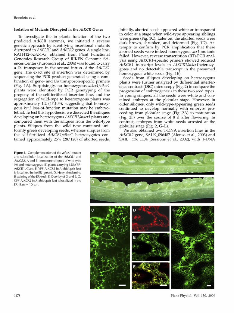
To investigate the in planta function of the twopredicted AtKCR enzymes, we initiated a reversegenetic approach by identifying insertional mutantsdisrupted in AtKCR1 and AtKCR2 genes. A single line,RATH12-5282-1-G, obtained from Plant FunctionalGenomics Research Group of RIKEN Genomic Sci-ences Center (Kuromori et al., 2004) was found to carrya Ds transposon in the second intron of the AtKCR1gene. The exact site of insertion was determined bysequencing the PCR product generated using a com-bination of gene- and Ds transposon-specific primers(Fig. 1A). Surprisingly, no homozygous atkcr1/atkcr1plants were identified by PCR genotyping of theprogeny of the self-fertilized insertion line, and thesegregation of wild-type to heterozygous plants wasapproximately 1:2 (47:103), suggesting that homozy-gous kcr1 loss-of-function mutation may be embryo-lethal. To test this hypothesis, we dissected the siliquesdeveloping on heterozygous AtKCR1/atkcr1 plants andcompared them with the siliques from the wild-typeplants. Siliques from the wild type contained uni-formly green developing seeds, whereas siliques fromthe self-fertilized AtKCR1/atkcr1 heterozygotes con-tained approximately 25% (28/120) of aborted seeds.
Initially, aborted seeds appeared white or transparentin color at a stage when wild-type appearing siblingswere green (Fig. 1C). Later on, the aborted seeds weredark brown, shrunken, and deformed (Fig. 1D). At-tempts to confirm by PCR amplification that theseaborted seeds were indeed homozygous kcr1 mutantsfailed. However, reverse transcription (RT)-PCR anal-ysis using AtKCR1-specific primers showed reducedAtKCR1 transcript levels in AtKCR1/atkcr1heterozy-gotes and no detectable transcript in the presumedhomozygous white seeds (Fig. 1E).
Seeds from siliques developing on heterozygousplants were further analyzed by differential interfer-ence contrast (DIC) microscopy (Fig. 2) to compare theprogression of embryogenesis in these two seed types.In young siliques, all the seeds were white and con-tained embryos at the globular stage. However, inolder siliques, only wild-type-appearing green seedscontinued to develop normally with embryos pro-ceeding from globular stage (Fig. 2A) to maturation(Fig. 2F) over the course of 8 d after flowering. Incontrast, embryos from white seeds arrested at theglobular stage (Fig. 2, G–L).
We also obtained two T-DNA insertion lines in theAtKCR2 gene, SALK_096487 (Alonso et al., 2003) andSAIL _536_H04 (Sessions et al., 2002), with T-DNA
Figure 5. Complementation of the atkcr1 mutantand subcellular localization of the AtKCR1 andAtKCR2. A and B, Immature siliques of wild-type(A) and heterozygous (B) plants carrying 35S:YFP-AtKCR1. C and E, YFP-AtKCR1 in Arabidopsis leafis localized in the ER (green). D, Hexyl rhodamineB staining of the ER (red). F, Overlay of D and E. G,CFP-AtKCR2 in Arabidopsis leaf is localized in theER. Bars = 10 mm.
Beaudoin et al.
1178 Plant Physiol. Vol. 150, 2009

insertions in the second and fourth exons, respectively(Fig. 1B). No visible developmental phenotypes wereobserved in homozygous atkcr2 progenies from eitherT-DNA mutant line.
AtKCR1 and AtKCR2 Expression Domains Overlap andThey Both Reside in the ER
To determine if the two apparent Arabidopsis ho-mologs are both constitutively expressed and todelineate their expression domains, we performedRT-PCR analyses and b-glucuronidase (GUS) assays intransgenic plants transformed with AtKCR1promoter-GUS and AtKCR2promoter-GUS constructs. As shownin Figure 3, both genes are expressed in green siliques,flowers, inflorescence stems, and leaves, with thehighest expression levels detected in siliques andflowers. High AtKCR1 transcript levels were alsopresent in roots, whereas AtKCR2 transcript was al-most undetectable in root tissues. These expressionresults were confirmed by GUS activity assays (Fig. 4).
In addition, AtKCR1promoter- and AtKCR2promoter-directed GUS activity was also detected in embryos ofdifferent stages (Fig. 4, A–F and L–R) and youngdeveloping seedlings (Fig. 4, G and S) but was absentfrom mature seeds (Fig. 4, K and W) and stem bases(data not shown).
To compare the subcellular localization of theAtKCR1 and AtKCR2, yellow fluorescent protein(YFP)-AtKCR1 and cyan fluorescent protein (CFP)-AtKCR2 fusion constructs were initially transientlyexpressed in tobacco (Nicotiana tabacum) under thecontrol of the 35S promoter. Visualization of tobaccoleaves revealed that both proteins were localized in theER (data not shown). We also introduced both fluores-cent protein fusion constructs into Arabidopsis. Toensure that the YFP-AtKCR1 is fully functional, weexpressed it in AtKCR1/atkcr1 heterozygous plants.Sixteen out of 20 recovered independent transgeniclines had siliques with only green wild-type-lookingseeds and no white seeds, indicating complementation(Fig. 5B) and demonstrating that the mutation in the
Figure 6. AtKCR1 complements fatty acid elongationin yeast. Gas chromatography-flame ionization de-tection analysis of FAMEs prepared from wild-type(WT) or mutant (ybr159D) yeast coexpressing AtFAE1(A) or IgELO9 (B) and either a vector control, yeastYBR159, AtKCR1, or AtKCR2 gene.
Arabidopsis b-Ketoacyl-Coenzyme A Reductase
Plant Physiol. Vol. 150, 2009 1179

AtKCR1 gene is responsible for the embryo lethality. Inall the complemented YFP-AtKCR1 transgenic linesexamined by confocal microscopy, YFP fluorescencelabeled a reticulate network typical of the ER (Fig. 5C).Because the disruption of AtKCR2 did not result in adetectable phenotype, we could not verify if the CFP-AtKCR2 fusion protein is active by complementation ofa loss-of-function mutant, but we decided to proceedwith its localization. For this purpose, we transformedthe 35Spromoter-CFP-AtKCR2 fusion construct intowild-type Arabidopsis. This experiment revealed thatthe AtKCR2 protein also resides in the ER (Fig. 5G).
Only AtKCR1 Complements the Yeast ybr159D Mutant
and Embryo Lethality of the Arabidopsis atkcr1 Mutant
AtKCR1 has been previously shown to restore het-erologous elongase activity of the yeast ybr159D mu-tant similar to the expression of native yeast KCR geneYBR159 (Beaudoin et al., 2002), indicating that theArabidopsis and yeast enzymes are functionallyequivalent and that AtKCR1 also likely catalyzes thereduction of the b-ketoacyl-CoA. This result was con-firmed in this study (Fig. 6). By contrast, the expressionof AtKCR2 failed to restore heterologous fatty acid
Figure 7. Morphological characterization ofAtKCR1-RNAi plants. A, C, and D, Fused rosetteleaves and inflorescences of 4-week-old T1 plants.B, Eight-week-old AtKCR1-RNAi T1 plant. E,Flowers from 6-week-old glossy T2 plants openedmanually. F, Mature siliques from 8-week-oldglossy T2 plants. G and H, Glossy (G) and non-glossy (H), but curly, 6-week-old T2 plants. I and J,Wild-type (I) and glossy (J) AtKCR1-RNAi T2seedlings showing changes in root morphology.
Beaudoin et al.
1180 Plant Physiol. Vol. 150, 2009

elongation mediated by either FAE1 or the Isochrysisgalbana D9-ELO-like elongating activity (Qi et al., 2002)in the ybr159D yeast mutant (Fig. 6) and also failed torescue the slow growth phenotype of ybr159D yeastcells associated with impaired endogenous elongasefunction (data not shown), suggesting that AtKCR2has no KCR activity in yeast.The fact that atkcr1 loss-of-function mutation is
embryo lethal suggests that either AtKCR2 is not afunctional KCR or that AtKCR2 is not expressed dur-ing embryogenesis. GUS activity assays in transgenicplants transformed with the AtKCR2promoter:GUSconstruct (Fig. 4, L–W) and global gene expressionanalyses during Arabidopsis embryogenesis (Schmidet al., 2005) indicate that AtKCR2 is transcribed at alltimes during embryo development, albeit at a muchlower level than AtKCR1. To determine if the lowexpression level of AtKCR2 was the reason whyAtKCR2 could not support embryogenesis in the ab-sence of AtKCR1 or whether the AtKCR2 was indeedinactive, we expressed AtKCR2 using the AtKCR1promoter in the AtKCR1/atkcr1 background. In con-trast to AtKCR1, which complemented the atkcr1 mu-tant (Fig. 5B), AtKCR2 could not rescue the embryolethality of atkcr1 even when expressed in the AtKCR1manner behind the AtKCR1 promoter (data notshown), confirming that it is not a functional KCRisoform.
RNAi-Suppressed and Cosuppressed AtKCR1 PlantsDisplay Fused Vegetative and Reproductive Organs and
an Abnormal Root Morphology
Embryo lethality prevented further phenotypicanalysis on homozygous atkcr1 lines, and no develop-mental differences or changes in seed VLCFA compo-sition or cuticular wax accumulation were detected inAtKCR1/atkcr1 heterozygotes in comparison withwild-type plants. We therefore generated transgenicplants in which AtKCR1was down-regulated by RNAito characterize their morphological and biochemicalphenotypes and determine the role of AtKCR1 in thedifferent VLCFA metabolic pathways.To test the specificity of our RNAi construct and
ensure that the phenotypes that we describe are due tosuppression of AtKCR1 alone, we performed RT-PCRanalysis of the RNAi suppressed plants using mRNAprepared from flowers that showed high KCR1 andKCR2 expression in the wild type in previous assays(Fig. 3). This experiment confirmed that the RNAi
construct specifically down-regulates the expres-sion of only AtKCR1, but not AtKCR2 (SupplementalFig. S2).
Twenty T1 RNAi transformants were chosen forfurther evaluation. These plants displayed a variety ofphenotypes, including growth retardation and fusedrosette leaves (Fig. 7, A and D). Individuals with themost severe morphological phenotype never formedrosettes, but instead produced incompletely devel-oped leaf-like structures (Fig. 7A). They were alsohighly sensitive to dehydration and needed to becultivated in a controlled environment with atmo-spheric humidity above 85% in wet soil at all times.Eleven T1 plants failed to grow to maturity, developednecrotic spots, and died. From the nine remainingplants, three (designated 4, 6, and 7) required mechan-ical organ separations to allow distorted leaves andinflorescence stems to develop (Fig. 7, C and D).
Soil-grown T2 progeny of these AtKCR-RNAi pri-mary transformants exhibited two predominant phe-notypes: glossy inflorescence stems (Fig. 7G) ortwisted and curled fiddlehead (fdh)-like stems thatwere not obviously glossy (Fig. 7H). Both glossy andnonglossy plants were present in the progeny of the 4,6, and 7 lines. These three lines, together with a fourthline (line 9, showing a less extreme phenotype), wereselected for a detailed morphological and biochemicalcharacterization. Examination of the flowers fromglossy plants revealed an unusual bushy stigma mor-phology with longer papillae and stamen with shorterfilaments unable to position anthers at the height of thestigma, similar to that reported for cer10 (Zheng et al.,2005; Fig. 7E). These plants also produced short andcrooked siliques, containing mostly aborted seeds,suggesting a problem with pollen viability/fertility(Fig. 7F). Cross-pollination of glossy plants with wild-type pollen resulted in longer siliques and a consider-ably increased number of seeds produced (data notshown). Similar short and crooked siliques were alsofound on the nonglossy plants. Interestingly, maturedried seeds collected from both glossy and nonglossyAtKCR-RNAi plants were considerably larger thanseeds collected from wild-type plants grown underidentical conditions (Table II).
An aspect of theAtKCR-RNAi phenotype that has notpreviously been reported in other mutants impaired inVLCFA biosynthesis is the abnormal root morphologyof the glossy plants (Fig. 7J). When grown horizontallyon plates, the roots of these plants ran on the surface ofthe gel matrix, lacked lateral branches, and had dramat-
Table II. Dry weight of seeds collected from glossy and nonglossy AtKCR RNAi plants compared tothe wild type
Each value is the mean of three independent measurements 6 SD. GL, Glossy; NGL, nonglossy.
Line Wild Type 4 GL 7 GL 7 NGL 9 NGL
Seed weight(mg/50 seeds)
21.77 6 1.0 36.47 6 0.8 34.53 6 1.0 40.47 6 1.4 43.20 6 1.1
Arabidopsis b-Ketoacyl-Coenzyme A Reductase
Plant Physiol. Vol. 150, 2009 1181

ically reduced root hairs (Fig. 7J, inset). Closer observa-tion revealed that lateral root primordia were formedbut did not elongate. In contrast, roots of nonglossyplants with curly inflorescences appeared wild-type.
To determine if the phenotypes observed were dueto AtKCR1 RNAi suppression, AtKCR1 transcriptlevels in transgenic lines were determined by RT-PCR in 15-d-old-seedlings and in a number of tissuesfrom 8-week-old T2 plants. As shown in Figure 8, inthe developing seedlings and in all of the tissuesanalyzed, the levels of AtKCR1 transcripts were lowerthan in the wild type. The reduction in KCR transcriptswas more pronounced in the glossy plants comparedwith the more limited reduction observed in the non-glossy lines. Within each line and for each plantphenotype (glossy or nonglossy), levels of AtKCR1transcripts were similar in all tissues. These resultsshow a clear correlation between reduced levels ofAtKCR1 transcripts and increased severity of the phe-notype in the T2 generation.
We also overexpressed the AtKCR1 cDNA under thecontrol of 35S promoter. About 50% of transgenic linesdisplayed abnormal phenotypes, similar to those of theAtKCR1-RNAi lines, including dwarfism, curly caulineleaves, fused flower buds, and bright green glossystems (Supplemental Fig. S3, A–C). To investigate ifthese phenotypes were caused by AtKCR1 cosuppres-sion, AtKCR1 transcript levels in a variety of tissues oftransgenic plants were determined by RT-PCR. Asshown in Supplemental Figure S3D, in cauline leaves,stems, and floral buds, the organs exhibiting the mostpronounced phenotypes, AtKCR1 transcript levelswere considerably lower than in the wild type.
RNAi-Suppressed AtKCR1 Plants Have Abnormal
Trichome and Epidermal Cell Morphology
As previously reported for cer10, fdh, wbc11, andwax2/pel6, both glossy and nonglossy AtKCR-RNAiplants display clearly altered trichome morphology(Fig. 9, A–F). As shown in Figure 9B, trichomes aresparsely distributed, have fused branches in glossyplants, and have shorter or bent branches in nonglossyplants (Fig. 9C). Scanning electron microscopy exam-ination showed that the basal cells of these trichomesare swollen and bulging out of the epidermal surface(Fig. 9, D–F). A further inspection of the surface ofyoung leaves revealed impaired integrity of the epi-dermis in glossy plants. Groups of cells appearedswollen to the point were they did not adhere tightly toeach other, resulting in deep intercellular grooves (Fig.9, G–I) and probably contributing to the extremesensitivity of glossy AtKCR1-RNAi plants to dehydra-tion. This type of abnormal epidermal structure couldnot be detected in nonglossy plants.
Cuticular Wax Load Is Reduced in AtKCR1-RNAi Lines
Organ fusions and stem glossiness observed in theAtKCR1-RNAi plants are typically associated with
defects in plant cuticles. Cuticle abnormalities can bedetected by a toluidine blue (TB) test, which results instaining of organs with permeable cuticles (Tanakaet al., 2004). We used the TB test to evaluate cuticles ofglossy line 4 and nonglossy line 9 individuals (Fig.10A). The test resulted in complete staining of line 4glossy seedlings, indicating severe cuticular abnor-malities of this RNAi line. Nonglossy line 9 seedlingsdisplayed a range of partially stained patterns, includ-ing patchy or random staining as observed for fdh andcer14 mutants (Fig. 10A; Tanaka et al., 2004).
Figure 8. Expression of Arabidopsis KCR genes. A, RT-PCR analysis ofthe expression levels of AtKCR1 in 2-week-old wild-type (WT) and T2AtKCR1-RNAi seedlings. The bottom panel shows the expression of atubulin gene as a loading control for the corresponding lanes in the toppanel. B, RT-PCR analysis of the expression levels of AtKCR1 in 15-d-old seedlings, flowers, stems, leaves, and roots of wild-type and T2AtKCR1-RNAi plants. GL, Glossy; NGL, nonglossy.
Beaudoin et al.
1182 Plant Physiol. Vol. 150, 2009

Because VLCFAs are precursors of all the aliphaticcomponents of cuticular wax, we suspected that cuti-cle permeability of AtKCR1-RNAi lines may at least inpart be due to reduced wax accumulation. We there-fore examined the glossy inflorescence stem surfacesof the AtKCR1-RNAi plants by scanning electron mi-croscopy. As shown in Figure 10B, wild-type stems aredensely covered with epicuticular wax crystals. Incontrast, glossy stem surfaces of AtKCR1-RNAi plantsare virtually devoid of wax crystals.Wax analyses confirmed major reductions in wax
loads in AtKCR1-RNAi plants. The total wax coverageon wild-type Columbia-0 (Col-0) inflorescence stemsunder the conditions used here was 1086 mg per gfresh weight. In contrast, wax loads measured inglossy AtKCR1-RNAi plants reached 124.2 mg per gfresh weight (11.4% of the wild type) in line 7 (7 GL)and 42.8 mg per g fresh weight (3.9% of the wild type)in line 6 (6 GL; Fig. 11). Quantification of wax com-ponents demonstrated that reduced wax loads weredue to decreased accumulation of all compoundclasses (Fig. 11). In nonglossy AtKCR1-RNAi plants,the decrease in wax load was far less pronounced,and it was proportional to the levels of AtKCR1transcripts detected in the stem of those plants (Fig.8). Wax loads measured for nonglossy plants fromlines 7 and 9 were 811 and 869 mg per g fresh weight(75% and 80% of the wild type), respectively (Fig. 11,inset). However, these data indicate that while lines 7and 9 were phenotypically classified as nonglossy,they do have a reduction in wax load, as might beexpected for plants with an impaired capacity tosynthesize VLCFAs.
VLCFA Content Is Decreased in Root Lipids and Seed
Triacylglycerols of AtKCR1-RNAi Lines
Since Arabidopsis leaves contain very low levels ofVLCFAs, we analyzed the fatty acid composition inthe roots of wild-type and AtKCR-RNAi plants thatexhibit abnormal root morphology (Fig. 7J). A clearreduction in the content of C20, C22, and C24 fatty acidswas detected in glossy lines 4 and 6 (Fig. 12, A and B).VLCFAs accounted for 5.6% of the total root fatty acidextracted from wild-type Col-0 plants grown in thisstudy, predominantly reported in phospholipids,such as phosphatidylethanolamine and phosphati-dylserine (Devaiah et al., 2006). The VLCFA contentmeasured in glossy plants was reduced to 1.4% and1.7% in lines 4 and 6 (24% and 29% of the wild type),respectively. The VLCFA content in nonglossy plantswas similar to that of wild-type plants with 5.3% and5.6% measured in lines 7 and 9, respectively. Theseresults correlated well with the levels of VLCFAsmeasured in the acyl-CoA pool in roots of the sameplants (Fig. 12C).
Surprisingly, in the root fatty acid methyl ester(FAME) extracts prepared from glossy plants, wedetected three novel peaks (Fig. 12, A and B, peaks1–3) that were either absent or low in wild-typeextracts. These peaks were tentatively identified bymass spectrometry as C16 and C18 a,v-dicarboxylicacids and an v-hydroxy fatty acid, compounds that areabundant in the cutin and suberin in Arabidopsis(Bonaventure et al., 2004, Franke et al., 2005, Beissonet al., 2007). The presence of these usually highlypolymerized constituents in the AtKCR1-RNAi FAME
Figure 9. Glossy AtKCR1-RNAi plantshave abnormal trichomes and epider-mal structures. A to C, Primary leavesof 2-week-old wild-type (A), line 4glossy (B), and line 9 nonglossy (C) T2
seedlings. D to F, Trichomes from thecauline leaves of wild-type (D) andglossy plants (E and F). G to I, Abnor-mal epidermal structure of the abaxialsurface of cauline leaves in glossyplants. In the scanning electron micro-graphs (D–I), bars = 50mm, except in E,where the bar = 10 mm.
Arabidopsis b-Ketoacyl-Coenzyme A Reductase
Plant Physiol. Vol. 150, 2009 1183

samples after mild methanolic acid extraction may
reflect the altered composition and/or structure of thesuberin when the synthesis of longer C20, C22, and C24compounds is reduced. The nonglossy plant extractsdid not contain the three additional compoundsfounds in the roots of glossy plants.
Because Arabidopsis seed oil also containsVLCFAs, we examined the fatty acid compositionand content of mature dry seeds. Compositionalanalyses of oil produced by the AtKCR1-RNAi seedsrevealed that only the oil from glossy plants wasdepleted in C20 and C22 saturated and monounsatu-rated fatty acids. The total VLCFAs content in theseseeds was about 30% lower compared to the wildtype (Table III; Fig. 13). The larger seeds collectedfrom both glossy and nonglossy AtKCR1-RNAi plantshad a higher fatty acid content than wild-type seeds(Tables II and III). Overall, the increase in total fattyacid content was largely proportional to the increasein seed size and did not result from a higher propor-tion of oil as a percentage of the seed weight (data notshown).
Sphingolipid Analyses
To evaluate the effect of reduced AtKCR1 expressionon VLCFA incorporation into sphingolipids, we ana-lyzed the complex sphingolipid composition in thestems of glossy and nonglossy AtKCR1-RNAi plantsthat all exhibit abnormal shoot morphology. All theexamined lines accumulated free long-chain bases(LCBs), which are characteristic of impaired sphingo-lipid metabolism as reported for the yeast FAE mu-tants (Kohlwein et al., 2001; Fig. 14A). In addition, amoderate but statistically significant decrease in al-most all the sphingolipid species was detected (Fig.14B). Conversely, neither the accumulation of freeLCBs nor the reduction in any of the sphingolipidspecies could be detected in the stems of morpholog-ically normal plants (data not shown). This suggeststhat the twisted shoot phenotype may also be causedby altered sphingolipid structure and not only thedefective cuticular lipid composition as proposed forthe fdh mutant.
Figure 10. AtKCR1-RNAi plants display cuticular defects. A, Two-week-old wild-type (WT) and AtKCR1-RNAi seedlings stained with TB. B,Epicuticular wax crystal structure on stem surfaces visualized by scanningelectronic microscopy. Shown are images of the wild type (left panel,bar =10 mm) and glossy AtKCR1-RNAi line 4 (right panel, bar = 2 mm).
Figure 11. Cuticular wax compositionon stems of wild-type (WT) andAtKCR1-RNAi plants. Each bar repre-sents the amount of a specific waxconstituent, labeled on the x axis by achemical class and a carbon chainlength. Each value is the mean of fourmeasurements. Error bars indicate SD.FW, Fresh weight; GL, glossy; NGL,nonglossy.
Beaudoin et al.
1184 Plant Physiol. Vol. 150, 2009

DISCUSSION
This study describes the functional characterizationof the Arabidopsis orthologs of the yeast KCRYBR159w and confirms the central role of this enzyme
activity in the synthesis of VLCFAs in plants, as well as
the essential nature of these fatty acids. Like maize,
Arabidopsis contains two KCR-like sequences (KCR1
and KCR2), but they do not share the same high degree
Figure 12. Fatty acid composition inthe root of wild-type and AtKCR1-RNAi T2 lines. Gas chromatography-flame ionization detection (FID) traceof FAMEs prepared from the roots ofwild-type and AtKCR1-RNAi 15-d-oldseedlings. In addition to the expectedreduction in C20+ fatty acids, severalelevated peaks were identified asC16:0 and C18:0 dicarboxylic acids(peaks 1 and 2, respectively) and anv-hydroxylated fatty acid (peak 3). Thestar indicates a phenolic compound,most likely ferulic acid. B, Quantitationof variation in root fatty acid composi-tion in wild-type and AtKCR1-RNAilines. WT, Wild type. C, Acyl-CoA pro-filing of the roots of wild-type andAtKCR1-RNAi 15-d-old seedlings.Each value is the mean of four mea-surements. Error bars indicate SD. GL,Glossy; NGL, nonglossy.
Arabidopsis b-Ketoacyl-Coenzyme A Reductase
Plant Physiol. Vol. 150, 2009 1185

of identity exhibited by the monocot genes. Our dataindicate that only KCR1 is a bona fide KCR of the FAEin Arabidopsis, based on several independent criteria.First, KCR1 but not KCR2 is capable of rescuing fattyacid elongation in the yeast ybr159wD mutant. Second,the expression of the KCR2 ORF under the control ofthe KCR1 promoter is unable to suppress the embryolethality of the kcr1 insertional mutant. Finally, whileinsertional mutations of KCR1 are embryo lethal, sim-ilar mutations of KCR2 have no obvious phenotypiceffect and do not affect the composition of lipids thatare known to accumulate or are derived fromVLCFAs,such as seed storage lipids, cuticular waxes, andsphingolipids. It is possible that this apparently nor-mal phenotype of the kcr2 mutant is a result of com-pensation by the KCR1 protein, but we have notdetected increased KCR1 transcript levels in the kcr2mutant to support this idea. The precise function ofKCR2 remains to be determined, although it is stilllikely to be as a member of the short-chain dehydro-
genases/reductase superfamily. Thus, Arabidopsisdiffers from maize, in which two closely relatedKCRs (GL8A and GL8B) both contribute to the activityof the microsomal elongase.
Since KCR1 represents the sole microsomal elongaseKCR, it is not surprising that disruption of the KCR1gene is embryo lethal. This also indicates that thecapacity to synthesize VLCFAs is crucial at early stagesof embryo development and is in agreement with therecent data of Bach et al. (2008) showing that thedehydratase PAS2, another core microsomal elongasecomponent, is essential during embryogenesis. It isinteresting to note that while PAS2 and KCR1 are bothessential single genes in Arabidopsis, the CER10 geneis not, even though it represents the sole ortholog ofthe yeast TSC13 ECR. This very likely indicates thepresence of structurally unrelated but functionallyequivalent forms of the ECR in Arabidopsis.
To evaluate the requirement for VLCFAs during thegrowth and development of Arabidopsis after embryo-
Table III. Fatty acid composition of seed storage lipids in wild-type and AtKCR-RNAi lines (mol %)
Each value is the mean of three independent measurements 6 SD. GL, Glossy; NGL, nonglossy.
Fatty Acid Wild Type Line 4 GL Line 6 GL Line 7 NGL Line 9 NGL
16:0 9.38 6 0.79 9.28 6 0.52 8.87 6 0.76 8.25 6 0.45 8.93 6 0.5816:1 0.52 6 0.07 0.53 6 0.14 0.52 6 0.19 0.54 6 0.06 0.52 6 0.0318:0 1.19 6 0.12 0.76 6 0.20 1.12 6 0.04 0.99 6 0.05 1.02 6 0.0018:1 16.61 6 0.63 16.44 6 0.95 18.19 6 0.25 17.96 6 0.52 17.88 6 0.3418:2 29.89 6 1.77 31.32 6 1.49 30.15 6 1.88 29.50 6 1.61 30.32 6 1.5818:3 20.55 6 2.26 21.41 6 1.16 20.34 6 2.48 20.41 6 1.02 20.48 6 0.9820:0 0.83 6 0.02 0.66 6 0.22 0.77 6 0.13 0.63 6 0.16 0.65 6 0.1920:1 17.28 6 1.11 12.99 6 1.24 13.66 6 1.10 18.14 6 1.34 16.87 6 0.9620:2 1.61 6 0.14 1.71 6 0.08 1.43 6 0.10 1.55 6 0.17 1.48 6 0.1620:3 0.49 6 0.01 0.50 6 0.01 0.44 6 0.08 0.43 6 0.07 0.42 6 0.0422:0 0.23 6 0.09 0.12 6 0.04 0.13 6 0.03 0.17 6 0.05 0.14 6 0.0422:1 1.43 6 0.13 0.94 6 0.20 0.98 6 0.25 1.44 6 0.33 1.28 6 0.16Total fatty acids
(nmole/seed)22.62 6 1.04 45.55 6 1.00 37.1 6 1.08 47.25 6 1.59 50.95 6 1.31
Total fatty acids(mg/seed)
6.44 6 0.30 12.89 6 0.28 10.51 6 0.31 13.46 6 0.45 14.49 6 0.37
Figure 13. Fatty acid composition of total seedlipids from mature wild-type (WT) and AtKCR1-RNAi T2 seeds. Each value is the mean of fourmeasurements. Error bars indicate SD.
Beaudoin et al.
1186 Plant Physiol. Vol. 150, 2009

genesis, we generated a number of transgenic lineswith reduced KCR1 transcript levels via RNAi-mediated silencing. Such plants displayed a numberof developmental and biochemical phenotypes thatcorrelated with the relative reduction in transcriptlevels for KCR1. In particular, lines with more pro-nounced reduction in KCR1 transcripts showed analmost complete loss of cuticular waxes and a strongfiddlehead-like phenotype, whereas less severe reduc-tion in KCR1 transcripts still generated an fdh pheno-type, but only a small reduction in waxes. Given thatthe KCSs required for wax synthesis (predominantlyCER6) is expressed at significantly higher levels thanthe FDH KCS (Joubes et al., 2008), the phenotypes thatresult from the knockdown of KCR1 may reflect notonly the reduction in this elongase component activity,but a perturbation to the normal stoichiometry ofdifferent KCS activities. Thus, in the absence of suffi-cient KCR to interactwith all the different KCSs presentin a cell, the most abundant KCS could titrate out otherless abundant KCSs. Such a model could explain theoccurrence of an fdh phenotype in the absence of aglossy phenotype. Alternatively, loss of KCR activitycould result in reduced activity of all KCSs, mediated
through a currently undefined homeostatic processleading to a more uniform reduction in the synthesisof VLCFAs.However, in both scenarios, the outcome is aperturbation (reduction) in the normal total synthesis ofVLCFAs as mediated by the activity of several differentKCSs (which could include both FAE-like KCSs andELO-like forms). Given our observations of discretephenotypes, such as fdh, in the absence of significantreduction in wax loads, we favor the titration model.
Our data confirm the essential nature of VLCFAs inArabidopsis development, though given the evidencethat these fatty acids accumulate in virtually all classesof lipids (sphingolipids, waxes, triacylglycerols, su-berin, and some phospholipids), the precise reason(s)for the observed embryo lethality in insertion mutantsand gross phenotypic perturbations in the KCR1-RNAiknockdowns requires further investigation. For exam-ple, there is clear evidence for an absolute requirementfor VLCFAs in Arabidopsis for the synthesis of sphin-golipids (Chen et al., 2006; Dietrich et al., 2008; Tenget al., 2008), similar to the situation observed in yeast(Dickson et al., 2006). However, recent studies haveshown that it is possible to generate viable yeast mu-tants with sphingolipids entirely devoid of VLCFAs
Figure 14. Sphingolipid composition of the stems of wild-type (WT) and AtKCR1-RNAi plants. A, Free LCBs in the stems of wild-type, glossy (GL), and nonglossy (NGL) AtKCR1-RNAi T2 plants. B, Sphingolipid species in the stems of wild-type, glossy, andnonglossy AtKCR1-RNAi T2 plants. GIPC, Glycosylinositolphosphoceramide. C, LCB composition of the stem sphingolipids inwild-type, glossy, and nonglossy AtKCR1-RNAi T2 plants. D, Fatty acid composition of the stem sphingolipids in wild-type,glossy, and nonglossy AtKCR1-RNAi T2 plants. dw, Dry weight.
Arabidopsis b-Ketoacyl-Coenzyme A Reductase
Plant Physiol. Vol. 150, 2009 1187

(Cerantola et al., 2007), even though the yeast doubleKCS mutant elo2D/elo3D, which is unable to synthesizeVLCFAs, is not viable (Oh et al., 1997). Thus, theassumption that the lack of VLCFAs critically impairsthe formation of essential sphingolipids may not holdtrue in all cases. It is also interesting to note that asuppressor screen for yeast mutants capable of over-coming an absence of sphingolipids recovered a mu-tant allele (slc1-1) of a phospholipid acyltransferaseSLC1, resulting in the abnormal accumulation ofVLCFAs in yeast phospholipids (Nagiec et al., 1993).These studies indicate that the cellular function ofVLCFAs, even in extensively studied single-cell modelsystems, is still only poorly understood.
The results obtained in our study do provide somenew insights into the role of VLCFAs in Arabidopsis.First, it is clear that the ability to make waxes isintimately linked to the capacity to synthesizeVLCFAs, with some of the KCR1-RNAi knockdownlines showing an almost complete absence of waxes.As previously reported, such wax-deficient mutantsare still viable, but they have to be grown at highhumidity. Thus, the essential nature of VLCFAs isunlikely to relate to defects in wax biosynthesis. Ourstudy also shows that the more severe KCR1-RNAiknockdown lines are not able to form lateral roots androot hairs, which correlates with a strong reduction inC20+ saturated fatty acids. In Arabidopsis roots, sat-urated VLCFAs are present in phospholipids, predom-inantly phosphatidylethanolamine, and are alsocomponents of suberin. Strikingly, in lines 4 and 6,which show the strongest defect in lateral root forma-tion, we detected the presence of several suberin-derived dicarboxyclic acids in simple fatty acidmethyl-ester extractions. Dicarboxyclic acids are notnormally extracted under such mild conditions andmay indicate a perturbation to the VLCFA-containingsuberin polymer in the root. Further work is needed todetermine if impaired root development is linked tophospholipid- or suberin-related VLCFA deficiency.
One final role of VLCFAs that needs to be consideredrelates to their potential involvement in the establish-ment of cellular polarity, most likely through the actionof VLCFA-containing sphingolipids. Sphingolipids, inconjunction with sterols, have been shown to be en-riched in Arabidopsis detergent-insoluble membranefractions (Borner et al., 2005), consistent with theirpresence in so-called lipid rafts. Auxin carrier proteins,such as the PINs, have promiscuous (apolar) targetingin plants that are disrupted in sterol metabolism, andthis has been hypothesized to relate to defects inendocytosis (Feraru and Friml, 2008). PIN1 has alsorecently been shown tobepresent indetergent-insolublemembranes (Titapiwatanakun et al., 2009). One sce-nario that cannot be excluded is that the KCR1 knock-down phenotypes result from perturbations to cellpolarity of some critical cell types. This may be due todisruption of membrane composition, either the for-mation of plasma membrane microdomains (e.g. lipidrafts) ormembrane trafficking as described for the ECR
mutant (Zheng et al., 2005). Currently almost nothing isknown in plants about the role of VLCFA-containinglipids in these processes, and as such, this represents atopic ripe for further research.
MATERIALS AND METHODS
Plant Material and Growth Conditions
The Ds transposon insertional line (RATH12-5282-1-G) was obtained from
Plant Functional Genomics ResearchGroup of RIKENGenomic Sciences Center
(Kuromori et al., 2004), whereas SALK_096487 and SAIL_536_H04 T-DNA
insertional lines were ordered from the Arabidopsis Biological Resource Center
(www.arabidopsis.org).TheseedsweregerminatedonAT-agarplates (Somerville
and Ogren, 1982) and grown in soil (Sunshine Mix 5; SunGro) at 20�C under
continuous light (90–120 mE m22 s21 photosynthetically active radiation).
After transformation with the KCR1-RNAi construct, T1 seeds were surface
sterilized, spread evenly onto agar plates containing kanamycin, stratified for
3 d in a cold room, and germinated at 22�C under continuous light (150 mE
m22 s21). After 2 weeks, kanamycin-resistant plant were transferred to soil and
grown in a controlled environment cabinet with a photoperiod of 16 h light
(150 mE m22 s21) at 22�C and 8 h dark at 18�C, keeping the soil damp and
atmospheric humidity above 80%.
Wax and Fatty Acid Extraction and Analysis
Cuticular wax was extracted and analyzed as described by Rowland et al.
(2006). For the determination of the seed fatty acid composition, FAMEs were
prepared by incubation with methanolic-HCl (Supelco) at 80�C for 2 h,
followed by extraction with hexane and analysis by gas-liquid chromatogra-
phy (Rossak et al., 2001).
Extraction and Analysis of Fatty Acids, Acyl-CoAs,and Sphingolipids
Total fatty acids from leaves and root tissues and from yeast cultures were
extracted and methylated as described before (Sayanova et al., 1997; Napier
et al., 1998). Total FAMEs were analyzed by gas chromatography and
identified by mass spectrometry. Acyl-CoA extraction and profiling was
carried out as described by Larson and Graham (2001). Sphingolipids were
extracted from root and shoot tissues and quantified as detailed by Markham
and Jaworski (2007).
Total RNA Isolation and SemiquantitativeRT-PCR Analysis
Total RNAs from aerial tissues of 2- to 6-week-old Arabidopsis (Arabidopsis
thaliana) plants were extracted using TRIZOL reagent (Invitrogen Life Tech-
nologies) following the manufacturer’s instructions. Total RNA from root
tissue was isolated from 14-d-old seedlings grown on AT-agar plates placed
vertically. For RT-PCR analysis of AtKCR1 mRNA levels in seeds from imma-
ture siliques, total RNA (2.5 mg) from each sample, treated with RNase-free
DNase (Promega), was used for reverse transcriptase reactions. First-strand
cDNAwas synthesizedwith randomhexamers using a SuperScript first-strand
synthesis system according to the manufacturer’s manual (Invitrogen Life
Technologies), and the constitutive expression gene glyceraldehyde-3-P dehy-
drogenase C subunit was used as a control for RT-PCR experiments. One
microliter of reverse transcription reaction mixture was used as a template in a
20-mL PCR. The primer sequences used were as follows: AtKCR1 forward
primer 5#-GGGGACAAGTTTGTACAAAAAAGCAGGCTTCATGGAGAT-
CTGCACTTACTTCAAAT-3# (bold sequences = directional attB sites) and
AtKCR1 reverse primer 5#-GGGGACCACTTTGTACAAGAAAGCTG-
GGTCTTCTTTCTTCATGGAGTCTTTTTGG-3#. The amplification conditions
were 94�C (30 s), 56�C (30 s), and 72�C (30 s), and the number of cycles varied
from 28 to 32 for different genes. Each RT-PCRwas repeated twice. For RT-PCR
analysis ofAtKCR1 andAtKCR2mRNA levels in 2- to 6-week-old plant tissues,
total RNA (2.5 mg) from each sample, treated with Turbo DNA-free DNase
(Ambion), was used for reverse transcriptase reactions. First-strand cDNAwas
synthesized using oligo(dT)12-18 primer and SuperScript II reverse transcriptase
Beaudoin et al.
1188 Plant Physiol. Vol. 150, 2009

according to the manufacturer’s instructions (Invitrogen). The constitutive
expression gene tubulin (a-1 tubulin) was used as a control for RT-PCR
experiments. Onemicroliter of reverse transcription reactionmixturewas used
as a template in a 25-mL PCR. The primer sequences used were as follows:
AtKCR1 forward primer 5#-GGCCGGTACCATGGAGATCTGCACTTAC-3#and AtKCR1 reverse primer 5#- GGCCCTCGAGTCATTCTTTCTTCATGGA-3#orAtKCR2 forwardprimer 5#-GGCCGGTACCATGCAGGGAGCATGCATCTC-3#andAtKCR2 reverseprimer5#-GGCCCTCGAGTTAAGATAAACTTCTTCTGC-3#.The amplification conditions were 94�C (30 s), 55�C (30 s), and 72�C (60 s), and the
numberof cyclesvariedfrom25to28 fordifferentgenes.EachRT-PCRwasrepeated
three times.
Plasmid Construction and Plant Transformation
The full-length cDNAs of AtKCR1 and AtKCR2 were obtained by RT-PCR
with primers containing Gateway (Invitrogen) recombination sites (bold se-
quences), AtKCR1 forward primer 5#-GGGGACAAGTTTGTACAAAA-
AAGCAGGCTTCATGGAGATCTGCACTTACTTCAAAT-3# and reverse
primer 5#-GGGGACCACTTTGTACAAGAAAGCTGGGTCTTCTTTCTTC-
ATGGAGTCTTTTTGG-3#; AtKCR2 forward primer 5#-GGGGACAAGTT-
TGTACAAAAAAGCAGGCTTCATGCAGGGAGCATGCATCTCCGAGA-3#and reverse primer 5#-GGGGACCACTTTGTACAAGAAAGCTGGGTCAG-
ATAAACTTCTTCTGCGAAGTCCG-3#, and subcloned into Gateway donor
vector (pDONR207; Curtis andGrossniklaus, 2003).After sequencing the coding
sequences of the AtKCR1 and AtKCR2 genes, the confirmed clones of AtKCR1
and AtKCR2 were transferred into the Gateway destination vectors pMDC32
(Curtis and Grossniklaus, 2003), for overexpression, or pEarleygate101 (Earley
et al., 2006), for determination of subcellular localizations of AtKCR1 and
AtKCR2. The destination vectors were introduced into 5-week-old relevant
Arabidopsis plants by Agrobacterium tumefaciens-mediated transformation
(Clough and Bent, 1998). The first 390 bp of AtKCR1 were amplified using
either primers KCR1RNAiXhoF (5#-GGCCCTCGAGATGGAGATCTGCA-
CTTAC-3#) and KCR1RNAiKpnR (5#-GGCCGGTACCATCTAATCCTTCAAT-
ACT-3#) or primers KCR1RNAiXbaF (5#-GGCCTCTAGAATGGAGATCTGC-
ACTTAC-3#) and KCR1RNAiClaR (5#-GGCCATCGATATCTAATCCTTCAAT-
ACT-3#). The amplified products were subcloned into the pCR4-TOPO vector
(Invitrogen) and sequenced. Verified inserts were restricted from pCR4-TOPO
using XhoI and KpnI or XbaI and ClaI (underlined in primer sequences) and
cloned in sense or antisense orientation, respectively, into the pKannibal vector
(Wesley et al., 2001). The hairpin RNA encoding cassette thus created was
restricted from pKannibal usingNotI and cloned into aNotI cut pART27 binary
vector. This construct was introduced into Arabidopsis (ecotype Col) by Agro-
bacterium-mediated transformation as described in Clough and Bent (1998).
Functional Complementation of the Yeast ybr159wDMutant by AtKCR1 or AtKCR2
Wild-type and ybr159wD mutant yeast strains expressing either AtFAE1 or
IgELO9 (Isochrysis galbana; IgASE1) pYES2 constructs have been described
before (Beaudoin et al., 2002, Qi et al., 2002). The AtKCR2 ORF was amplified
by PCR using primers AtKCR2-Fwd (5#-GGCCGGATCCATGCAGGGAG-
CATGCATCTC-3#) and AtKCR2-Rev (5#-GGCCGGTACCTTAAGATAAA-
CTTCTTCTGC-3#). The amplified sequence was restricted using BamHI and
KpnI (underlined in the forward and reverse primers, respectively), purified
using the Qiagen PCR purification kit, and ligated into a BamHI/KpnI cut
pESC-TRP plasmid vector (Stratagene). For functional complementation
studies, the ybr159wD strains described above were cotransformed with
pESC-TRP constructs containing either YBR159w, AtKCR1 (Beaudoin et al.,
2002), or AtKCR2 (this study). The empty pESC-TRP vector was used as
negative control. Yeast cells were cultivated and the transgenes induced in the
presence or absence of exogenously supplied fatty acid substrate as described
previously (Beaudoin et al., 2002).
GUS Activity Assays
A length of 780 bp of the 5# promoter region of AtKCR1was amplified from
genomic DNA by PCR using forward primer 5#-GGGGACAAGTTTGTA-
CAAAAAAGCAGGCTTCACCCTTTGGACTTACCAACG-3# and reverse
primer 5#-GGGGACCACTTTGTACAAGAAAGCTGGGTCAGGTTGA-
GACTTTGGAGATGAGA-3#. The amplified product was introduced into
Gateway entry vector by Gateway BP Clonase enzyme mix (Invitrogen). The
confirmed entry clones were transferred into destination vector pMDC163
(Curtis and Grossniklaus, 2003) carrying the GUS gene. The generated
construct was introduced into wild-type Col-0 plants by Agrobacterium-
mediated transformation.
For GUS activity assays, 1- or 2-week-old seedlings, leaves, flowers, and
siliques from 4-week-old plants, and embryos of different developmental stages
were incubated in GUS assay buffer (50 mM sodium phosphate, pH 7.0, 0.5 mM
potassium ferricyanide, 0.5mMpotassium ferrocyanide, 0.1% [v/v]TritonX-100,
and0.5mg/mL5-bromo-4-chloro-3-indolyl-b-D-glucuronide) at 37�Covernight.
Then, samples were cleared in 70% ethanol and visualized by light microscopy.
TB Test
For rapid visualization of leaf cuticle defects, we used the TB test described
by Tanaka et al. (2004) with the following differences. After surface steriliza-
tion, seeds were sown on plate containing half-concentration Murashige and
Skoog (1962) medium solidified with 0.5% (w/v) phytagel (Sigma-Aldrich).
Microscopy
For the YFP or CFP fusion protein localization analysis, leaf samples of
transgenic plants were examined using confocal laser scanning microscope as
described by Zheng et al. (2005). Excitation light wavelength was 458 to 514
nm, the emission filter set at 505 to 530 nm, and autofluorescence was detected
using an emission filter set at 600 to 650 nm.
Siliques of different developmental stages from heterozygous plants were
dissected. Ovules from individual siliques were mounted on slides in Hoyer’s
clearing solution (chloral hydrate, water, glycerol, 8:2:1 v/v) and cleared
overnight at 4�C (Liu andMeinke, 1998). The cleared ovule was observed with
a LSM 510 Meta DIC microscope (Carl Zeiss).
For electron microscopy, sections of stem and leaves were mounted on Al
cryo stubs using Optimal Cutting Temperature compound (Ted Pella) and
plunge frozen in slushed liquid nitrogen. The samples were then transferred
to the cryo chamber stage under vacuum and etched for 2 min at 295�C. Thestage temperature was returned to 2175�C, and the samples were coated for
90 s with Au Pd. The samples were then loaded on a JEOL 6700 FEG scanning
electron microscope chamber stage maintained at 2160�C for imaging using
the on board software.
Bioinformatics
For routine sequence comparison, BLASTwas used (Altschul et al., 1990).
The amino acid sequences were aligned using the ClustalW program
(Thompson et al., 1994). Transmembrane spanning helices were predicted
using the DAS transmembrane prediction server (http://www.sbc.su.se/
~miklos/DAS/). Motifs were predicted using the ELPH program (http://
www.cbcb.umd.edu/software/ELPH). The FASTA sequence comparison pro-
gram (http://wrpmg5c.bioch.virginia.edu/fasta_www2/fasta_www.cgi)was
used for sequence identity and similarity.
Sequence data from this article can be found in the GenBank/EMBL data
libraries under accession numbers NY143811 (AtKCR1), NM102292
(AtKCR2), AY557868 (Ybr159), AF302098 (GL8a), and AF527771 (GL8b).
Supplemental Data
The following materials are available in the online version of this article.
Supplemental Figure S1. Sequence comparisons of AtKCR1 and AtKCR2
with homologs from other species.
Supplemental Figure S2. Expression of AtKCR1 and AtKCR2 in AtKCR1-
RNAi plants.
Supplemental Figure S3. The phenotypes of cosuppressed plants.
ACKNOWLEDGMENTS
We thank the Genomic Analysis Laboratory of the Salk Institute and
RIKEN Genomic Sciences Center for providing Arabidopsis T-DNA insertion
mutants, the Bioimaging Facility at the University of British Columbia for
providing microscopy and technical support, and Jean Devonshire and the
Arabidopsis b-Ketoacyl-Coenzyme A Reductase
Plant Physiol. Vol. 150, 2009 1189

Centre for Bioimaging at Rothamsted who helped with the scanning electron
microscopy work. We are also grateful to Louise Michaelson and Teresa Dunn
for helpful discussions.
Received February 20, 2009; accepted April 28, 2009; published May 13, 2009.
LITERATURE CITED
Alonso JM, Stepanova AN, Leisse TJ, Kim CJ, Chen H, Shinn P, Stevenson
DK, Zimmerman J, Barajas P, Cheuk R, et al (2003) Genome-wide inser-
tional mutagenesis of Arabidopsis thaliana. Science 301: 653–657
Altschul SF, Gish W, Miller W, Myers EW, Lipman DJ (1990) Basic local
alignment search tool. J Mol Biol 215: 403–410
Bach L, Michaelson LV, Haslam R, Bellec Y, Gissot L, Marion J, Da Costa
M, Boutin JP, Miquel M, Tellier F, et al (2008) The very-long-chain
hydroxy fatty acyl-CoA dehydratase PASTICCINO2 is essential and
limiting for plant development. Proc Natl Acad Sci USA 105: 14727–
14731
Beaudoin F, Gable K, Sayanova O, Dunn T, Napier AJ (2002) A Saccha-
romyces cerevisiae gene required for heterologous fatty acid elongase
activity encodes a microsomal b-keto-reductase. J Biol Chem 29: 11481–
11488
Beisson F, Li Y, Bonaventure G, Pollard M, Ohlrogge JB (2007) The
acyltransferase GPAT5 is required for the synthesis of suberin in seed
coat and root of Arabidopsis. Plant Cell 19: 351–368
Bernards MA (2002) Demystifying suberin. Can J Bot 80: 227–240
Bonaventure G, Beisson F, Ohlrogge J, Pollard M (2004) Analysis of the
aliphatic monomer composition of polyesters associated with Arabi-
dopsis epidermis: occurrence of octadeca-cis-6, cis-9-diene-1,18-dioate
as the major component. Plant J 40: 920–930
Borner GH, Sherrier DJ, Weimar T, Michaelson LV, Hawkins ND,
Macaskill A, Napier JA, Beale MH, Lilley KS, Dupree P (2005)
Analysis of detergent-resistant membranes in Arabidopsis. Evidence
for plasma membrane lipid rafts. Plant Physiol 137: 104–116
Cerantola V, Vionnet C, Aebischer OF, Jenny T, Knudsen J, Conzelmann
A (2007) Yeast sphingolipids do not need to contain very long chain fatty
acids. Biochem J 401: 205–216
Chen M, Han G, Dietrich CR, Dunn TM, Cahoon EB (2006) The essential
nature of sphingolipids in plants as revealed by the functional identi-
fication and characterization of the Arabidopsis LCB1 subunit of serine
palmitoyltransferase. Plant Cell 18: 3576–3593
Cinti DL, Cook L, Nagi MN, Suneja SK (1992) The fatty acid chain
elongation system of mammalian endoplasmic reticulum. Prog Lipid
Res 31: 1–51
Clough SJ, Bent AF (1998) Floral dip: a simplified method for Agrobacte-
rium-mediated transformation of Arabidopsis thaliana. Plant J 16: 735–743
Curtis MD, Grossniklaus U (2003) A gateway cloning vector set for high-
throughput functional analysis of genes in planta. Plant Physiol 133:
462–469
Denic V, Weissman JS (2007) A molecular caliper mechanism for deter-
mining very long-chain fatty acid length. Nature 130: 663–677
Devaiah SP, Roth MR, Baughman E, Li M, Tamura P, Jeannotte R, Welti R,
Wang X (2006) Quantitative profiling of polar glycerolipid species from
organs of wild-type Arabidopsis and a phospholipase Dalpha1 knock-
out mutant. Phytochemistry 67: 1907–1924
Dickson RC, Sumanasekera C, Lester RL (2006) Functions and metabolism
of sphingolipids in Saccharomyces cerevisiae. Prog Lipid Res 45: 447–465
Dietrich CR, Han G, ChenM, Berg RH, Dunn TM, Cahoon EB (2008) Loss-
of-function mutations and inducible RNAi suppression of Arabidopsis
LCB2 genes reveal the critical role of sphingolipids in gametophytic and
sporophytic cell viability. Plant J 54: 284–298
Dietrich CR, Perera MADN, Yandeau-Nelson MD, Meeley BM, Nikolau
BJ, Schanable PS (2005) Characterization of two GL8 paralogs reveals
that the 3-ketoacyl reductase component of fatty acid elongase is
essential for maize (Zea mays L.) development. Plant J 42: 844–861
Dunn TM, Lynch DV, Michaelson LV, Napier JA (2004) A post-genomic
approach to understanding sphingolipid metabolism in Arabidopsis
thaliana. Ann Bot (Lond) 93: 483–497
Earley KW, Haag JR, Pontes O, Opper K, Juehne T, Song K, Pikaard CS
(2006) Gateway-compatible vectors for plant functional genomics and
proteomics. Plant J 45: 616–629
Eigenbrode SD, Espelie KE (1995) Effects of plant epicuticular lipids on
insect herbivores. Annu Rev Entomol 40: 171–194
Feraru E, Friml J (2008) PIN polar targeting. Plant Physiol 147: 1553–1559
Fiebig A, Mayfield JA, Miley NL, Chau S, Fischer RL, Preuss D (2000)
Alterations in CER6, a gene identical to CUT1, differentially affect long-
chain lipid content on the surface of pollen and stems. Plant Cell 12:
2001–2008
Franke R, Briesen I, Wojciechowski T, Faust A, Yephremov A, Nawrath C,
Schreiber L (2005) Apoplastic polyesters in Arabidopsis surface
tissues—a typical suberin and a particular cutin. Phytochemistry 66:
2643–2658
Franke R, Schreiber L (2007) Suberin—a biopolyester forming apoplastic
plant interfaces. Curr Opin Plant Biol 10: 252–259
Gable K, Garton S, Napier JA, Dunn TM (2004) Functional characteriza-
tion of the Arabidopsis thaliana orthologue of Tsc13p, the enoyl reductase
of the yeast microsomal fatty acid elongating system. J Exp Bot 55:
543–545
Han G, Gable K, Kohlwein SD, Beaudoin F, Napier JA, Dunn TM (2002)
The Saccharomyces cerevisiae YBR159w gene encodes the 3-ketoreductase
of the microsomal fatty acid elongase. J Biol Chem 277: 35440–35449
Jenks MA, Joly RJ, Peters PJ, Rich PJ, Axtell JD, Ashworth EA (1994)
Chemically induced cuticle mutation affecting epidermal conductance
to water vapor and disease susceptibility in Sorghum bicolor (L.) Moench.
Plant Physiol 105: 1239–1245
Joubes J, Raffaele S, Bourdenx B, Garcia C, Laroche-Traineau J, Moreau P,
Domergue F, Lessire R (2008) The VLCFA elongase gene family in
Arabidopsis thaliana: phylogenetic analysis, 3D modelling and expression
profiling. Plant Mol Biol 67: 547–566
Karimi R, Brumfield T, Brumfield F, Safaiyan F, Stein S (2007) Zellweger
syndrome: A genetic disorder that alters lipid biosynthesis and metab-
olism. Internet J Pharmacol 5: 1–15
Kohlwein SD, Eder S, Oh CS, Martin CE, Gable K, Bacikova D, Dunn T
(2001) Tsc13p is required for fatty acid elongation and localizes to a
novel structure at the nuclear-vacuolar interface in Saccharomyces
cerevisiae. Mol Cell Biol 21: 109–125
Koornneef M, Hanhart CJ, Thiel F (1989) A genetic and phenotypic
description of eceriferum (cer) mutants in Arabidopsis thaliana. J Hered 80:
118–122
Kunst L, Samuels AL (2003) Biosynthesis and secretion of plant cuticular
wax. Prog Lipid Res 42: 51–80
Kunst L, Taylor DC, Underhill EW (1992) Fatty acid elongation in devel-
oping seeds of Arabidopsis thaliana. Plant Physiol Biochem 30: 425–434
Kuromori T, Hirayama T, Kiyosue Y, Tkabe H, Mizukado S, Sakurai T,
Akiyama K, Kamiya A, Ito T, Shinozaki K (2004) A collection of 11 800
single-copy Ds transposon insertion lines in Arabidopsis. Plant J 37:
897–905
Larson TR, Graham IA (2001) A novel technique for the sensitive quan-
tification of acyl-CoA esters from plant tissues. Plant J 25: 115–125
Leonard AE, Kelder B, Bobik EG, Chuang LT, Lewis CJ, Kopchick JJ,
Mukerji P, Huang YS (2002) Identification and expression of mamma-
lian long-chain PUFA elongation enzymes. Lipids 37: 733–740
Liu CM, Meinke DW (1998) The titan mutants of Arabidopsis are
disrupted in mitosis and cell cycle control during seed development.
Plant J 16: 21–31
Markham JE, Jaworski JG (2007) Rapid measurement of sphingolipids from
Arabidopsis thaliana by reverse-phase high-performance liquid chromatog-
raphy coupled to electrospray-ionization tandem mass-spectrometry.
Rapid Commun Mass Spectrom 21: 1304–1314
McMahon A, Butovich I, Mata NL, Klein M, Ritter R, Richardson J, Birch
DG, Edwards AO, Kedzierski W (2007) Retinal pathology and skin
barrier defect in mice carrying a Stargardt disease-3 mutation in
elongase of very long chain fatty acids-4. Mol Vis 13: 258–272
Millar AA, Clemens S, Zachgo S, Giblin EM, Taylor DC, Kunst L (1999)
CUT1, an Arabidopsis gene required for cuticular wax biosynthesis and
pollen fertility, encodes a very-long-chain fatty acid condensing en-
zyme. Plant Cell 11: 825–838
Millar AA, Kunst L (1997) Very-long-chain fatty acid biosynthesis is
controlled through the expression and specificity of the condensing
enzyme. Plant J 12: 121–131
Murashige T, Skoog F (1962) A revised medium for rapid growth and
bioassays with tobacco tissue cultures. Physiol Plant 15: 473–497
Nagiec MM, Wells GB, Lester RL, Dickson RC (1993) A suppressor gene
that enables Saccharomyces cerevisiae to grow without making sphingo-
Beaudoin et al.
1190 Plant Physiol. Vol. 150, 2009

lipids encodes a protein that resembles an Escherichia coli fatty acyl-
transferase. J Biol Chem 268: 22156–22163
Napier JA, Hey SJ, Lacey DJ, Shewry PR (1998) Identification of a
Caenorhabditis elegans D6-fatty-acid-desaturase by heterologous expres-
sion in Saccharomyces cerevisiae. Biochem J 330: 611–614
Nugteren DH (1965) The enzymic chain elongation of fatty acids by rat-
liver microsomes. Biochim Biophys Acta 106: 280–290
Oh CS, Toke DA, Mandala S, Martin CE (1997) ELO2 and ELO3, homo-
logues of the Saccharomyces cerevisiae ELO1 gene, function in fatty acid
elongation and are required for sphingolipid formation. J Biol Chem
272: 17376–17384
Paul S, Gable K, Beaudoin F, Cahoon E, Jaworski J, Napier JA, Dunn TM
(2006) Members of the Arabidopsis FAE1-like 3-ketoacyl-CoA synthase
gene family substitute for the Elop proteins of Saccharomyces cerevisiae.
J Biol Chem 281: 9018–9029
Poulos A, Beckman K, Johnson DW, Paton BC, Robinson BS, Sharp P,
Usher S, Singh H (1992) Very long-chain fatty acids in peroxisomal
disease. Adv Exp Med Biol 318: 331–340
Preuss D, Lemieux B, Yen G, Davis RW (1993) A conditional sterile
mutation eliminates surface components from Arabidopsis pollen and
disrupts cell signalling during fertilization. Genes Dev 7: 974–985
Qi B, Beaudoin F, Fraser T, Stobart AK, Napier JA, Lazarus CM (2002)
Identification of a cDNA encoding a novel C18-Delta(9) polyunsatu-
rated fatty acid-specific elongating activity from the docosahexaenoic
acid (DHA)-producing microalga, Isochrysis galbana. FEBS Lett 510:
159–165
Rossak M, Smith M, Kunst L (2001) Expression of the FAE1 gene and FAE1
promoter activity in developing seeds of Arabidopsis thaliana. Plant Mol
Biol 46: 717–725
Rowland O, Zheng H, Hepworth SR, Lam P, Jetter R, Kunst L (2006) CER4
encodes an alcohol-forming fatty acyl-Coenzyme A reductase involved
in cuticular wax production in Arabidopsis. Plant Physiol 142: 866–877
Sayanova O, Smith MA, Lapinskas P, Stobart AK, Dobson G, Christie
WW, Shewry PR, Napier JA (1997) Expression of a borage desaturase
cDNA containing an N-terminal cytochrome b5 domain results in the
accumulation of high levels of D6-desaturated fatty acids in transgenic
tobacco. Proc Natl Acad Sci USA 94: 4211–4216
Schmid M, Davison TS, Henz SR, Pape UJ, Demar M, Vingron M,
Scholkopf B, Weigel D, Lohmann JU (2005) A gene expression map of
Arabidopsis thaliana development. Nat Genet 37: 501–506
Schneiter R, Brugger B, Amann CM, Prestwich GD, Epand RF, Zellnig G,
Wieland FT, Epand RM (2004) Identification and biophysical charac-
terization of a very-long-chain-fatty-acid-substituted phosphatidylino-
sitol in yeast subcellular membranes. Biochem J 381: 941–949
Schneiter R, Hitomi M, Ivessa AS, Fasch EV, Kohlwein SD, Tartakoff AM
(1996) A yeast acetyl coenzyme A carboxylase mutant links very-long-
chain fatty acid synthesis to the structure and function of the nuclear
membrane-pore complex. Mol Cell Biol 16: 7161–7172
Sessions A, Burke E, Presting G, Aux G, McElver J, Patton D, Dietrich B,
Ho P, Bacwaden J, Ko C, et al (2002) A high-throughput Arabidopsis
reverse genetics system. Plant Cell 14: 2985–2994
Somerville CR, Ogren WL (1982) Isolation of photorespiratory mutants of
Arabidopsis. In RB Hallick, NH Chua, eds, Methods in Chloroplast
Molecular Biology. Elsevier, New York, pp 129–139
Sprecher H (1974) The influence of dietary alterations, fasting and com-
petitive interactions on the microsomal chain elongation of fatty acids.
Biochim Biophys Acta 360: 113–123
Tanaka T, Tanaka H, Machida C, Watanabe M, Machida Y (2004) A new
method for rapid visualization of defects in leaf cuticule reveals five
intrinsic patterns of surface defects in Arabidopsis. Plant J 37: 139–146
Tehlivets O, Scheuringer K, Kohlwein SD (2007) Fatty acid synthesis and
elongation in yeast. Biochim Biophys Acta 1771: 255–270
Teng C, Dong H, Shi L, Deng Y, Mu J, Zhang J, Yang X, Zuo J (2008) Serine
palmitoyltransferase, a key enzyme for de novo synthesis of sphingo-
lipids, is essential for male gametophyte development in Arabidopsis.
Plant Physiol 146: 1322–1332
Thompson JD, Higgins DG, Bibson TJ (1994) CLUSTALW: improving the
sensitivity of progressive multiple sequence alignment through se-
quence weighting, position-specific gap penalties and weight matrix
choice. Nucleic Acids Res 22: 4673–4680
Titapiwatanakun B, Blakeslee JJ, Bandyopadhyay A, Yang H, Mravec J,
Sauer M, Cheng Y, Adamec J, Nagashima A, Geisler M, et al (2009)
ABCB19/PGP19 stabilises PIN1 in membrane microdomains in Arabi-
dopsis. Plant J 57: 27–44
Toulmay A, Schneiter R (2007) Lipid-dependent surface transport of the
proton pumping ATPase: a model to study plasma membrane biogen-
esis in yeast. Biochimie 89: 249–254
Trenkamp S, Martin W, Tietjen K (2004) Specific and differential inhibition
of very-long-chain fatty acid elongases from Arabidopsis thaliana by
different herbicides. Proc Natl Acad Sci USA 101: 11903–11908
Vasireddy V, Uchida Y, Salem N, Kim SY, Mandal MN, Reddy GB,
Bodepudi R, Alderson NL, Brown JC, Hama H, et al (2007) Loss of
functional ELOVL4 depletes very long-chain fatty acids (. or =C28) and
the unique omega-O-acylceramides in skin leading to neonatal death.
Hum Mol Genet 16: 471–482
von Wettstein-Knowles P (1982) Elongase and epicuticular wax biosyn-
thesis. Physiol Veg 20: 797–809
von Wettstein-Knowles P (1993) Waxes, cutin and suberin. In T Moore, ed,
Lipid Metabolism in Plants. CRC Press, Boca Raton, FL, pp 127–166
Wesley SV, Helliwell CA, Smith NA, Wang MB, Rouse DT, Liu Q,
Gooding PS, Singh SP, Abbott D, Stoutjesdijk PA, et al (2001) Con-
struct design for efficient, effective and high-throughput gene silencing
in plants. Plant J 27: 581–90
Westerberg R, Tvrdik P, Unden AB, Mansson JE, Norlen L, Jakobsson A,
Holleran WH, Elias PM, Asadi A, Flodby P, et al (2004) Role for
ELOVL3 and fatty acid chain length in development of hair and skin
function. J Biol Chem 279: 5621–5629
Wertz PW, Downing DT (1983) Ceramides of pig epidermis: structure
determination. J Lipid Res 24: 759–765
Xu X, Dietrich CR, Delledonne M, Xia Y, Wen TJ, Robertson DJ, Nikolau
BJ, Schnable PS (1997) Sequential analysis of the cloned glossy8 gene of
maize suggests that it may code for a b-ketoacyl reductase required for
the biosynthesis of cuticular waxes. Plant J 15: 501–510
Zhang K, Kniazeva M, Han M, Li W, Yu Z, Yang Z, Li Y, Metzker ML,
Allikmets R, Zack DJ, et al (2001) A 5-bp deletion in ELOVL4 is
associated with two related forms of autosomal dominant macular
dystrophy. Nat Genet 27: 89–93
Zheng H, Rowland O, Kunst L (2005) Disruptions of the Arabidopsis enoyl-
CoA reductase gene reveal an essential role for very-long-chain fatty
acid synthesis in cell expansion during plant morphogenesis. Plant Cell
17: 1467–1481
Arabidopsis b-Ketoacyl-Coenzyme A Reductase
Plant Physiol. Vol. 150, 2009 1191