function, evolution & experimental methods - cbs · center for biological sequence...
TRANSCRIPT

CENTER FOR BIOLOGICAL SEQUENCE ANALYSIS TECHNICAL UNIVERSITY OF DENMARK DTU
Thomas Blicher, Center for Biological Sequence AnalysisAnne Mølgaard, Kemisk Institut, Københavns Universitet
Details of Protein Structure
Function, evolution &experimental methods

CENTER FOR BIOLOGICAL SEQUENCE ANALYSIS TECHNICAL UNIVERSITY OF DENMARK DTU
Learning Objectives
Outline the basic levels of protein structure.
Outline key differences between X-raycrystallography and NMR spectroscopy.
Identify relevant parameters for evaluatingthe quality of protein structures determinedby X-ray crystallography and NMRspectroscopy.
CENTER FOR BIOLOGICAL SEQUENCE ANALYSIS TECHNICAL UNIVERSITY OF DENMARK DTU
Outline
Protein structure evolution and functionInferring function from structure.Modifying function
Experimental techniquesX-ray crystallographyNMR spectroscopy
Structure validation

CENTER FOR BIOLOGICAL SEQUENCE ANALYSIS TECHNICAL UNIVERSITY OF DENMARK DTU
Watson, Crick and DNA, 1952

CENTER FOR BIOLOGICAL SEQUENCE ANALYSIS TECHNICAL UNIVERSITY OF DENMARK DTU
"We wish to suggest a structure for the salt ofdeoxyribose nucleic acid (D.N.A.). This structurehas novel features which are of considerablebiological interest….…It has not escaped our notice that the specificpairing we have postulated immediatelysuggests a possible copying mechanism for thegenetic material."
J.D. Watson & F.H.C. Crick (1953) Nature, 171, 737.
DNA Conclusions
CENTER FOR BIOLOGICAL SEQUENCE ANALYSIS TECHNICAL UNIVERSITY OF DENMARK DTU
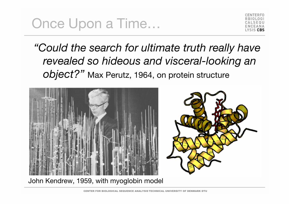
“Could the search for ultimate truth really haverevealed so hideous and visceral-looking anobject?” Max Perutz, 1964, on protein structure
John Kendrew, 1959, with myoglobin model
Once Upon a Time…

CENTER FOR BIOLOGICAL SEQUENCE ANALYSIS TECHNICAL UNIVERSITY OF DENMARK DTU
They provide a detailed picture ofinteresting biological features, such asactive site, substrate specificity, allostericregulation etc.
They aid in rational drug design and proteinengineering.
They can elucidate evolutionaryrelationships undetectable by sequencecomparisons.
Why are Protein Structures soInteresting?

CENTER FOR BIOLOGICAL SEQUENCE ANALYSIS TECHNICAL UNIVERSITY OF DENMARK DTU
In evolution structure is conserved longer thanboth function and sequence.
Structure > Function > Sequence
Structure & Evolution

CENTER FOR BIOLOGICAL SEQUENCE ANALYSIS TECHNICAL UNIVERSITY OF DENMARK DTU
Rhamnogalacturonanacetylesterase
(A. aculeatus) (1k7c)
Platelet activatingfactor acetylhydrolase
(B. Taurus) (1WAB)
Serine esterase(S. scabies) (1ESC)
Structure & Evolution

CENTER FOR BIOLOGICAL SEQUENCE ANALYSIS TECHNICAL UNIVERSITY OF DENMARK DTU
COOH
NH2
Asp His Ser Topological switchpoint
Inferring biologicalfeatures from the structure
1DEO
Structure to Function

CENTER FOR BIOLOGICAL SEQUENCE ANALYSIS TECHNICAL UNIVERSITY OF DENMARK DTU
Platelet activatingfactor acetylhydrolase
Serine esterase
Rhamnogalacturonanacetylesterase
Mølgaard, Kauppinen & Larsen (2000) Structure, 8, 373-383.
Structure & Evolution

CENTER FOR BIOLOGICAL SEQUENCE ANALYSIS TECHNICAL UNIVERSITY OF DENMARK DTU
Why Fold?
Hydrophobic collapseHydrophobic residues cluster to “escape” interactionswith water.
Indirect effect of attraction between water molecules.
Polar backbone groups form secondary structure tosatisfy hydrogen bonding donors and acceptors.Interactions withInitially formed structure is in molten globule state(ensemble).Molten globule condenses to native fold via transitionstate
CENTER FOR BIOLOGICAL SEQUENCE ANALYSIS TECHNICAL UNIVERSITY OF DENMARK DTU
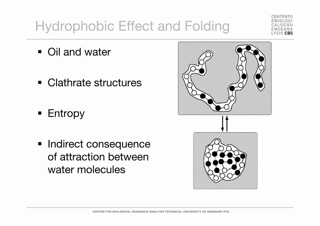
Hydrophobic Effect and Folding
Oil and water
Clathrate structures
Entropy
Indirect consequenceof attraction betweenwater molecules

CENTER FOR BIOLOGICAL SEQUENCE ANALYSIS TECHNICAL UNIVERSITY OF DENMARK DTU
Hydrophobic Core
Hydrophobic side chains go into the core ofthe molecule – but the main chain is highlypolar.The polar groups (C=O and NH) areneutralized through formation of H-bonds.
Myoglobin
Surface Interior
CENTER FOR BIOLOGICAL SEQUENCE ANALYSIS TECHNICAL UNIVERSITY OF DENMARK DTU
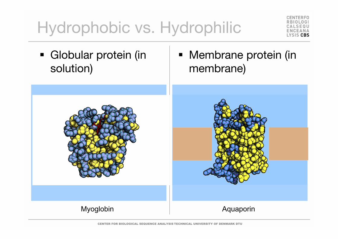
Hydrophobic vs. Hydrophilic
Globular protein (insolution)
Membrane protein (inmembrane)
Myoglobin Aquaporin

CENTER FOR BIOLOGICAL SEQUENCE ANALYSIS TECHNICAL UNIVERSITY OF DENMARK DTU
Hydrophobic vs. Hydrophilic
Globular protein (insolution)
Membrane protein (inmembrane)
Myoglobin Aquaporin
Cross-section Cross-section

CENTER FOR BIOLOGICAL SEQUENCE ANALYSIS TECHNICAL UNIVERSITY OF DENMARK DTU
Characteristics of Helices
Aligned peptideunits DipolarmomentIon/ligand bindingSecondary andquaternarystructure packingCapping residuesThe helix(i i+4)Other helix types!(310, )
N
C

CENTER FOR BIOLOGICAL SEQUENCE ANALYSIS TECHNICAL UNIVERSITY OF DENMARK DTU
-Sheets
Multiple strands sheet
Parallel vs. antiparallelTwist
FlexibilityVs. helicesFoldingStructure propagation(amyloids)Other…
Thioredoxin

CENTER FOR BIOLOGICAL SEQUENCE ANALYSIS TECHNICAL UNIVERSITY OF DENMARK DTU
-Sheets
Multiple strands sheet
Parallel vs. antiparallelTwist
FlexibilityVs. helicesFoldingStructure propagation(amyloids)Other…

CENTER FOR BIOLOGICAL SEQUENCE ANALYSIS TECHNICAL UNIVERSITY OF DENMARK DTU
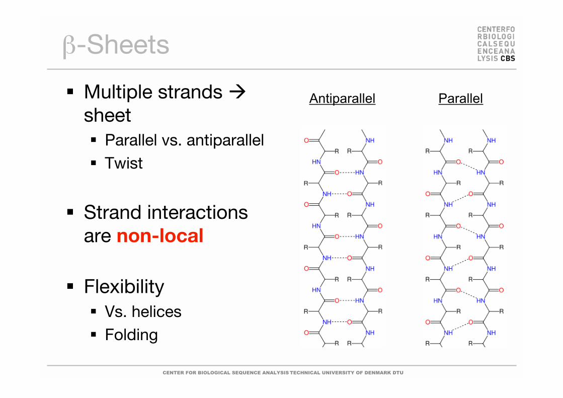
-Sheets
Multiple strands sheet
Parallel vs. antiparallelTwist
FlexibilityVs. helicesFoldingStructure propagation(amyloids)Other…

CENTER FOR BIOLOGICAL SEQUENCE ANALYSIS TECHNICAL UNIVERSITY OF DENMARK DTU
-Sheets
Multiple strands sheet
Parallel vs. antiparallelTwist
FlexibilityVs. helicesFoldingStructure propagation(amyloids)Other…
CENTER FOR BIOLOGICAL SEQUENCE ANALYSIS TECHNICAL UNIVERSITY OF DENMARK DTU
-Sheets
Multiple strands sheet
Parallel vs. antiparallelTwist
Strand interactionsare non-local
FlexibilityVs. helicesFolding
Antiparallel Parallel

CENTER FOR BIOLOGICAL SEQUENCE ANALYSIS TECHNICAL UNIVERSITY OF DENMARK DTU
Turns, Loops & Bends Revisited
Between helicesand sheets
On protein surface
Intrinsically“unstructured”proteins

CENTER FOR BIOLOGICAL SEQUENCE ANALYSIS TECHNICAL UNIVERSITY OF DENMARK DTU
Structure LevelsPrimary structure = Sequence
Secondary Structure = Helix,sheets/strands, loops & turns
Structural Motif = Small,recurrent arrangement ofsecondary structure, e.g.
Helix-loop-helixBeta hairpinsEF hand (calcium binding motif)Etc.
Tertiary structure = Arrangementof Secondary structure elements
MSSVLLGHIKKLEMGHS…

CENTER FOR BIOLOGICAL SEQUENCE ANALYSIS TECHNICAL UNIVERSITY OF DENMARK DTU
Myoglobin
Hemoglobin
Quaternary Structure
Assembly ofmonomers/subunitsinto protein complex
Backbone-backbone,backbone-side-chain &side-chain-side-chaininteractions:
Intramolecular vs.intermolecular contacts.For ligand binding sidechains may or may notcontribute. For the latter,mutations have littleeffect.

CENTER FOR BIOLOGICAL SEQUENCE ANALYSIS TECHNICAL UNIVERSITY OF DENMARK DTU
Grouping Amino Acids
Livingstone & Barton, CABIOS, 9, 745-756, 1993
A – AlaC – CysD – AspE – GluF – PheG – GlyH – HisI – IleK – LysL – Leu
M – MetN – AsnP – ProQ – GlnR – ArgS – SerT – ThrV – ValW – TrpY - Tyr

CENTER FOR BIOLOGICAL SEQUENCE ANALYSIS TECHNICAL UNIVERSITY OF DENMARK DTU
http://www.ch.cam.ac.uk/magnus/molecules/amino/

CENTER FOR BIOLOGICAL SEQUENCE ANALYSIS TECHNICAL UNIVERSITY OF DENMARK DTU
Proteins Are PolypeptidesThe peptide bond A polypeptide chain
CENTER FOR BIOLOGICAL SEQUENCE ANALYSIS TECHNICAL UNIVERSITY OF DENMARK DTU
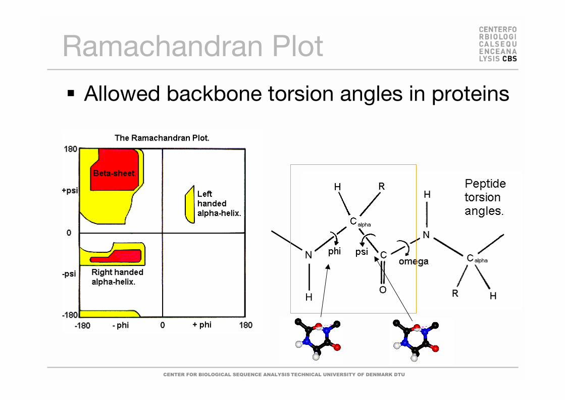
Ramachandran Plot
Allowed backbone torsion angles in proteins
N
H
CENTER FOR BIOLOGICAL SEQUENCE ANALYSIS TECHNICAL UNIVERSITY OF DENMARK DTU
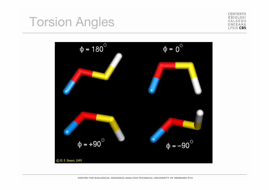
Torsion Angles

CENTER FOR BIOLOGICAL SEQUENCE ANALYSIS TECHNICAL UNIVERSITY OF DENMARK DTU
Ramachandran Plots
CENTER FOR BIOLOGICAL SEQUENCE ANALYSIS TECHNICAL UNIVERSITY OF DENMARK DTU
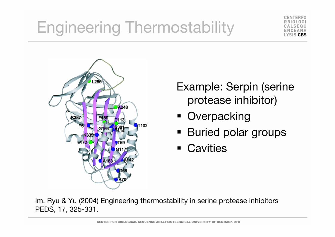
Im, Ryu & Yu (2004) Engineering thermostability in serine protease inhibitorsPEDS, 17, 325-331.
Engineering Thermostability
Example: Serpin (serineprotease inhibitor)OverpackingBuried polar groupsCavities

CENTER FOR BIOLOGICAL SEQUENCE ANALYSIS TECHNICAL UNIVERSITY OF DENMARK DTU
Experimental Methods
Crystallography&
NMR spectroscopy
CENTER FOR BIOLOGICAL SEQUENCE ANALYSIS TECHNICAL UNIVERSITY OF DENMARK DTU
X-ray crystallographyNuclear Magnetic Resonance (NMR)Modelling techniques
More exotic techniquesCryo electron microscopy (Cryo EM)Small angle X-ray scattering (SAXS)Neutron scattering
Methods for StructureDetermination

CENTER FOR BIOLOGICAL SEQUENCE ANALYSIS TECHNICAL UNIVERSITY OF DENMARK DTU
X-ray Crystallography
No size limitation.Protein molecules are ”stuck” in a crystallattice.Some proteins seem to be uncrystallizable.Slow.
Especially suited for studying structuraldetails.

CENTER FOR BIOLOGICAL SEQUENCE ANALYSIS TECHNICAL UNIVERSITY OF DENMARK DTU
X-rays
Fourier transform
CENTER FOR BIOLOGICAL SEQUENCE ANALYSIS TECHNICAL UNIVERSITY OF DENMARK DTU
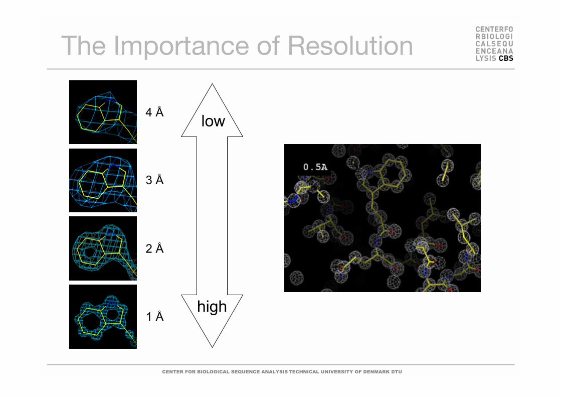
The Importance of Resolution
high
low4 Å
2 Å
3 Å
1 Å
CENTER FOR BIOLOGICAL SEQUENCE ANALYSIS TECHNICAL UNIVERSITY OF DENMARK DTU
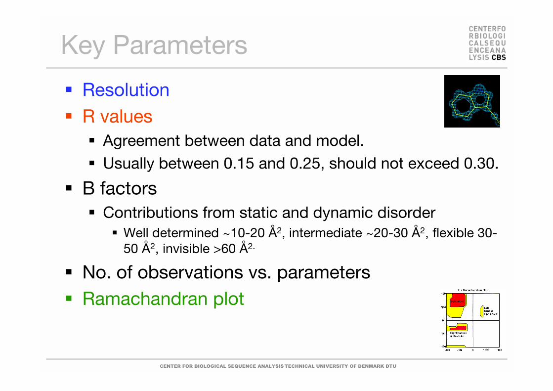
Key Parameters
ResolutionR values
Agreement between data and model.Usually between 0.15 and 0.25, should not exceed 0.30.
B factorsContributions from static and dynamic disorder
Well determined ~10-20 Å2, intermediate ~20-30 Å2, flexible 30-50 Å2, invisible >60 Å2.
No. of observations vs. parametersRamachandran plot

CENTER FOR BIOLOGICAL SEQUENCE ANALYSIS TECHNICAL UNIVERSITY OF DENMARK DTU
NMR Spectroscopy
Upper limit for structure determinationcurrently ~50 kDa.Protein molecules are in solution.Dynamics, protein folding.Slow.
Especially suited for studies of proteindynamics of small to medium size proteins.
CENTER FOR BIOLOGICAL SEQUENCE ANALYSIS TECHNICAL UNIVERSITY OF DENMARK DTU
NMR Basics
NMR is nuclear magnetic resonance
NMR spectroscopy is done on proteins INSOLUTION
Only atoms 1H, 13C, 15N (and 31P) can be detectedin NMR experiments
Proteins up to 30 kDa
Proteins stable at high concentration (0.5-1mM),preferably at room temperature

CENTER FOR BIOLOGICAL SEQUENCE ANALYSIS TECHNICAL UNIVERSITY OF DENMARK DTU
NMR Spectroscopy
CENTER FOR BIOLOGICAL SEQUENCE ANALYSIS TECHNICAL UNIVERSITY OF DENMARK DTU
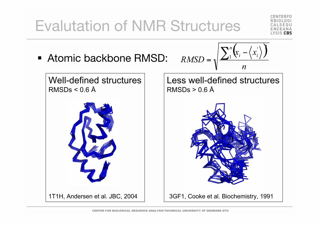
Well-defined structuresRMSDs < 0.6 Å
Evalutation of NMR Structures
Atomic backbone RMSD:
Less well-defined structuresRMSDs > 0.6 Å
3GF1, Cooke et al. Biochemistry, 19911T1H, Andersen et al. JBC, 2004
( )n
xxRMSD
n
ii
= 1
2'
CENTER FOR BIOLOGICAL SEQUENCE ANALYSIS TECHNICAL UNIVERSITY OF DENMARK DTU
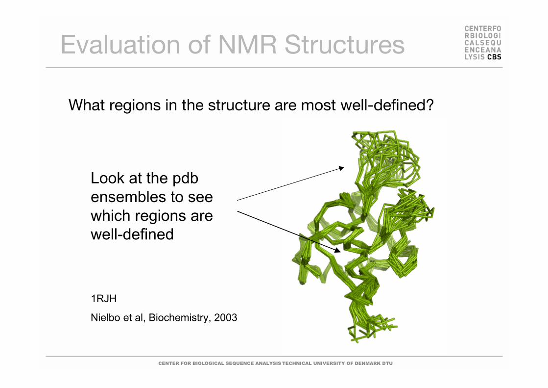
Evaluation of NMR Structures
What regions in the structure are most well-defined?
Look at the pdbensembles to seewhich regions arewell-defined
1RJH
Nielbo et al, Biochemistry, 2003

CENTER FOR BIOLOGICAL SEQUENCE ANALYSIS TECHNICAL UNIVERSITY OF DENMARK DTU
Which Structural Model?
Normally NMR structure models are listedaccording to the total energy and thenumber of violations.Model 1 in the PDB file is often the one withlowest energy and fewest violations.Use that model as template for modelling.

CENTER FOR BIOLOGICAL SEQUENCE ANALYSIS TECHNICAL UNIVERSITY OF DENMARK DTU
NMR versus X-ray Crystallography
Hydrogen atoms are observed!
Only 13C,15N and 1H are observed
Study of proteins in solution
Only proteins up to 30-40 kDa
No total “map” of the structure
Information used is incomplete and used as restraints
An ensemble of structures is submitted to PDB
The solved structure can be used for further dynamicscharacterization with NMR

CENTER FOR BIOLOGICAL SEQUENCE ANALYSIS TECHNICAL UNIVERSITY OF DENMARK DTU
Holdings of the Protein DataBank (PDB):
The PDB also containsnucleotide and nucleotideanalogue structures.
PDB
Sep. 2001 May 2006 Oct. 2007X-ray 13116 30860 39706NMR 2451 5368 6862Other 338 200 250Total 15905 36428 46818

CENTER FOR BIOLOGICAL SEQUENCE ANALYSIS TECHNICAL UNIVERSITY OF DENMARK DTU
Summary
In evolution structure is conserved longerthan both function and sequence.
X-ray crystallographyProteins in crystallatticeMany details – onemodelResolution, R-values,Ramchandran plot
NMR spectroscopyProteins in solutionFewer details – manymodelsViolations, RMSD,Ramachandran plot
CENTER FOR BIOLOGICAL SEQUENCE ANALYSIS TECHNICAL UNIVERSITY OF DENMARK DTU
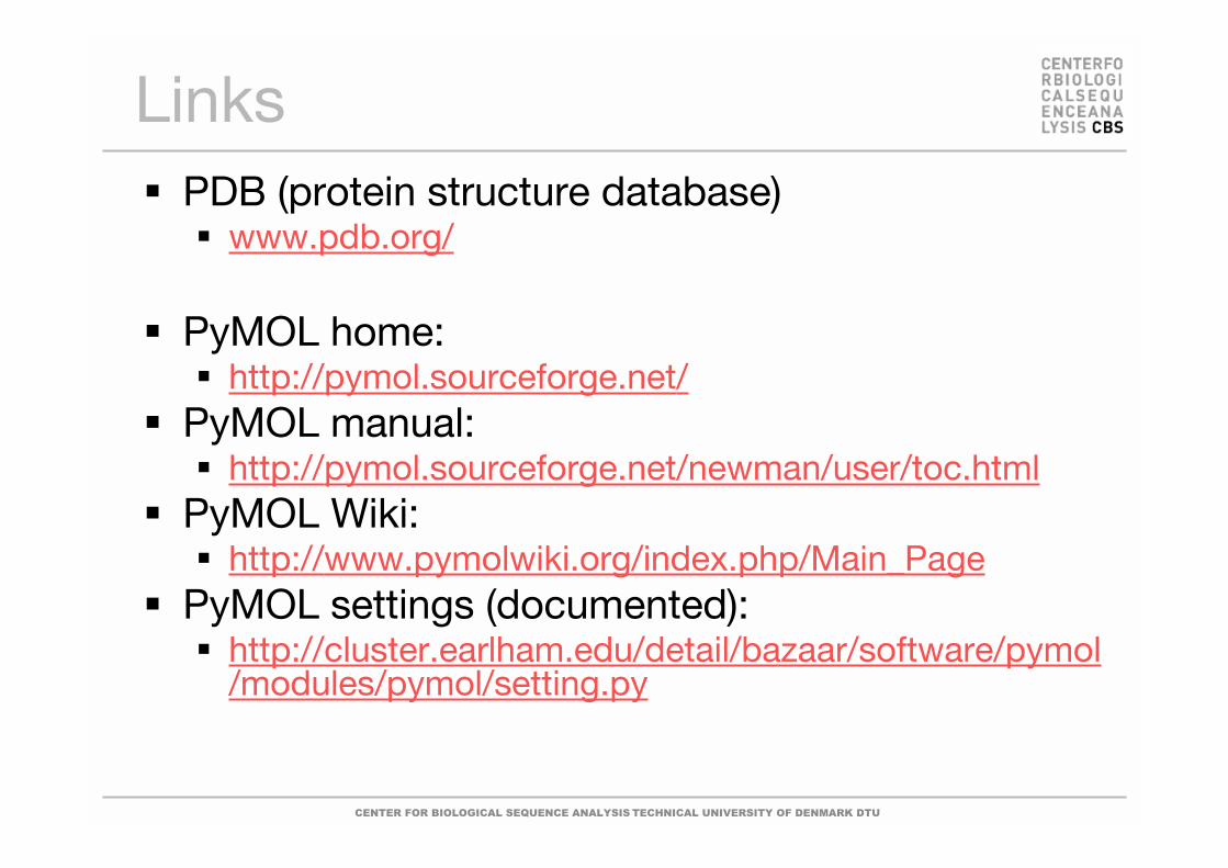
LinksPDB (protein structure database)
www.pdb.org/
PyMOL home:http://pymol.sourceforge.net/
PyMOL manual:http://pymol.sourceforge.net/newman/user/toc.html
PyMOL Wiki:http://www.pymolwiki.org/index.php/Main_Page
PyMOL settings (documented):http://cluster.earlham.edu/detail/bazaar/software/pymol/modules/pymol/setting.py
CENTER FOR BIOLOGICAL SEQUENCE ANALYSIS TECHNICAL UNIVERSITY OF DENMARK DTU
Other Courses
27617Protein Structure and Computational BiologyMaster’s level course13 weeks, spring semester5 ECTSMax. 40 students