fulltext: english, pdf
TRANSCRIPT

† Presented at the 10th Congress of the Croatian Society of Biochemistry and Molecular Biology held in Opatija, Croatia,
September 15–18, 2010. * Author to whom correspondence should be addressed. (E-mail: [email protected])
CROATICA CHEMICA ACTA CCACAA, ISSN 0011-1643, e-ISSN 1334-417X
Croat. Chem. Acta 85 (2) (2012) 201–208. http://dx.doi.org/10.5562/cca1813
Original Scientific Article
Relevance of DPP IV/CD26 among the Gut-brain Axis during Experimental Colitis†
Lara Batičić Pučar,a Dijana Detel,a Sunčica Buljević,a Ester Pernjak Pugel,b Natalia Kučić,c and Jadranka Varljena,*
aDepartment of Chemistry and Biochemistry, School of Medicine, University of Rijeka, Braće Branchetta 20, HR-51000 Rijeka, Croatia
bDepartment of Histology and Embriology, School of Medicine, University of Rijeka, Braće Branchetta 20, HR-51000 Rijeka, Croatia
cDepartment of Physiology and Immunology, School of Medicine, University of Rijeka, Braće Branchetta 20, HR-51000 Rijeka, Croatia
RECEIVED DECEMBER 10, 2010; REVISED JULY 7, 2011; ACCEPTED JULY 26, 2011
Abstract. Inflammatory bowel diseases (IBD) represent a group of chronic conditions of the gastrointestinal tract of unknown etiology. Latest knowledge accentuates the bidirectional connection between the central and enteric nervous systems. An important role of peptidases has been proposed in maintaining the homeostasis in the gut. One of them is dipeptidil-peptidase IV (DPP IV/CD26), a multifunctional glycoprotein found in both soluble and membrane-bound form in living organisms. In order to evaluate the relevance of DPP IV/CD26 among the gut-brain axis, a TNBS (Crohn-like) model of colitis has been induced in CD26 defi-cient and wild type mice. Results of this study showed that CD26 deficient mice show specificity in histolog-ical damage compared to wild type mice. A decreased DPP IV/CD26 activity was found in serum, colon and brain in wild type mice with colitis, while CD26 protein expression was increased in colon of those mice. DPP IV/CD26-like activity was decreased only in colon of CD26 deficient mice. Changes occurring during inflammatory processes in colon reflected on investigated parameters in brain. Therefore, our results indicate the importance of the gut-brain axis in the pathogenesis of IBD. (doi: 10.5562/cca1813)
Keywords: dipeptidyl-peptidase IV, CD26 molecule, CD26 deficient mice, inflammatory bowel diseases, TNBS-colitis, gut-brain axis
INTRODUCTION
Inflammatory bowel diseases (IBD) represent a group of idiopathic, chronic inflammatory conditions that could affect different segments of the gastrointestinal tract, with numerous complications and extraintestinal mani-festations.1 Two main pathomorphological entities of IBD are Crohn's disease (CD) and ulcerative colitis (UC), sharing some similarities, but also having indi-vidual characteristics in disease manifestations. The etiology of IBD, despite of many investigations, still remains unknown.2 It is generally accepted that IBD occurs in genetically susceptible individuals, under influence of environmental and microbiological fac- tors, as an overexpressed immunological response to antigens of unknown origin.3 Latest knowledge regarding IBD pathogenesis includes the influence of neuroimmunological parameters, which are driven
by the hypothesis that neurogenic inflammation could be involved in inflammatory processes in the gut.4 Given the connection of enteric and central nervous system and the bidirectional communication between the gut and the brain, a term "gut-brain-axis" has been introduced and proposed as an important aspect in the etiology of IBD.5
In order to elucidate the pathogenesis of IBD, dif-ferent animal models of gastrointestinal inflammation have been developed.6 One of the most accepted exper-imental model of Crohn-like-colitis is the trinitrobenzenesulfonic acid-induced (TNBS) colitis, which resembles many histological, immunological and clinical manifestations of CD in humans.7 Animal mod-els of IBD have given insight in different processes at the molecular level which lead to development of in-flammatory changes and have revealed the importance of many molecules involved in IBD etiology.8

202 L. Batičić Pučar et al., DPP IV/CD26 in Experimental Colitis
Croat. Chem. Acta 85 (2012) 201.
In the last decade, proteases have been proposed as important factors in inflammatory processes, given their ability to metabolize different biologically active substrates involved in maintaining the integrity of the mucosal barrier.9 One of them is dipeptidyl-peptidase IV, known also as CD26 molecule (DPP IV/CD26). DPP IV/CD26 is a highly glycosylated type II transmembrane sialoglycoprotein comprising two iden-tical subunits of approximately 110 kDa.10 DPP IV/CD26 was originally characterized as a T-cell differ-entiation antigen and was reported to be expressed on various cell types, having multiple functions in various biological processes, including immunological regula-tions.11 A soluble form circulating in body fluids is found in living organisms. As a serine protease, it pos-sesses a specific peptidase function with unique features in substrate processing: it cleaves dipeptides from the N-terminus of polypeptides where proline or alanine is at the penultimate position. Since N-termini containing Xaa-Pro are not easily cleaved by other proteases, the action of DPP IV/CD26 is a rate-limiting step in the degradation of many polypeptides.12
Different cytokines, chemokines and neuropep-tides involved in inflammatory events are found among DPP IV/CD26 substrates.13 Our hypothesis was that CD26 deficient mice would express differences in in-flammatory processes and disease manifestations. This study was undertaken in order to have an insight in histological and biochemical alterations at local and systemic levels during development and resolution of colitis under conditions of DPP IV/CD26 deficiency in comparison with wild type mice. Furthermore, we wanted to evaluate changes in DPP IV/CD26 and DPP IV/CD26-like activity at local and systemic levels, likewise DPP IV/CD26 protein expression in targeted tissues during colitis development and resolution. Given the potential importance of the gut-brain axis in the development of inflammatory processes, experiments were focused on changes occurring at local site of in-flammation - in colon, and in the central nervous system - in brain.
EXPERIMENTAL
Experimental Animals
This study was performed using two mice strains: wild type mice strain C57BL/6 and mice with inactivated gene for molecule CD26 (C57BL/6 Jbom-ob, CD26–/–), generated on a C57BL/6 genetic background. CD26 deficient mice were kindly provided by Dr. Didier Marguet, Centre d’Immunologie Marseille-Luminy, France. Generation of CD26 deficient mice has been described previously.14 Male, 8–10 week old mice were used in this study. Animals were housed and bred under
standard conditions at the Central Animal Facility of the School of Medicine, University of Rijeka. Laboratory animals were housed in plastic cages, fed with standard pellet food (MK, Complete Diet for Laboratory Rats and Mice, Slovenia) and given tap water ad libitum. They were maintained under a 12/12 hours dark/light cycle at constant temperature (20 ± 1) °C and humidity (50 ± 5) %. Each study group comprised 8–10 animals. Animal handling, experimental procedures and anesthe-sia were performed in accordance with the general prin-ciples contained in the Guide for the Care and Use of Laboratory Animals (National Academic Press). The Ethical Committee of the School of Medicine, Universi-ty of Rijeka, approved all experimental procedures. Colitis Induction
Crohn-like-colitis (TNBS-colitis) was induced by rectal administration of 5 % (mass concentration) 2,4,6-tri- nitrobenzenesulfonic acid solution (TNBS, Sigma-Aldrich, Germany) dissolved in 50 % ethanol (Kemika, Croatia). Each animal received 0.1 mL of TNBS-ethanol solution, using a vinyl catheter that was posi-tioned 4 cm from the anus, as described previously.7 Two control groups of mice were used for each mice strain. Control mice underwent identical procedures, but were instilled equal volumes of saline (NaCl, γ = 0.9 %) or ethanol solution. Administration of TNBS, saline or ethanol solution was performed in mice anesthetized with ketamine/xylazine. Experimental Procedures
Animals were sacrificed by cervical dislocation after 2, 7, 15 and 30 days upon administration of TNBS, saline or ethanol solution. Peripheral blood samples were tak-en and serum samples were collected by centrifugation at 3000 rpm for 10 minutes. Colons were freed from adhering tissue and macroscopic changes were noted. The colon lumen was carefully washed with ice-cold saline and its weights and lengths was measured. One part of the colon was used for histological analyses while the remaining segments of the colon were slit open longitudinally and the mucosa was scraped using a glass microscope slide. Colonic mucosal samples were prepared from obtained mucosal scrapings according to Ahnen et al.15 Obtained mucosal aliquot samples were stored at –80 °C until further analyses.
Brains were separated immediately after sacrifice, washed in ice-cold saline and frozen in liquid nitrogen then stored at –80 °C until further analyses. Brains were homogenized on ice in a volume of 0.4 mL of 0.1 M TRIS-HCl buffer (pH = 8). Homogenates were then centrifuged at 14000 rpm for 20 minutes at +4 °C. Result-ing supernatants were measured for total protein concen-trations according to the method of Bradford et al.16

L. Batičić Pučar et al., DPP IV/CD26 in Experimental Colitis 203
Croat. Chem. Acta 85 (2012) 201.
Histological Analyses
Colon tissues were collected and fixed in 4 % formalin for 24 h. Samples were processed and embedded in paraffin wax. Two-micrometer sections were stained with hematoxylin and eosin for histological analyses. An experienced pathologist blinded to treatment alloca-tion scored microscopical changes, including overall severity of damage, number of crypts of Lieberkühn and their depth and width. Microscopic analysis was carried out using an Olympus BX40 microscope (Tokyo, Japan) and the Pulnix TMC 76S digital camera (Tokyo, Japan). Analyses of digital images were performed using Issa software package (VAMS, Zagreb, Croatia). DPP IV/CD26 Activity Assay
The DPP IV/CD26 (in C57BL/6) and DPP IV/CD26-like enzymatic activities (in CD26 deficient mice) in serum, brain and colon homogenates were measured according to the protocol of Kreiser et al.17 by detection of free 4-nitroaniline in an assay mixture containing 0.1 M Tris-HCl buffer (pH = 8), and 2 · 10–3 M Gly-Pro p-nitroanilide as the substrate (Sigma-Aldrich, Germa-ny) in a total volume of 0.20 mL. The reaction was stopped by addition of 0.80 mL of sodium acetate buffer (1 mol dm–3, pH = 4.5) after 30 minutes of incubation at 37 °C. The amounts of hydrolyzed substrate were meas-ured in duplicates spectrophotometrically at λ = 450 nm using a Varian Cary UV/Vis Spectrophotometer. En-zyme activities are expressed according to the Nomen-clature Committee of the International Union of Bio-chemistry. Serum DPP IV/CD26 and DPP IV/CD26-like enzyme activities are expressed as amount of sub-strate hydrolyzed per liter of serum (nkat L–1). DPP IV/CD26 and DPP IV/CD26-like enzyme activities in brain and colon homogenates are expressed as the amount of substrate hydrolyzed per mg of protein (nkat mg–1 of protein). CD26 Western Blot Analysis
Brain and colon samples for Western blot analyses of CD26 protein expression were homogenized on ice using RIPA lysis buffer including inhibitors of proteases and phosphatases (Santa Cruz Biotechnology Inc., CA). Homogenates were then centrifuged at 14000 rpm for 20 minutes at +4 °C. Resulting supernatants were meas-ured for total protein concentrations according to the method of Bradford et al.16 All samples were heated for 5 min at 95 °C in a sample buffer. Prestained molecular weight markers (Kaleidoscope Prestained Standards, BIO-RAD and Fermentas Life Sciences PageRulerTM) were used as standards. Equal amounts of total proteins (50 μg per lane) were separated by sodium dodecyl sul-phate-polyacrylamide gel electrophoresis on 10 % gels. Samples were electrophorezed at 50 V for 6 h on ice.
Proteins were transferred from the polyacrylamide gels to polyvinylidenedifluoride membranes by electroblotting in a semi-dry transfer apparatus at 0.22 mA for 45 min. Membranes were blocked for 2 hours at 4 °C in phosphate-buffered saline containing 0.05 % Tween 20 (PBS-T, Amersham Biosciences, USA), con-taining 5 % (mass concentration) nonfat milk powder (Santa Cruz Biotechnology Inc., CA). Membranes were treated overnight with primary anti-CD26 antibody (Santa Cruz Biotechnology Inc., CA, 1:200 in blocking buffer). Blots were washed three times for 15 min in PBS-T. As a secondary antibody, horseradish peroxi-dase-conjugated mouse-anti-rabbit IgG in a dilution of 1 : 2000 (Santa Cruz Biotechnology Inc., CA) was used. After the second washing procedure, CD26 molecule (110 kDa) was detected by chemiluminescent Amer-sham ECL-plus Western blotting detection reagents (Amersham, Little Chalfont, UK), which enabled visu-alization of bends after exposure to photosensitive films (AGFA Ortho CP-G plus). Equal total protein loading was ensured with use of the primary mouse ß-actin antibody (Chemicon International, USA), in a dilution of 1 : 40000, and secondary horseradish peroxidase-conjugated goat-anti-mouse IgG in a dilution of 1 : 2000 (Santa Cruz Biotechnology Inc., CA). Statistical Analyses
Results are expressed as mean ± standard deviation. Statistical comparisons were made using STATISTICA version 8.0 (StatSoft Inc., TULSA, USA). Testing dif-ferences between groups has been performed using ANOVA and post-hoc Scheffe test. The level of P<0.05 was defined as statistically significant.
RESULTS AND DISCUSSION
TNBS-induced colitis in mice is an experimental model of IBD which shares many similarities with CD in hu-mans at molecular levels and in clinical manifestations of disease and therefore represents a valuable tool in IBD research.7 Macroscopic signs of inflammation char-acteristic for TNBS-induced colitis comprise marked oedema and mucosal thickening, which are accompa-nied by increased colon weight.18 Results of our study are in agreement with these observations. The presence of an accentuated oedema in the colon was determined, with marked inflammatory changes that were most conspicuous the second day following TNBS-ethanol administration. Therefore, the second day after colitis induction was classified as acute phase of colitis, which is in agreement with previously reported findings.7 Shortening and thickening of the colon was also ob-served in mice which received TNBS-ethanol solution. Intrarectal application of TNBS-ethanol solution in mice
204 L. Batičić Pučar et al., DPP IV/CD26 in Experimental Colitis
Croat. Chem. Acta 85 (2012) 201.
induces a transmural, granulomatous inflammation with infiltration of inflammatory cells mainly located in the distal part of the colon.7 Histopathological changes observed in colon tissue sections of both wild type and CD26 deficient mice confirmed the presence of inflam-matory processes and accomplishment of colitis induc-tion. It should be emphasized that to date, no TNBS-induced colitis in CD26 deficient mice was described in available literature to our knowledge, and scarce results are published regarding investigation using this mice strain.
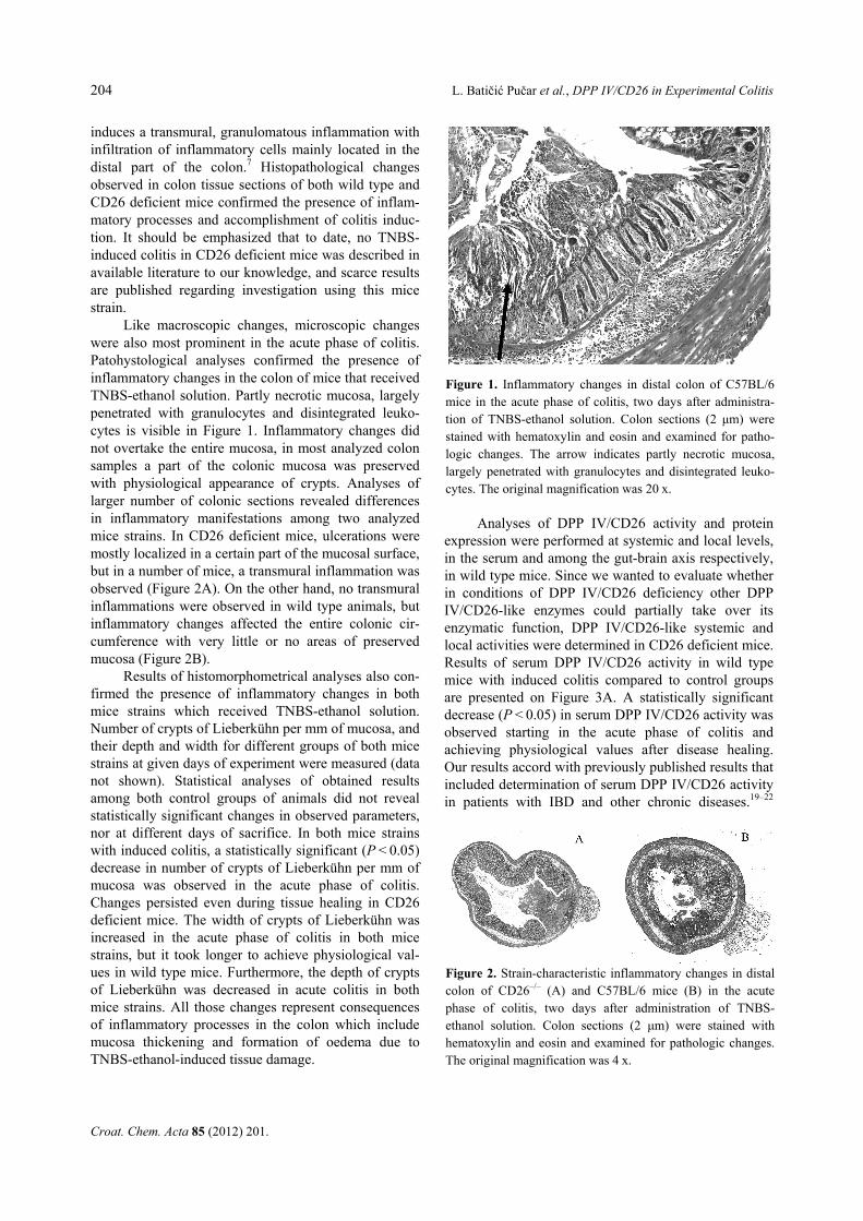
Like macroscopic changes, microscopic changes were also most prominent in the acute phase of colitis. Patohystological analyses confirmed the presence of inflammatory changes in the colon of mice that received TNBS-ethanol solution. Partly necrotic mucosa, largely penetrated with granulocytes and disintegrated leuko-cytes is visible in Figure 1. Inflammatory changes did not overtake the entire mucosa, in most analyzed colon samples a part of the colonic mucosa was preserved with physiological appearance of crypts. Analyses of larger number of colonic sections revealed differences in inflammatory manifestations among two analyzed mice strains. In CD26 deficient mice, ulcerations were mostly localized in a certain part of the mucosal surface, but in a number of mice, a transmural inflammation was observed (Figure 2A). On the other hand, no transmural inflammations were observed in wild type animals, but inflammatory changes affected the entire colonic cir-cumference with very little or no areas of preserved mucosa (Figure 2B).
Results of histomorphometrical analyses also con-firmed the presence of inflammatory changes in both mice strains which received TNBS-ethanol solution. Number of crypts of Lieberkühn per mm of mucosa, and their depth and width for different groups of both mice strains at given days of experiment were measured (data not shown). Statistical analyses of obtained results among both control groups of animals did not reveal statistically significant changes in observed parameters, nor at different days of sacrifice. In both mice strains with induced colitis, a statistically significant (P < 0.05) decrease in number of crypts of Lieberkühn per mm of mucosa was observed in the acute phase of colitis. Changes persisted even during tissue healing in CD26 deficient mice. The width of crypts of Lieberkühn was increased in the acute phase of colitis in both mice strains, but it took longer to achieve physiological val-ues in wild type mice. Furthermore, the depth of crypts of Lieberkühn was decreased in acute colitis in both mice strains. All those changes represent consequences of inflammatory processes in the colon which include mucosa thickening and formation of oedema due to TNBS-ethanol-induced tissue damage.
Analyses of DPP IV/CD26 activity and protein expression were performed at systemic and local levels, in the serum and among the gut-brain axis respectively, in wild type mice. Since we wanted to evaluate whether in conditions of DPP IV/CD26 deficiency other DPP IV/CD26-like enzymes could partially take over its enzymatic function, DPP IV/CD26-like systemic and local activities were determined in CD26 deficient mice. Results of serum DPP IV/CD26 activity in wild type mice with induced colitis compared to control groups are presented on Figure 3A. A statistically significant decrease (P < 0.05) in serum DPP IV/CD26 activity was observed starting in the acute phase of colitis and achieving physiological values after disease healing. Our results accord with previously published results that included determination of serum DPP IV/CD26 activity in patients with IBD and other chronic diseases.19–22
Figure 2. Strain-characteristic inflammatory changes in distalcolon of CD26–/– (A) and C57BL/6 mice (B) in the acutephase of colitis, two days after administration of TNBS-ethanol solution. Colon sections (2 μm) were stained withhematoxylin and eosin and examined for pathologic changes.The original magnification was 4 x.
Figure 1. Inflammatory changes in distal colon of C57BL/6mice in the acute phase of colitis, two days after administra-tion of TNBS-ethanol solution. Colon sections (2 μm) werestained with hematoxylin and eosin and examined for patho-logic changes. The arrow indicates partly necrotic mucosa,largely penetrated with granulocytes and disintegrated leuko-cytes. The original magnification was 20 x.

L. Batičić Pučar et al., DPP IV/CD26 in Experimental Colitis 205
Croat. Chem. Acta 85 (2012) 201.
Therefore, a clinical relevance of serum DPP IV/CD26 activity as a marker of disease severity in IBD was pro-posed.21 Its role in inflammatory events includes degra-dation of biologically active substrates which have a crucial role in inflammation. Our results are in accord-ance with the observation that serum DPP IV/CD26 activity correlates inversely with disease severity in patients with IBD.21
In order to evaluate changes in serum DPP IV/CD26-like activities during colitis development and healing, CD26 deficient mice were analyzed. Results of our study show that CD26 deficient mice express ap-proximately 10 % of total serum DPP IV/CD26 activity detected in wild type mice. Figure 3B shows results of serum DPP IV/CD26-like activity in CD26 deficient mice with induced colitis compared to their control groups. It could be seen that no statistically significantly differences were found in serum DPP IV/CD26-like activity between groups of CD26 deficient animals with colitis and control animals. Therefore, the significance of DPP IV/CD26 over DPP IV/CD26-like serum activi-ty is proposed.
In the last decade, growing scientific evidence emphasizes neuroimmunomodulation as an important factor in the occurrence of inflammation.23 Due to the causal connection between central and enteric nervous system, the term gut-brain axis has been proposed.5 DPP IV/CD26 has previously been proven to play an im-portant role in metabolism of important bioactive neuro- and immunopeptides, as well as in the costimulation of immune cells.11 Given its localization in the nervous system and among the digestive tract, likewise on the surface of important inflammatory cells,24 our aim was to investigate changes in DPP IV/CD26 activity and protein expression at sight of inflammation and reveal if those changes reflect on examined parameters in the brain.
Figure 3. Serum DPP IV/CD26 activity in C57BL/6 mice (A)and serum DPP IV/CD26-like activity in CD26–/– mice (B) during colitis development and resolution compared to controlgroup. Statistically significantly different values (a) compared to control group (P < 0.05). 0 – control group, physiological condition; 2, 7, 15, 30 days after administration of TNBS-ethanol solution (colitis group) or ethanol solution (controlgroup).
Figure 4. DPP IV/CD26 activity in colon of C57BL/6 mice(A) and DPP IV/CD26-like activity in colon of CD26–/– mice(C). CD26 protein expression in colon of C57BL/6 mice dur-ing colitis development and resolution compared to controlgroup (B). Statistically significantly different values (a) com-pared to control group (P < 0.05). 0 – control group, physio-logical condition; 2, 7, 15, 30 days after administration ofTNBS-ethanol solution (colitis group) or ethanol solution(control group).

206 L. Batičić Pučar et al., DPP IV/CD26 in Experimental Colitis
Croat. Chem. Acta 85 (2012) 201.
Our results showed an accentuated decrease in DPP IV/CD26 activity in the inflamed colon in wild type animals compared to their control groups (Figure 4A). On the other hand, Western blotting technique revealed an increased CD26 protein expression in the acute phase of disease, as shown on Figure 4B. This phenomenon could, besides a regulatory mechanism at the enzymatic level, also be partially explained as a compensatory mechanism, since a part of the decreased DPP IV/CD26 activity in the colon of wild type mice is a consequence of severe mucosal damage induced by TNBS-ethanol application. Therefore, enhanced CD26 protein expression in the acute phase of disease could represent an attempt to accomplish a compensatory role due to damaged DPP IV/CD26 conformation and im-proper enzyme activity in inflamed tissue. An enhanced DPP IV/CD26 mRNA production has previously been shown under inflammatory environment.25 Consequent-ly, a higher CD26 protein expression could be expected in inflammation. Our results are in agreement with pre-viously reported observations regarding enhanced CD26 protein production in inflamed tissue.
Investigations on DPP IV/CD26-like activity in the colon of CD26 deficient mice shown that, in physio-logical conditions, CD26 deficient mice express less than 2 % of total DPP IV/CD26 activity detected in the colon of wild type mice. Statistically significantly (P < 0.05) decreased DPP IV/CD26 activity in inflamed colon homogenates was also found in CD26 deficient mice compared to their controls (Figure 4C). However, this could also partially be a consequence of tissue dam-age and not only an intrinsic regulation mechanism which downregulates the activity of DPP IV/CD26-like enzymes in inflammatory processes.
Interesting results were found when analyzing DPP IV/CD26 activity in brain homogenates during colitis development and resolution in wild type mice. It was found that DPP IV/CD26 activity in brain is statis-tically significantly decreased (P < 0.05) in the acute phase of colitis compared to control groups (Figure 5A), while the activity of DPP IV/CD26-like enzymes does not change (Figure 5C). On the other hand, CD26 pro-tein expression, as confirmed by Western blot technique (Figure 5B) remains constant. A possible explanation of this phenomenon could be a regulatory mechanism which control DPP IV/CD26 activity in brain, inde-pendently of its protein expression. Likewise, since there is no local inflammation in the brain which could destroy DPP IV/CD26 enzymatic function like in the inflamed colon, there is no need for a potential compen-satory mechanism which would try to compensate lower DPP IV/CD26 activity by its higher protein expression. Therefore, lower DPP IV/CD26 activity and its un-changed protein expression in the brain most probably represents an intrinsic regulatory mechanism at the enzymatic level.
Moreover, results of this study indicate that changes which occur during inflammatory processes at local level, in the colon, exhibit a reflection on investi-gated parameters in the central nervous system. There-fore, a decreased DPP IV/CD26 activity in the brain is most probably causally connected with its changes in the colon during inflammatory events. Hence, our re-sults indicate the importance of the gut-brain axis in the pathogenesis of IBD. This study reveals new data about DPP IV/CD26 activity and protein expression in a mod-el of Crohn-like colitis in mice. Likewise, due to very little available results of colitis investigation under con-ditions of CD26 deficiency, our study gives new in-sights in inflammatory manifestations induced by TNBS-ethanol administration in CD26 deficient mice.
Figure 5. DPP IV/CD26 activity in brain of C57BL/6 mice(A) and DPP IV/CD26-like activity in brain of CD26-/- mice(C). CD26 protein expression in brain of C57BL/6 mice dur-ing colitis development and resolution compared to controlgroup (B). Statistically significantly different values (a) com-pared to control group (P < 0.05). 0 – control group, physio-logical condition; 2, 7, 15, 30 days after administration ofTNBS-ethanol solution (colitis group) or ethanol solution(control group).

L. Batičić Pučar et al., DPP IV/CD26 in Experimental Colitis 207
Croat. Chem. Acta 85 (2012) 201.
Inhibition of DPP IV/CD26 has been proposed as potential therapy for chronic inflammatory diseases.26 This hypothesis is supported by higher values of anti-inflammatory cytokines obtained in CD26 deficient mice.27 Furthermore, it has recently been shown that inhibition of DPP IV/CD26 partially ameliorates colitis in mice.28 IBD is characterized by imbalances in innate and acquired immune response, where two dysregulated T cell subsets are crucial: activated effector T cells and regulatory T cells. They are characterized by a strong expression of DPP IV/CD26, which has been confirmed to play a significant role in the control of immune acti-vation and in regulating cellular communication by hydrolyzing bioactive polypeptides.29 Positive results in the regulation of immune and non-immune processes in IBD have been proved in vitro as well in vivo with DPP IV/CD26 and alanyl-aminopeptidase inhibitors.30
Nevertheless, results of our study showed that CD26 deficient mice are not protected from chemically induced Crohn-like colitis, but exhibit some differences in histological damage compared to wild type mice. Otherwise, CD26 deficient mice show a normal pheno-type and behavior as wild type mice, but are known to posses and enhanced insulin secretion and improved glucose tolerance.14 This knowledge lead to develop-ment of DPP IV/CD26 inhibitors in the therapy of dia-betes mellitus type II which are already in clinical use.31–33 However, further studies should be done in order to test if DPP IV/CD26 inhibition could be an acceptable therapeutic approach in chronic inflammato-ry disease alleviation, as new drug candidates for the treatment of diseases associated with an imbalanced T cell response. CONCLUSIONS
This study reveals new data about DPP IV/CD26 activi-ty and protein expression in a model of TNBS (Crohn-like) colitis in mice. Due to meager available results of colitis investigation under conditions of CD26 deficien-cy, we established a TNBS-ethanol induced colitis in CD26 deficient mice. Our study gives new insights in Crohn-like colitis inflammatory manifestations in CD26 deficient mice. Results of our study showed that CD26 deficient mice are not protected from chemically in-duced Crohn-like colitis, but show specificity in histo-logical damage compared to wild type mice. A de-creased DPP IV/CD26 activity was found in serum, colon and brain in C57BL/6 mice with colitis, while DPP IV/CD26-like activity is decreased only in colon of CD26 deficient mice. An enhanced protein expression of CD26 molecule was found in colon of wild type mice in the acute phase of colitis. It was noticed that changes occurring during inflammatory processes in the colon, reflect on investigated parameters in the central nervous
system. Therefore, our results indicate the importance of the gut-brain axis in the pathogenesis of IBD.
Acknowledgements. This study was supported by the Croa-tian Ministry of Science, Education and Sports (grant No. 062-0061245-0213). We gratefully acknowledge Dr. Didier Marguet (Centre d’Immunologie Marseille-Luminy, France), for providing CD26 deficient mice. Many thanks to professor Siniša Volarević, PhD, head of the department of Molecular Medicine and Biotechnology and professor Stipan Jonjić, PhD, head of the department of Histology and Embriology, School of Medicine, University of Rijeka, for allowing us to complete a part of experiments using the equipment at their departments. REFERENCES
1. S. B. Hanauer and D. W. Hommes, Expert Rev. Clin. Immunol. 6 (2010) 499–500.
2. D. Q. Shih and S. R. Targan, Curr Gastroenterol Rep. 11 (2009) 473–480.
3. R. S. Blumberg, Dig. Dis. 27 (2009) 455–464. 4. Y. Takami, C. R. Mantyh, T. N. Pappas, T. Takahashi, K. Koda,
and M. Miyazaki, Hepatogastroenterology 56 (2009) 682–686. 5. J. A. Romijn, E. P. Corssmit, L. M. Havekes, and H. Pijl, Curr.
Opin. Clin. Nutr. Metab. Care 11 (2008) 518–521. 6. H. H. Uhlig and F. Powrie, Eur. J. Immunol. 39 (2009) 2021–
2026. 7. F. Scheiffele and I. J. Fuss, Current Protocols in Immunology
2002, Chapter 15, Unit 15.19. 8. S. Wirtz and M. F. Neurath, Adv. Drug Deliv. Rev. 59 (2007)
1073–1083. 9. A. Ravi, P. Garg, and S. V. Sitaraman, Inflamm. Bowel. Dis. 13
(2007) 97–107. 10. M. D. Gorrell, X. M. Wang, J. Park, K. Ajami, D. M. Yu, H.
Knott, D. Seth, and G. W. McCaughan Adv. Exp. Med. Biol. 575 (2006) 45–54.
11. M. Vanderheyden, J. Bartunek, M. Goethals, S. Verstreken, A. M. Lambeir, I. De Meester, and S. Scharpe, Clin Chem Lab Med 47 (2009) 248–252.
12. M. D. Gorrell, Clin. Sci. 108 (2005) 277–292. 13. R. Mentlein, Regul. Pept. 85 (1999) 9–24. 14. D. Marguet, L. Baggio, T. Kobayashi, A. M. Bernard, M.
Pierres, P. F. Nielsen, U. Ribel, T. Watanabe, D. J. Drucker, and N. Wagtmann, Proc. Natl. Acad. Sci. U. S. A. 97 (2000) 6874–6879.
15. D. J. Ahnen, N. A. Santiago, J. P. Cezard, and G. M. Gray, J. Biol. Chem. 257 (1982) 12129–12135.
16. M. M. Bradford, Anal. Biochem. 72 (1976) 248–254. 17. W. Kreisel, R. Heussner, B. Volk, R. Buchsel, W. Reutter, and
W. Gerok, FEBS Lett. 147 (1982) 85–88. 18. L. Camoglio, A. A. te Velde, A. de Boer, F. J. ten Kate, M. Kopf,
and S. J. van Deventer, Eur. J. Immunol. 30 (2000) 1486–1495. 19. M. Hildebrandt, M. Rose, J. Ruter, A. Salama, H. Monnikes, and
B. F. Klapp, Scand. J. Gastroenterol. 36 (2001) 1067–1072. 20. H. Kobayashi, O. Hosono, T. Mimori, H. Kawasaki, N. H. Dang,
H. Tanaka, and C. J Morimoto, Rheumatol. 29 (2002) 1858–1866.
21. J. Varljen, B. Mijandrušić Sinčić, L. Batičić, N. Varljen, D. Detel, and A. Lekić, Croat. Chem. Acta 78 (2005) 427–432.
22. N. Busso, N. Wagtmann, C. Herling, V. Chobaz-Peclat, A. Bischof-Delaloye, A. So, and E. Grouzmann, Am. J. Pathol. 166 (2005) 433–442.

208 L. Batičić Pučar et al., DPP IV/CD26 in Experimental Colitis
Croat. Chem. Acta 85 (2012) 201.
23. L. Ohman and M. Simren, Nat. Rev. Gastroenterol. Hepatol. 7 (2010) 163–173.
24. E. Matteucci and O. Giampietro, Curr. Med. Chem. 16 (2009) 2943–2951.
25. E. Nemoto, S. Sugawara, H. Takada, S. Shoji, and H. Horiuch, Infect. Immun. 67 (1999) 6225–6233.
26. U. Aytac and N. H. Dang, Curr. Drug Targets: Immune, Endocr. Metab. Disord. 4 (2004) 11–18.
27. M. S. Geier, D. Tenikoff, R. Yazbeck, G. W. McCaughan, C. A. Abbott, and G. S. Howarth, J. Cell Physiol. 204 (2005) 687–692.
28. R. Yazbeck, G. S. Howarth, M. S. Geier, H. U. Demuth, and C. A. Abbott, Front. Biosci. 13 (2008) 6850–6858.
29. S. Ansorge, U. Bank, A. Heimburg, M. Helmuth, G. Koch, J. Tadje, U. Lendeckel, C. Wolke, K. Neubert, J. Faust, P. Fuchs, D. Reinhold, A. Thielitz, and M. Tager, Clin. Chem. Lab. Med. 47 (2009) 253–261.
30. U. Bank, U. R. Bohr, D. Reinhold, U. Lendeckel, S. Ansorge, P. Malfertheiner, and M. Tager, Front. Biosci. 13 (2008) 3699–3713.
31. I. De Meester, S. Scharpe, and A. M. Lambeir, Clin. Chem. Lab. Med. 47 (2009) 245–247.
32. D. M. Riche, H. E. East, and K. D. Riche, Am. J. Med. Sci. 337 (2009) 321–328.
33. N. I. Siddiqui, Mymensingh Med. J. 18 (2009) 113–124.