from postmortem homogeneous cells to microarray … postmortem homogeneous cells to microarray...
TRANSCRIPT

APPLICATION NOTE Applied Biosystems ArcturusXT System
From postmortem homogeneous cells to microarray analysis: an optimized protocol
Materials• 4.5 inch forceps (VWR Cat. No. 82027-392)• ArcturusXT Microdissection System (Cat. No. ARCTURUSXT)• Applied Biosystems™ Turbo Labeling™ Kit with Biotin
(Cat. No. KIT0608)• Applied Biosystems™ CapSure™ HS LCM Caps
(Cat. No. LCM0214)• Applied Biosystems™ CapSure™ Macro LCM Caps
(Cat. No. LCM0211)• Experion™ Automated Electrophoresis Station
(Bio-Rad Cat. No. 7007001)• Experion™ RNA HighSens Chips, 10 chips
(Bio-Rad Cat. No. 7007155)• Experion™ RNA HighSens Reagents, 10 chips
(Bio-Rad Cat. No. 7007156)• Experion™ RNA StdSens Chips, 10 chips
(Bio-Rad Cat. No. 7007153)• Experion™ RNA StdSens Reagents, 10 chips
(Bio-Rad Cat. No. 7007154)• Falcon™ Polypropylene Conical Tube (Becton-Dickinson Labware
Cat. No. 352070)• Applied Biosystems™ GeneAmp™ Thin-Walled Reaction Tube with
Domed Cap (Cat. No. N8010611)• GeneChip™ Human X3P Array, 6 chips
(Affymetrix Cat. No. 900516)• Applied Biosystems™ HistoGene™ LCM Frozen Section Staining
Kit (Cat. No. KIT0401)• Micro Slide Box (VWR Cat. No. 48444-004)• Thermo Scientifi c™ Microm™ HM 505E Cryostat
or equivalent• Microprocessor Controlled 280 Series Water Bath Model 282
(Thermo Electron Corporation Cat. No. 51221048)• Molecular Sieves, Type A (EMD Cat. No. MX1583L-1)
(Continued on next page)
IntroductionWe describe a process for using the Applied Biosystems™
ArcturusXT™ Microdissection System and the Applied Biosystems™ PicoPure™ RNA Isolation Kit to obtain mRNA of good quality and quantity from pyramidal neurons obtained from postmortem human brain tissue for microarray studies. We also provide an overview of considerations that should be taken into account during the preparation of samples for laser capture microdissection (LCM) and include a few modifi cations to the company-provided protocols to further preserve RNA integrity and to obtain maximum mRNA yield.
It is well known that the human cerebral cortex is an exceptionally heterogeneous structure that is comprised of diverse regions, layers, and cell types, each of which is characterized by distinct cellular and molecular compositions. LCM enables us to circumvent confounding effects from surrounding structures and thereby obtain gene expression profi les specifi c to a homogeneous cell population. Microarray gene expression profi ling also has innate advantages, such as enabling the user to discover unanticipated relationships normally overlooked in hypothesis-driven experiments. This wealth of data allows for the formation of new hypotheses relating to disease states [1]. However, extenuating factors often affect the integrity of postmortem tissue, making microarray studies from single cell populations diffi cult. These extenuating factors include disease treatment effects, co-morbidity, and limited sample sizes. In addition, postmortem interval, tissue pH, and other tissue-processing effects are factors that can affect RNA integrity [1].
Despite these diffi culties, this protocol provides a method to isolate good quality mRNA from single cell populations obtained from postmortem tissue in order to achieve specifi c cell type–based profi ling. This will allow us the molecular resolution to begin to generate hypotheses as to how neuronal connectivities and their molecular signaling control might be altered or compromised within and between cells. This method has the potential to lead to new insights into the development of rational therapeutic strategies by directly correcting or recalibrating neural circuit disturbances.
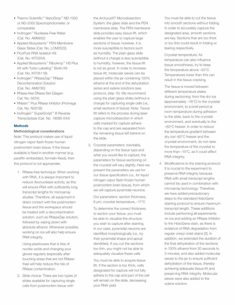
• Thermo Scientific™ NanoDrop™ ND-1000 or ND-2000 Spectrophotometer, or comparable
• Invitrogen™ Nuclease-Free Water (Cat. No. AM9932)
• Applied Biosystems™ PEN Membrane Glass Slides (Cat. No. LCM0522)
• PicoPure RNA Isolation Kit (Cat. No. KIT0204)
• Applied Biosystems™ RiboAmp™ HS Plus Kit with Turbo Labeling™ Biotin Kit (Cat. No. KIT0511B)
• Invitrogen™ RNaseZap™ RNase Decontamination Solution (Cat. No. AM9780)
• RNase-free DNase Set (Qiagen Cat. No. 9254)
• RNasin™ Plus RNase Inhibitor (Promega Cat. No. N261B)
• Invitrogen™ SuperScript™ III Reverse Transcriptase (Cat. No. 18080-044)
Methods
Methodological considerations
Note: This protocol makes use of liquid
nitrogen vapor flash-frozen human
postmortem brain tissue. If the tissue
available is fixed in another manner (e.g.,
paraffin-embedded, formalin-fixed), then
this protocol is not appropriate.
1. RNase-free technique: When working with RNA, it is always important to reduce ribonuclease activity, as this will ensure RNA with sufficiently long transcript lengths for microarray studies. Therefore, all equipment in direct contact with the postmortem tissue and the workspace should be treated with a decontamination solution, such as RNaseZap solution, followed by wiping down with absolute ethanol. Whenever possible, working on ice will also help ensure RNA integrity.
Using plasticware that is free of nucleic acids and changing your gloves regularly (especially after touching areas that are not RNase-free) will help reduce the risk of RNase contamination.
2. Slide choice: There are two types of slides available for capturing single cells from postmortem tissue with
the ArcturusXT Microdissection System: the glass slide and the PEN membrane slide. The PEN membrane slide provides easy tissue lift, which enables the user to capture large sections of tissue; however, it is more susceptible to factors such as humidity. The plain glass slide (without a charge) is less susceptible to humidity; however, the tissue lift is not as good. In order to increase tissue lift, molecular sieves can be placed within the jar containing 100% ethanol at the end of the dehydration series and xylene solutions (see protocol, step 15). We recommend using the plain glass slides (without a charge) for capturing single cells (i.e., small sections of tissue). Note: Tissue lift refers to the process during laser capture microdissection in which cells marked for capture adhere to the cap and are separated from the remaining tissue left behind on the slide.
3. Cryostat parameters: Inevitably, depending on the tissue type and what you would like to capture, the parameters for tissue sectioning on the cryostat will vary slightly. Here we present the parameters we use for our tissue specification (i.e., for liquid nitrogen vapor flash-frozen human postmortem brain tissue), from which we will capture pyramidal neurons.
Our parameters: section thickness, 8 µm; cryostat temperature, –17°C
To determine the correct thickness to section your tissue, you must be able to visualize the structure you wish to capture after staining. In our case, pyramidal neurons are identified morphologically (i.e., by their pyramidal shape and apical dendrites). If you cut the sections too thin, you might not be able to adequately visualize these cells
You must be able to acquire tissue lift. If the section is too thick, cells designated for capture will not fully adhere to the cap and part of the cell will remain on the slide, decreasing your RNA yield.
You must be able to cut the tissue into smooth sections without folding. In order to accurately capture the designated area, smooth sections are key. Sections that are too thick or too thin could result in folding or tearing respectively.
Cryostat temperature: As temperature can also influence tissue smoothness, try to keep the temperature above –20°C. Temperatures lower than this can result in the tissue cracking.
The tissue is moved between different temperature states during sectioning: from the dry ice (approximately –78°C) to the cryostat environment, to a brief period at room temperature during adhesion to the slide, back to the cryostat environment, and eventually to the –80°C freezer. In order to reduce the temperature gradient between dry ice/–80°C freezer and the cryostat environment, do not raise the temperature of the cryostat to more than –15°C, as it could affect RNA integrity.
4. Modifications to the staining protocol: It is crucial to the experiment to preserve RNA integrity because RNA with small transcript lengths cannot be used in combination with microarray technology. Therefore, we have added precautionary steps to the standard HistoGene staining protocol to ensure maximum transcript length. These additions include performing all experiments on ice and adding an RNase inhibitor to the HistoGene stain, as there is evidence of RNA degradation from regular cresyl violet stains [2]. In addition, we extended the duration of the final dehydration of the sections in 100% ethanol from 30 seconds to 3 minutes, and also added molecular sieves to the jar to ensure sufficient dehydration of the tissue, thereby achieving adequate tissue lift and preserving RNA integrity. Molecular sieves were also added to the xylene solution.

5. LCM cap choice: The two LCM caps available with the ArcturusXT Microdissection System—the CapSure Macro Cap and the CapSure HS Cap—each have distinct advantages, depending on the objective of the experiment. When making the choice of which cap to use, consider the following: You must be able to acquire tissue lift. Sometimes, during humid weather or particularly aqueous procedures, such as immunohistochemistry, the tissue is not completely dry. In this case, if additional dehydration does not help, you should use the CapSure Macro Cap as it is in direct contact with the tissue, and therefore adheres to the cells more easily. Keep in mind, however, that this cap often picks up unwanted tissue “debris” for the same reason. You should use the prep strips that are provided, because they help to remove the debris from the surface of the section prior to microdissection. The use of the CapSure HS cap is more advantageous if the section of the tissue is somewhat folded because the HS cap is fitted with rails that allow the thermoplastic film to be raised above the tissue section. If the cap is in direct contact with the section of tissue that has small folds, the cap will tilt, resulting in variable IR spot sizes. This is particularly important when capturing individual cells, as the inclusion of surrounding tissue will contaminate your sample
Cap choice is also dependent on the size of the structure you wish to capture. The Macro cap has a larger capture area, making it appropriate for capturing larger tissue structures, such as tumors. Even though the HS cap is made for capturing single cells, it only has a small area designated for capture, which can limit the section area from which you would like to capture. To alleviate this issue, keep the settings on the AutoScanXT software on Macro cap while using the HS cap, in order to capture cells from a larger section area.
Note: If using this approach, do not use the CapSure device for the extraction of material from the HS Cap. The CapSure device concentrates biomolecule extraction on the material collected in center region of the HS cap. Any material outside the center region will not be accessed for extraction if using the CapSure device. When using this approach, follow the protocol for the CapSure Macro Cap.
6. Spot size: When proceeding to laser capture, IR spot size (determined by LCM laser strength and duration) is an important variable that can influence whether you are able to achieve tissue lift and/or cell specificity. The spot size should be specific to your cell size (i.e., not too big that it is nonspecific, and small enough to be as specific as possible). At the same time, the laser strength should still be strong enough to be able to capture the cell. Conversely, if the laser strength is too high, the spot size will enlarge and might not be specific to the cell of interest. After identifying the cell of interest, adjust the laser strength and duration so that the spot created has a crisp, dark ring surrounding the cell. If it is too light, the cell will not adhere to the cap.
Do not include cells that have other cell types directly next to them, as these may also be captured and you will no longer have a homogeneous cell population. Also try to limit the amount of surrounding tissue you capture with the cell of interest by decreasing the spot size while still ensuring adequate capture of that cell.
Our IR laser parameters with the HS cap: strength, 70 mW; duration, 16 msec = spot size of 25 µm.
Note: These parameters are specific for our tissue type and fixation method (i.e., postmortem human brain tissue that was liquid nitrogen vapor flash-frozen), and is meant only as a rough guide. Be sure to
customize these parameters for your tissue type. These parameters may also vary slightly per section so adjust accordingly.
Protocol1. Remove possible RNase
contamination on the plain glass slide (with no charge) with a decontamination solution such as RNaseZap solution, followed by wiping it down with absolute ethanol.
2. Remove the tissue block (approximately 3 mm thick) stored in the –80°C freezer, and place it in a container with dry ice for transportation to the cryostat.
3. Place a micro slide box in the container.
4. Adjust the cryostat temperature to –17°C and the slice thickness to 8 µm, and wipe down with 100% ethanol.
5. Wipe down the sectioning blade with 100% ethanol before sectioning.
6. Place the platform (chuck) in the cryostat environment to acclimate.
7. After adhering the tissue block to the platform, let the block acclimate to the cryostat environment (temperature) for approximately 10– 15 minutes. This will prevent damage to the tissue block during sectioning in addition to ensuring smooth sections.
8. After acquiring a suitable section, adhere it to the glass slide (towards the center of the slide) at room temperature and place immediately thereafter inside the cryostat (–17°C).
9. Acquire another suitably smooth section and adhere it to the same slide, also towards the center of the slide.
10. Place the slide containing the two sections into the micro slide box on dry ice.
11. Cut enough sections for your entire experiment. In our case, four sections (or two slides) per case were sufficient.

12. Proceed to the next tissue block (case).
13. When you are finished sectioning, place the micro slide box containing the sections into the –80°C freezer until needed for LCM.
14. Prepare the ethanol dehydration series (2 x 75%, 2x nuclease-free water, 95%, 100%), adding 25 mL of each into the appropriate jars.
15. Add molecular sieves to the 100% ethanol jar and the xylene jar under the fume hood. Add just enough molecular seives to cover the bottom of the jar.
16. Place the first 75% ethanol jar into the –20°C freezer.
17. Place all other jars on ice in an ice bucket.
18. Add 4 µL of RNasin Plus RNase Inhibitor to 400 µL of HistoGene stain in an RNase-free 1.5 mL microcentrifuge tube, and place the tube on ice until needed.
19. Switch on the water bath and set to 42°C.
20. Place a rack containing a 50 mL Falcon tube in the water bath.
21. Remove two slides with two sections each from the –80°C freezer and defrost on Kimwipes™ tissues for 30 seconds or until the corners of the slides have defrosted. Note: Transport the slides from one jar to the next with forceps treated with RNaseZap solution.
22. Transfer the slides into the 75% ethanol jar that was originally placed in the –20°C freezer for 30 seconds.
23. After 30 seconds in the second jar containing nuclease-free water, place the slides on Kimwipes tissues.
24. Circle the sections on the slide with a PAP pen, and aliquot 90 µL of the HistoGene stain with RNase Inhibitor per section. Incubate at room temperature for 20 seconds.
25. Proceed with the dehydration series as outlined in the provided protocol until the final 100% ethanol dehydration step.
26. At this point, leave the slides in the final 100% ethanol jar containing the molecular sieves for 3 minutes.
27. Proceed to the fume hood and place the slides in the xylene jar containing the molecular sieves for 5 minutes.
28. Air-dry on Kimwipes tissues under the fume hood for 5 minutes.
29. Proceed immediately with LCM, capturing approximately 500 cells. Note: In our experiment, capturing approximately 500 pyramidal neurons from postmortem human brain tissue results in approximately 500 pg of total RNA per case.
30. Following LCM, proceed with RNA isolation using the PicoPure RNA Isolation Kit and using the CapSure Macro Cap protocol. During RNA isolation, follow the appendix protocol for DNase treatment of the sample, as the elimination of genomic DNA is critical for accurate downstream applications such as real-time PCR.
31. After total RNA isolation, check the total RNA quality by evaluating the 18S/28S peaks displayed on an electropherogram and virtual gel with the Experion RNA HighSens chips and reagents. Partial degradation is expected when working with postmortem tissue, but both peaks should be visible.
32. Amplify the mRNA (two rounds) with the RiboAmp HS Plus Kit and provided protocol. Our lab has made one slight modification to the manufacturer’s protocol for the second-strand cDNA synthesis. After the sample has been transferred from the tube to the purification column, centrifuge the tube originally containing the sample and transfer the extra microliter obtained after centrifugation into the purification column. Repeat this process during the second round of amplification. Note: We recommend performing the amplification procedure using the 0.5 mL tubes provided and, subsequently, performing the thermal
cycling with a corresponding 0.5 mL block to increase the RNA yield.
33. To check the amplified mRNA quality, perform two control steps: 1) check mRNA quality using Experion RNA StdSens chips and reagents to ensure that the transcript length exceeds the 600 nucleotides required for microarray hybridization, and 2) check the concentration and purity (A
260/A280 ratio) with the NanoDrop Spectrophotometer. Note: Our lab chose the Experion and NanoDrop systems to evaluate the quality and quantity of amplified RNA, but there are other comparable systems available that perform the same function. Regardless of the method you employ, it is critical you perform quality control measures before proceeding with labeling for microarray analysis.
34. If the mRNA meets the necessary requirements (see Results section), then proceed with biotin labeling using the Turbo Labeling Kit with Biotin. Our lab has made one modification to the manufacturer’s protocol. We recommend adding 1–5 µL extra RNase-free water to the spin column and another round of full-speed centrifugation (1 minute) to ensure that all the RNA is eluted from the column. Note: As some mRNA can be lost during the labeling protocol, add 3–5 µg more mRNA than required for the microarray chip.
35. Follow the hybridization procedure for the microarray chip. We used the Affymetrix GeneChip Human X3P Array, which was developed for degraded starting material such as postmortem tissue.
36. After hybridization, each array is scanned twice and images are visually inspected for artifacts. The Affymetrix™ Microarray Suite 5.1 Software averages the two images to compute an intensity value for each probe cell within each probe set.

Quality control of results, using metrics such as percent call, is paramount in microarray experiments. A low percent call may indicate poor RNA quality or unfragmented RNA, which reduces the efficiency of hybridization. In order to determine the percent calls, we employed the dChip software’s built-in function. www.hsph.harvard.edu/~cli/complab/dchip/
ResultsThis optimized protocol should enable the user to obtain enough good quality mRNA from a homogeneous cell population to perform microarray experiments. Figure 1 illustrates the critical documentation that should take place during any microdissection experiment. By taking pictures before and after microdissection, the user may effectively track progress and ensure sufficient collection of the desired material. After isolating the RNA from your cell population, it is important to check the quality of the total RNA obtained. Figure 2 illustrates good-quality total RNA obtained from cells captured from human postmortem tissue. Even though there is some degradation due to the nature of human postmortem tissue, both the 18S and 28S peaks are visible. This is reflected both in the electropherogram and in the virtual gel generated by the Experion RNA StdSens chips and reagents. If the curve is biased towards the beginning of the electropherogram (i.e., peaks at shorter lengths), and no 18S or 28S peaks are visible, this indicates that the RNA is too degraded to continue.
After two rounds of amplification, another set of controls helps determine whether the amplified mRNA is of good enough quality for microarray hybridization. In Figure 3, we see another electropherogram and virtual gel of a representative sample obtained with Experion RNA StdSens chips and reagents, indicating good mRNA transcript length extending past the 600-nucleotide minimum required for microarray hybridization.
Figure 1. Laser capture microdissection process. (A) The HistoGene stain allows pyramidal neurons to be identified morphologically. (B) After laser capture, cells adhere to the cap, leaving surrounding tissue behind. (C) A homogeneous cell population adhering to the CapSure HS Cap.
Figure 2. Total RNA quality assessment reveals good-quality mRNA. Electropherogram and accompanying virtual gel of total RNA generated using the Experion RNA StdSens chips and reagents. Both the 18S and 28S peaks are visible.
Figure 3. mRNA quality assessment. Electropherogram and accompanying virtual gel showing mRNA transcript lengths after two rounds of linear amplification. The mRNA transcript lengths extend up to the 3,000-nucleotide range, past the minimum length required for microarray studies (600 nucleotides).

Table 1. Quantity and quality of amplified mRNA. Mean concentration and purity of
the mRNA in control and treatment groups obtained after two rounds of amplification as
determined with a NanoDrop Spectrophotometer.
Control Treatment
mRNAµg/µL
Purity(A260/A280)
mRNA(μg/μL)
Purity(A260/A280)
1.4 2.54 2.1 2.49
Table 2. Microarray hybridization efficiency. Mean probe intensity and percent calls
of control and treatment groups after hybridization to the Affymetrix GeneChip Human
X3P Array.
Control Treatment
Probeintensity
Percentcall
Probeintensity
Percentcall
83.1 25.6 69.8 27.5
Find out more at thermofisher.com/lcm For Research Use Only. Not for use in diagnostic procedures. © 2015 Thermo Fisher Scientific Inc. All rights reserved. All trademarks are the property of Thermo Fisher Scientific and its subsidiaries unless otherwise specified. Experion is a trademark of Bio-Rad Laboratories. Falcon is a trademark of Corning, Inc. GeneChip is a trademark of Affymetrix, Inc. Kimwipes is a trademark of Kimberly-Clark Worldwide, Inc. RNasin is a trademark of Promega Corp. CO019849 1216
Table 1 gives the average amount of mRNA obtained per microliter of the 30 µL final elution—more than enough for microarray hybridization and subsequent downstream validation experiments such as real-time PCR. The purity results from the A260/A280 ratio of approximately 2.5 obtained from duplicate readings using the NanoDrop Spectrophotometer indicate good-quality mRNA samples. Table 2 summarizes the microarray hybridization efficiency with percent calls over 25%.
ConclusionsOur results indicate that we have enough good-quality mRNA obtained from a homogeneous cell population, and that hybridization onto the Affymetrix GeneChip Human X3P Array resulted in adequate percent calls. This protocol allows the user to obtain a specific molecular signature of a homogeneous cell population extracted from postmortem human brain tissue, without interference from surrounding tissue structures. Combining laser capture microdissection of human postmortem tissue with microarray analysis, therefore, has the potential to help generate new hypotheses, based on neuronal networks, of various neurological disease states.
References1. Mirnics K, Levitt P, Lewis DA (2006) Critical appraisal of DNA microarrays in psychiatric genomics. Biol Psychiatry
60:163–176.
2. Stemmer K, Ellinger-Ziegelbauer H, Lotz K et al. (2006) Establishment of a protocol for the gene expression analysis of laser microdissected rat kidney samples with Affymetrix GeneChips. Toxicol Appl Pharmacol 217:134–142.
3. Pietersen CY, Lim MP, Woo TUW (2009) Obtaining High Quality RNA from Single Cell Populations in Human Postmortem Brain Tissue. JoVE. http://www.jove.com/index/details.stp?id=1444. doi: 10.3791/1444.