femtosecond laser-assisted cvintal topoplasty · apresentamos um relato de astigmatismo tardio...
TRANSCRIPT

200 CASE REPORT
1Second-year resident at the Ophthalmology Department of Benjamin Constant Institute - Rio de Janeiro/RJ, Brazil.2Member of the Cornea Unit of Benjamin Constant Institute - Rio de Janeiro/RJ, Brazil.3Member of Renato Ambrosia Eye Institute – Rio de Janeiro/RJ, Brazil.4Associate Professor at the Ophthalmic Post-graduate Programme of the Federal University of São Paulo (UNIFESP) - São Paulo/SP, Brazil; CatholicUniversity of Rio de Janeiro (PUC-Rio) - Rio de Janeiro/RJ, Brazil; Renato Ambrósio Eye Institute - Rio de Janeiro/RJ, Brazil; Consultant at Oculus.
Study conducted at Benjamin Constant Institute and Renato Ambrósio Eye Institute – Rio de Janeiro/RJ, Brazil.
Femtosecond laser-assisted Cvintal topoplastyTopoplastia de Cvintal assistida por laser de femtossegundo
Alexandre Takayoshi Ishizaki1, Frederico Guerra2, Issac Ramos3, Renato Ambrosio Jr.4
The authors declare no conflicts of interest
Received for publication: 21/3/2012 - Accepted for publication: 3/9/2012
RESUMO
Apresentamos um relato de astigmatismo tardio progressivo pós-transplante de córnea para ceratocone, associado à afinamentoperiférico na junção doador-receptor, o que presumidamente pode ser considerado como recorrência da ectasia. O caso foi tratadopor meio de Topoplastia de Cvintal assistida por laser de femtossegundo para a confecção da incisão com geometria "top hat",seguido de sutura com ajuste per-operatório guiado por ceratoscopia.
Descritores: Astigmatismo/etiologia; Córnea/patologia; Ceratoplastia penetrante/efeitos adversos; Ceratocone/cirurgia; Te-rapia a laser/métodos; Complicações pós-operatórias; Relatos de casos
ABSTRACT
We present a case of late high progressive astigmatism following penetrating keratoplasty for keratoconus, which was associated withperipheral thinning in the donor-receptor area, which may be recognized as recurrence of ectasia. Treatment was accomplished withCvintal's Topoplasty assisted by femtosecond laser for a "top hat", followed by resuture with peroperative adjustment guided byceratoscopy.
Keywords: Astigmatism/etiology; Cornea/pathology; keratoplasty, penetrating/adverse effects; keratoconus/surgery; Lasers therapy/methods; Postoperative complications; Case reports
Rev Bras Oftalmol. 2013; 72 (3): 200-3

201
INTRODUCTION
Astigmatism is the most common cause of low visual acuityafter corneal transplantation. According to the literature,the average degree of astigmatism is 2.76 dioptres (D)
at 24 months after the procedure, and in 15% of cases it is greaterthan 5 D.(1) However, irregular astigmatism associated withhigher-order aberrations limits visual results as it cannot be fullycorrected with glasses.(2,3) Thus, the use of special contact lensesplays an important role in the visual rehabilitation of thesepatients.
Controlling astigmatism after transplantation is a majorchallenge for any cornea surgeon. The attempt to controlastigmatism begins during surgery, by performing a stable suturewith balanced tension in every axis of the keratoplasty. Selectiveremoval of separate sutures based on topographic progressionplays a key role in the management of post-PK astigmatism.
Surgical options for correcting astigmatism after atransplant include relaxing incisions, photoablative refractivesurgery with laser (LASIK or surface ablation), insertion ofintrastromal corneal ring segments, wedge resection withcompression suture, toric phakic intraocular lens implantation andretransplantation.(4)
The advent of the femtosecond (FS) laser has increasedthe accuracy and efficiency of corneal surgery.(5) FS laser-assistedcorneal transplantation provides a lower rate of intraoperativecomplications and can facilitate the control of postoperativeastigmatism.(5,6)
Topoplasty, a technique described by Dr. Tadeu Cvintal, isanother surgical option in cases of high astigmatism after cornealtransplantation.(7) The aim of this paper is to describe the firstcase of FS laser-assisted Cvintal topoplasty in a patient with highastigmatism associated with thinning of the host cornea,presumably related to the relapse of ectasia after PK.
CASE REPORT
A 54-year-old white female patient was undergoingophthalmic follow-up at the cornea unit of Benjamin ConstantInstitute. The patient reported a history of keratoconus, havingundergone penetrating keratoplasty in the right eye 16 yearsago and in the left eye 8 years ago. Transplantation in the lefteye progressed with immune rejection and glaucoma, with lossof transparency. The patient reported a progressive loss of visionin the right eye and did not adapt to rigid contact lenses or glasses.
On examination, uncorrected visual acuity (VA) was 20/200 in the right eye and hand motion in the left eye. Correctedvisual acuity in the right eye was 20/80 with manifest refractionof -2.75 -8.00 x 8°. Visual acuity in the left eye did not improvewith correction.
Biomicroscopy of the right eye showed a transparentcorneal graft slightly decentred toward the nose, thinning of thedonor-host junction and thinning of the host cornea (Figure 1).Biomicroscopy of the left eye showed an opaque corneal graftwith diffuse oedema.
The axial (sagittal) curvature map obtained by Placidotopography in the right eye (Figure 2) showed corneal astigmatismgreater than 14 D. The curvature and elevation maps obtainedby tomography with rotating Scheimpflug photography(Pentacam HR, Oculus, Germany) showed similar findings, withhigher toricity of the anterior and posterior surfaces of the cornea.In addition, corneal tomography showed inferior corneal thinning,which was consistent with the slit lamp examination (Figure 3).(8)
Total aberrometry or wavefront analysis (iTrace, TraceyTechnologies Corp. USA) showed findings consistent with those
Figure 1. Biomicroscopy (RE): transparent corneal button 15 yearsafter a penetrating keratoplasty to treat keratoconus. The button isslightly off-centre toward the nose, with thinning (presumably dueto a relapse of ectasia) of the donor-host junction in the flattest andmost elevated meridian on tomography
Figure 2. Topography (RE) showing a high degree of cornealastigmatism
Rev Bras Oftalmol. 2013; 72 (3): 200-3
Femtosecond laser-assisted Cvintal topoplasty
of topography and tomography, with a similar pattern ofastigmatism. Specular microscopy found 888 endothelial cells persquare millimetre.
Since the patient could not see well with glasses and couldnot adapt to contact lenses, a surgical solution was necessary.Two cornea surgeons indicated a new penetrating transplant dueto the severe astigmatism and corneal thinning, as well as therelatively low endothelial cell counts. However, based on thefindings, we opted for femtosecond laser-assisted topoplasty asdescribed by Cvintal.
Summary of the traditional surgical techniqueThe surgical technique of Cvintal Topoplasty(7) will be only
briefly described here. It is a combination of four surgicalprocedures:
- Marginal keratotomy;- Selective marginal keratectomy with drill;- Deep delamination of surgical margins, toward the centre in
the steepest meridians and toward the periphery in the flattest ones;
202
Figure 3. Corneal tomography with Pentacam and Scheimpflug imageshowing inferior corneal thinning (RE)
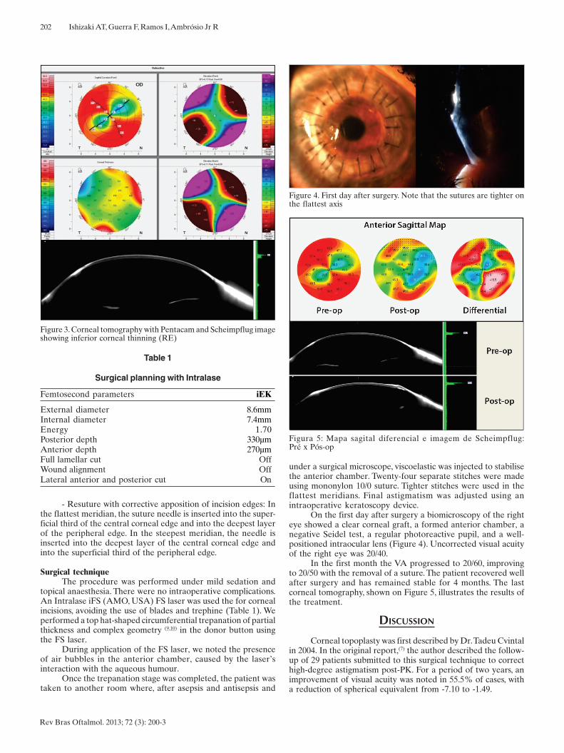
Figure 4. First day after surgery. Note that the sutures are tighter onthe flattest axis
Figura 5: Mapa sagital diferencial e imagem de Scheimpflug:Pré x Pós-op
Table 1
Surgical planning with Intralase
Femtosecond parameters iEK
External diameter 8.6mmInternal diameter 7.4mmEnergy 1.70Posterior depth 330µmAnterior depth 270µmFull lamellar cut OffWound alignment OffLateral anterior and posterior cut On
Rev Bras Oftalmol. 2013; 72 (3): 200-3
Ishizaki AT, Guerra F, Ramos I, Ambrósio Jr R
- Resuture with corrective apposition of incision edges: Inthe flattest meridian, the suture needle is inserted into the super-ficial third of the central corneal edge and into the deepest layerof the peripheral edge. In the steepest meridian, the needle isinserted into the deepest layer of the central corneal edge andinto the superficial third of the peripheral edge.
Surgical techniqueThe procedure was performed under mild sedation and
topical anaesthesia. There were no intraoperative complications.An Intralase iFS (AMO, USA) FS laser was used the for cornealincisions, avoiding the use of blades and trephine (Table 1). Weperformed a top hat-shaped circumferential trepanation of partialthickness and complex geometry (9,10) in the donor button usingthe FS laser.
During application of the FS laser, we noted the presenceof air bubbles in the anterior chamber, caused by the laser’sinteraction with the aqueous humour.
Once the trepanation stage was completed, the patient wastaken to another room where, after asepsis and antisepsis and
under a surgical microscope, viscoelastic was injected to stabilisethe anterior chamber. Twenty-four separate stitches were madeusing mononylon 10/0 suture. Tighter stitches were used in theflattest meridians. Final astigmatism was adjusted using anintraoperative keratoscopy device.
On the first day after surgery a biomicroscopy of the righteye showed a clear corneal graft, a formed anterior chamber, anegative Seidel test, a regular photoreactive pupil, and a well-positioned intraocular lens (Figure 4). Uncorrected visual acuityof the right eye was 20/40.
In the first month the VA progressed to 20/60, improvingto 20/50 with the removal of a suture. The patient recovered wellafter surgery and has remained stable for 4 months. The lastcorneal tomography, shown on Figure 5, illustrates the results ofthe treatment.
DISCUSSION
Corneal topoplasty was first described by Dr. Tadeu Cvintalin 2004. In the original report,(7) the author described the follow-up of 29 patients submitted to this surgical technique to correcthigh-degree astigmatism post-PK. For a period of two years, animprovement of visual acuity was noted in 55.5% of cases, witha reduction of spherical equivalent from -7.10 to -1.49.

203
Corresponding Author:Instituto de Olhos Renato AmbrósioRua Conde de Bonfim, nº 211/712 – TijucaCEP 20520-050 - Rio de Janeiro – (RJ), BrazilTel/Fax: +5521 2234 4233/2264 4430
Rev Bras Oftalmol. 2013; 72 (3): 200-3
Femtosecond laser-assisted Cvintal topoplasty
Topoplasty is indicated in many clinical situations, especiallyin cases of high-degree astigmatism post-PK and those with anirregular corneal structure.(7) The technique aims to reduce oreliminate the high toricity of the transplanted cornea, attemptingto shape it into a more spherical surface.
The introduction of the FS laser in the field of cornealtransplant has provided better clinical results and fewercomplications.(6) Different incision shapes can be used with thistype of laser, of which the most common are the top hat, themushroom, the zigzag and the Christmas tree. (9,10) Such complexincisions improve the fitting and stability of the donor-hostjunction.(6) Thus, in theory, fewer sutures are necessary and theycan be removed earlier.
Several studies report on different techniques to reduceastigmatism resulting from corneal transplant. Chamberlain etal. studied postoperative astigmatism in a series of patientssubmitted to PK, comparing the manual technique with the FSlaser-assisted procedure.(11) The latter provided better control ofastigmatism in the early postoperative period. However, after 6months of follow-up there was no significant difference inpostoperative astigmatism between the two groups.
FS laser is an option for arcuate wedge-shaped resectionaimed to correct post-PK astigmatism. The laser-guided procedureprovides a controlled and precise excision of tissue. Reductionsof 14.5 D in corneal astigmatism have been described with thisprocedure.(12)
Nubile et al. studied the efficacy of FS laser-guidedastigmatic keratotomy to treat post-keratoplasty astigmatism.(13)
The procedure consists of incisions in the steepest cornealmeridian on the periphery of the graft at a depth of 90% thetotal thickness of the stroma. The results showed a reduction ofastigmatism from 7.16 D (±3.07) to 2.23 D (±1.55) and refractivestability. Similar outcomes have been reported by otherauthors.(14)
In this case report we presented a patient that had beenpreviously submitted to bilateral PK due to keratoconus.Keratoconus is an ectatic and degenerative corneal disorder, andits progression is rare at the patient’s age. The left eye progressedwith graft failure, which explains its opacification. The donor buttonof the right eye showed good transparency; however, cornealthinning was evident at the donor-host junction, indicating arelapse of keratoconus.
The high degree of astigmatism in this case is due not onlyto the recurrence of keratoconus but also to the probableoccurrence of post-transplant astigmatism. The relapse of ectasiacontributes to disorganising the corneal structure, increasing thedegree and irregularity of the astigmatism. The low visual acuityin the right eye reflects the impact of this refractive error on theoptical system.
We opted for FS laser-assisted topoplasty because it is asimpler, more accurate procedure which leads to a greaterreduction of astigmatism compared with other techniques, andalso because of the availability of the device. However, thistechnique requires expensive equipment which is available onlyin a few operating rooms in Brazil.
CONCLUSION
The recurrence of keratoconus in eyes previously submittedto PK can be considered as an indication for a new cornealtransplantation. However, performing a new PK is a very complexapproach. The surgical technique described here preserves thecorneal tissue graft, thus avoiding exposure to new antigens thatcould trigger an immune rejection. Another important aspect ofthe technique is that it leads to more regular astigmatism andcorneal topography.
On the first day after surgery an improvement of the visu-al, refractive, and topographic outcome was noted. During thefollow-up period there were no complications or biomicroscopicor topographic signs of progression of corneal ectasia.
Thus, FS laser-assisted topoplasty facilitates themanagement of astigmatism and the treatment of recurrentectasia in previously-grafted eyes, with excellent postoperativeresults.
This procedure was effective in the treatment of highastigmatism associated with relapse of keratoconus after PK.The patient had a significant improvement of visual acuity andshowed refractive and topographic stability one month after theprocedure; after 4 months, the results had remained stable. Upto this date, with one year of follow-up, there was no need for anew corneal transplant.
REFERÊNCIAS
1. Olson RJ, Pingree M, Ridges R, Lundergan ML, Alldredge C Jr, ClinchTE. Penetrating keratoplasty for keratoconus: a long-term review ofresults and complications. J Cataract Refract Surg. 2000;26(7):987-91.
2. Krachmer JH, Fenzl RE. Surgical correction of high postkeratoplastyastigmatism. Relaxing incisions vs wedge resection. Arch Ophthalmol.1980;98(8):1400-2.
3. Rajan MS, O'Brart DP, Patel P, Falcon MG, Marshall J. Topography-guided customized laser-assisted subepithelial keratectomy for thetreatment of postkeratoplasty astigmatism. J Cataract Refract Surg.2006;32(6):949-57.
4. Lavery GW, Lindstrom RL, Hofer LA, Doughman DJ. The surgicalmanagement of corneal astigmatism after penetrating keratoplasty.Ophthalmic Surg. 1985;16(3):165-9.
5. Ambrosio Júnior R. A revolução dos lasers de femtossegundo naoftalmologia. Rev Bras Oftalmol. 2011;70(4):207-10.
6. Yoo SH, Hurmeric V. Femtosecond laser-assisted keratoplasty. Am JOphthalmol. 2011;151(2):189-91.
7. Cvintal T. Topoplastia. In: Cvintal T. Complicações do transplante decórnea. São Paulo: Santos Editora; 2004. p. 283-8.
8. Ambrósio R Jr, Belin MW. Imaging of the cornea: topography vs to-mography. J Refract Surg. 2010;26(11):847-9.
9. Bahar I, Kaiserman I, McAllum P, Rootman D. Femtosecond laser-assisted penetrating keratoplasty: stability evaluation of differentwound configurations. Cornea. 2008;27(2):209-11.
10. Ignacio TS, Nguyen TB, Chuck RS, Kurtz RM, Sarayba MA. Top hat woundconfiguration for penetrating keratoplasty using the femtosecond laser: alaboratory model. Cornea. 2006;25(3):336-40.
11. Chamberlain WD, Rush SW, Mathers WD, Cabezas M, FraunfelderFW. Comparison of femtosecond laser-assisted keratoplasty versusconventional penetrating keratoplasty. Ophthalmology.2011;118(3):486-91.
12. Ghanem RC, Azar DT. Femtosecond-laser arcuate wedge-shapedresection to correct high residual astigmatism after penetrating kerato-plasty. J Cataract Refract Surg. 2006;32(9):1415-9.
13. Nubile M, Carpineto P, Lanzini M, Calienno R, Agnifili L, CiancagliniM, et al. Femtosecond laser arcuate keratotomy for the correction ofhigh astigmatism after keratoplasty. Ophthalmology.2009;116(6):1083-92.
14. Kumar NL, Kaiserman I, Shehadeh-Mashor R, Sansanayudh W,Ritenour R, Rootman DS. IntraLase-enabled astigmatic keratotomyfor post-keratoplasty astigmatism: on-axis vector analysis. Ophthal-mology. 2010;117(6):1228-35.e1.