feather degrading bacillis thuringiensis s3kubot
TRANSCRIPT
Isolation and characterization of a feather degrading bacterium from soil
V.P. PottyPrincipal ScientistCEPC Kollam
G.M. NairProf. & HeadDepartment of Plant SciencesCentral University of Kerala
Resmi Raj LResearch ScholarDepartment of BotanyUniversity of Kerala

Introduction
Feather as poultry waste.
Disposal of waste feathers.
Protein wastage and environmental pollution.

Feathers represent over 90% protein.
β-keratin
Structural rigidity due to disulfide, hydrogen and hydrophobic bonds.
Resistance to degradation by common microbial proteases.
Degradation by inducible enzyme - keratinase.
Only a minor quantity of waste feathers is being used, even though wide industrial applications are possible.
Pretreatment methods hydrolyze feather to corresponding amino acids and small peptides.
Drawbacks of the pretreatment methods gave a way to use microbial keratinases.
Thus there is a requirement of isolation of keratinases from natural sources to meet the industrial and environmental demand.

ObjectivesIsolation of feather degrading bacteria
from various poultry-waste dumping sites of Thiruvananthapuram.
Selection by screening of the maximum feather degrading strain from among the isolates.
Characterization of the selected strain by cultural and biochemical characters; molecular and microscopic techniques.

Materials & Methods
Collection of soil samples from different poultry-waste dumping sites.
Plating of the bacterial suspension on feather agar plates.
Incubation of the plates at 37º C for 24 to 48 hrs.
Selection of single colonies and maintenance at 4ºC for further work.

The basic medium used for isolation and fermentation of feather degrading micro organisms contained:
NaCl - 0.5 g/LKH2PO4 - 0.7 g/LK2HPO4 - 1.4 g/LMgSO4 - 0.1 g/LFeather - 10 g/L
1ml of 24hr grown culture (4.25×10⁵ CFU/ml) in LB broth was used as inoculum.
Keratinolytic activity determination
0.5 % (w/v) soluble keratin was used as substrate for keratinolytic activity determination (Wawrzkiewicz et al., 1987).
Crude enzyme was obtained from inoculated medium after 96 hrs of cultivation and done enzyme assay according to Cai et al., 2008 with uninoculated medium as control.
One unit of enzyme activity (U/ml) was defined as an increase of corrected absorbance at 280 nm with that of the control for 0.01/minute.

Taxonomic studies and identification of the microorganism with potential keratinolytic activity
The cultural, morphological and biochemical characteristics of the selected bacterial strain were compared with the data from Bergey’s Manual of Determinative Bacteriology (Robert S. Breed et al., 1957).
Molecular characterization of 16S rRNA gene was carried out. Bacterial 16S rDNA was amplified by using the universal primers:
Forward primer: 8F 5’ AGAGTTTGATCCTGGCTCAG 3’ Reverse primer: 1492R 5’CGGTTACCTTGTTACGACTT
3’
Sequence obtained was aligned using BioEdit sequence alignment software and verified for correctness using the BLAST option available at: http://www.ncbi.nlm.nih.gov/
The 16S rRNA sequence of the strain was then submitted to GenBank and obtained the accession number. A phylogenetic tree was constructed using TREECONW 3.1 software package.

(a)Light microscopy
Sporulated culture was stained and examined in a light microscope (Olympus BX51 Image Analyser) to study the crystal morphology. The slide was then heat fixed and stained (0.133% CBB R-250 stain in 50% acetic acid), rinsed in distilled water, dried and observed under light microscope using a 100x oil immersion objective.
Microscopic studies

(b) Phase Contrast microscopySpore-crystal mixture was prepared in sterile
distilled water after heat treatment for 1hr and a wet mount of the sample was observed in phase contrast microscope (Zeiss Observer A1, AX10) under 100x oil immersion objective.
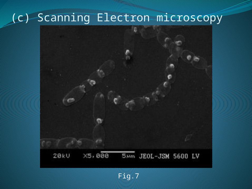
(c) Scanning Electron microscopyA thin smear of the spore-crystal suspension
was prepared on glass mounts and then coated with gold in JEOL-JFC 1200 sputtering unit and photographed in JEOL-JSM 5600LV scanning electron microscope operated at a voltage of 20 kV.

Results & DiscussionIsolation of keratinolytic microorganisms
Six morphologically different bacteria were isolated from the collected soil samples. All the strains were maintained in Nutrient Agar for elucidating their keratinolytic activity as well as feather degrading capacity. S3KUBOT showed maximum activity as per the keratinolytic activity determining assay.

Fig.1 Growth of the isolate S3KUBOT on (a) nutrient agar medium (b) feather agar medium
Fig.2 (a) uninoculated control medium (b) inoculated with S3KUBOT after 48hrs.
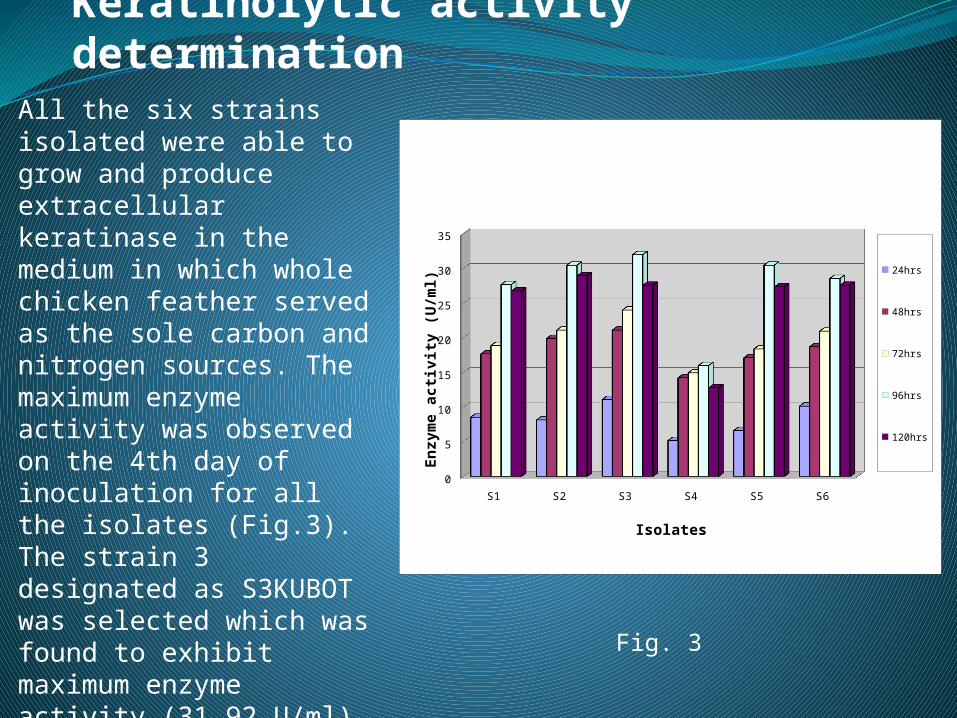
Keratinolytic activity determination
All the six strains isolated were able to grow and produce extracellular keratinase in the medium in which whole chicken feather served as the sole carbon and nitrogen sources. The maximum enzyme activity was observed on the 4th day of inoculation for all the isolates (Fig.3). The strain 3 designated as S3KUBOT was selected which was found to exhibit maximum enzyme activity (31.92 U/ml) on the basis of the assay. All the strains except S4 showed the morphological and cultural characteristics of Bacillus species.
S1 S2 S3 S4 S5 S6
0
5
10
15
20
25
30
35
24hrs
48hrs
72hrs
96hrs
120hrs
Isolates
Enz
yme
acti
vity
(U
/ml)
Fig. 3
Taxonomic studies and identification
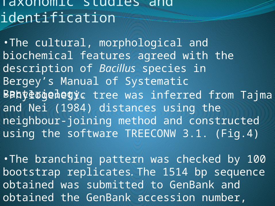
•Phylogenetic tree was inferred from Tajma and Nei (1984) distances using the neighbour-joining method and constructed using the software TREECONW 3.1. (Fig.4)
•The branching pattern was checked by 100 bootstrap replicates. The 1514 bp sequence obtained was submitted to GenBank and obtained the GenBank accession number, HQ832565.
•The cultural, morphological and biochemical features agreed with the description of Bacillus species in Bergey’s Manual of Systematic Bacteriology.

Morphological characteristics InferenceFormGram stainSpore
Rod Positive Spore forming
Cultural characteristicsFeather Agar and Nutrient Agar Creamy white colonies
Biochemical Test Inference
CatalaseOxidaseO-FMRVP CitrateIndolePhenyl alanineNitrate reductionGelatin liquefactionGelatinase AmylaseLecithinaseMotility
PositivePositivePositive PositiveNegativePositiveNegativeNegativePositiveRapid liquefactionPositivePositivePositivePositive
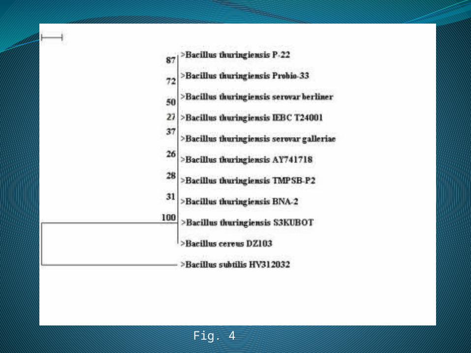
Fig. 4

Microscopic studies
(a) Light microscopy
Fig. 5(a) Fig. 5(b)

(b) Phase Contrast microscopy
Fig. 6
(c) Scanning Electron microscopy
Fig.7

ConclusionThe strain S3KUBOT has been reported to produce keratinases during vegetative phase which makes the degradation of feather possible within 96 hrs of incubation.
The inefficiency of the 16S rRNA gene analysis may be the prime reason that a lot much of isolates belonging to the Bacillus cereus group are still been identified as Bacillus species within the GenBank.
The isolate S3KUBOT was confirmed as Bacillus thuringiensis by electron microscopy to possess a parasporal body.

References
• Cheng-gang Cai, Bing-gan Lou, Xiao-dong Zheng. (2008) Keratinase production and keratin degradation by a mutant strain of Bacillus subtilis. J Zhejiang Univ Sci B 9(1): 60-67.
• Robert, S. Breed, E. G. D. Murray and Nathan, R. Smith. (1957) Bergey’s Manual of Determinative Bacteriology. Seventh Edition, Baltimore. The Williams & Wilkins Company.
• Wawrzkiewicz, K., Lobarewski, J. and Wolski, T. (1987) Intracellular keratinases of Trichophyton gallinae. J. Med. Vet. Mycol. 25: 261–268.

Thank You