eyeblink artifact suppression from eeg signal using ... · eyeblink artifact suppression from eeg...
TRANSCRIPT
IOSR Journal of VLSI and Signal Processing (IOSR-JVSP)
Volume 7, Issue 2, Ver. I (Mar. - Apr. 2017), PP 01-09
e-ISSN: 2319 – 4200, p-ISSN No. : 2319 – 4197
www.iosrjournals.org
DOI: 10.9790/4200-0702010109 www.iosrjournals.org 1 | Page
Eyeblink Artifact Suppression from EEG Signal using Lifting
Wavelet Transform
Mst. Jannatul Ferdous1,2,*
, Md. Sujan Ali1,2
, Md. Ekramul Hamid1 and
Md. Khademul Islam Molla1
1Signal Processing and Computational Neuroscience Lab, Department of Computer Science and Engineering,
University of Rajshahi, Rajshahi, Bangladesh 2Department of Computer Science and Engineering, Jatiya Kabi Kazi Nazrul Islam University, Mymensingh,
Bangladesh
*E-mail: [email protected]
Abstract: In this paper we proposed a technique to remove eye blink artifact from electroencephalogram
(EEG) using lifting wavelet transform (LWT). The LWT has been successfully used in eye blink artifact
suppression form the recorded electroencephalography (EEG) signals using a data-adaptive subband filtering
approach. The LWT is applied to decompose EEG signal into a finite set of subbands. The energy based
subband filtering is implemented to separate the lower frequency noise components to clean the EEG signal.
The energies of individual subbands respectively for EEG and fGn that of contaminated EEG are compared to
derive the energy based threshold for the suppression of eyeblink effects. We adopt two de-noising algorithms
based on stationary subspace analysis (SSA), and lifting wavelet transform (LWT) for comparison purpose.
Through using contaminated EEG signals from BCI database, we evaluate the artifact correction results by
means of SAR and MSE, and conclude that LWT algorithm is the suitable one for de-noising EEG signal.
Keywords: electroencephalography, artifact reduction, stationary subspace analysis, lifting wavelet transform
I. Introduction Brain-computer interface (BCI) has been one of the most outstanding biomedical engineering research
fields for decades. It provides a promising technology allowing humans to control external devices by
modulating their brain waves.The quality of electroencephalography (EEG) is degraded by the non-cerebral
signal sources are known as artifacts. Among all the artifacts, ocular artifacts are the most prevailing. Ocular
artifacts occur through eye movements and blink which generates a signal greater in magnitude than EEG
signals, allowing it to travel throughout the scalp, covering and distorting EEG signals [1–4]. In order to achieve
higher quality EEG signals, these artifacts must be removed with-out distorting or removing any of the
underlying EEG data. There are various methods to control ocular artifacts. One of the modest methods is
confining the eye movements and eye blinking of the subject by keeping on a fixed point. Though it is difficult
for the subject to perform this task especially for infants or persons with certain disabilities during experiments.
Moreover, the effort of performing the task can have a significant effect on the attained EEG data [5].
In previous research, many simple and complex methods have been proposed for detecting and
removing artifacts. The simple signal processing filter, known as Butterworth bandpass filter is used to remove
the artifact. The clean EEG signal is easily achieved from raw EEG by applying 4th
order of bandpass
Butterworth filter. However, this type of filter does not suitable for EEG signal processing because some of the
original information are loss during the filtration process [18]. ICA has become a popular method in removing
artifacts from EEG data. The disadvantage of ICA is that the components do not necessarily only contain artifact
data, but also contains underlying EEG data [2]. Removing the contaminated component will thus lead to loss of
EEG data.
To address the weakness of ICA-based artifact correction methods another effective BSS method,
stationary subspace analysis (SSA) is used [6,7]. The EOG signals are considered as low frequency and high
energy trend in the recorded EEG signals, the SSA is performed better to suppress it from mixing. The
components that are determined to be related to the ocular artifacts are projected back to be subtracted from
EEG signals, producing the clean EEG data eventually. Hence it is very evident to employ SSA for eyeblink
artifact identification and suppression. It is used for multiple channel data to partition the whole space into
stationary and non-stationary subspaces [8].
The discrete wavelet transform has been extensively studied over the past 20 years. Many applications
have been proposed but the power of wavelet transform lies in its performance in compression and denoising
schemes. The existence of fast algorithms for its computation is another important factor. The well-known
Eyeblink Artifact Suppression from EEG Signal using Lifting Wavelet Transform
DOI: 10.9790/4200-0702010109 www.iosrjournals.org 2 | Page
Mallat’s algorithm (DWT– Discrete Wavelet Transform) employs two-channel filter bank iteratively and the
filter bank can be equally represented by a polyphase lifting scheme. Iterative application of the lifting scheme
(LWT–Lifting Wavelet Transform) results in the same coefficients as the DWT does. The lifting scheme was
rediscovered by Sweldens in [9] and according to [10], every wavelet filter bank can be decomposed into a
lifting scheme.
It is presented that the realization of a lifting wavelet processor which is used an integer type Haar
lifting wavelet transform (LWT) to measure ECG signals in real time [11]. It is also adopted Db4 lifting wavelet
transform to reconstruct ECG signals [12]. It is also proposed a lifting-based discrete wavelet transform to de-
noise ECG signals. Through testing with Haar, Db4, Db6, Filter (9-7), and Cubic B-splines, Db4 decomposition
in 4 scales with level dependent threshold estimator can get best SNR and visual inspection in de-noising [13].
In this paper, lifting wavelet transform (LWT) approach is introduced to separate low frequency
artifacts from contaminated EEG. The signal is decomposed using wavelet transform yielding the subbands. The
individual subband is thresholded and added together to obtain the clean signal. Also, this paper aims to shows
the comparison when EEG signal filter with lifting wavelet transform (LWT) and stationary subspace analysis
(SSA). The comparative study presented that LWT approach is more effective to extract the EEG signals after
suppressing the contaminated artifacts.
The remaining parts of the paper are organized as- the algorithms used in the paper (SSA, and LWT)
are described in Section II, the experimental result and discussion of the artificially contaminated EEG data and
performance metrics are explained in Section III, and finally Section IV gives a brief conclusion to the paper.
II. Methods The recorded EEG is modeled as linear combination of pure EEG and EOG artifact defined as:
)1()()()( tAtPtS EOGEEGEEG
where , , and are measured EEG, pure EEG and EOG artifact respectively. The
measured EEG signal is the superposition of original EEG signal due to brain activity and the fraction of EOG
signal due to eye blink activity. The aim of the proposed scheme is to extract required EEG signal
from the measured signal which consists of the required signal plus the ocular artifact
signal . In this paper, multichannel is used as the primary input to suppress its EOG artifact
using SSA and wavelet based techniques. No reference channel is used to estimate the pure EEG signals.
A. Stationary Subspace Analysis
Stationary Subspace Analysis (SSA) is a blind source separation algorithm which factorizes a
multivariate time series into stationary and non-stationary components. In EEG analysis, the electrodes on the
scalp record the activity of a large number of sources located inside the brain. These sources can be stationary or
non-stationary, but they are not discernible in the electrode signals, which are a mixture of these sources. SSA
allows the separation of the stationary from the non-stationary sources in an observed time series.
According to the SSA model [8], the observed multivariate time series s(t) is assumed to be generated as a linear
superposition of stationary sources and non-stationary sources as follows:
)(
)(][)()(
ts
tsMMtMnts
n
s
ns
where, is an unknown but time-constant mixing matrix; and are the basis of the stationary and non-
stationary subspace respectively. Given samples from the time series )(ts the aim of SSA is to estimate the
inverse mixing matrix1M separating the stationary from non-stationary sources in the mixture.
Using the estimated mixing matrix, ]ˆˆ[ˆns MMM given by SSA, these artifactual components )(tX art
are projected back to EEG channel, and artifacts in EEG data )(tsart are calculated as
)(ˆ)( tXMts artnart
Eyeblink Artifact Suppression from EEG Signal using Lifting Wavelet Transform
DOI: 10.9790/4200-0702010109 www.iosrjournals.org 3 | Page
Finally, this ocular activity is removed from the EEG recording to yield the clean EEG data ts by the
following formula:
)2()()()(ˆ tststs art
where, )()(ˆ tSts EEG .Thus the pure EEG of any channel is reconstructed using Eq. (2).Using the above
calculating formula we get the clean EEG from artificially contaminated EEG and the process is depicted in Fig.
1.
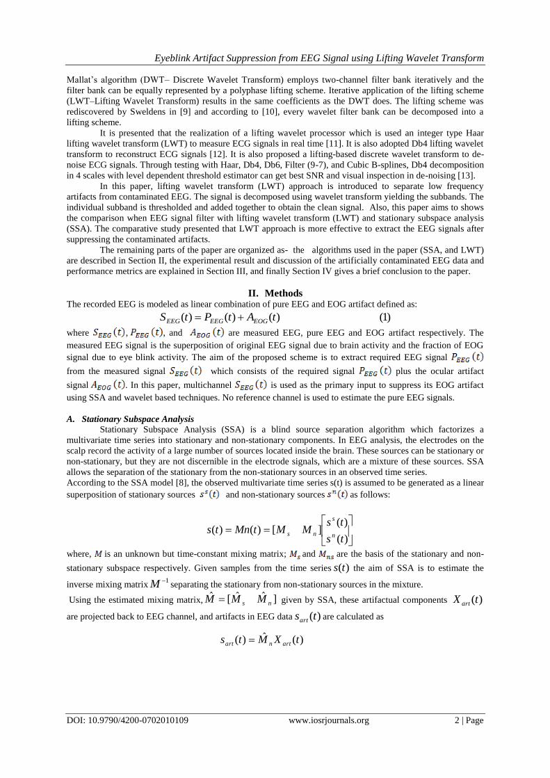
Fig. 1. Block diagram of stationary subspace analysis approach.
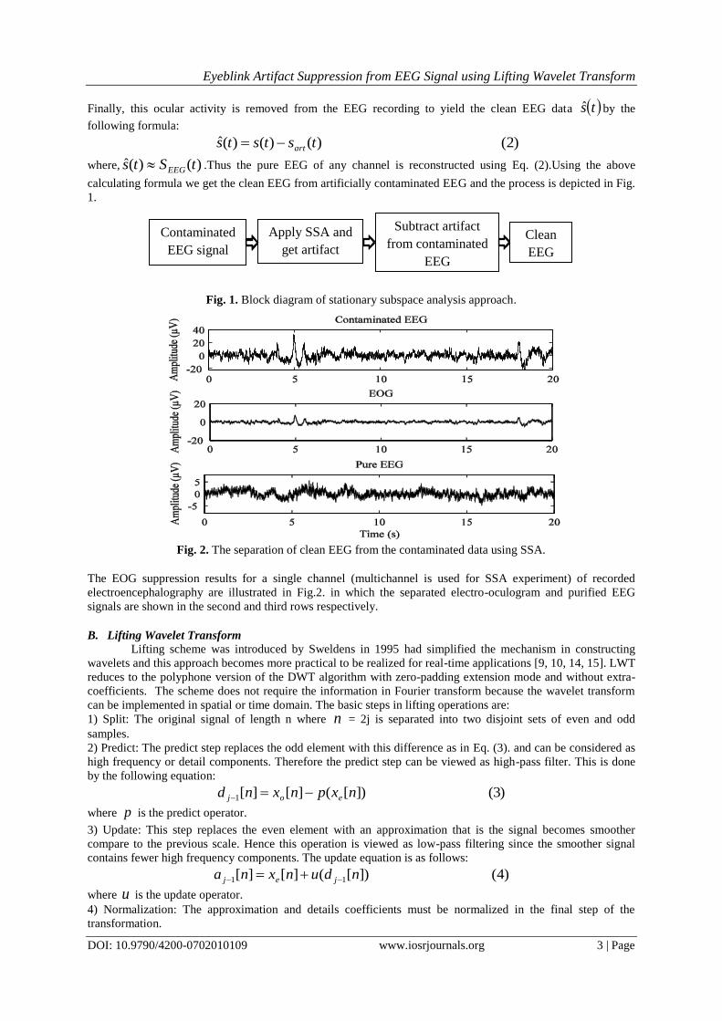
The EOG suppression results for a single channel (multichannel is used for SSA experiment) of recorded
electroencephalography are illustrated in Fig.2. in which the separated electro-oculogram and purified EEG
signals are shown in the second and third rows respectively.
B. Lifting Wavelet Transform
Lifting scheme was introduced by Sweldens in 1995 had simplified the mechanism in constructing
wavelets and this approach becomes more practical to be realized for real-time applications [9, 10, 14, 15]. LWT
reduces to the polyphone version of the DWT algorithm with zero-padding extension mode and without extra-
coefficients. The scheme does not require the information in Fourier transform because the wavelet transform
can be implemented in spatial or time domain. The basic steps in lifting operations are: 1) Split: The original signal of length n where n = 2j is separated into two disjoint sets of even and odd
samples.
2) Predict: The predict step replaces the odd element with this difference as in Eq. (3). and can be considered as
high frequency or detail components. Therefore the predict step can be viewed as high-pass filter. This is done
by the following equation:
)3(])[(][][1 nxpnxnd eoj
where p is the predict operator.
3) Update: This step replaces the even element with an approximation that is the signal becomes smoother
compare to the previous scale. Hence this operation is viewed as low-pass filtering since the smoother signal
contains fewer high frequency components. The update equation is as follows:
)4(])[(][][ 11 ndunxna jej
where u is the update operator.
4) Normalization: The approximation and details coefficients must be normalized in the final step of the
transformation.
Fig. 2. The separation of clean EEG from the contaminated data using SSA.
Contaminated
EEG signal
Apply SSA and
get artifact
Subtract artifact
from contaminated
EEG
Clean
EEG

Eyeblink Artifact Suppression from EEG Signal using Lifting Wavelet Transform
DOI: 10.9790/4200-0702010109 www.iosrjournals.org 4 | Page
Fig. 3. (a)One step forward lifting operation. (b) Inverse lifting step
The lifting step is depicted in Fig.3.(a) for the decomposition or analysis of the forward wavelet transform. The
update and predict stages can become a pair but sometimes they may not be together in a lifting step.
To obtain the signal back, the operations can be undone by just reversing them and change the signs as shown in
Fig. 3.(b).The operation is working backwards from the forward lifting operation.
If more steps are required, they can be added singly. The first generation wavelets can be converted into lifting
steps by factoring its h-coefficients as shown in many texts [9,10,14,15].In this study, a noise assisted LWT
based approach is implemented here to reduce the low frequency noise from single channel EEG. At the end of
the decomposition, the reconstructed signal is represented as
5)()(~1
1
tqtsL
b
b
where, ts~ ≈x(t). The LWT based filtering method of EEG signal is shown in Fig. 4. In this Fig., the clean
EEG signal is easily achieved from raw EEG by applying LWT .Then the clean EEG is evaluate by the same
evaluation parameter used in SSA. The db4 mother wavelet is choosing for this filter. The lower frequency EEG
signal of the single channel can be estimated by summing up the lower order subbands as:
61
tCtsD
b
b
where, bC (t) is the bth
subband of the channel. Here, the subject is to find the critical (threshold) subband with
index D such that the subbands of indices 1,2,3,....,D are responsible for relatively lower frequency pure EEG
component.
Fig. 4. Block diagram of lifting wavelet transform approach.
It is a well-recognized wavelet transform of a 1-D signal and inverts the transform to demonstrate perfect
reconstruction. The original signal, reconstructed signal using LWT method and the reconstruction error are
illustrated in Fig.6.
Contaminated
EEG signal
Apply
LWT
Apply subband
energy based
threshold
Clean
EEG

Eyeblink Artifact Suppression from EEG Signal using Lifting Wavelet Transform
DOI: 10.9790/4200-0702010109 www.iosrjournals.org 5 | Page
Fig. 5. The subband decomposition of contaminated EEG using LWT.
III. Results and Discussion In this section, the LWT based method is tested with the artificially corrupted EEG signal and after that
the signal is comprised with SSA. For comparison purposes, the time series contaminated EEG data is
decomposed into multiple subbands using SSA and lifting wavelet transforms (LWT).
The SSA is used for multiple channel data and its decomposition is produced stationary and non-
stationary components. More precisely, the target EEG is considered as stationary with respect to artifacts of
higher order non-stationary. To get the desired clean EEG data, the artifactual component is subtracted from
contaminated EEG [8]. The experimental result for the first channel is illustrated in Fig.2. It is observed that the
purified EEG signal holds some artifact and the EOG is not separated properly but includes some signal
components of EEG signal.
Another potential multiresolution decomposition approach, LWT which is analyzing a non-stationary
signal like EEG. In this study,“db4” is used with 7 level decomposition. It is another approach for EOG artifact
reduction.To get artifact free EEG the LWT based filter is used. The raw EEG signal and a reference signal fGn
are decomposed up to level 7 using db4 mother wavelet. After decompose, we got 8 subbands. The subband
decomposition of recorded electroencephalography data and fractional Gaussian noise are shown in Fig. 5 and
Fig. 7 respectively. The algorithm for LWT to find the index D of the threshold subband for the channel is
following:
1) Decompose the analyzing EEG signal together with the fGn ( in Fig.7.) into a finite set of subbands using
LWT.
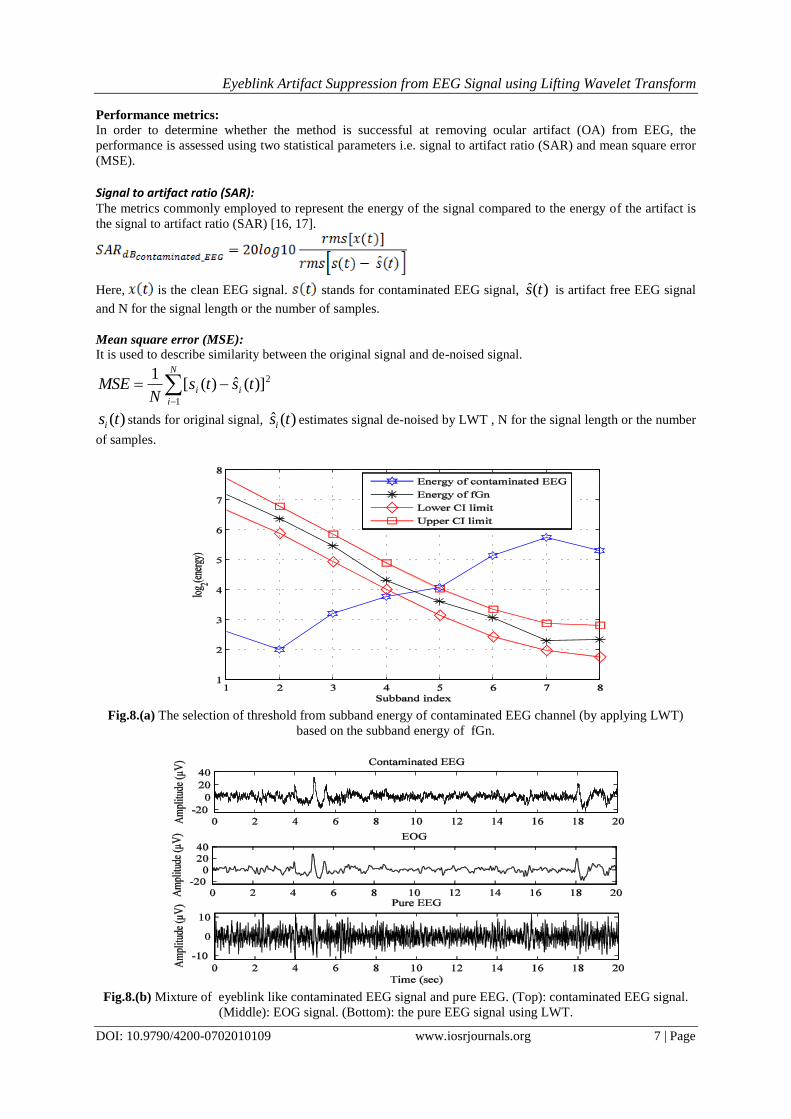
2) Calculate the energies of the subbands of fGn and its 95% confidence interval (CI).
3) Compute the energies of all subbands of contaminated EEG. Then find the lowest order subband with energy
exceeding the upper limit of CI derived in step 2 say it is the nth subband. The selected nth
(in Fig.8(a), n = 5)
subband is the starting index to reconstruct electro-oculogram signal.
4) The EEG is separated by summing up the subband coefficient starting from 1 to Dth
subband of
electroencephalography signals using Eq. (6).
The determination of threshold subband based on the subband energy is illustrated in Fig.8.(a). After computing
the index D of threshold subband (for EEG channel), the pure EEG of that channel is separated using Eq. (6).

Eyeblink Artifact Suppression from EEG Signal using Lifting Wavelet Transform
DOI: 10.9790/4200-0702010109 www.iosrjournals.org 6 | Page
Fig.6. Reconstruction of contaminated EEG (top) and reconstruction error (bottom) using LWT.
It is observed in Fig. 8.(a) that the 5th
subband is the first subband index that exceeds the upper limit of
confidence interval and the total number of subbands are 8. The 5th
subband coefficient is the starting point of
lower frequency components. The electro-oculogram is separated by summing the subband coefficients 5 to 8.
By subtracting electro-oculogram from raw electroencephalography, we get the purified electroencephalography
that reflects the actual neural activities. The electro-oculogram suppression results for a single channel of
recorded electroencephalography are illustrated in Fig.8.(b) in which the separated electro-oculogram and
purified electroencephalography signals are shown in the second and third rows respectively. From Fig. 8.(b), it
is observed that the purified EEG signal contains more original information although the artifact has cancel out.
It is apparent that using SSA for artifact correction, underlying EEG or low frequency cerebral data may be lost.
In order to reduce the data loss LWT method is used.
Fig. 9. depict the contaminated EEG and clean EEG for the two methods. From this Fig., it is observed that the
LWT baesd method is best for reduce the EOG from contaminated EEG without cutting the information and
assist to get clean EEG. The pure EEG do not show any large EOG.As a result, the purified EEG signal is found
as completely artifact free in LWT method.
Fig. 7. The subband decomposition of fGn using LWT.
Eyeblink Artifact Suppression from EEG Signal using Lifting Wavelet Transform
DOI: 10.9790/4200-0702010109 www.iosrjournals.org 7 | Page
Performance metrics:
In order to determine whether the method is successful at removing ocular artifact (OA) from EEG, the
performance is assessed using two statistical parameters i.e. signal to artifact ratio (SAR) and mean square error
(MSE).
Signal to artifact ratio (SAR): The metrics commonly employed to represent the energy of the signal compared to the energy of the artifact is
the signal to artifact ratio (SAR) [16, 17].
Here, is the clean EEG signal. stands for contaminated EEG signal, )(ˆ ts is artifact free EEG signal
and N for the signal length or the number of samples.
Mean square error (MSE):
It is used to describe similarity between the original signal and de-noised signal.
N
i
ii tstsN
MSE1
2)](ˆ)([1
)(tsi stands for original signal, )(ˆ tsi estimates signal de-noised by LWT , N for the signal length or the number
of samples.
Fig.8.(a) The selection of threshold from subband energy of contaminated EEG channel (by applying LWT)
based on the subband energy of fGn.
Fig.8.(b) Mixture of eyeblink like contaminated EEG signal and pure EEG. (Top): contaminated EEG signal.
(Middle): EOG signal. (Bottom): the pure EEG signal using LWT.
Eyeblink Artifact Suppression from EEG Signal using Lifting Wavelet Transform
DOI: 10.9790/4200-0702010109 www.iosrjournals.org 8 | Page
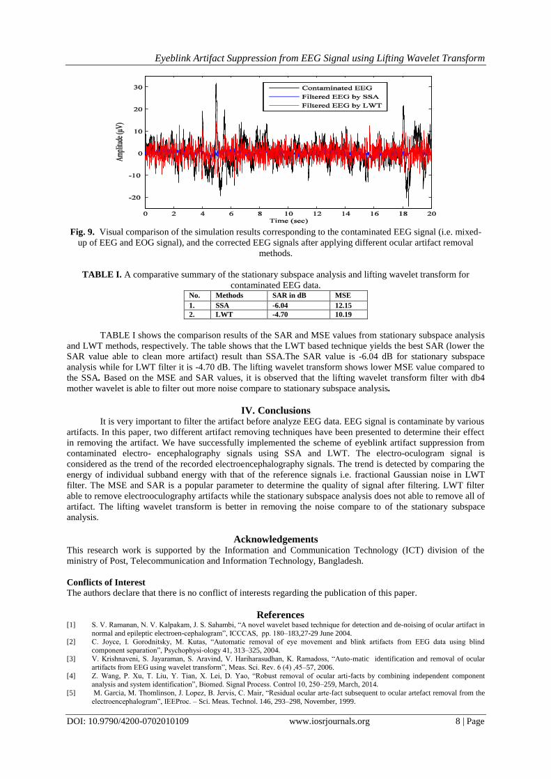
Fig. 9. Visual comparison of the simulation results corresponding to the contaminated EEG signal (i.e. mixed-
up of EEG and EOG signal), and the corrected EEG signals after applying different ocular artifact removal
methods.
TABLE I. A comparative summary of the stationary subspace analysis and lifting wavelet transform for
contaminated EEG data. No. Methods SAR in dB MSE
1. SSA -6.04 12.15
2. LWT -4.70 10.19
TABLE I shows the comparison results of the SAR and MSE values from stationary subspace analysis
and LWT methods, respectively. The table shows that the LWT based technique yields the best SAR (lower the
SAR value able to clean more artifact) result than SSA.The SAR value is -6.04 dB for stationary subspace
analysis while for LWT filter it is -4.70 dB. The lifting wavelet transform shows lower MSE value compared to
the SSA. Based on the MSE and SAR values, it is observed that the lifting wavelet transform filter with db4
mother wavelet is able to filter out more noise compare to stationary subspace analysis.
IV. Conclusions It is very important to filter the artifact before analyze EEG data. EEG signal is contaminate by various
artifacts. In this paper, two different artifact removing techniques have been presented to determine their effect
in removing the artifact. We have successfully implemented the scheme of eyeblink artifact suppression from
contaminated electro- encephalography signals using SSA and LWT. The electro-oculogram signal is
considered as the trend of the recorded electroencephalography signals. The trend is detected by comparing the
energy of individual subband energy with that of the reference signals i.e. fractional Gaussian noise in LWT
filter. The MSE and SAR is a popular parameter to determine the quality of signal after filtering. LWT filter
able to remove electrooculography artifacts while the stationary subspace analysis does not able to remove all of
artifact. The lifting wavelet transform is better in removing the noise compare to of the stationary subspace
analysis.
Acknowledgements This research work is supported by the Information and Communication Technology (ICT) division of the
ministry of Post, Telecommunication and Information Technology, Bangladesh.
Conflicts of Interest
The authors declare that there is no conflict of interests regarding the publication of this paper.
References [1] S. V. Ramanan, N. V. Kalpakam, J. S. Sahambi, “A novel wavelet based technique for detection and de-noising of ocular artifact in
normal and epileptic electroen-cephalogram”, ICCCAS, pp. 180–183,27-29 June 2004. [2] C. Joyce, I. Gorodnitsky, M. Kutas, “Automatic removal of eye movement and blink artifacts from EEG data using blind
component separation”, Psychophysi-ology 41, 313–325, 2004. [3] V. Krishnaveni, S. Jayaraman, S. Aravind, V. Hariharasudhan, K. Ramadoss, “Auto-matic identification and removal of ocular
artifacts from EEG using wavelet transform”, Meas. Sci. Rev. 6 (4) ,45–57, 2006.
[4] Z. Wang, P. Xu, T. Liu, Y. Tian, X. Lei, D. Yao, “Robust removal of ocular arti-facts by combining independent component analysis and system identification”, Biomed. Signal Process. Control 10, 250–259, March, 2014.
[5] M. Garcia, M. Thomlinson, J. Lopez, B. Jervis, C. Mair, “Residual ocular arte-fact subsequent to ocular artefact removal from the
electroencephalogram”, IEEProc. – Sci. Meas. Technol. 146, 293–298, November, 1999.

Eyeblink Artifact Suppression from EEG Signal using Lifting Wavelet Transform
DOI: 10.9790/4200-0702010109 www.iosrjournals.org 9 | Page
[6] P. V. Bunau, F. C. Meinecke, F. C. Kiraly, and K. R. Muller, “Finding stationary subspaces in multivariate time series,” Physical
Review Letters, vol. 103, no.21, Nov. 2009.
[7] S. Hara, Y. Kawahara, T. Washio, P. von Bunau, and T. Tokunaga, “Separation of stationary and non-stationary sources with a generalized eigenvalue problem,” Neural Networks, vol. 33, pp.7–20, 2012.
[8] H. Zeng and A. Song, “Removal of EOG artifacts from EEG recordings using stationary subspace analysis,” The Scientific World
Journal, vol. 2014, pp. 1-9, 2014. [9] W. Sweldens, “The lifting scheme: A construction of second generation wavelets,” SIAM J. Math. Anal., vol. 29, no. 2, pp. 511–
546, 1997.
[10] I. Daubechies and W. Sweldens, “Factoring wavelet transforms into lifting steps,” J. Fourier Anal. Appl., vol. 4, no. 3, pp. 245–267, 1998.
[11] Kuzume K, Niijima K, “Takano S, FPGA-based lifting wavelet processor for real-time signal detection,” SIGNAL PROCESSING,
2004, 84(10), pp. 1931-1940. [12] Sai, R.Ben, Messaoud, M.,Kachouri,A.,Sellami, F., “Analysis of the electrocardiogram signal by the lifting scheme,” International
Symposium on Control, Communications and Signal Processing,ISCCSP, pp. 263-266, 2004.
[13] Erelebi, E., “Electrocardiogram signals de-noising using lifting-based discrete wavelet transform,” Computers in Biology and Medicine, 34 (6), pp. 479-493, 2004.
[14] W. Sweldens and P. Schroder, “Building your own wavelets at home,”Wavelets in Computer Graphics, ACM Siggraph Course
Notes, pp. 15-87. 1996. [15] A. Jensen and la Cour-Harbo, “A. ripples in mathematics: discrete wavelet transform,” Germany: Springer-Verlag, 2001.
[16] S. Romero, M. A. Mañanas, and M. J. Barbanoj, “A comparative study of automatic techniques for ocular artifact reduction in
spontaneous EEG signals based on clinical target variables: a simulation case comput.” Biol. Med., vol.38, pp.348–60,2008. [17] M. H. Soomro, N. Badruddin, M. Z. Yusoff, and A. S. Malik, “A method for automatic removal of eye blink artifacts from EEG
based on EMD-ICA,” in Proc. IEEE 9th Int. Colloq. Signal Process. Appl., pp. 129-134.March 2013.
[18] M. J. Ferdous, M.S. Ali, M. E. Hamid and M. K. I. Molla, “A comparison of butterworth bandpass filter and discrete wavelet transform filter for the suppression of ocular artifact from EEG signal”, International Journal of Research in Engineering
Technology, vol. 1 Issue 4, May – June 2016.