extracellular fibrils and contact-mediated cell interactions in … · relatively new technique of...
TRANSCRIPT

JOURNAL OF BACTERIOLOGY, Dec. 1991, p. 7810-7821 Vol. 173, No. 240021-9193/91/247810-11$02.00/0Copyright © 1991, American Society for Microbiology
Extracellular Fibrils and Contact-Mediated Cell Interactionsin Myxococcus xanthus
RICHARD M. BEHMLANDER AND MARTIN DWORKIN*Department of Microbiology, University of Minnesota, Minneapolis, Minnesota 55455-0312
Received 4 June 1991/Accepted 15 October 1991
Contact-mediated cell-cell interactions play an important role in the social life-style of Myxococcus xanthus.Previous investigations have demonstrated that fimbriae (also referred to as pili) and extracellular fibrils areinvolved in these social interactions (L. J. Shimkets, Microbiol. Rev. 54:473-501, 1990). We have used therelatively new technique of low-voltage scanning electron microscopy (an ultra-high-resolution scanningtechnique that allows for the nanometer resolution of biological materials) to observe the topological details ofcell-cell interactions in M. xanthus. Our observations indicated that the fibrils (which measure approximately30 nm in diameter) are produced most extensively by cells that are in close contact with each other and areaberrantly produced by the cohesion-deficient dsp mutants. Immunogold analysis identified an antigen whichis located exclusively on the extracellular fibrils. Western blots (immunoblots) of this antigen (designated FA-1for fibrillar antigen 1) indicated that it is composed of several immunoreactive bands (molecular size range, 90to 14 kDa), all of which are sensitive to protease digestion. A technique for fibril isolation was developed byusing FA-1 as a fibril-specific marker. Low-voltage scanning electron microscope observations of swarmingcells demonstrated that the expression of fibrils is differentially regulated between adventurous (individual) andsocially (group) motile cells. The differential expression of fibrils suggests the existence of a mechanism for theregulation of fibril biosynthesis that functions within the overall system governing social interactions in M.xanthus.
Myxococcus xanthus is a gram-negative prokaryote thatbelongs to a unique group of soil eubacteria, the myxobac-teria. The myxobacteria belong to the delta subdivision ofthe Proteobacteria (31) and are distinguished from otherbacteria by three characteristics. All myxobacteria translo-cate by gliding motility, exhibit complex social interactions,and when deprived of nutrients enter into a complex devel-opmental cycle that results in the formation of a fruitingbody containing thousands of resistant myxospores (23). It isthis formation of a multicellular structure that makes themyxobacteria unique among the prokaryotes. Research inseveral laboratories indicates that contact-mediated cell-cellinteractions play an important role in the biology of themyxobacteria. Since it has not been possible to determinethat M. xanthus is able to respond chemotactically to agradient (10), contact interactions and the exchange ofsignals may be the basis of their social behavior and directedmotility.During the vegetative phase, myxobacteria exhibit coop-
erative growth, a process in which the growth rate oninsoluble macromolecules (i.e., protein, the primary nutrientsource of M. xanthus) is dependent on cell density (25). Thisgrowth strategy may have been the evolutionary impetus forthe formation of social behaviors in the myxobacteria (11).In addition, there are a number of other cell-cell interactionsof M. xanthus that have been described (27), e.g., rippling(28), the motility-dependent interchange of the C signal (19),and social motility (16).The gliding motility of M. xanthus is regulated by two
genetically distinct systems, termed A (for adventurous) andS (for social) (16). The A motility is described as the motilityexhibited by cells moving as individuals, and it has beennoted that adventurous cells inevitably return to the swarm
* Corresponding author.
(27). The S motility system governs the movement of cells asgroups (16). Kaiser and Crosby have demonstrated that cellsmust be in close (within 5 ,um) proximity to express Smotility (18). Because close apposition, rather than directcell-cell contact, is sufficient to allow S motility, it has beensuggested that extracellular appendages mediate these inter-actions (18).The cells of M. xanthus possess two types of extracellular
appendages, fimbriae (pili) and fibrils. Fimbriae were firstdescribed in M. xanthus by MacRae and McCurdy as8.5-nm-thick structures observed by electron microscopy(20). Two classes of fimbriae (rigid and flaccid) were de-scribed and found to be composed of protein with anapparent subunit molecular size of 8,000 Da (9). Kaiser alsoobserved fimbriae (which were referred to as pili) anddescribed them as 10-nm appendages that were polarlyoriented (17). Fimbriae have also been associated with Smotility through the study of a group of contact-stimulated Smotility (tgl) mutants (17).
Extracellular fibrils were first described by Fluegel (13)and later shown to be lateral, branching appendages with anaverage width of approximately 50 nm (2). Arnold andShimkets were the first to demonstrate that the extracellularfibrils played a role in cell-cell cohesion and the socialbehaviors of M. xanthus (1). It was also demonstrated thatthe diazo dye Congo red prevented the formation of thesefibrils (1). Congo red (and several other dyes) binds to theextracellular polysaccharides of several species of bacteria,indicating that the fibrils of M. xanthus might be composedof polysaccharides (27). A class of mutants was isolated(termed dsp for dispersed growing) that was shown to beincapable of S motility and which lack the ability to formcell-cell cohesions. Cells of this mutant class did not possessextracellular fibrils (1).
In this study of M. xanthus and its fibrils, we report
7810
on June 20, 2020 by guesthttp://jb.asm
.org/D
ownloaded from

EXTRACELLULAR FIBRILS FROM M. XANTHUS 7811
observations obtained with ultra-high-resolution scanningelectron microscopy, a technique that allows for nanometerresolution of the surfaces of biological materials (22). Wehave demonstrated that fibril expression by cells undergoingA or S motility is differentially regulated. Procedures havebeen developed for the isolation of the extracellular fibrils,and an antigen, recognized by monoclonal antibody 2105(MAb 2105) (14), has been localized to the fibrils. Thisantigen has been named FA-1 (fibrillar antigen 1). Thisarticle reports the beginning of our attempts to understandthe nature of the fibrils and the role they play in mediatingthe contact interactions of M. xanthus.
MATERIALS AND METHODS
Bacterial strains and growth conditions. M. xanthus wasgrown in CT broth (24) at 32°C with shaking at 300 rpm. Themedium contained kanamycin (70 ,ug/ml) when required forTn5-containing strains. Two strains of M. xanthus were usedthroughout the investigation, the wild-type strain MD 207(DK 1622) and the dsp mutant strain MD 1000 (DK 3468,provided by L. J. Shimkets) which contains a TnS markerlinked to the dsp locus (26).
Electron microscopy. Cells were deposited on glass chips(4 by 8 mm; cut from a standard microscope slide) placed onthe bottom of the wells of 24-well tissue culture dishes. Eachglass chip was covered with 1 ml of a suspension of cells ata density of 5 x 10⁸ cells per ml of CT broth and incubatedfor times that varied from 15 to 90 min. The dsp strains,which are not adherent to glass, required that the glass chipsbe coated with a 0.1% solution of poly-L-lysine (SigmaChemical Co., St. Louis, Mo.) according to the manufactur-er's directions. For the observation of swarm edges, 107 cellswere spotted in 2-gl drops onto CTT agar (24) next to stripsof sterile dialysis membrane (3 by 7 mm) that had been driedonto the agar surface. The resultant swarms were allowed toglide and grow onto the dialysis tubing for 36 to 48 h.Samples (cells on either the glass chips or the dialysis
membrane) were rinsed twice in 0.1 M K2HPO4 (pH 7.6) andthen fixed for 90 min with 2.5% glutaraldehyde in 0.1 MK2HPO4. The samples were then dehydrated through agraded series of increasing concentrations of ethanol anddried at the critical point of CO2 by use of a TousimisSamdri-780A critical-point drier. After the critical-point dry-ing, the dialysis membrane samples were affixed to glasschips by use of a cyanoacrylate adhesive. The dried sampleswere coated with platinum by using an Ion-Tech model 705Microsputterer ion beam sputtering unit at 10 kV and 4 mAfor 6 min, roughly calibrated to give a thickness of 10 A (1nm) of platinum. Samples were viewed with a Hitachi S-900low-voltage, high-resolution scanning electron microscope(LV-SEM [221).For immunogold visualization of antigens with the LV-
SEM, cells were deposited on glass chips and fixed asdescribed above. Samples were blocked for 45 min with0.1% bovine serum albumin (BSA; 99% globulin-free;Sigma) in 0.1 M K2HPO4 and then probed for 30 min with theprimary antibody (MAb 2105 [14]). The primary antibodywas washed off with three changes of 0.1 M K2HPO4, and 19KI of a 1:50 dilution of the immunogold conjugate (15 nm ofgold conjugated to protein A; Biocell Research Laborato-ries, Cardiff, United Kingdom) was placed directly on thechip. Samples were washed three times in 0.1 M K2HPO4and then processed as described above. Visualization of thegold particles was accomplished by use of a molecular-mass
backscatter detector on the Hitachi S-900 which was modi-fied for low-voltage operation by the findings of Autrada (4).SDS-PAGE and immunoblots. The procedures for sodium
dodecyl sulfate-polyacrylamide gel electrophoresis (SDS-PAGE) and electroblotting to nitrocellulose are describedelsewhere (15). In all experiments, 10% gels were used.Probing of Western blots (immunoblots) and dot blots withmonoclonal antibodies was performed by the method ofHarlow and Lane (15). The conjugate for these experimentswas horseradish peroxidase linked to a goat anti-mouseimmunoglobulin G (Jackson Inimunoresearch, Westgrove,Pa.). The production of hybridomas and characterizations ofmonoclonal antibodies used in this study were describedpreviously (14). The protein content of samples was assayedby the BCA method (29).
Isolation of extracellular fibrils. Shaken liquid cultures ofthe wild-type strain of M. xanthus were grown to highdensity in CT broth and transferred to CTT agar (24). Theagar cultures were incubated at 32°C for 48 h which allowedfor the formation of dense lawns of cells. Cells were har-vested by gently scraping them into TNE (10 mM Tris [pH7.5], 100 mM NaCl, 5 mM EDTA) and resuspended bystirring on ice. The cell suspension was adjusted to a densityof approximately 109 cells per ml by the addition of TNEbuffer. The cells were solubilized by the addition of SDS ata final concentration in the cell slurry of 0.5%. Stirring wascontinued at room temperature until the solution hadcleared. The cell lysate was centrifuged at 12,000 x g for 10min at 20°C, and the supernatant fluid was discarded. Thesoft pellet was washed by resuspension in 1 volume of TNEand by centrifugatioti at 12,000 x g for 10 min; washes wererepeated three times.The amount of FA-1 relative to total protein present in the
different fractions was determined by using endpoint dilutiondot blots (see Fig. 8). SDS-PAGE and Western blots wereperformed as described above. For the observation of iso-lated fibrillar material by LV-SEM, samples were depositedon poly-L-lysine-coated glass chips for 90 min at 25°C.Samples were prepared for immunoelectron microscopy asdescribed above, with the exception that the fixation stepwas eliminated.
RESULTS
High-resolution observation of the surface of M. xanthus.The Hitachi S-900 LV-SEM allows for ultrahigh resolutionof the surface details of biological materials. With thismicroscope, the surface topology of prokaryotic cells can beobserved with a level of resolution comparable to thatobtained with thin sections viewed in a transmission electronmicroscope (22). Figure 1 shows wild-type cells of M.xanthus from a submerged stationary culture observed withan LV-SEM (for comparison to images of M. xanthus by useof conventional SEM, see reference 27).
Figure 1A shows a single cell with fibrillar connections toneighboring cells and to the substrate. The dimensions ofthis representative cell (0.4 by 5.6 ,um) are slightly less thanthe 0.5 by 7 ,um normally taken as the actual size of M.xanthus; some cells were observed to be as short as 4 ,um.This apparent discrepancy may be due to shrinkage duringcritical-point drying, a factor that may also account for therugose appearance of the cell surface. The dimensions of thefibrils observed ranged from several nanometers to tens ofmicrometers in length, with a relatively constant width of 30nm. When the LV-SEM was used, we did not see fimbriae onM. xanthus. However, fixation and drying procedures dif-
VOL. 173, 1991
on June 20, 2020 by guesthttp://jb.asm
.org/D
ownloaded from

FIG. 1. LV-SEM images of vegetative cells of M. xanthus grown on a solid surface in submerged culture. (A) Individual cells and fibrils.(B) A cluster of cells with extensive fibrillar interconnections. (C) Cells from submerged culture. These cells are anchored to the substrateat only one end, the free end is shown. (D) High magnification of the cell surface and fibril anchoring points. Note the branched nature of thefibrils.
7812
on June 20, 2020 by guesthttp://jb.asm
.org/D
ownloaded from

EXTRACELLULAR FIBRILS FROM M. XANTHUS 7813
FIG. 2. LV-SEM images of dsp mutants displaying what appear to be aberrant fibrils. Only rarely were such fibrils observed to connecttwo cells. Cells were never seen in clusters. The arrows point to debris that might be the aberrant fibrillar material that had dissociated fromthe cells.
fered from those used to demonstrate fimbriae (20). Inaddition, the original observations of fimbriae were donewith negatively stained material, a technique that we havenot used.
In Fig. 1B, a cluster of cells is shown, demonstrating theextensive networks of fibrils produced by the cells. Thesenetworks are most extensive when cells are in close contactwith each other. We also noted that the amount of fibril-lar material increased with the length of time that the cellsspent on the surface (before fixation) and with the number ofcells. Cells fixed before deposition had no fibrils, indicatingthat the fibrils were not produced during growth in shakenliquid culture (micrographs not shown). High-magnification(x 100,000) observation of the cell surface (Fig. 1D) demon-strates the frequently branched nature of the fibrils and theattachments of fibrils to the cell surface. Observation ofnumerous samples has indicated that there is no apparentpolar orientation of the fibrils on cells grown in a submergedculture.The extracellular appendages of M. xanthus have been
suggested as the mediators of cell-cell cohesion (1), primarilyon the basis of the apparent absence of these structures onthe cohesion-deficient dsp mutants (1). Because we areinterested in the potential role played by the extracellularfibrils in cell-cell interactions, dsp mutants were observed byusing LV-SEM to determine if they possessed the fibrilsobserved on wild-type cells. Ultra-high-resolution analysisof the dsp mutant strain (MD 1000) indicated that these cellsproduced what appeared to be aberrant fibrils. Debris re-peatedly found near dsp cells (Fig. 2, arrows) appears to be
material that had dissociated from the cells. Consistent withthe inability of this strain to form cell-cell cohesions (1), noclusters of cells were observed in any of the samplesprepared. Because the dsp strains were anchored to the glasschips by using poly-L-lysine, there was a possibility thatfibril production was affected by this compound. As acontrol, wild-type cells were anchored in the same fashionand were found to produce a normal complement of extra-cellular fibrils (data not shown).
Observation of cells displaying individual and group motil-ity. The micrographs of cells among densely populated areas(Fig. 1) indicated that the cells are interconnected to the restof the population by the extracellular fibrils. The degree offibrillar interconnections might form the physical basis forthe difference between A (individual) and S (group) motility.To determine the difference in fibril expression betweenindividually motile cells and cells moving in groups, samplesfrom an active swarm edge (containing both individual andgrouped cells) were prepared for observation by LV-SEM.The results of one such analysis are shown in Fig. 3: panel Ais a low-magnification view of a swarm flare and panels B toD are fields of higher magnification from regions of that flare.
Figure 3B shows adventurously motile cells from the mostdistal portion of the flare. Very few fibrils are expressed onsuch cells even when they are in clusters of up to three cells.Those fibrils that are evident do not interconnect cells butrather are in contact with the substrate. Figure 3C showswhat is an apparent transition zone between nonfibrillatedand fibrillated cells. The cells that are located at the center of
VOL. 173, 1991
on June 20, 2020 by guesthttp://jb.asm
.org/D
ownloaded from

FIG. 3. A swarm of wild-type M. xanthus cells showing both adventurous and socially motile cells. (A) Low-magnification image of a flarefrom the swarm edge. Arrows denote cells near the center of the three subsequent panels. (B) Adventurously motile cells from the portionof the flare distal to the population. (C) Cells from the flare at a transition point between low (upper left) to higher (lower right) cell density.Note the sharp line at which fibrillar interconnections begin. (D) Cells from the most densely populated region of the flare.
7814
on June 20, 2020 by guesthttp://jb.asm
.org/D
ownloaded from
EXTRACELLULAR FIBRILS FROM M. XANTHUS 7815
TABLE 1. Fibril expression in socially (grouped) motile andadventurously (ungrouped) motile cells of M. xanthus
Type of cells No. of cells" % of total
In groupsbWith fibrils 441 85With no fibrils 78 15
Individual'With fibrils 0 0With no fibrils 21 100
A total of six fields were counted from two different sample sets.b Cells not separated from other cells by more than one cell length.C Cells separated from other cells by more than one cell length.
the most densely populated region of the flare (Fig. 3D)express a number of fibrillar connections.To quantitate the observed trend in fibril expression
between the two cell types, cells from six different LV-SEMfields were counted. Each cell was counted as either indi-viduals (cells separated by more than one cell length) orgrouped (cells not separated by more than one cell length).The cells from each group were then scored as fibrillated ornonfibrillated. The results (Table 1) confirm the observationthat while grouped cells were usually fibrillated, individualcells were never found to possess fibrils. The increase infibrillar interconnections from the low-density region to thehigh-density region indicates that cell density plays a role infibril expression.To further test the notion that cell density, and not simply
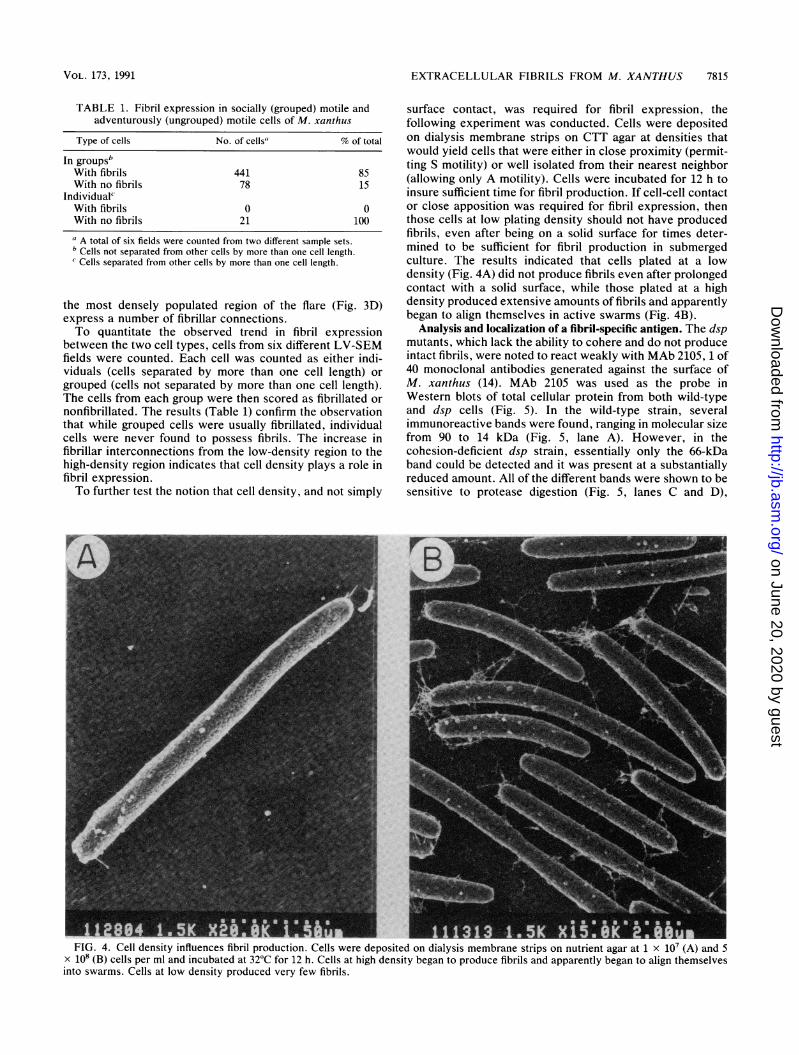
surface contact, was required for fibril expression, thefollowing experiment was conducted. Cells were depositedon dialysis membrane strips on CTT agar at densities thatwould yield cells that were either in close proximity (permit-ting S motility) or well isolated from their nearest neighbor(allowing only A motility). Cells were incubated for 12 h toinsure sufficient time for fibril production. If cell-cell contactor close apposition was required for fibril expression, thenthose cells at low plating density should not have producedfibrils, even after being on a solid surface for times deter-mined to be sufficient for fibril production in submergedculture. The results indicated that cells plated at a lowdensity (Fig. 4A) did not produce fibrils even after prolongedcontact with a solid surface, while those plated at a highdensity produced extensive amounts of fibrils and apparentlybegan to align themselves in active swarms (Fig. 4B).
Analysis and localization of a fibril-specific antigen. The dspmutants, which lack the ability to cohere and do not produceintact fibrils, were noted to react weakly with MAb 2105, 1 of40 monoclonal antibodies generated against the surface ofM. xanthus (14). MAb 2105 was used as the probe inWestern blots of total cellular protein from both wild-typeand dsp cells (Fig. 5). In the wild-type strain, severalimmunoreactive bands were found, ranging in molecular sizefrom 90 to 14 kDa (Fig. 5, lane A). However, in thecohesion-deficient dsp strain, essentially only the 66-kDaband could be detected and it was present at a substantiallyreduced amount. All of the different bands were shown to besensitive to protease digestion (Fig. 5, lanes C and D),
FlU. 4. Cell density influences fibril production. Cells were deposited on dialysis membrane strips on nutrient agar at 1 x 10' (A) and 5x 10⁸ (B) cells per ml and incubated at 32°C for 12 h. Cells at high density began to produce fibrils and apparently began to align themselvesinto swarms. Cells at low density produced very few fibrils.
VOL. 173, 1991
on June 20, 2020 by guesthttp://jb.asm
.org/D
ownloaded from

7816 BEHMLANDER AND DWORKIN
FIG. 5. Western blots of whole-cell extracts (probed with MAb2105) from cells of wild-type M. xanthus (MD 207; lane A) and a dspmutant (MD 1000; lane B). Cell extracts of the wild-type strain weredigested with a protease cocktail containing pronase E (1 mg/ml),proteinase K (0.2 mg/ml), trypsin (0.01 mg/ml), and chymotrypsin(0.1 mg/ml). These samples were incubated at pH 7.5 and 37°C for 0and 60 min (lanes C and D, respectively). Digestions were stoppedby the addition of SDS-PAGE sample buffer (15).
indicating that the antigen recognized by MAb 2105 iscomposed, at least in part, of protein. Interestingly, the90-kDa band was present only on cells grown in shakenliquid culture and was lost when cells were grown on a
surface (Fig. 6).To localize the antigens recognized by MAb 2105, cells
were labeled with the antibody and a 15-nm gold-labeledprotein A conjugate for analysis with the LV-SEM. TheHitachi S-900 LV-SEM is equipped with a backscatterdetector that permits the detection of metals of a differentmolecular mass than the primary coating metal (i.e., the gold
* .se, . jf b~~~~~4 k FS
- ~
FIG. 6. Western blots of whole-cell extracts (probed with MAb2105) grown in liquid culture (lane A) or on a solid surface (lane B).The arrow indicates the 90-kDa band.
of the conjugate versus the platinum coating metal). Figure 7shows that MAb 2105 reacts almost exclusively with theextracellular fibrils. What appears to be background labelingin the backscatter image is predominantly the labeling offibrillar fragments anchored to the substratum, as can beseen in the secondary electron image. Control samples,which were probed only with the conjugate, indicated verylittle background labeling. Because MAb 2105 labels onlyfibrils and not the cell surface, we have named these antigensFA-1.
Isolation of extracellular fibrils from M. xanthus. From theimmunogold localization results, it was apparent that MAb2105 could be used as a marker in the isolation of theextracellular fibrils from M. xanthus. The scheme devel-oped, essentially the centrifugal sedimentation of solubiliza-tion-resistant material, has proven a useful and reproduciblemeans of fibril isolation.The diazo dye Congo red had been shown to inhibit
cell-cell interactions in M. xanthus, essentially causing wild-type strains to behave as dsp mutants (2). It was also shownthat Congo red (5 ,ug/ml) inhibited the formation of fibrils (1),suggesting that the binding site of Congo red was on thefibrils. The dye was also used in attempts to isolate theCongo red receptor (fibrils) of M. xanthus (3). In light ofthese findings, initial fibril isolations were performed withCongo red in the wash buffers at a concentration of 5 ,ug/ml.When the suspended fibrillar material was then centrifuged,the resulting pellet had bound most of the dye, effectivelyremoving it from the supernatent fluids. This effect was notquantitated, the Congo red dye was found not to be requiredfor fibril isolation, and, accordingly, the dye was not addedin later isolations.Dot blots of whole cells and isolated fibrils probed with
MAb 2105 as a fibril-specific marker showed that there is a16-fold enrichment of the fibril-specific antigens recognizedby MAb 2105 in the material isolated from the whole cells(Fig. 8). SDS-polyacrylamide gels stained for total proteinand Western blots showed that, of the 14 protein bandspresent in the isolated fibrils, 10 reacted with MAb 2105 (Fig.9, lanes C and D). These data indicated that the isolatedfraction was indeed an enrichment of fibrillar material.
Isolated fibrils were probed with MAb 2105 and subjectedto immunogold analysis with the Hitachi S-900 LV-SEM.The results (Fig. 10) showed quite clearly that the fibrilsremained intact and that reactivity with MAb 2105 wasmaintained. Controls, in which fibrils were probed withconjugate only (omitting MAb 2105), showed an extremelylow level of background labeling (Fig. lOB). These resultsdemonstrate that the isolation procedure yielded fibrils thatare structurally intact and that those fibrils apparently main-tained their biochemical integrity (as evidenced by thebinding of MAb 2105).
DISCUSSION
On the basis of chemical and genetic evidence, the extra-cellular fibrils of M. xanthus have been shown to be themediators of cell-cell cohesion and as such they are requiredfor S (group) motility (2). Our observation of cells of M.xanthus by LV-SEM have revealed that fibril formation isregulated by cell density and contact with the substratum;under the appropriate conditions, a matrix is formed aroundthe cells consisting of lateral, branching fibrils measuring 30nm in diameter. The size, morphology, and absence of thesefibrils on dsp mutants have indicated to us that they are thesame as those reported by Arnold and Shimkets (1).
J. BACTERIOL.
A ~-
on June 20, 2020 by guesthttp://jb.asm
.org/D
ownloaded from

EXTRACELLULAR FIBRILS FROM M. XANTHUS 7817
FIG. 7. Immunoelectron localization of FA-1. Cells were probed with MAb 2105 and a 15-nm gold conjugate (protein A). (A) Secondaryelectron image; (B) backscatter electron image. The bright spots in the backscatter image are the gold particles. Note the fibril fragmentsvisible on the substrate in the secondary electron image. These micrographs show slightly less surface detail than others because they aretaken from images formed at an accelerating voltage of 15 kV rather than the 1.5 to 4.0 kV normally used.
In this article we also demonstrate the utility of theultra-high-resolution technique of LV-SEM for the study ofcell-cell interactions. With this technique, it has been possi-ble to examine the surface of the cells of M. xanthus with afar higher degree of resolution than has previously beenpossible with conventional SEM or transmission electronmicroscopy. In addition, our observations, along with im-proved analytical techniques (i.e., the incorporation of crit-ical-point drying of samples [5]), lend support to the viewthat the extracellular polysaccharides (glycocalyces) ofmany microorganisms are organized as fibrils (8).
Cells in a swarm differentially regulate the expression offibrils. Those cells moving as individual cells (presumablythe adventurously motile cells) do not express fibrillar con-nections, whereas a majority of the cells moving in groups
(presumably the socially motile cells) form intercellularconnections. Further experiments demonstrated that celldensity, rather than simple cell contact with the substratum,is required for fibril expression. These findings suggest theexistence of a mechanism for the regulation of fibril expres-sion (biosynthesis) that functions within the overall systemgoverning S and A motility behaviors in M. xanthus.
FA-1, recognized by MAb 2105, was identified as a seriesof protein antigens localized exclusively on the extracellularfibrils of M. xanthus. (Our laboratory has also used immu-nogold LV-SEM to localize a developmentally expressedfibrillar antigen, dFA-1 [7], and a cell surface antigen, CSA302 [6].) The recognition of several bands on a Western blotby a single monoclonal antibody can by accounted for in one
of two ways: either MAb 2105 recognizes different forms ofa single gene product (produced either through posttransla-tional modification or degradation) or the antibody recog-nizes a common epitope (either protein or carbohydrate) onseveral different proteins. Until the nature of the differentimmunoreactive bands is resolved, the designation FA-1 willbe retained for all of them. The amount of MAb 2105 boundby the cohesion-deficient dsp mutants is consistent with theobservation that these mutants produce a small amount ofwhat appears to be aberrantly formed fibrillar material.Attempts to label this material with MAb 2105 for immu-nogold analysis have been unsuccessful.The observation of cells of M. xanthus by LV-SEM
revealed that, under the appropriate conditions of cell den-sity and contact with the substratum, a matrix is formedaround the cells consisting of lateral, branching fibrils mea-suring 30 nm in diameter. The fibrils actually appear to be ofdifferent diameters, but the diameters of the fibrils at theirinsertion points at the cell surface are consistent. This mightsuggest that the fibrils are capable of cohering to each otherin a longitudinal fashion. The size, morphology, and absenceof these fibrils on dsp mutants indicated that they are thesame as those reported by Arnold and Shimkets (1).By using FA-1 as a fibril-specific marker, a technique for
the isolation of the 30-nm fibrils was developed. Observationof the isolated fibrils by using immunogold LV-SEM indi-cates that it is indeed composed of the 30-nm fibrils thatmediate cohesion (1) and make up at least part of theextracellular matrix. The isolated fibrils of M. xanthus bind
VOL. 173, 1991
on June 20, 2020 by guesthttp://jb.asm
.org/D
ownloaded from

7818 BEHMLANDER AND DWORKIN
*0*,
42%
0A f7
FIG. 9. Western blots probed with MAb 2105 and SDS-PAGEstained with Ponceau S (for total protein) of whole cells (lanes A andB) and isolated fibrils (lanes C and D). Each lane contained 25 mg ofprotein. Lanes A and C were stained with Ponceau S; lanes B and Dwere probed with MAb 2105.
FIG. 8. Enrichment of FA-1 in isolated fibrils. Endpoint dilutiondot blots of whole cells (column A) and isolated fibrils (column B)from M. xanthus were done. Each spot is a twofold dilution of theprevious spot. The final dilution displaying immunoreactivity isindicated with an arrow. The inverse of the dilution factor for thelast immunoreactive spot gave the number of relative immunoreac-tive units in the sample. Each of the initial spots (labeled 0)contained 25 mg of protein. Blots were probed with MAb 2105.
Congo red and remove the dye from solution, suggesting thatthey constitute what Arnold and Shimkets referred to as theCongo red receptor (1). On the basis of an analogy to theextracellular fibrils of other bacteria that bind Congo red, itis likely that the fibrils of M. xanthus contain substantialamounts of polysaccharides. Indeed, extracellular fibrilscomposed of polysaccharides are the apparent mediators ofcohesion in several other prokaryotes, including Agrobacte-rium tumefaciens (21), Zooglea ramigera (12), and Rhizo-bium leguminosum (30), among others (27).
Sutherland and Thomson isolated exopolysaccharide fromseveral genera of myxobacteria as an acetone-precipitablefraction released from intact cells by shear forces (33).Exopolysaccharide from several species of the genus Myxo-coccus constitutes 5 to 10% of the dry weight of the startingmaterial and is predominantly composed of carbohydrates(neutral and amino sugars) (32). While it seems likely that the30-nm extracellular fibrils that have been isolated from M.xanthus comprise at least part of the extracellular matrix,complete compositional analysis has yet to be done. Thepossible existence of amorphous polysaccharides (not de-
tected by LV-SEM) must also be explained. A more com-plete structural and compositional analysis of the isolatedfibrils will allow more detailed conclusions to be drawnabout their precise function in M. xanthus.By using the ultra-high-resolution technique of LV-SEM,
new insights have been gained with regard to the cell-cellinteractions of M. xanthus. We have demonstrated that theproduction of extracellular matrix fibrils is differentiallyregulated between cells expressing A and S motility behav-iors. That the expression of extracellular fibrils is dependenton cell density indicates the existence of a mechanism for theperception of cell density and the regulation of fibril produc-tion, a system which seems to operate within the overallsystem governing A and S behaviors in M. xanthus. Byisolating the extracellular fibrils and purifying the proteinsuniquely associated with them, we hope to gain a betterunderstanding of their structure and function. It is possiblethat the extracellular fibrils are not only the mediators ofcell-cell cohesion but also function as an integral part of acontact-mediated signaling system used to sense and regu-late cell density in M. xanthus.
ACKNOWLEDGMENTS
We thank Chris Fretham for instruction in the use of the HitachiS-900 LV-SEM. We also thank Christina M. Chance, Deborah A.Eastman, and Ellis Brockman for helpful insights and discussions.
This work was supported by Public Health Service grant GMS19957 from the National Institutes of Health to M.D. R.M.B. was apredoctoral fellow supported by NRSA Institutional Training Grant5T32CA09138. The Hitachi S-900 LV-SEM was obtained with theassistance of grant 1 S10 RRO 4973 from the National Institutes ofHealth to Stanley Erlandsen.
J. BACTERIOL.
.l .:
on June 20, 2020 by guesthttp://jb.asm
.org/D
ownloaded from

VOL. 173, 1991 EXTRACELLULAR FIBRILS FROM M. XANTHUS 7819
FIG. 10. Immunogold labeling of FA-1 on isolated fibrils. (A) Fibrils labeled with MAb 2105; (B) fibrils labeled with conjugate only as acontrol for background binding. Both micrographs are from backscatter images taken at an accelerating voltage of 4.0 kV.
REFERENCES1. Arnold, J. W., and L. J. Shimkets. 1988. Inhibition of cell-cell
interactions in Myxococcus xanthus by Congo red. J. Bacteriol.170:5765-5770.
2. Arnold, J. W., and L. J. Shimkets. 1988. Cell surface propertiescorrelated with cohesion in Myxococcus xanthus. J. Bacteriol.170:5771-5777.
3. Arnold, J. W., and L. J. Shimkets. Personal communication.4. Autrada, R. 1989. Backscattered electron imaging using single
crystal scintillator detectors. Scanning Microsc. 3:739-763.5. Cagle, G. D. 1974. Critical-point drying: rapid method for the
determination of bacterial extracellular polymer and surfacestructures. Appl. Microbiol. 28:312-316.
6. Chance, C. M. Personal communication.7. Clemans, D. L. 1991. Ph.D, thesis. University of Minnesota,
Minneapolis.8. Costerton, J. W., R. T. Irvin, and K. Cheng. 1981. The bacterial
glycocalyx in nature and disease. Annu. Rev. Microbiol. 35:299-324.
9. Dobson, W. J., and H. D. McCurdy. 1979. The function offimbriae in Myxococcus xanthus. I. Purification and propertiesof Myxococcus xanthus fimbriae. Can J. Microbiol. 25:1152-1160.
10. Dworkin, M., and D. Eide. 1983. Myxococcus xanthus does notrespond chemotactically to moderate concentration gradients.J. Bacteriol. 154:437-442.
11. Dworkin, M., and D. Kaiser. 1985. Cell interactions in myxo-bacterial growth and development. Science 234:18-24.
12. Easson, D. D. J., Jr., A. J. Sinskey, and 0. P. Peoples. 1987.Isolation of Zooglea ramigera I-16-M exopolysaccharide bio-synthetic genes and evidence for instability within this region. J.Bacteriol. 169:4518-4524.
13. Fluegel, W. 1963. Simple method for demonstrating myxobac-terial slime. J. Bacteriol. 85:1173-1174.
14. Gill, J., E. Stellwag, and M. Dworkin. 1985. Monoclonal anti-bodies against cell surface antigens of developing cells ofMyxococcus xanthus. Ann. Inst. Pasteur/Microbiol. (Paris)136A:11-18.
15. Harlow, E., and D. Lane. 1988. Antibodies: A laboratorymanual. Cold Spring Harbor Laboratory, Cold Spring Harbor,N.Y.
16. Hodgkin, J., and D. Kaiser. 1979. Genetics of gliding motility inMyxococcus xanthus (Myxobacterales): two gene systems con-trol movement. Mol. Gen. Genet. 171:177-191.
17. Kaiser, D. 1979. Social gliding is correlated with the presence ofpili in Myxococcus xanthus. Proc. Natl. Acad. Sci. USA 76:5952-5956.
18. Kaiser, D., and C. Crosby. 1983. Cell movement and its coor-dination in swarms of Myxococcus xanthus. Cell Motil. 3:227-245.
19. Kim, S. K., and D. Kaiser. 1990. C-factor: a cell-cell signallingprotein required for fruiting body morphogenesis of Myxococ-cus xanthus. Cell 61:19-26.
20. MacRae, T. H., and H. D. McCurdy. 1976. Evidence formotility-related fimbriae in the gliding microorganism Myxococ-cus xanthus. Can. J. Microbiol. 22:1589-1593.
21. Matthysse, A. G. 1983. Role of bacterial cellulose fibrils inAgrobacterium tumefaciens infection. J. Bacteriol. 154:906-915.
22. Nagatani, T. 1989. The ultra-high resolution scanning electronmicroscope and some applications to biological sciences. Bio-Techniques 7:270-275.
23. Reichenbach, H. 1985. Myxobacteria: a most peculiar group ofsocial prokaryotes, p. 1-50. In E. Rosenberg (ed.), Myxobacte-ria: development and cell interactions. Springer-Verlag, NewYork.
24. Rosenberg, E. (ed.). 1984. Myxobacteria. Development and cellinteractions. Springer-Verlag, New York.
on June 20, 2020 by guesthttp://jb.asm
.org/D
ownloaded from

7820 BEHMLANDER AND DWORKIN J. BACTERIOL.
25. Rosenberg, E., K. H. KeUer, hnd M. Dworkin. 1977. Celldensity-dependent growth of Myxococcus xanthus on casein. J.Baternol. 129:770-777.
26. Shlmkets, L. J. 1986. Correlation of energy-dependent cellcohesion with social motility in Myxococcus xanthus. J. Bacte-riol. 166:837-841.
27. Shlmkets, L. J. 1990. Social and developmental biology of themyxobacteria. Microbiol. Rev. 54:473-501.
28. Shlmkets, L. J., and D. Kaiser. 1982. Induction of coordinatedmovement of Myxococcus xanthus cells. J. Bacteriol. 152:451-461.
29. Sigma Chemical Co. 1991. Sigma manual. Sigma Chemical Co.,St. Louis, Mo.
30. Smit, G., J. N. Kjne, and B. J. Lugtenberg. 1987. Involvement
of both cellulose fibrils and a Ca2' dependent adhesin in theattachment of Rhizobium leguminosarum to pea root hair tips.J. Bacteriol. 169:4294-4301.
31. Stackebrandt, E., R. G. E. Murray, and H. G. Truper. 1988.Proteobacteria classis nov., a name for the phylogenetic taxonthat includes the "purple bacteria and their relatives." Int. J.Syst. Bacteriol. 38:321-325.
32. Sutherland, I. W. 1979. Polysaccharides produced by Cystobac-ter, Archangium, Sorangium and Stigmatella species. J. Gen.Microbiol. 111:211-216.
33. Sutherland, I. W., and S. Thomson. 1975. Comparison ofpolysaccharides produced by Myxococcus strains. J. Gen. Mi-crobiol. 89:124-132.
on June 20, 2020 by guesthttp://jb.asm
.org/D
ownloaded from