exosomes: vesicular carriers for intercellular ... exosomes: vesicular carriers for intercellular...
TRANSCRIPT
REVIEW
Exosomes: vesicular carriers for intercellular communicationin neurodegenerative disorders
Anja Schneider & Mikael Simons
Received: 10 January 2012 /Accepted: 5 April 2012 /Published online: 19 May 2012# The Author(s) 2012. This article is published with open access at Springerlink.com
Abstract The intercellular transfer of misfolded proteinshas received increasing attention in various neurodegenera-tive diseases characterized by the aggregation of specificproteins, as observed in Alzheimer’s, Parkinson’s andHuntington’s disease. One hypothesis holds that intercellu-lar dissemination of these aggregates within the centralnervous system results in the seeded assembly of the cog-nate soluble protein in target cells, similar to that proposedfor transmissible prion diseases. The molecular mechanismsunderlying the intercellular transfer of these proteinaceousaggregates are poorly understood. Various transfer modes of
misfolded proteins including continuous cell-cell contactssuch as nanotubes, unconventional secretion or microve-sicle/exosome-associated dissemination have been sug-gested. Cells can release proteins, lipids and nucleic acidsby vesicular exocytosis pathways destined for horizontaltransfer. Encapsulation into microvesicular/exosomalvehicles not only protects these molecules from degradationand dilution in the extracellular space but also facilitatesdelivery over large distances, e.g. within the blood flow orinterstitial fluid. Specific surface ligands might allow thehighly efficient and targeted uptake of these vesicles byrecipient cells. In this review, we focus on the cell biologyand function of neuronal microvesicles/exosomes and dis-cuss the evidence for pathogenic intercellular protein trans-fer mediated by vesicular carriers.
Keywords Exosomes . Dementia . Spreading . Transfer .
Aggregopathy
Cell biology of microvesicles and exosomes
Microparticles have been isolated from various body fluidssuch as urine, ascites, saliva, breast milk and blood by ultracen-trifugation, ultrafiltration or immunoprecipitation (Simpsonet al. 2009). A consensus regarding the nomenclature of theseheterogeneous vesicular populations is still missing because ofexperimental difficulties in separating and distinguishing thevarious extracellular vesicles based on their biochemical ormorphological properties. The terminology mainly refers tothe cellular origin (e.g. aggrosomes, prostasomes, promino-somes), their attributed function (e.g. apoptotic body), size(ranging from 40 nm to 4 μm) or subcellular origin (exosomes,shedding vesicles; see Table 1). Whereas exosomes are builtwithin the endosomal system, shedding vesicles (or ectosomes)
A. Schneider (*)Department of Psychiatry and Psychotherapy,University Medicine Goettingen,Von-Siebold-Str.5,37075 Goettingen, Germanye-mail: [email protected]
A. Schneider :M. SimonsDFG Research Center for Molecular Physiology of the Brain,CMPB,Goettingen, Germany
A. SchneiderGerman Center for Neurodegenerative Diseases (DZNE),Goettingen,Von-Siebold-Str.5,37075 Goettingen, Germany
A. Schneider :M. SimonsMax-Planck-Institute for Experimental Medicine,Hermann-Rein-Str.3,37075 Goettingen, Germany
M. SimonsDepartment of Neurology, University Medicine Goettingen,Robert-Koch-Str. 40,37075 Goettingen, Germany
Cell Tissue Res (2013) 352:33–47DOI 10.1007/s00441-012-1428-2
bud directly from the plasma membrane into the extracellularspace. Shedding vesicles can be further divided into micro-vesicles, with variable diameters of 0.1 to 1 μm and the largerapoptotic bodies.
Exosomes
Exosomes are generated within the (late) endosomal com-partments by inward vagination and fission of the limitingmembrane. Endosomes that are filled with these intralumi-nal vesicles (ILV) are termed multivesicular endosomes(MVE). ILVs can serve as storage compartments forproteins and signalling complexes and can re-enter the cy-tosol by backfusion with the MVE limiting membrane(Abrami et al. 2004; Le Blanc et al. 2005; Dobrowolskiand De Robertis 2011). In addition to a mere storage func-tion, the MVE can either fuse with the lysosome, followedby the degradation of ILVs, or with the plasma membrane torelease the ILVs as exosomes into the extracellular space.Whether these different pathways correspond to distinctsubclasses of MVEs or whether each MVE can switchbetween the different itineries described above is unknown.Exosomes contain cytosol and feature a membrane topologythat is inverse to the endosomal membrane. The inner exo-somal membrane leaflet faces the cytosol, whereas the outerleaflet adjoins the extracellular space. Exosomes are secret-ed by a variety of cells in vitro and in vivo under physio-logical and pathological conditions. On transmission ofelectron or cryo-electron microscopic images, exosomesappear as vesicles of 40–100 nm in diameter with a charac-teristic round or cup-shaped morphology (Thery et al. 2006;Conde-Vancells et al. 2010). Exosomes differ in their originand in their protein and lipid composition. Depending ontheir cellular ancestry, they carry cell-type-specific proteins,such as major histocompatibility complex (MHC) whenreleased from antigen-presenting cells, or myelin proteins,when derived from oligodendrocytes (Kramer-Albers et al.2007; Thery et al. 2001). Several proteins are specificallyenriched in exosomes and serve as marker proteins. Theseinclude the integrins and tetraspanins CD63, CD89, CD81,CD9 and CD82, the MVE proteins alix and tsg101, theendosomal and endosome maturation-related proteinsflotillin and annexin and the heat shock proteins hsp70 andhsp90 (Simons and Raposo 2009). Proteins derived from thenucleus, mitochondria or endoplasmic reticulum are mainlyexcluded from the exosomal pathway.
Shedding vesicles
Shedding vesicles (or ectosomes) are generated by shedding atthe plasma membrane and include microvesicles with a het-erogeneous size range from 100 nm to 1 μm and apoptoticbodies. Apoptotic bodies are released from the plasma mem-brane during the breakdown of apoptotic cells. They carryDNA, histones, organelles and surface markers that allowtheir recognition and internalization by phagocytic and othersubsequent cells, thereby preventing the release of intracellu-lar content and inflammatory reactions (Nunez et al. 2010;Thery et al. 2001). Their diameter varies between 1 and 4 μm.Shedding particles with a diameter of 100 nm cannot bedistinguished from endosomally derived exosomes on a mor-phological or biochemical basis, including density gradientcentrifugation. Some authors refer to these vesicles as exo-somes derived from the direct pathway as compared withexosomes that stem from the endosomal indirect pathway(Booth et al. 2006; Simons and Raposo 2009). Further com-plexity is added by the finding that several proteins can budeither into exosomes or shedding vesicles in a cell-type-dependent manner (Shen et al. 2011a). Throughout our re-view, we will therefore use the umbrella term “exosomes andmicrovesicles” (EMV) to describe extracellular vesicles thatare of 40-100 nm in size and that are generated within bothpathways as suggested by Shen et al. (2011a). Despite theexperimental difficulties in distinguishing between exo-somes and microvesicles, they might still represent distinctentities with different properties and functions.
Physiological function of EMVs
Exosomes were first identified as a pathway for shuttlingsuperfluous material out of the cell, especially from cells withlow lysosomal activity or lysosome number. Only recently hastheir role as an alternative exocytosis pathway for cytosolic ortransmembrane proteins and their function in the targeted de-livery of molecules destined for intercellular communicationand signalling been recognized (Mathivanan et al. 2010b).Targeting mechanisms for the selective sorting of proteins,lipids, mRNA and small non-coding RNA are under intenseinvestigation since certain cellular subsets of these moleculesare specifically enriched in EMVs. There is ample evidence fora role of EMVs in intercellular communication; however, themechanisms for target cell recognition, entry and the intracel-lular itinery in recipient cells are far from being understood.
Table 1 Extracellular vesiclesand their characteristics Microparticles Origin Size Flotation
Exosomes Multivesicular endosome 40–100 nm 1.13–1.19 g/ml
Shedding vesicles(ectosomes)
Microvesicle Plasma membrane 0.1–1 μm
Apoptotic body Plasma membrane 1–4 μm 1.24–1.28 g/ml
34 Cell Tissue Res (2013) 352:33–47
Regarding the central nervous system (CNS), EMV release hasbeen shown in vitro for oligodendrocyte, microglia, astrocyteand neuronal cell cultures (Faure et al. 2006; Kramer-Albers etal. 2007; Potolicchio et al. 2005; Taylor et al. 2007).
Neuronal EMVs
Origin
Primary neurons release vesicles which can be isolated fromconditioned medium in vitro (Faure et al. 2006). Their sizeand morphology as assessed by gradient centrifugation andelectron microscopy closely resemble EMVs and the prep-arations are positive for exosomal marker proteins, such ashsp70 and flotillin (Bulloj et al. 2010; Faure et al. 2006;Lachenal et al. 2011). Because of the lack of specific exo-somal marker proteins, difficulties abound when trying toestablish whether these vesicles represent bona fide exo-somes derived from the indirect endosomal pathway. Re-cently, Lachenal et al. (2011) have demonstrated thepresence of tetanus toxin in EMV preparations derived fromneuronal culture medium. Tetanus toxin is endocytosedfrom the cell surface and is present in endosomes. Theauthors therefore speculate that these tetanus-toxin-positiveEMVs originate from the indirect pathway (Lachenal et al.2011). However, the presence of tetanus toxin does notexclude direct budding from the plasma membrane, sincetetanus toxin primarily binds to membrane gangliosides andwould also be expected in vesicles that bud directly from theplasma membrane.
Neuronal MVEs are predominantly distributed within thesomatodendritic compartment where they are 50 times moreabundant than in the axon (for a review, see Von Bartheld andAltick 2011). The accumulation of MVEs at the postsynapseindicates that MVE fusion and exosome release might occurfrom dendritic spines. Electron-microscopic examination ofstimulated primary neuronal cultures has demonstrated vesic-ular structures with the size and morphology of exosomes inclose proximity to somatodendritic compartments (Lachenalet al. 2011).More experiments, e.g. with chamber systems, areneeded to improve the characterization of the sites of EMVrelease in polarized neurons. In addition, knowledge of wheth-er MVEs released from different neuronal subcompartmentsare distinct with regard to their molecular composition andcargo would be of interest.
Function
Neuronal MVEs have been shown to carry glutamate receptor(GluR2) subunits. MVE-mediated release could therefore be amechanism to eliminate α-amino-3-hydroxy-5-methyl-4-iso-xazoleproprionic acid (AMPA) receptors in response to
glutamatergic stimulation (Lachenal et al. 2011). Thus, exo-somes released from the postsynaptic site might modulate syn-aptic transmission and plasticity. This notion is furthersupported by the finding that the number of dendritic MVEsand EMV release increase in electrically stimulated neurons(Kadota et al. 1994; Kraev et al. 2009). Likewise, prolongedpotassium-induced depolarization of neuronal cultures potenti-ates EMV secretion (Faure et al. 2006). Further evidence foractivity-dependent EMVrelease has been provided by Lachenalet al. (2011) who have demonstrated that neuronal EMV secre-tion is regulated by calcium influx and glutamatergic activity.Not only treatment with ionomycin to raise intracellular calciumconcentrations but also increased glutamatergic activity afterpharmacological inhibition of γ-aminobutyric acid (GABA)-Areceptors results in enhanced EMV secretion from neuronalcultures. Interestingly, treatment with AMPA- or N-methyl D-aspartate (NMDA)-receptor antagonists counteract the glutama-tergic effect on EMV release. Hence, the authors speculate thatneurons modulate their number of ionotropic postsynapticreceptors, synaptic plasticity and strength by activity-dependent EMV release (Lachenal et al. 2011).
In vivo evidence of neuronal exosome release and itsfunctional significance is still lacking. The transduction ofwnt signalling by exosome-like vesicular structures hasbeen reported in Drosophila. The palmitoylated wnt pro-teins are membrane-bound and thus unlikely to be releasedas soluble proteins to the extracellular space. Instead, theDrosophila wnt1 homolog wingless (wg) has been shown tobe transported trans-synaptically with vesicles resemblingexosomes, followed by the binding of wg to Drosophilafrizzled 2 (DFz2) receptors at the postsynapse (Korkutet al. 2009). Further in vivo evidence for neuronally derivedEMVs is based on their presence in cerebrospinal fluid(CSF). Vella et al. (2008) have described the isolation ofmicroparticles, which are enriched in the native prion proteinPrPc, from ovine CSF. Harrington et al. (2009) have identi-fied, in human CSF, nanostructures including exosome-likevesicles that can be labelled with antibodies against variousexosomal marker proteins in immuno-transmission electronmicroscopy. Whereas these vesicles might be derived fromCSF immune cells or ventricular ependymal cells, we havebeen able to fractionate, from human CSF, exosome-shapedvesicles positive for GluR2, indicating their neuronal origin(own unpublished data).
Exosomes in neurodegenerative diseases
Although definitive evidence for intercellular EMV transferwithin the CNS is still lacking, EMVs have been repeatedlydiscussed as potential carriers in the dissemination of diseasepathology in neurodegenerative disorders (for a review, seeAguzzi and Rajendran 2009).
Cell Tissue Res (2013) 352:33–47 35
Prions
This hypothesis evolved first in the context of the interneu-ronal spreading of transmissible prion disorders such as thenew variant of Creutzfeld-Jacob disease (CJD), bovinespongiform encephalitis (BSE) and scrapie. Prions exist intwo different conformational states: the natively folded PrPc
and the disease-associated misfolded PrPsc. PrPsc is charac-terized by an abnormal conformation, which can serve as atemplate to induce the misfolding of PrPc (a mechanismcalled permissive templating). In infectious prion diseases,PrPsc can enter the organism by the gut, followed by theinvasion of lymphoid tissue from where it spreads into theperipheral nervous system and finally the CNS. In additionto intercellular transfer by tunneling nanotubes, as discussedby Gousset et al. (2009), a role for exosomes as a carrier forPrPsc in this intercellular dissemination has been proposed.Tunnelling nanotubes are transient membranous connec-tions that can connect cells over distances of up to 100μm. Two types of nanotubes can be distinguished basedupon their diameter and cytoskeleton, which includes eitheractin or actin and microtubules. The transport of vesiclesand organelles has been demonstrated within nanotubes thatcan bridge the distance between numerous cell types (Gurkeet al. 2008). PrPsc-bearing exosomes can travel either withthe blood stream or after internalization within blood cells toreach their target cells. This hypothesis has been triggeredby the finding that cell culture medium from a scrapie-infected hypothalamic GT1 cell line can induce PrPsc for-mation in recipient cells, indicating a cell-free transfer mode(Schatzl et al. 1997). Both PrPc and PrPsc are released fromcells expressing ovine PrP together with vesicles that, basedon their morphology, biochemical properties and proteincomposition, closely resemble exosomes (Fevrier et al.2004). Exosomal PrPsc and PrPc secretion from an endoge-nously PrP-expressing neuronal cell line has been reportedupon infection with PrPsc (Veith et al. 2009; Vella et al.2007). Incubation of target cells with exosome preparationsfrom prion-infected neuronal cells is sufficient to induce theconformational shift to PrPsc in various target cell lines. Fur-thermore, intracerebral injection of PrPsc-positive exosomalmembranes triggers neurodegeneration and death in recipientmice transgenic for ovine PrP (Fevrier et al. 2004). Both PrPc
and PrPsc have been detected in late endosomes and MVEs onan ultrastructural level, indicating an exosomal pathway(Ersdal et al. 2009; Godsave et al. 2008; Laine et al. 2001;Marijanovic et al. 2009).
The subcellular compartment in which the conformation-al shift from PrPc to PrPsc takes place remains unclear;however, speculation that the MVE/EMV system is in-volved via local protein enrichment, favourable pH and thelipid environment is tempting. Macromolecular crowdinghas been shown to promote the conversion to β-sheet
structure and the oligomerization of prions (Huang et al.2010). Exosomal enrichment of PrPc and PrPsc might gen-erate a high local concentration and close proximity betweentemplate and PrPc, thereby facilitating the conformationalshift to PrPsc. Furthermore, the conversion of PrPc to PrPsc
requires the partitioning of PrP into sphingolipid- andcholesterol-rich membrane domains, which are present inexosomal membranes (Baron et al. 2002; Laulagnier et al.2004; Subra et al. 2007). Along this line, the in vitro gen-eration of infectious PrPsc from bacterially expressed recom-binant PrPc has been shown to require the presence of lipidcofactors, such as the synthetic anionic phospholipid POPG(1-palmitoyl-2-oleoylphosphatidylglycerol; Wang et al.2010). In addition, several studies have indicated that con-version takes place in acidic endosomal compartments, ar-guing again for a conversion within the late endosome/MVE(Peters et al. 2003). Alternatively, the fusion of PrPsc-posi-tive exosomes with the recipient cell membrane might in-duce the conversion of PrPc at the target cell surface, as hasbeen indicated by Baron et al. (2002) who have shown thatthe conversion of PrPc to PrPsc requires the insertion of PrPsc
into target cell membranes and the formation of a contiguousmembrane layer.
AA-amyloidosis
Similar to transmissible prion diseases, an exosome-mediated transfer of misfolded proteins has been shownfor systemic AA-amyloidosis in vivo. Serum amyloid-A(SAA) proteins are apolipoproteins that are expressed inthe liver and that circulate in the blood stream bound tohigh density lipoproteins. Under inflammatory conditionsand interleukin-1 and -6 and tumor necrosis factor stimula-tion, the expression of these acute phase proteins is in-creased up to 1000-fold. During chronic inflammationsuch as rheumatoid arthritis, high concentrations of SAAeventually lead to the formation of a nucleus and polymer-ization of otherwise soluble SAA proteins into amyloidfibrils. Deposits of SAA fibrils can be found in the intersti-tial space of many organs. Similar to prion protein misfold-ing, this SAA fibrillation involves a conformational shift ofSAA protein into a β-sheet structure followed by aggrega-tion. Mouse models of experimental AA-amyloidosis devel-op systemic amyloid deposits under chronic inflammatoryconditions triggered by the intravenous, intraperitoneal ororal application of SAA-containing tissue or circulatingblood monocytes derived from murine SAA mouse models.This process is reminiscent of transmissible prion diseases(Axelrad et al. 1982; Werdelin and Ranlov 1966). The“seeding” factor, also termed amyloid-enhancing factor(AEF), has been shown to consist in either SAA oligomersor SAA fibrils (Lundmark et al. 2002; Senthilkumar et al.2008; Sponarova et al. 2008). Tasaki et al. (2010) have
36 Cell Tissue Res (2013) 352:33–47
demonstrated that blood and plasma derived from experi-mental murine SAA amyloidosis models can induce pathol-ogy in recipient animals and that freeze-thaw cycles abolishthe seeding activity of these plasma samples. The authorshave been able not only to show that plasma EMVs isolatedfrom mice with SAA amyloidosis carry oligomeric andprefibrillar SAA but also that these EMVs are sufficient totransmit disease pathology to recipient animals (Tasaki et al.2010). Noteworthy, though, is that not all exosome prepa-rations possess seeding capacity, which might be a result ofshearing forces or the clumping of EMVs during the prep-aration process. Another possible explanation is that onlyEMVs derived from SAA-positive organs can induceamyloidosis in recipient mice and that these EMVs arenot present in the plasma in sufficiently high numbersall the time.
An oral transmission of SAA amyloidosis among cheetahs,which secrete SAA fibrils in their faeces, has been reported(Zhang et al. 2008). Potentially infectious SAA fibrils havealso been detected in foie gras (Solomon et al. 2007). Severallines of evidence point to an uptake of exogenous SAAamyloid seeds via the epithelial cells in Peyer’s plaques,followed by transepithelial transport, internalization intofollicular dendritic cells and transfer to the spleen whereamyloid replication and deposition occurs (for a review, seeWestermark and Westermark 2009). This itinery most likelyreflects a selective targeting pathway rather than randomuptake and release of free circulating fibrils. The exosomaltransfer of SAA aggregates could help to explain this repro-ducible route of seed propagation attributable to tissue- or cell-specific uptake signals on the surface of EMVs. Similar totransmissible prion diseases, cells from the lymphomoncyticlineage could mediate amyloid transport by the uptake ofSAA-positive EMVs via specific receptors, followed by trans-port within the circulation and release through another roundof exocytosis in the target tissue. In support of this notion,macrophages have been shown in vitro to be able to internal-ize AEF from the culture medium and SAA has been detectedin various endocytic compartments (Kluve-Beckerman et al.2001). Immunoelectron analysis has revealed fibrillar SAAprotein in lysosomes and LAMP-positive structures in mono-cytoid cells from SAA amyloidosis mice (Chronopoulos et al.1994). Taken together, these findings are compatible withendocytic uptake, transport with the blood stream and theexocytosis and transfer of SAA aggregates via the MVE/EMV pathway.
Neurodegenerative aggregopathies
The transneuronal spreading of oligomers or fibrillar aggre-gates is increasingly recognized in a variety of neurodegener-ative disorders including tau protein and amyloid-β peptide inAlzheimer’s disease, superoxide dismutase 1 (SOD1) in
amyotrophic lateral sclerosis (ALS), huntingtin in Hunting-ton’s disease (HD) and α-synuclein in Parkinson’s disease(PD). Aggregopathies do not belong to the class of priondiseases, since infectious transmission between two individu-als has never been observed. However, intra- and inter-individual spreading of disease pathology in several of theseaggregopathies has led to their classification as possible prio-noid disorders (Aguzzi and Rajendran 2009). Strikingly, allproteins involved in the pathogenesis of these diseases seem tobe present in EMVs.
α-Synuclein
PD is characterized by intracellular aggregates of α-synuclein,which are refered to as Lewy bodies. Lewy bodies appear firstin the brainstem followed by the subsequent deposition ofaggregates in higher brain regions. The spatial distribution ofLewy body pathology over time follows a predictable anatom-ical course that reflects patterns of neuronal connectivity(Braak et al. 2004). Similarily, anatomically connected spread-ing patterns have been observed in prion models of the Syrianhamster after the oral uptake of prions, starting in the dorsalvagus nerve and followed by the medulla, pons, midbrain andcerebellum (Natale et al. 2011). Likewise, after the injection ofinfectious prions into the eye, the pathology develops along theoptical tracts (Liberski et al. 1990). The hypothesis of inter-neuronal disease propagation in synucleinopathies has beenfuelled by the finding that transplanted fetal neurons in PDpatients accumulate intraneuronal α-synuclein aggregates, in-dicating a possible transfer of pathology from substantia nigrahost neurons to grafted striatal neurons (Kordower et al. 2008;Li et al. 2008). In a similar fashion, host to graft transmission ofα-synuclein has been observed in an α-synuclein transgenicmouse model in which green-fluorescent-protein-labelled neu-ronal stem cell transplants incorporate the host’s transgenicallyexpressed α-synuclein (Desplats et al. 2009). The induction ofα-synuclein aggregation and the worsening of behaviour and/or motor phenotype have been demonstrated in transgenicmice after the intracerebral injection of brain extracts derivedfrom older littermates that exhibited α-synuclein aggregates(Mougenot et al. 2011). Interneuronal transfer of α-synucleinaggregates could serve as a seed to induce aggregation in thehost neuron and contribute to the dissemination of aggregatesthroughout the brain, similar to prion-like self-propagation.Intercellular transfer and the induction of disease pathologyhave recently been described for PrPsc. Intercellular propaga-tion of α-synuclein seeds could either be mediated by tunnel-ling nanotubes, which connect neighbouring neurons, byextracellular α-synuclein species passively released fromdying neurons or by active secretion, including EMV-basedrelease (Fig. 1; Agnati et al. 2010; Danzer et al. 2011;Emmanouilidou et al. 2010). An atypical secretion mechanismhas been discussed, as has passive release from dying neurons,
Cell Tissue Res (2013) 352:33–47 37
to explain the extracellular presence of this cytosolic protein,which lacks conventional secretion signals. Extracellular non-vesicular α-synuclein has been detected in tissue culture me-dium and in CSF and its concentration is increased undercellular stress conditions suggesting a regulated release mech-anism (Jang et al. 2010). In addition, α-synuclein has beendemonstrated in EMVs derived from neuronal cultures(Emmanouilidou et al. 2010). To date, the form of extracellularα-synuclein that is relevant for the disease pathology and theway that the cytosolic protein can be actively secreted fromcells are unknown. EMVs could act as “Trojan horses” in thetransneuronal propagation of α-synuclein aggregates (Brundinand Olsson 2011). Speculation that α-synuclein-containingEMVs are internalized into target cells at a much higher effi-ciency than non-vesicular α-synuclein species is tempting. Inaddition, the exosomal compartment could favour the aggrega-tion of α-synuclein by increased local protein concentrations,pH and high membrane curvature, similar to the situation thatwe discussed for the case of prion protein transconformation.Aggregates ofα-synuclein are well established to be able to actas seeds to trigger the aggregation of the monomeric protein.For example, Hansen et al. (2011) have demonstrated cellularrelease, endocytic uptake, co-dimerization and aggregate for-mation of α-synuclein in recipient cells within a co-culturesystem. The transfer of α-synuclein is independent of directcell-cell contacts; however, despite the presence of α-synucleinin EMVs, they have yet to be shown to be the carriers forintercellular α-synuclein transfer. In vivo evidence of a func-tionally active uptake of exosomes into postmitotic neurons has
recently been provided by Alverez-Eviti et al. (2011), althoughonly with exosomes that have been produced in transgenic cellsthat transgeneously express a rabies glycoprotein construct thatis sorted into exosomes and confers neuroglia-specific uptake.Alternatively, α-synuclein might reach the target cell uponunconventional secretion or passive release from dying cells(Nickel and Rabouille 2009). The trans-synaptic transmissionof toxic α-synuclein oligomers has been demonstrated in tissueculture models (Danzer et al. 2011). The proportion of extra-cellular α-synuclein that is localized in EMVs and the form(free or EMV-encapsulated) of α-synuclein that confers toxic-ity and/or seeding capacity remain unknown.
Tau
In AD and other tauopathies such as corticobasal degener-ation, progressive supranuclear palsy and a subgroup offrontotemporal dementias, intracellular aggregates of themicrotubule-associated protein tau are assumed to mediateneuronal dysfunction and subsequently neurodegeneration.Tau aggregates in AD emerge first in the entorhinal cortexfollowed by propagation to hippocampal regions, temporallobes and more distant neocortical areas (Bancher et al.1993). Recently, the interneuronal transmission of tau pa-thology was reported in vitro whereby exogenously addedtau fibrils were internalized into host cells and induced theaggregation of endogenous tau protein (Frost et al. 2009;Guo and Lee 2011). In addition, tau aggregates have beenshown to be transferred between cells in a co-culture system
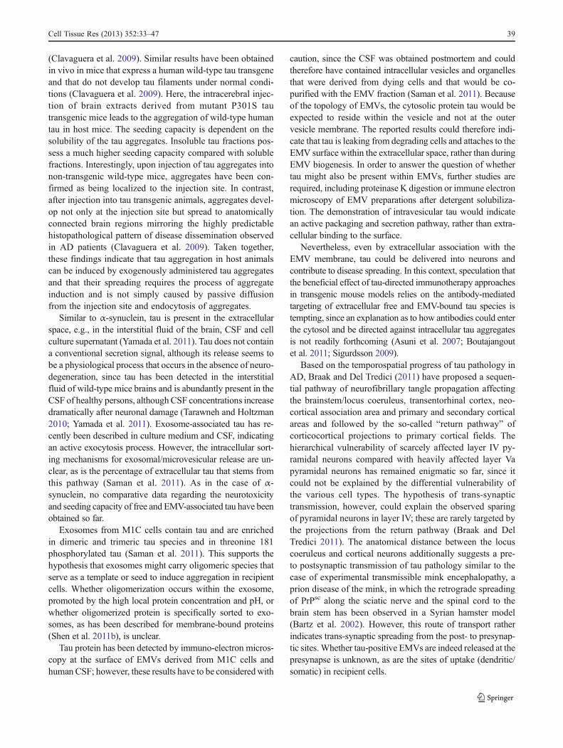
Fig. 1 Mechanisms ofintercellular transfer ofaggregates inneurodegenerative disorders.Misfolded proteins could eitherbe transported via tunnellingnanotubes between cells, withinEMVs or by unconventionalsecretion of free protein.Extracellular misfolded proteinmoieties could be cleared by themicroglia or internalized intoneurons where they might serveas seeds to induceprotein aggregation
38 Cell Tissue Res (2013) 352:33–47
(Clavaguera et al. 2009). Similar results have been obtainedin vivo in mice that express a human wild-type tau transgeneand that do not develop tau filaments under normal condi-tions (Clavaguera et al. 2009). Here, the intracerebral injec-tion of brain extracts derived from mutant P301S tautransgenic mice leads to the aggregation of wild-type humantau in host mice. The seeding capacity is dependent on thesolubility of the tau aggregates. Insoluble tau fractions pos-sess a much higher seeding capacity compared with solublefractions. Interestingly, upon injection of tau aggregates intonon-transgenic wild-type mice, aggregates have been con-firmed as being localized to the injection site. In contrast,after injection into tau transgenic animals, aggregates devel-op not only at the injection site but spread to anatomicallyconnected brain regions mirroring the highly predictablehistopathological pattern of disease dissemination observedin AD patients (Clavaguera et al. 2009). Taken together,these findings indicate that tau aggregation in host animalscan be induced by exogenously administered tau aggregatesand that their spreading requires the process of aggregateinduction and is not simply caused by passive diffusionfrom the injection site and endocytosis of aggregates.
Similar to α-synuclein, tau is present in the extracellularspace, e.g., in the interstitial fluid of the brain, CSF and cellculture supernatant (Yamada et al. 2011). Tau does not containa conventional secretion signal, although its release seems tobe a physiological process that occurs in the absence of neuro-degeneration, since tau has been detected in the interstitialfluid of wild-type mice brains and is abundantly present in theCSF of healthy persons, althoughCSF concentrations increasedramatically after neuronal damage (Tarawneh and Holtzman2010; Yamada et al. 2011). Exosome-associated tau has re-cently been described in culture medium and CSF, indicatingan active exocytosis process. However, the intracellular sort-ing mechanisms for exosomal/microvesicular release are un-clear, as is the percentage of extracellular tau that stems fromthis pathway (Saman et al. 2011). As in the case of α-synuclein, no comparative data regarding the neurotoxicityand seeding capacity of free and EMV-associated tau have beenobtained so far.
Exosomes from M1C cells contain tau and are enrichedin dimeric and trimeric tau species and in threonine 181phosphorylated tau (Saman et al. 2011). This supports thehypothesis that exosomes might carry oligomeric species thatserve as a template or seed to induce aggregation in recipientcells. Whether oligomerization occurs within the exosome,promoted by the high local protein concentration and pH, orwhether oligomerized protein is specifically sorted to exo-somes, as has been described for membrane-bound proteins(Shen et al. 2011b), is unclear.
Tau protein has been detected by immuno-electron micros-copy at the surface of EMVs derived from M1C cells andhuman CSF; however, these results have to be consideredwith
caution, since the CSF was obtained postmortem and couldtherefore have contained intracellular vesicles and organellesthat were derived from dying cells and that would be co-purified with the EMV fraction (Saman et al. 2011). Becauseof the topology of EMVs, the cytosolic protein tau would beexpected to reside within the vesicle and not at the outervesicle membrane. The reported results could therefore indi-cate that tau is leaking from degrading cells and attaches to theEMV surface within the extracellular space, rather than duringEMV biogenesis. In order to answer the question of whethertau might also be present within EMVs, further studies arerequired, including proteinase K digestion or immune electronmicroscopy of EMV preparations after detergent solubiliza-tion. The demonstration of intravesicular tau would indicatean active packaging and secretion pathway, rather than extra-cellular binding to the surface.
Nevertheless, even by extracellular association with theEMV membrane, tau could be delivered into neurons andcontribute to disease spreading. In this context, speculation thatthe beneficial effect of tau-directed immunotherapy approachesin transgenic mouse models relies on the antibody-mediatedtargeting of extracellular free and EMV-bound tau species istempting, since an explanation as to how antibodies could enterthe cytosol and be directed against intracellular tau aggregatesis not readily forthcoming (Asuni et al. 2007; Boutajangoutet al. 2011; Sigurdsson 2009).
Based on the temporospatial progress of tau pathology inAD, Braak and Del Tredici (2011) have proposed a sequen-tial pathway of neurofibrillary tangle propagation affectingthe brainstem/locus coeruleus, transentorhinal cortex, neo-cortical association area and primary and secondary corticalareas and followed by the so-called “return pathway” ofcorticocortical projections to primary cortical fields. Thehierarchical vulnerability of scarcely affected layer IV py-ramidal neurons compared with heavily affected layer Vapyramidal neurons has remained enigmatic so far, since itcould not be explained by the differential vulnerability ofthe various cell types. The hypothesis of trans-synaptictransmission, however, could explain the observed sparingof pyramidal neurons in layer IV; these are rarely targeted bythe projections from the return pathway (Braak and DelTredici 2011). The anatomical distance between the locuscoeruleus and cortical neurons additionally suggests a pre-to postsynaptic transmission of tau pathology similar to thecase of experimental transmissible mink encephalopathy, aprion disease of the mink, in which the retrograde spreadingof PrPsc along the sciatic nerve and the spinal cord to thebrain stem has been observed in a Syrian hamster model(Bartz et al. 2002). However, this route of transport ratherindicates trans-synaptic spreading from the post- to presynap-tic sites.Whether tau-positive EMVs are indeed released at thepresynapse is unknown, as are the sites of uptake (dendritic/somatic) in recipient cells.
Cell Tissue Res (2013) 352:33–47 39
Amyloid-beta
Intracerebral injection of human AD brain extracts or extractsprepared from human amyloid precursor protein (APP) trans-genic mouse brains containing aggregated amyloid-β into thebrains of APP transgenic mice induces the formation ofamyloid-β plaques in the host brains (Eisele et al. 2010;Meyer-Luehmann et al. 2006). A seeding mechanism is likely,since the immunodepletion of amyloid-β or denaturation byformic acid abolishes the capacity of extracts to induce plaqueformation. Furthermore, the induction of amyloid-β deposi-tion requires the combination of human APP transgenic hostmice and human APP-derived amyloid-β assemblies in theextract. These experiments also indicate the possibility ofinterneuronal cell-autonomous disease propagation, since theinduction of amyloid-β deposits is not restricted to the injec-tion site but includes axonally connected areas that are notadjacent to each other (Eisele et al. 2010; Meyer-Luehmannet al. 2006). The concept of disease spreading is furthersupported by the finding that the intraperitoneal administra-tion of brain extracts is sufficient to trigger amyloid-β aggre-gation in the APP transgenic mouse brain (Eisele et al. 2010).Initiation of rapid amyloid-β assembly by exogenous seedscontaining amyloid-β aggregates has also been described afterinoculation into the brains of primates (Baker et al. 1994).
In vitro assembled aggregates of either synthetic amyloid-β40 or 42 fail to induce seeding and a so far unknown co-factor isprobably required to induce misfolding into aggregates withseeding properties (Meyer-Luehmann et al. 2006). Interesting-ly, EMVs carry proteins involved in the generation of amyloid-β (Sharples et al. 2008). APP is cleaved by the sequential actionof two secretases (β -and γ-secretase, which release amyloid-βfrom APP). β-Secretase cleavage produces the APP C-terminalfragment (CTF-β), which can be further processed by γ-secretase (presenilin complex) to APP CTF-γ and amyloid-βpeptide. EMV preparations contain full-length APP and CTFs(Sharples et al. 2008). The implications of these findings onAPP processing are not clear and whether APP or APP CTFcleavage occurs within the exosome/microvesicle membrane isunknown. A small portion of about 1 %–2 % of total extracel-lular amyloid-β peptide in the medium of the neuronal cell lineN2a has been found to be attached to the surface of exosomes(Rajendran et al. 2006). The exosomal surface could serve as aseed to induce a conformational shift, thereby triggeringamyloid-β aggregation. In addition, exosomes could carryamyloid-β peptides to other neurons. However, the impact onoligomerization and interneuronal spreading of amyloid-β pa-thology clearly needs further investigation.
Superoxide dismutase 1
In amyotrophic lateral sclerosis (ALS), aggregates of misfoldedsuperoxide dismutase 1 (SOD1) propagate in a spatiotemporal
manner linking upper and lower motor neurons (Ravits and LaSpada 2009). SOD1 or TDP43 (TARDNA-binding protein 43)inclusions are the two most common neuropathological hall-marks of the disease (Lagier-Tourenne and Cleveland 2009).The export of misfolded SOD1 and uptake into recipient cellshave been shown in vitro (Urushitani et al. 2008). Aggregationof endogenous SOD1 can be induced in cell culture by theexogenous addition of misfolded SOD1 seeds and this templat-ing process continues even after removal of the seed from theculture medium (Grad et al. 2011). Munch et al. (2011) havesubsequently been able to demonstrate the interneuronal trans-fer of SOD1 between cultured cells and the induction of SOD1assembly in target cells. Some evidence for the in vivo transferof SOD1 between astrocytes and motor neurons has beenprovided by recent work of Haidet-Phillips et al. (2011). Theseauthors have isolated progenitor cells from ALS autopsy brainsand differentiated them into astrocytes. Co-culturing or theaddition of this astrocyte-derived medium induces toxicity inexposed mouse motor neuron cultures; this can be alleviatedupon short interfering RNA (siRNA)-mediated SOD1 down-regulation in the astrocytes. As has previously been shown instable motor-neuron-like cell lines expressing wild-type orvarious SOD1 mutants, SOD1 is at least partially secretedtogether with EMVs (Gomes et al. 2007). Similar to tau andα-synuclein, experimental data on the toxicity, transfer effi-ciency and seeding capacity of EMV versus membrane-freeSOD1 are lacking.
Cross-seeding
Cross-seeding between amyloid-β and α-synuclein, α-synuclein and tau, or prion and amyloid-β has been reportedin vitro. Indeed, an overlap of disease pathology has oftenbeen seen at the histopathological level, e.g. α-synucleinaggregates in AD or tau in Lewy body dementia (LBD). Inaddition, tau pathology has been genetically linked to PD andLBD. Since both α-synuclein and tau have been detected inEMVs (although definitive evidence that they are present inthe same vesicle is absent), these vesicles might represent thesite in which cross-seeding occurs.
Open questions
In vivo significance and regulation of EMV release
In vivo evidence is needed to answer the question of whetherEMVs do indeed confer toxicity and induce seeding in animalmodels, as has been shown in SAA amyloidosis. The study ofthe in vivo significance of EMV-mediated disease propagationis hampered by the lack of specific agents to interfere withEMV release or uptake; such agents would enable in vivostudies on the spread of disease pathology. The cell biology of
40 Cell Tissue Res (2013) 352:33–47
protein sorting and EMV release is still not resolved. Aninteraction with the endosomal sorting complex required fortransport (ESCRT) machinery has been described for mono-ubiquitinated transmembrane proteins; this machinery regu-lates their sorting into ILVs. Ubiquitin-interacting motifs me-diate the binding of ESCRT 0 to cargo destined for sorting intoMVBs. The bound cargo is sequentially transported to ESCRTcomplexes I and II at the late endosomal membrane fromwhere invagination and fission into the endosomal lumenoccurs with the help of ESCRT-III (Henne et al. 2011). Incontrast, the intra-endosomal budding of other proteins, suchas the proteolipid protein PLP, occurs independently of theESCRT machinery and requires ceramide (Trajkovic et al.2008). Cytosolic proteins can be sorted into exosomes by theirassociation with lipids and/or transmembrane proteins at theMVE surface or plasmamembranemicrodomains destined foroutward budding. In light of the putative role of EMVs in thepathogenesis of aggregopathies, interestingly, higher-orderoligomerization induced by antibody-mediated cross-linkingpromotes the microvesicular release of various transmem-brane proteins such as transferrin-receptor, MHC-I andCD43 (Muntasell et al. 2007; Vidal et al. 1997). Furthermore,the introduction of oligomerization domains to a membranelocalization sequence is sufficient to induce ESCRT-independent exosomal release (Fang et al. 2007). The tetra-spanin CD63 governs another sorting mechanism into MVEs,a mechanism that is independent of ESCRTand ceramide (vanNiel et al. 2011). Strikingly, CD63-dependent sorting ofpigment-cell-specific integral membrane glycoprotein(PMEL) targets the protein from the MVE (premelanosome)membrane into ILVs. Here, PMEL is cleaved by two site-specific proteases into the C-terminal fragment and the lumi-nal domain (Kummer et al. 2009). Cleavage is followed bypolymerization into physiological PMEL amyloid fibrils with-in the MVE/premelanosome. This process is reminiscent ofthe proposed mechanism of intravesicular amyloid-formation.The viral oncogene latent membrane protein LMP1 is anotherprotein that relies on CD63-dependent sorting into a subtypeof ILVs that are characterized by low cholesterol and areexosomally secreted (Verweij et al. 2011).
Exosomes can either be secreted in a constitutive orregulated process. An increase in intracellular calcium cantrigger MVE fusion and exosome release in various celltypes, including neurons, via a mechanism similar to thatdescribed for secretory lysosomes (Faure et al. 2006; Savinaet al. 2003). The latter process requires synaptotagmin VII,rab27, Munc13-4, AP3 and VAMP7 (Lakkaraju andRodriguez-Boulan 2008). However, whether these mole-cules are also involved in MVE fusion and subsequentexosome release is unclear. The secretion of exosomesinvolves tethering, docking and fusion of the MVE at theplasma membrane. Several regulatory factors of this ma-chinery have been identified, including rab11, the rhoA
effector citron kinase, rab27 and rab35 (Loomis et al. 2006;Savina et al. 2002; Ostrowski et al. 2010; Hsu et al. 2010).Calcium enhances exosome release probably by stimulatingthe fusion of MVEs with the plasma cell membrane in a V-ATPase V0-subunit-dependent manner (Liegeois et al. 2006;Marshansky and Futai 2008). Changes in intracellular ionconcentrations after the P2X7-receptor-induced activation ofthe ATP-gated ion channel have been described to trigger therelease of exosomes in immune cells (Qu and Dubyak 2009).Other stimulatory factors, such as DNA damage and (oxida-tive) stress also promote exosome release, consistent with arole for exosomes in the removal of toxic molecules from thecell (Lespagnol et al. 2008).
Microparticles shed from the plasma membrane are depen-dent on the calcium-induced reorganization of the cytoskele-ton and membrane lipid asymmetry. The outer membraneleaflet of microparticles is enriched in aminophospholipidssuch as phoshatidylserine (PS) and phosphatidylethanolamine(PE) and the asymmetric distribution of these lipids has beenproposed as a mechanism to trigger membrane bending be-cause of their conical shape (Basse et al. 1993; Wehman et al.2011). Lipid asymmetry is, among other factors, created bythe enzymatic activity of scramblase, which translocates andenriches PS and PE from the inner to the outer membraneleaflet (Contreras et al. 2010). This is illustrated by the defi-ciency of procoagulatory platelet microvesiculation observedin Scott’s syndrome in which the lipid asymmetry of the outerplasma membrane is dysregulated and PE and PS are mainlyrestricted to the inner leaflet of the bilayer (Lhermusier et al.2011). Recently, the transmembrane flippase TAT-5 has beenshown, in Caenorhabditis elegans, selectively to enrich PEwithin the inner leaflet without affecting PS asymmetry(Wehman et al. 2011). A deficiency in TAT-5 results in PEenrichment within the outer leaflet and vesicle shedding,whereas TAT-1 mutations, which lead to the accumulation ofPS within the outer leaflet, have no impact on vesicle release.In addition, Wehman et al. (2011) have identified rab11 andthe ESCRT complex as promoting microvesicle formation.Whether the conical shape of PE mediates the outward bend-ing or whether the relative decrease of PE at the inner leafletshifts the net charge in favour of the anionic PS, which couldenhance ESCRT binding followed by vesiculation of themembrane, remains unclear (Wehman et al. 2011).
Microglial clearance and target cell selectivity
Exosomes can transport obsolete cellular content out of thecell (Pan et al. 1985). This has led to the assumption that theprimary function of exosomesmight be the disposal of cellulardebris and toxic molecules as an alternative to lysosomalprocessing in cells with low degradative capacity. In the lipidstorage disorder Niemann-Pick type C, exosomal release isupregulated and contributes to shuttling excess cholesterol out
Cell Tissue Res (2013) 352:33–47 41
of the cells (Strauss et al. 2010). Other examples include theshedding of microvesicles to remove complement attack com-plexes from opsonized cells (Pilzer et al. 2005). Cells canhandle protein aggregates by interaction with chaperonesand by degradation in the proteasome, lysosome or autopha-gosome. Exosomal release of toxic or aggregated proteinsmight serve as an alternative pathway for the cell to removeunwanted content, followed by microglia clearance. For ex-ample, microglia cells internalize oligodendrocytic EMVs bymacropinocytosis in vitro and in vivo and might therebyestablish a clearance mechanism (Fitzner et al. 2011; Zhuanget al. 2011). Activated microglia reside next to amyloid pla-ques and have been extensively discussed in the context ofplaque clearance (Jantzen et al. 2002). Microglia dysfunctionhas been observed in neurodegenerative diseases and either adeficiency of microglia/myeloid cell function and/or an over-load of their endocytosis capacities might enable the intra-neuronal uptake of EMV-packed aggregates, which finallymight result in the spreading of pathology. The ganciclovir-induced ablation of microglia in an APP mouse model hasbeen shown by Grathwohl et al. (2009) to exert no effect onamyloid plaque formation. The authors therefore speculatethat microglia might not have a prominent role in amyloidplaque clearance. However, microglia ablation is induced onlyafter the onset of plaque formation. An effect of microglialfunction on intercellular disease propagation could be studiedin seeding experiments in microglia ablated APP mice. Itwould be interesting to examine whether microglia deficiencycan enhance seeding and interneuronal spreading after theintracerebral injection of amyloid-laden brain extracts.
Of note, several tau or alpha-synuclein aggregopathies arenot restricted to the neuronal cell type but can start in the glialcell lineage. An EMV-based transfer mechanism is a feasibleexplanation of these findings. However, in vivo evidence foroligodendroglial/neuronal EMV transfer is still lacking.
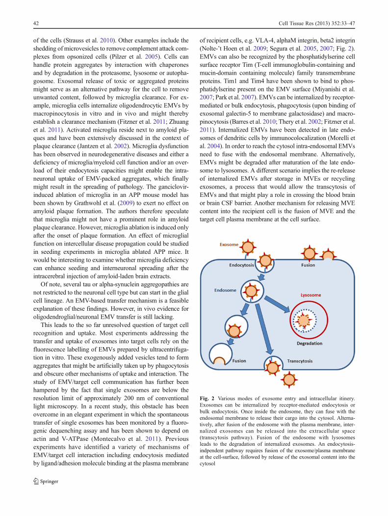
This leads to the so far unresolved question of target cellrecognition and uptake. Most experiments addressing thetransfer and uptake of exosomes into target cells rely on thefluorescence labelling of EMVs prepared by ultracentrifuga-tion in vitro. These exogenously added vesicles tend to formaggregates that might be artificially taken up by phagocytosisand obscure other mechanisms of uptake and interaction. Thestudy of EMV/target cell communication has further beenhampered by the fact that single exosomes are below theresolution limit of approximately 200 nm of conventionallight microscopy. In a recent study, this obstacle has beenovercome in an elegant experiment in which the spontaneoustransfer of single exosomes has been monitored by a fluoro-genic dequenching assay and has been shown to depend onactin and V-ATPase (Montecalvo et al. 2011). Previousexperiments have identified a variety of mechanisms ofEMV/target cell interaction including endocytosis mediatedby ligand/adhesion molecule binding at the plasma membrane
of recipient cells, e.g. VLA-4, alphaM integrin, beta2 integrin(Nolte-’t Hoen et al. 2009; Segura et al. 2005, 2007; Fig. 2).EMVs can also be recognized by the phosphatidylserine cellsurface receptor Tim (T-cell immunoglobulin-containing andmucin-domain containing molecule) family transmembraneproteins. Tim1 and Tim4 have been shown to bind to phos-phatidylserine present on the EMV surface (Miyanishi et al.2007; Park et al. 2007). EMVs can be internalized by receptor-mediated or bulk endocytosis, phagocytosis (upon binding ofexosomal galectin-5 to membrane galactosidase) and macro-pinocytosis (Barres et al. 2010; Thery et al. 2002; Fitzner et al.2011). Internalized EMVs have been detected in late endo-somes of dendritic cells by immunocolocalization (Morelli etal. 2004). In order to reach the cytosol intra-endosomal EMVsneed to fuse with the endosomal membrane. Alternatively,EMVs might be degraded after maturation of the late endo-some to lysosomes. A different scenario implies the re-releaseof internalized EMVs after storage in MVEs or recyclingexosomes, a process that would allow the transcytosis ofEMVs and that might play a role in crossing the blood brainor brain CSF barrier. Another mechanism for releasing MVEcontent into the recipient cell is the fusion of MVE and thetarget cell plasma membrane at the cell surface.
Fig. 2 Various modes of exosome entry and intracellular itinery.Exosomes can be internalized by receptor-mediated endocytosis orbulk endocytosis. Once inside the endosome, they can fuse with theendosomal membrane to release their cargo into the cytosol. Alterna-tively, after fusion of the endosome with the plasma membrane, inter-nalized exosomes can be released into the extracellular space(transcytosis pathway). Fusion of the endosome with lysosomesleads to the degradation of internalized exosomes. An endocytosis-indpendent pathway requires fusion of the exosome/plasma membraneat the cell-surface, followed by release of the exosomal content into thecytosol
42 Cell Tissue Res (2013) 352:33–47
Clinical implications
Infectious prion diseases are characterized by inter-individualdisease transfer via the natural environment, whereas prionoidtransfer is characterized by intra-individual spreading (Aguzziand Rajendran 2009). For none of the above-mentioned aggre-gopathies has an infectious transmission between animals orhumans been demonstrated. One exception, however, is SAAamyloidosis among cheetahs, which secrete AA fibrils intotheir faeces and for which an oral transmission has beenreported (Zhang et al. 2008). However, no epidemiologicalor experimental data so far have suggested that aggregopathiescan be transferred from one individual to the other. Withoutfurther experimental data, the clinical implications of thesefindings are still uncertain but nevertheless evoke the questionas to whether AD pathology might be transmitted via bloodtransfusion, organ transplants or surgical instruments (Walkerand Jucker 2011).
In light of the observed Lewy body pathology in trans-planted fetal neurons in PD, stem-cell-based therapy strategiesneed to be reconsidered. One possibility of escaping theseeding of pathological aggregation in stem cell grafts wouldbe the use of genetically engineered cells that do not expressthe aggregating protein.
In addition to their putative contribution to disease pathology,EMVs could be employed as a biomarker or as a therapeutic toolin degenerative diseases. Exosomal proteome ormicroRNAome(miRNAome) profiling is a common approach in the develop-ment of novel diagnostic or prognostic biomarkers, especially inoncology (for reviews, see Mathivanan et al. 2010a, 2012). In asimilar fashion, CSF or blood exosomes could serve as adiagnostic tool in aggregopathies, especially since several ofthe aggregating proteins are associated with EMVs.
The potential of EMVs for the targeted delivery of thera-peutic drugs is currently under investigation. This emergingconcept has been boosted by a recent publication on the tar-geted exosomal delivery of siRNA-directed againstβ-secretasein an Alzheimer mouse model (Alvarez-Erviti et al. 2011). Onemajor obstacle of miRNA, miRNA inhibitors or siRNA as atherapeutic approach in various diseases is the challenge oftarget tissue specificity. In the above-mentioned example ofAD, transfer of the therapeutic substance across the blood-brainbarrier has to be ensured. Both can be achieved by exploitingexosomes as a transport vesicle, as they carry a neuron-specifictargeting signal (Alvarez-Erviti et al. 2011).
Acknowledgments Grants to A.S.: CMPB, DFG Research CenterMolecular Physiology of the Brain, German Research FoundationGrants SCHN1265/2-1 and SCHN1265/1-1.
Open Access This article is distributed under the terms of the Crea-tive Commons Attribution License which permits any use, distribution,and reproduction in any medium, provided the original author(s) andthe source are credited.
References
Abrami L, Lindsay M, Parton RG, Leppla SH, Goot FG van der (2004)Membrane insertion of anthrax protective antigen and cytoplas-mic delivery of lethal factor occur at different stages of theendocytic pathway. J Cell Biol 166:645–651
Agnati LF, Guidolin D, Baluska F, Leo G, Barlow PW, Carone C,Genedani S (2010) A new hypothesis of pathogenesis based onthe divorce between mitochondria and their host cells: possiblerelevance for Alzheimer’s disease. Curr Alzheimer Res 7:307–322
Aguzzi A, Rajendran L (2009) The transcellular spread of cytosolicamyloids, prions, and prionoids. Neuron 64:783–790
Alvarez-Erviti L, Seow Y, Yin H, Betts C, Lakhal S, Wood MJ (2011)Delivery of siRNA to the mouse brain by systemic injection oftargeted exosomes. Nat Biotechnol 29:341–345
Asuni AA, Boutajangout A, Quartermain D, Sigurdsson EM (2007)Immunotherapy targeting pathological tau conformers in a tanglemouse model reduces brain pathology with associated functionalimprovements. J Neurosci 27:9115–9129
Axelrad MA, Kisilevsky R, Willmer J, Chen SJ, Skinner M (1982)Further characterization of amyloid-enhancing factor. Lab Invest47:139–146
Baker HF, Ridley RM, Duchen LW, Crow TJ, Bruton CJ (1994) Induc-tion of beta (A4)-amyloid in primates by injection of Alzheimer’sdisease brain homogenate. Comparisonwith transmission of spongi-form encephalopathy. Mol Neurobiol 8:25–39
Bancher C, Braak H, Fischer P, Jellinger KA (1993) Neuropath-ological staging of Alzheimer lesions and intellectual statusin Alzheimer’s and Parkinson’s disease patients. NeurosciLett 162:179–182
Baron GS, Wehrly K, Dorward DW, Chesebro B, Caghey B (2002)Conversion of raft associated prion protein to the protease-resistant state requires insertion of PrP-res (PrP(Sc)) into contig-uous membranes. EMBO J 21:1031–1040
Barres C, Blanc L, Bette-Bobillo P, Andre S, Mamoun R, Gabius HJ,Vidal M (2010) Galectin-5 is bound onto the surface of ratreticulocyte exosomes and modulates vesicle uptake by macro-phages. Blood 115:696–705
Bartz JC, Kincaid AE, Bessen RA (2002) Retrograde transport oftransmissible mink encephalopathy within descending motortracts. J Virol 76:5759–5768
Basse F, Gaffet P, Rendu F, Bienvenue A (1993) Translocation of spin-labeled phospholipids through plasma membrane duringthrombin- and ionophore A23187-induced platelet activation.Biochemistry 32:2337–2344
Booth AM, Fang Y, Fallon JK, Yang JM, Hildreth JE, Gould SJ (2006)Exosomes and HIV Gag bud from endosome-like domains of theT cell plasma membrane. J Cell Biol 172:923–935
Boutajangout A, Ingadottir J, Davies P, Sigurdsson EM (2011) Passiveimmunization targeting pathological phospho-tau protein in amouse model reduces functional decline and clears tau aggregatesfrom the brain. J Neurochem 118:658–667
Braak H, Del Tredici K (2011) Alzheimer’s pathogenesis: is thereneuron-to-neuron propagation? Acta Neuropathol 121:589–595
Braak H, Ghebremedhin E, Rub U, Bratzke H, Del Tredici K (2004)Stages in the development of Parkinson’s disease-related pathol-ogy. Cell Tissue Res 318:121–134
Brundin P, Olsson R (2011) Can alpha-synuclein be targeted in noveltherapies for Parkinson’s disease? Expert Rev Neurother 11:917–919
Bulloj A, Leal MC, Xu H, Castano EM, Morelli L (2010) Insulin-degrading enzyme sorting in exosomes: a secretory pathway for akey brain amyloid-beta degrading protease. J Alzheimers Dis19:79–95
Chronopoulos S, Laird DW, Ali-Khan Z (1994) Immunolocalization ofserum amyloid A and AA amyloid in lysosomes in murine
Cell Tissue Res (2013) 352:33–47 43
monocytoid cells: confocal and immunogold electron microscopicstudies. J Pathol 173:361–369
Clavaguera F, Bolmont T, Crowther RA, Abramowski D, Frank S,Probst A, Fraser G, Stalder AK, Beibel M, Staufenbiel M,Jucker M, Goedert M, Tolnay M (2009) Transmission andspreading of tauopathy in transgenic mouse brain. Nat CellBiol 11:909–913
Conde-Vancells J, Rodigruez-Suarez E, Gonzalez E, Berisa A, Gil D,Embade N, Valle M, Luka Z, Elortza F, Wagner C, Lu SC, MatoJM, Falcon-Perez M (2010) Candidate biomarkers in exosome-likevesicles purified from rat and mouse urine samples. Proteomics ClinAppl 4:416–425
Contreras FX, Sanchez-Magraner L, Alonso A, Goni FM (2010)Transbilayer (flip-flop) lipid motion and lipid scrambling in mem-branes. FEBS Lett 584:1779–1786
Danzer KM, Ruf WP, Putcha P, Joyner D, Hashimoto T, Glabe C,Hyman BT, McLean P (2011) Heat-shock protein 70 modulatestoxic extracellular a-synuclein oligomers and rescues trans-synaptic toxicity. FASEB J 25:326–336
Desplats P, Lee HJ, Bae EJ, Patrick C, Rockenstein E, Crews L,Spencer B, Masliah E, Lee SJ (2009) Inclusion formation andneuronal cell death through neuron-to-neuron transmission ofalpha-synuclein. Proc Natl Acad Sci USA 106:13010–13015
Dobrowolski R, De Robertis EM (2011) Endocytic control of growthfactor signalling: multivesicular bodies as signalling organelles.Nat Rev Mol Cell Biol 13:53–60
Eisele YS, Obermuller U, Heilbronner G, Baumann F, Kaeser SA,Wolburg H, Walker LC, Staufenbiel M, Heikenwalder M, JuckerM (2010) Peripherally applied Abeta-containing inoculates inducecerebral beta-amyloidosis. Science 330:980–982
Emmanouilidou E, Melachroinou K, Roumeliotis T, Garbis SD,Ntzouni M, Margaritis LH, Stefanis L, Vekrellis K (2010) Cell-produced alpha-synuclein is secreted in a calcium-dependentmanner by exosomes and impacts neuronal survival. J Neurosci30:6838–6851
Ersdal C, Goodsir CM, Simmons MM, McGovern G, Jeffrey M (2009)Abnormal prion protein is associated with changes of plasmamembranes and endocytosis in bovine spongiform encephalopa-thy (BSE)-affected cattle brains. Neuropathol Appl Neurobiol35:259–271
Fang Y,WuN, Gan X, YanW,Morrell JC, Gould SJ (2007) Higher-orderoligomerization targets plasma membrane proteins and HIV gag toexosomes. PLoS Biol 5:e158
Faure J, Lachenal G, Court M, Hirrlinger J, Chatellard-Causse C, BlotB, Grange J, Schoehn G, Goldberg Y, Boyer V, Kirchhoff F,Raposo G, Garin J, Sadoul R (2006) Exosomes are released bycultured cortical neurones. Mol Cell Neurosci 31:642–648
Fevrier B, Vilette D, Archer F, Loew D, Faigle W, Vidal M, Laude H,Raposo G (2004) Cells release prions in association with exo-somes. Proc Natl Acad Sci USA 101:9683–9688
Fitzner D, Schnaars M, Rossum D van, Krishnamoorthy G, Dibaj P,Bakhti M, Regen T, Hanisch UK, Simons M (2011) Selectivetransfer of exosomes from oligodendrocytes to microglia by mac-ropinocytosis. J Cell Sci 124:447–458
Frost B, Jacks RL, Diamond MI (2009) Propagation of tau misfoldingfrom the outside to the inside of a cell. J Biol Chem 284:12845–12852
Godsave SF, Wille H, Kujala P, Latawiec D, DeArmond SJ, Serban A,Prusiner SB, Peters PJ (2008) Cryo-immunogold electron micros-copy for prions: toward identification of a conversion site. J Neuro-sci 28:12489–12499
Gomes C, Keller S, Altevogt P, Costa J (2007) Evidence for secretionof Cu, Zn superoxide dismutase via exosomes from a cell modelof amyotrophic lateral sclerosis. Neurosci Lett 428:43–46
Gousset K, Schiff E, Langevin C,Marijanovic Z, Caputo A, BrowmanDT,Chenouard N, Chaumont F de, Martino A, Enninga J, Olivo-Marin
JC, Mannel D, Zurzolo C (2009) Prions hijack tunnelling nanotubesfor intercellular spread. Nat Cell Biol 11:328–336
Grad LI, Guest WC, Yanai A, Pokrishevsky E, O’Neill MA, Gibbs E,Semenchenko V, Yousefi M, Wishart DS, Plotkin SS, CashmanNR (2011) Intermolecular transmission of superoxide dismutase 1misfolding in living cells. Proc Natl Acad Sci USA 108:16398–16403
Grathwohl SA, Kalin RE, Bolmont T, Prokop S, Winkelmann G,Kaeser SA, Odenthal J, Radde R, Eldh T, Gandy S, Aguzzi A,Staufenbiel M, Mathews PM, Wolburg H, Heppner FL, Jucker M(2009) Formation and maintenance of Alzheimer’s disease beta-amyloid plaques in the absence of microglia. Nat Neurosci12:1361–1363
Guo JL, Lee VM (2011) Seeding of normal Tau by pathological Tauconformers drives pathogenesis of Alzheimer-like tangles. J BiolChem 286:15317–15331
Gurke S, Barroso JF, Gerdes HH (2008) The art of cellular communi-cation: tunnelig nanotubes bridge the divide. Histochem Cell Biol129:539–550
Haidet-Phillips AM, Hester ME, Miranda CJ, Meyer K, Braun L,Frakes A, Song S, Likhite S, Murtha MJ, Foust KD, Rao M,Eagle A, Kammesheidt A, Christensen A, Mendell JR, BurghesAH, Kaspar BK (2011) Astrocytes from familial and sporadicALS patients are toxic to motor neurons. Nat Biotechnol 29:824–828
Hansen C, Angot E, Bergstrom AL, Steiner JA, Pieri L, Paul G,Outeiro TF, Melki R, Kallunki P, Fog K, Li JY, Brundin P(2011) alpha-Synuclein propagates from mouse brain to grafteddopaminergic neurons and seeds aggregation in cultured humancells. J Clin Invest 121:715–725
Harrington MG, Fonteh AN, Oborina E, Liao P, Cowan RP, McCombG, Chavez JN, Rush J, Biringer RG, Huhmer AF (2009) Themorphology and biochemistry of nanostructures provide evidencefor synthesis and signaling functions in human cerebrospinalfluid. Cerebrospinal fluid Res 6:10
Henne WM, Buchkovich NJ, Emr SD (2011) The ESCRT pathway.Dev Cell 21:77–91
Hsu C, Morohashi Y, Yoshimura S, Manrique-Hoyos N, Jung S,Lauterbach MA, Bakhti M, Gronborg M, Mobius W, Rhee J, BarrFA, Simons M (2010) Regulation of exosome secretion by Rab35and its GTPase-activating proteins TBC1D10A-C. J Cell Biol189:223–232
Huang L, Jin R, Li J, Luo K, Huang T, Wu D, Wang W, Chen R, XiaoG (2010) Macromolecular crowding converts the human recom-binant PrPC to the soluble neurotoxic beta-oligomers. FASEB J24:3536–3543
Jang A, Lee HJ, Suk JE, Jung JW, Kim KP, Lee SJ (2010) Non-classical exocytosis of alpha-synuclein is sensitive to foldingstates and promoted under stress conditions. J Neurochem113:1263–1274
Jantzen PT, Connor KE, DiCarlo G, Wenk GL, Wallace JL, RojianiAM, Coppola D, Morgan D, Gordon MN (2002) Microglialactivation and beta-amyloid deposit reduction caused by a nitricoxide-releasing nonsteroidal anti-inflammatory drug in amyloidprecursor protein plus presenilin-1 transgenic mice. J Neurosci22:2246–2254
Kadota T, Mizote M, Kadota K (1994) Dynamics of presynaptic endo-somes produced during transmitter release. J Electron Microsc43:62–71
Kluve-Beckerman B, Manaloor J, Liepnieks JJ (2001) Binding, traf-ficking and accumulation of serum amyloid A in peritoneal mac-rophages. Scand J Immunol 53:393–400
Kordower JH, Chu Y, Hauser RA, Freeman TB, Olanow CW(2008) Lewy body-like pathology in long-term embryonicnigral transplants in Parkinson’s disease. Nat Med 14:504–506
44 Cell Tissue Res (2013) 352:33–47
Korkut C, Ataman B, Ramachandran P, Ashley J, Barria R, GherbesiN, Budnik V (2009) Trans-synaptic transmission of vesicular Wntsignals through Evi/Wntless. Cell 139:393–404
Kraev IV, Godukhin OV, Patrushev IV, Davies HA, Popov VI, StewartMG (2009) Partial kindling induces neurogenesis, activates astro-cytes and alters synaptic morphology in the dentate gyrus of freelymoving adult rats. Neuroscience 162:254–267
Kramer-Albers EM, Bretz N, Tenzer S, Winterstein C, MobiusW, BergerH, Nave KA, Schild H, Trotter J (2007) Oligodendrocytes secreteexosomes containing major myelin and stress-protective proteins:trophic support for axons? Proteomics Clin Appl 1:1446–1461
Kummer MP, Maruyama H, Huelsmann C, Baches S, Weggen S, KooEH (2009) Formation of Pmel17 amyloid is regulated by juxta-membrane metalloproteinase cleavage, and the resulting C-terminal fragment is a substrate for gamma-secretase. J Biol Chem284:2296–2306
Lachenal G, Pernet-Gallay K, Chivet M, Hemming FJ, Belly A, BodonG, Blot B, Haase G, Goldberg Y, Sadoul R (2011) Release ofexosomes from differentiated neurons and its regulation by syn-aptic glutamatergic activity. Mol Cell Neurosci 46:409–418
Lagier-Tourenne C, Cleveland DW (2009) Rethinking ALS: the FUSabout TDP-43. Cell 136:1001–1004
Laine J, Marc ME, Sy MS, Axelrad H (2001) Cellular and subcellularmorphological localization of normal prion protein in rodentcerebellum. Eur J Neurosci 14:47–56
Lakkaraju A, Rodriguez-Boulan E (2008) Itinerant exosomes: emerg-ing roles in cell and tissue polarity. Trends Cell Biol 18:199–209
Laulagnier K, Grand D, Dujardin A, Hamdi S, Vincent-Schneider H,Lankar D, Salles JP, Bonnerot C, Perret B, Record M (2004)PLD2 is enriched on exosomes and its activity is correlated tothe release of exosomes. FEBS Lett 572:11–14
Le Blanc I, Luyet PP, Pons V, Ferguson C, Emans N, Petiot A, MayranN, Demaurex N, Faure J, Sadoul R, Parton RG, Gruenberg J(2005) Endosome-to-cytosol transport of viral nucleocapsids.Nat Cell Biol 7:653–664
Lespagnol A, Duflaut D, Beekman C, Blanc L, Fiucci G, Marine JC,Vidal M, Amson R, Telerman A (2008) Exosome secretion,including the DNA damage-induced p53-dependent secretorypathway, is severely compromised in TSAP6/Steap3-null mice.Cell Death Differ 15:1723–1733
Lhermusier T, Chap H, Payrastre B (2011) Platelet membrane phos-pholipid asymmetry: from the characterization of a scramblaseactivity to the identification of an essential protein mutated inScott syndrome. J Thromb Haemost 9:1883–1891
Li JY, Englund E, Holton JL, Soulet D, Hagell P, Lees AJ, Lashley T,Quinn NP, Rehncrona S, Bjorklund A, Widner H, Revesz T,Lindvall O, Brundin P (2008) Lewy bodies in grafted neuronsin subjects with Parkinson’s disease suggest host-to-graft diseasepropagation. Nat Med 14:501–503
Liberski PP, Yanagihara R, Gibbs CJ Jr, Gajdusek DC (1990) Spread ofCreutzfeldt-Jakob disease virus along visual pathways after intra-ocular inoculation. Arch Virol 111:141–147
Liegeois S, Benedetto A, Garnier JM, Schwab Y, Labouesse M (2006)The V0-ATPase mediates apical secretion of exosomes containingHedgehog-related proteins in Caenorhabditis elegans. J Cell Biol173:949–961
Loomis RJ, Holmes DA, Elms A, Solski PA, Der CJ, Su L (2006)Citron kinase, a RhoA effector, enhances HIV-1 virion productionby modulating exocytosis. Traffic 7:1643–1653
Lundmark K, Westermark GT, Nystrom S, Murphy CL, SolomonA, Westermark P (2002) Transmissibility of systemic amy-loidosis by a prion-like mechanism. Proc Natl Acad Sci USA99:6979–6984
Marijanovic Z, Caputo A, Campana V, Zurzolo C (2009) Identificationof an intracellular site of prion conversion. PLoS Pathog 5:e1000426
Marshansky V, Futai M (2008) The V-type H+-ATPase in vesiculartrafficking: targeting, regulation and function. Curr Opin Cell Biol20:415–426
Mathivanan S, Ji H, Simpson RJ (2010a) Exosomes: extracellularorganelles important in intercellular communication. J Proteomics73:1907–1920
Mathivanan S, Lim JW, Tauro BJ, Ji H, Moritz RL, Simpson RJ (2010b)Proteomics analysis of A33 immunoaffinity-purified exosomes re-leased from the human colon tumor cell line LIM1215 reveals atissue-specific protein signature. Mol Cell Proteomics 9:197–208
Mathivanan S, Fahner CJ, Reid GE, Simpson RJ (2012) ExoCarta2012: database of exosomal proteins, RNA and lipids. NucleicAcids Res 40:D1241–D1244
Meyer-Luehmann M, Coomaraswamy J, Bolmont T, Kaeser S, SchaeferC, Kilger E, Neuenschwander A, Abramowski D, Frey P, Jaton AL,Vigouret JM, Paganetti P, Walsh DM, Mathews PM, Ghiso J,Staufenbiel M, Walker LC, Jucker M (2006) Exogenous inductionof cerebral beta-amyloidogenesis is governed by agent and host.Science 313:1781–1784
Miyanishi M, Tada K, Koike M, Uchiyama Y, Kitamura T, Nagata S(2007) Identification of Tim4 as a phosphatidylserine receptor.Nature 450:435–439
Montecalvo A, Larregina AT, Shufesky WJ, Stolz DB, Sullivan ML,Karlsson JM, Baty CJ, Gibson GA, Erdos G, Wang Z, MilosevicJ, Tkacheva OA, Divito SJ, Jordan R, Lyons-Weiler J, WatkinsSC, Morelli AE (2011) Mechanism of transfer of functionalmicroRNAs between mouse dendritic cells via exosomes. Blood119:756–766
Morelli AE, Larregina AT, Shufesky WJ, Sullivan ML, Stolz DB,Papworth GD, Zahorchak AF, Logar AJ, Wang Z, Watkins SC, FaloLD Jr, Thomson AW (2004) Endocytosis, intracellular sorting, andprocessing of exosomes by dendritic cells. Blood 104:3257–3266
Mougenot AL, Nicot S, Bencsik A, Morignat E, Verchere J, Lakhdar L,Legastelois S, Baron T (2011) Prion-like acceleration of a synuclein-opathy in a transgenic mouse model. Neurobiol Aging (in press)
Munch C, O’Brien J, Bertolotti A (2011) Prion-like propagation ofmutant superoxide dismutase-1 misfolding in neuronal cells. ProcNatl Acad Sci USA 108:3548–3553
Muntasell A, Berger AC, Roche PA (2007) T cell-induced secretion ofMHC class II-peptide complexes on B cell exosomes. EMBO J26:4263–4272
Natale G, Ferrucci M, Lazzeri G, Paparelli A, Fornai F (2011) Trans-mission of prions within the gut and towards the central nervoussystem. Prion 5::142–149
Nickel W, Rabouille C (2009) Mechanisms of regulated unconvention-al protein secretion. Nat Rev Mol Cell Biol 10:148–155
Nolte-’t Hoen EN, Buschow SI, Anderton SM, Stoorvogel W, WaubenMH (2009) Activated T cells recruit exosomes secreted by den-dritic cells via LFA-1. Blood 113:1977–1981
Nunez R, Sancho-Martinez SM, Novoa JM, Lopez-Hernandez FJ(2010) Apoptotic volume decrease as a geometric determinant forcell dismantling into apoptotic bodies. Cell Death Differ 17:1665–1671
Ostrowski M, Carmo NB, Krumeich S, Fanget I, Raposo G, Savina A,Moita CF, Schauer K, Hume AN, Freitas RP, Goud B, Benaroch P,Hacohen N, Fukuda M, Desnos C, Seabra MC, Darchen F, Amigor-ena S, Moita LF, Thery C (2010) Rab27a and Rab27b controldifferent steps of the exosome secretion pathway. Nat Cell Biol12:19–30
Pan BT, Teng K, Wu C, Adam M, Johnstone RM (1985) Electronmicroscopic evidence for externalization of the transferrin receptorin vesicular form in sheep reticulocytes. J Cell Biol 101:942–948
Park D, Tosello-Trampont AC, Elliott MR, Lu M, Haney LB, Ma Z,Klibanov AL, Mandell JW, Ravichandran KS (2007) BAI1 is anengulfment receptor for apoptotic cells upstream of the ELMO/Dock180/Rac module. Nature 450:430–434
Cell Tissue Res (2013) 352:33–47 45
Peters PJ, Mironov A Jr, Peretz D, Donselaar E van, Leclerc E, Erpel S,DeArmond SJ, Burton DR, Williamson RA, Vey M, Prusiner SB(2003) Trafficking of prion proteins through a caveolae-mediatedendosomal pathway. J Cell Biol 162:703–717
Pilzer D, Gasser O, Moskovich O, Schifferli JA, Fishelson Z(2005) Emission of membrane vesicles: roles in complementresistance, immunity and cancer. Springer Semin Immunopa-thol 27:375–387
Potolicchio I, Carven GJ, XuX, Stipp C, Riese RJ, Stern LJ, SantambrogioL (2005) Proteomic analysis of microglia-derived exosomes: meta-bolic role of the aminopeptidase CD13 in neuropeptide catabolism. JImmunol 175:2237–2243
Qu Y, Dubyak GR (2009) P2X7 receptors regulate multiple types ofmembrane trafficking responses and non-classical secretion path-ways. Purinergic Signal 5:163–173
Rajendran L, Honsho M, Zahn TR, Keller P, Geiger KD, Verkade P,Simons K (2006) Alzheimer’s disease beta-amyloid peptides arereleased in association with exosomes. Proc Natl Acad Sci USA103:11172–11177
Ravits JM, La Spada AR (2009) ALS motor phenotype heterogeneity,focality, and spread: deconstructing motor neuron degeneration.Neurology 73:805–811
Saman S, Kim W, Raya M, Visnick Y, Miro S, Saman S, Jackson B,McKee AC, Alvarez VE, Lee NC, Hall GF (2011) Exosome-associated tau is secreted in tauopathy models and is selectivelyphosphorylated in cerebrospinal fluid (CSF) in early Alzheimerdisease. J Biol Chem 287:3842–3849
Savina A, Vidal M, Colombo MI (2002) The exosome pathway inK562 cells is regulated by Rab11. J Cell Sci 115:2505–2515
Savina A, Furlan M, Vidal M, Colombo MI (2003) Exosome release isregulated by a calcium-dependent mechanism in K562 cells. JBiol Chem 278:20083–20090
Schatzl HM, Laszlo L, Holtzman DM, Tatzelt J, DeArmond SJ, WeinerRI, Mobley WC, Prusiner SB (1997) A hypothalamic neuronalcell line persistently infected with scrapie prions exhibits apoptosis.J Virol 71:8821–8831
Segura E, Nicco C, Lombard B, Veron P, Raposo G, Batteux F,Amigorena S, Thery C (2005) ICAM-1 on exosomes from maturedendritic cells is critical for efficient naive T-cell priming. Blood106:216–223
Segura E, Guerin C, Hogg N, Amigorena S, Thery C (2007) CD8+
dendritic cells use LFA-1 to capture MHC-peptide complexesfrom exosomes in vivo. J Immunol 179:1489–1496
Senthilkumar S, Chang E, Jayakumar R (2008) Diffusible amyloidoligomers trigger systemic amyloidosis in mice. Biochem J 415:207–215
Sharples RA, Vella LJ, Nisbet RM, Naylor R, Perez K, Barnham KJ,Masters CL, Hill AF (2008) Inhibition of gamma-secretase causesincreased secretion of amyloid precursor protein C-terminal frag-ments in association with exosomes. FASEB J 22:1469–1478
Shen B, Fang Y, Wu N, Gould SJ (2011a) Biogenesis of the posteriorpole is mediated by the exosome/microvesicle protein-sortingpathway. J Biol Chem 286:44162–44176
Shen B, Wu N, Yang JM, Gould SJ (2011b) Protein targeting toexosomes/microvesicles by plasma membrane anchors. J BiolChem 286:14383–14395
Sigurdsson EM (2009) Tau-focused immunotherapy for Alzheimer’sdisease and related tauopathies. Curr Alzheimer Res 6:446–450
Simons M, Raposo G (2009) Exosomes–vesicular carriers for intercellu-lar communication. Curr Opin Cell Biol 21:575–581
Simpson RJ, Lim JW, Moritz RL, Mathivanan S (2009) Exosomes:proteomic insights and diagnostic potential. Expert Rev Proteomics6:267–283
Solomon A, Richey T, Murphy CL, Weiss DT, Wall JS, WestermarkGT, Westermark P (2007) Amyloidogenic potential of foie gras.Proc Natl Acad Sci USA 104:10998–11001
Sponarova J, Nystrom SN, Westermark GT (2008) AA-amyloidosis canbe transferred by peripheral blood monocytes. PLoS One 3:e3308
Strauss K, Goebel C, Runz H, Mobius W, Weiss S, Feussner I, SimonsM, Schneider A (2010) Exosome secretion ameliorates lysosomalstorage of cholesterol in Niemann-Pick type C disease. J BiolChem 285:26279–26288
Subra C, Laulagnier K, Perret B, Record M (2007) Exosome lipidomicsunravels lipid sorting at the level of multivesicular bodies. Biochimie89:205–212
Tarawneh R, Holtzman DM (2010) Biomarkers in translational re-search of Alzheimer’s disease. Neuropharmacology 59:310–322
Tasaki M, Ueda M, Ochiai S, Tanabe Y, Murata S, Misumi Y, Su Y,Sun X, Shinriki S, Jono H, Shono M, Obayashi K, Ando Y (2010)Transmission of circulating cell-free AA amyloid oligomers inexosomes vectors via a prion-like mechanism. Biochem BiophysRes Commun 400:559–562
Taylor AR, Robinson MB, Gifondorwa DJ, Tytell M, Milligan CE(2007) Regulation of heat shock protein 70 release in astrocytes:role of signaling kinases. Dev Neurobiol 67:1815–1829
Thery C, BoussacM, Veron P, Ricciardi-Castagnoli P, Raposo G, Garin J,Amigorena S (2001) Proteomic analysis of dendritic cell-derivedexosomes: a secreted subcellular compartment distinct from apopto-tic vesicles. J Immunol 166:7309–7318
Thery C, Zitvogel L, Amigorena S (2002) Exosomes: composition,biogenesis and function. Nat Rev Immunol 2:569–579
Thery C, Amigorena S, Raposo G, Clayton A (2006) Isolation andcharacterization of exosomes from cell culture supernatants andbiological fluids. Curr Protoc Cell Biol Chapter 3:Unit 3.22
Trajkovic K, Hsu C, Chiantia S, Rajendran L, Wenzel D, Wieland F,Schwille P, Brugger B, SimonsM (2008) Ceramide triggers buddingof exosome vesicles into multivesicular endosomes. Science319:1244–1247
Urushitani M, Ezzi SA, Matsuo A, Tooyama I, Julien JP (2008) Theendoplasmic reticulum-Golgi pathway is a target for translocationand aggregation of mutant superoxide dismutase linked to ALS.FASEB J 22:2476–2487
van Niel G, Charrin S, Simoes S, Romao M, Rochin L, Saftig P, MarksMS, Rubinstein E, Raposo G (2011) The tetraspanin CD63 reg-ulates ESCRT-independent and -dependent endosomal sortingduring melanogenesis. Dev Cell 21:708–721
Veith NM, Plattner H, Stuermer CA, Schulz-Schaeffer WJ, Burkle A(2009) Immunolocalisation of PrPSc in scrapie-infected N2amouse neuroblastoma cells by light and electron microscopy.Eur J Cell Biol 88:45–63
Vella LJ, Sharples RA, Lawson VA, Masters CL, Cappai R, Hill AF(2007) Packaging of prions into exosomes is associated with anovel pathway of PrP processing. J Pathol 211:582–590
Vella LJ, Greenwood DL, Cappai R, Scheerlinck JP, Hill AF (2008)Enrichment of prion protein in exosomes derived from ovinecerebral spinal fluid. Vet Immunol Immunopathol 124:385–393
Verweij FJ, Eijndhoven MA van, Hopmans ES, Vendrig T, Wurdinger T,Cahir-McFarland E, Kieff E, Geerts D, Kant R van der, Neefjes J,Middeldorp JM, Pegtel DM (2011) LMP1 association with CD63 inendosomes and secretion via exosomes limits constitutive NF-kappaB activation. EMBO J 30:2115–2129
Vidal M, Mangeat P, Hoekstra D (1997) Aggregation reroutes mole-cules from a recycling to a vesicle-mediated secretion pathwayduring reticulocyte maturation. J Cell Sci 110:1867–1877
Von Bartheld CS, Altick AL (2011) Multivesicular bodies in neurons:distribution, protein content, and trafficking functions. Prog Neuro-biol 93:313–340
Walker LC, Jucker M (2011) Amyloid by default. Nat Neurosci14:669–670
Wang F, Wang X, Yuan CG, Ma J (2010) Generating a prion withbacterially expressed recombinant prion protein. Science 327:1132–1135
46 Cell Tissue Res (2013) 352:33–47
Wehman AM, Poggioli C, Schweinsberg P, Grant BD, Nance J (2011)The P4-ATPase TAT-5 inhibits the budding of extracellularvesicles in C. elegans embryos. Curr Biol 21:1951–1959
Werdelin O, Ranlov P (1966) Amyloidosis in mice produced bytransplantation of spleen cells from casein-treated mice. ActaPathol Microbiol Scand 68:1–18
Westermark GT, Westermark P (2009) Serum amyloid A and proteinAA: molecular mechanisms of a transmissible amyloidosis. FEBSLett 583:2685–2690
Yamada K, Cirrito JR, Stewart FR, Jiang H, Finn MB, Holmes BB,Binder LI, Mandelkow EM, Diamond MI, Lee VM, Holtzman DM(2011) In vivo microdialysis reveals age-dependent decrease of
brain interstitial fluid tau levels in P301S human tau transgenicmice. J Neurosci 31:13110–13117
Zhang B, Une Y, Fu X, Yan J, Ge F, Yao J, Sawashita J, Mori M,Tomozawa H, Kametani F, Higuchi K (2008) Fecal transmis-sion of AA amyloidosis in the cheetah contributes to highincidence of disease. Proc Natl Acad Sci USA 105:7263–7268
Zhuang X, Xiang X, Grizzle W, Sun D, Zhang S, Axtell RC, Ju S, MuJ, Zhang L, Steinman L, Miller D, Zhang HG (2011) Treatment ofbrain inflammatory diseases by delivering exosome encapsulatedanti-inflammatory drugs from the nasal region to the brain. MolTher 19:1769–1779
Cell Tissue Res (2013) 352:33–47 47