evaluation of serum autotaxin as a novel noninvasive ...jmscr.igmpublication.org/v4-i11/100...
TRANSCRIPT

Mahmoud A. Al-Azoony et al JMSCR Volume 4 Issue 11 November 2016 Page 14128
JMSCR Vol||04||Issue||11||Page 14128-14139||November 2016
Evaluation of Serum Autotaxin as a Novel Noninvasive Marker for
Assessment of Hepatic Fibrosis in Chronic Hepatitis C Patients
Authors
Mahmoud A. Al-Azoony1, Jehan H.Sabry
2, Hisham A. Issa
3, Maha Z. Omar
4,
Abdelmoneam Ahmed5, Doaa M. Abd Almoneim
6
1,2,3Dept of Clinical and Chemical Pathology, Benha Faculty of Medicine, Benha University, Egypt
4Dept of Hepatology, Gastroenterology and Infectious Diseases, Benha Faculty of Medicine, Benha
University, Egypt 5Department of Internal Medicine, Benha Faculty of Medicine, Benha University, Egypt
6Department of Clinical Pathology, Benha Health Insurance Hospital, Egypt
Abstract
Egypt has a high prevalence of HCV infection which causes hepatic fibrosis. Assessment of the degree of
hepatic fibrosis is essential for decision of antiviral therapy. Untill now, liver biopsy is the gold standard for
assessment of hepatic fibrosis but it is an invasive procedure. The aim of this study was to assess the utility of
serum autotaxin level as a marker of liver fibrosis in chronic hepatitis C patients in comparison with liver
biopsy.Sixty HCV patients with hepatic fibrosis confirmed with liver biopsy and20 healthy subjects were
inrolled in the current study.Lab assessment included CBC, fasting serum glucose, liver and kidney function
tests, serum AFP, hepatitis markers (HCVAb, HBsAg), PCR for HCV RNA and serum autotaxin (using
sandwich ELISA).Results revealed that serum ATX levels were significantly increased in HCV group versus
control group and were positively correlated with the stage of fibrosis. Serum autotaxin level at cutoff value
(<115.8 ng/ml) was the best parameter in detection of F0 with AUC (0.79), at a cutoff value (≥180.3 ng/ml)
was the best parameter in detection of advanced fibrosis (≥ F3) with AUC (0.82) and at a cutoff value
(≥189.2 ng/ml) was the best parameter in detection of F4 with AUC (0.85).
It was concluded that serum autotaxin level is a valuable test for exclusion of fibrosis and detection of
advanced fibrosis and cirrhosis but lacks discrimination of intermediate fibrosis stages.
Keywords: Hepatitis C, autotaxin, hepatic fibrosis.
Introduction
Hepatitis C is a major health problem. Chronic
hepatitis C is a slowly progressive inflammatory
disease which can lead to cirrhosis with all its
complications [1]
. In Egypt, the prevalence rate
reaches 15% in rural areas, with some age groups
suffering from prevalence rates up to 50%. 13.6%
of volunteer blood donors have anti-HCV
antibodies, this high seropositive rate among
Egyptians is about 35 folds higher than other
countries [2]
.
Liver fibrosis is the excessive accumulation of
extracellular matrix proteins including collagen
that results from chronic damage to the liver [3]
.
The assessment of the stage of liver fibrosis is
essential for prognosis and for deciding on
antiviral treatment [4]
.
Liver biopsy is recommended as the gold
standard method for determining fibrosis stage,
prognosis and therapeutic indications in patients
with chronic liver disease. However, liver biopsy
is an invasive procedure and is associated with
www.jmscr.igmpublication.org
Impact Factor 5.244
Index Copernicus Value: 83.27
ISSN (e)-2347-176x ISSN (p) 2455-0450
DOI: https://dx.doi.org/10.18535/jmscr/v4i11.100

Mahmoud A. Al-Azoony et al JMSCR Volume 4 Issue 11 November 2016 Page 14129
JMSCR Vol||04||Issue||11||Page 14128-14139||November 2016
many complications [1]
. Approximately 1–3% of
patients require hospitalization for complications,
and a quarter of them report pain after
percutaneous liver biopsy. The diagnostic
accuracy of liver biopsy for assessment of hepatic
fibrosis is influenced by the quality of the biopsy
samples. In addition, there are many absolute or
relative contraindications to liver biopsy,
including severe coagulopathy [5]
. Hence, several
noninvasive tests have been proposed in an
attempt to predict the degree of fibrosis and
replace liver biopsy [6]
.
Autotaxin (ATX), is a secreted glycoprotein that
belongs to the ectonucleotidepyrophosphatase/
phosphodiesterase (NPP) family [7]
. ATX
hydrolyzes lysophosphatidylcholine to produce
lysophosphtidic acid, a multifunctional bioactive
lipid mediator [8]
. ATX is a vital enzyme which is
needed for early embryological development [9]
.
Physiologically, the most important role of ATX
after birth is probably in wound healing and tissue
remodeling. LPA is a potent activator of platelet
aggregation and it also stimulates the growth and
migration of fibroblasts, vascular smooth muscle
cells, endothelial cells and keratinocytes [10]
.
Lysophosphatidic acid (LPA) activates hepatic
stellate cells (HSC) which are the main cell type
involved in development of liver fibrosis,
stimulates their contraction and inhibits their
apoptosis [11]
.
Elevated serum ATX levels are an indicator for
activation of HSC during development of fibrosis
and cirrhosis [12]
. As ATX is cleared by sinusoidal
endothelial cells, dysfunctional endothelium may
be a cause for higher blood concentrations of
ATX in cirrhotic patients [13]
. Recently, a
connection between liver fibrosis and serum or
plasma LPA and ATX emerged in patients with
chronic HCV infection [14]
. However, serum ATX
in different stages of liver cirrhosis and its
prognostic value has not yet been investigated [15]
.
We aimed in this study to investigate the utility of
serum autotaxin level as a noninvasive marker of
hepatic fibrosis and if serum ATX can be an
indicator for the degree of liver fibrosis compared
to liver biopsy in chronic HCV patients.
Subjects and methods
Study population
The current case control study was carried out on
80 subjects. All included candidates were chosen
from attendants to the Department of Hepatology,
Gastroenterology and Infectious Diseases in
Benha University Hospital during the period from
July 2014 to July 2015.All included candidates
gave a written informed consent for using their
serum samples. They were categorized into 2
groups.
Group1 (patients' group): included 60chronic
hepatitis C patients, 39 males (65%) and
21females (35%).Chronic HCV diagnosis was
based on elevated serum transaminase levels for at
least six months and positive HCV antibody by
enzyme-linked immunosorbent assay and
confirmed by quantitative detection of circulating
HCV RNA using polymerase chain reaction
(PCR). Liver biopsywas taken from every patient
for staging of fibrosis. Patients were subgrouped
into five subgroups according to stage of liver
fibrosis (from F0 to F4).Inclusion criteria were:
age > 18 years old and positive HCV Abs
confirmed by HCV RNA detection by PCR.
Exclusion criteria were: age < 18 years old, HBV
infected patients, patients with hepatocellular
carcinoma (HCC) and other causes of liver disease
e.g alcoholism and autoimmune hepatitis. Group 2
(control group): included 20 healthy subjects (free
from liver diseases), they were 10 males (50%)
and 10 females (50%).
Ethical consideration
The study protocol was approved by the ethical
committee of Benha faculty of medicine and its
university hospital.
Spicemen collection
Blood samples were collected by peripheral
venipuncture under complete aseptic precautions.
Then each sample was divided into 3 parts: (1.8)
ml blood for each 0.2 ml Na citrate for
measurement of PT and international normalized
ratio, 2 ml on EDTA for complete blood count
and the rest of the sample was used for separation

Mahmoud A. Al-Azoony et al JMSCR Volume 4 Issue 11 November 2016 Page 14130
JMSCR Vol||04||Issue||11||Page 14128-14139||November 2016
of serum . Serum was separated by allowing
samples to clot for 30 minutes at room
temperature, then the tube was centrifuged for 15
minutes at 1500 r.p.m. then the sera were divided
into aliquots.
Lab investigations
Fasting serum glucose, liver function tests (AST,
ALT, ALP, total bilirubin, direct bilirubin, serum
albumin), kidney function tests (serum creatinine
and urea) were performed using Biosystem A15
auto-analyzer.
Serum AFP was measured using CanAg AFP
EIA kit provided by (FUJIREBIO Diagnostic,
Inc., SE-402 42 G ِ teborg, Sweden) [16]
.
Serum autotaxin was measured using Quantikine
Human ENPP-2/Autotaxin ELISA kit
(manufactured and distributed by R&D systems,
inc. USA & Canada) according to the
recommendations of the manufacturer.
Autotaxin assay is a sandwich ELISA in which a
monoclonal antibody specific for ENPP-2 has
been pre-coated onto a microplate. Then,
Standards and samples are pipetted into the wells,
then washed. An enzyme-linked polyclonal
antibody specific for ENPP-2 is added to the
wells. After washing, a substrate solution is added
to the wells and color develops. The color
development is stopped and the intensity of the
color is measured [17]
.
All laboratory investigations were done in the
department of Clinical and Chemical Pathology,
Benha University hospital.
Calculation of laboratory scores
APRI (Aspartate/platelet ratio index) was
calculated according to the formula:
APRI=[(AST/ULN) / PLT(× 109/L)] × 100
(Wai et al [18]
).
API (Age platelet index)was calculated
according to the formula:
Age score + platelet count score
Age: < 30=5; 30-39=1; 40-49=2; 50-59=3; 60-
69=4; ≥ 70=5.
Platelet count (× 109 /L): ≥ 225=0; 200 – 224=1;
175- 199=2; 150 – 174=3; 125 – 149=4; < 125=5
(Poynard and Bedossa [19]
).
Histopathological Assessment of liver biopsies:
All biopsies were obtained by trucut needle
(biopsy gun, G16) with a core length of at least 1
cm including a minimum of 6 portal tracts. All
were stained by haematoxylin-eosin (H & E)as
well as Masson Trichrome stains. The METAVIR
scoring system according to Bedossa and Poynard [20]
was applied by the same experienced
hitopathologist. Histological staging based on
degree of fibrosis has 5 stages (F0 – F4) as
following (F0: No fibrosis; F1: portal fibrosis
without septa; F2 : portal fibrosis with rare septa.
F3 : portal fibrosis with numerous septa without
cirrhosis; F4 : cirrhosis).
Statistical Analysis
The collected data were tabulated and analyzed
using SPSS version 16 software (SpssInc,
Chicago, ILL Company). Categorical data were
presented as number and percentages while
quantitative data were expressed as mean and
standard deviation or median and Inter
Quartilerange (IQR). Chi square test (X2), Fisher's
test, Spearman’s correlation coefficient (rho),
St."t", Man Whitney U test and Krauskal Wallis
test were the used tests of significance. ROC
curve was used to determine cutoff values with
optimum sensitivity and specificity of the studied
markers in detection of different stages of fibrosis.
Results
This study is a case control study that included 60
hepatitis C patients with hepatic fibrosis proved
with liver biopsy, their ages ranged from 21 - 58
years with mean of 43.98years and 20 healthy
subjects (free from liver diseases) as control group
their ages ranged from 24 - 62 years with mean of
41.95years. Patients were divided into five groups
according to METAVIR scoring system (from F0
to F4). They were 6 patients in F0, 15 patients in
F1, 18 patients in F2, 14 patients in F3, 7 patients
in F4.
Lab investigations in the form of CBC, PT, INR,
fasting serum glucose, liver function tests (AST,
ALT, ALP, total bilirubin, direct bilirubin, serum
albumin), kidney function tests (serum creatinine
Mahmoud A. Al-Azoony et al JMSCR Volume 4 Issue 11 November 2016 Page 14131
JMSCR Vol||04||Issue||11||Page 14128-14139||November 2016
and urea) were recorded in table (1) where all data
were expressed as mean ± SD while serum AFP,
serum ATX, APRI, API were expressed as median
and IQR.
The current study revealed that there was high
statistically significant increase in serum autotoxin
in patients' group (median=162.9) versus control
group (median=36.8) (P value <0.001) (table
1).While there was no significant difference
between males and females as regarding the mean
values of serum autotaxin level (P values were
0.75 for patients’ group and 0.14 for control group
(table 2).
High statistically significant increase in PT and
statistically significant increase in INR, ALT,
AFP, APRI, API were found in patients' group
versus control group (table 1).
The stage of fibrosis showed highly significant
positive correlations with AST, AFP, serum
autotaxin (P value <0.001), APRI (P value
<0.001), and API (P value 0.001) scores (figure 1
a, b, c), significant positive correlations with age ,
PT , INR , serum direct bilirubin , ALT , ALP ( P
values were 0.008 , 0.042 , 0.016 , 0.047 , 0.043 ,
0.012 respectively) and significant negative
correlations with platelets and serum albumin (P
values were 0.025, 0.007 respectively). While it
showed no correlation with Hb, WBCs, fasting
serum glucose, total bilirubin, urea, creatinine and
PCR results.
Also in the current study, serum autotaxin level
showed a significant positive correlation with age,
fasting serum glucose, ALP, urea, API, AST
(figure 2 a, b) (P values were 0.012, 0.025, 0.002,
0.005, 0.014, 0.008 respectively) and highly
significant positive correlation with AFP (P value
<0.001).
Serum autotaxin level at cutoff value (<115.8
ng/ml) was the best parameter in detection of F0
with AUC (0.79), sensitivity (83.3%), specificity
(74.1%) and accuracy (79.2%) (P value=0.02)
(table3, figure 3).
Serum autotaxin at cutoff value (<150.4 ng/ml)
had AUC (0.64), sensitivity (73.3%), specificity
(60%) and accuracy (64.1%)(P value=0.1)in
detection of F1.Serum autotaxin at cutoff value
(<162.9 ng/ml) had AUC (0.58), sensitivity
(61.1%), specificity (54.8%) and accuracy
(57.5%) (P value=0.35) in detection of F2.
APRI at cutoff value (≥0.444) was the best
parameter in detection of significant fibrosis (≥
F2) with AUC (0.75), sensitivity (74.4%),
specificity (85.7%) and accuracy (75.4%) (P
value=0.001) followed by Serum autotaxin whose
cutoff value (≥124.7 ng/ml) had AUC=0.75,
sensitivity (79.5%), specificity (71.4%) and
accuracy (75%) (P value=0.002) in detection of
significant fibrosis (≥ F2).
API at cutoff value (≥3.5) was the best parameter
in detection of F3 with AUC (0.73), sensitivity
(64.3%), specificity (67.4%) and accuracy
(73.4%) (P value=0.008) while serum autotaxin
level at cutoff value (≥162.9 ng/ml) had AUC
(0.71), sensitivity (78.6%), specificity (58.7%)
and accuracy (71.4%) in detection of F3(P
value=0.016).
Serum autotaxin level at cutoff value(≥180.3
ng/ml) was the best parameter in detection of
advanced fibrosis (≥ F3)with AUC (0.82),
sensitivity (71.4%), specificity (84.6%) and
accuracy (82.2%)(P value=<0.001) followed by
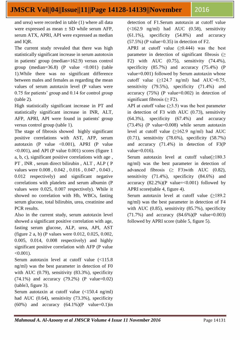
APRI score(table 4, figure 4).
Serum autotaxin level at cutoff value (≥189.2
ng/ml) was the best parameter in detection of F4
with AUC (0.85), sensitivity (85.7%), specificity
(71.7%) and accuracy (84.6%)(P value=0.003)
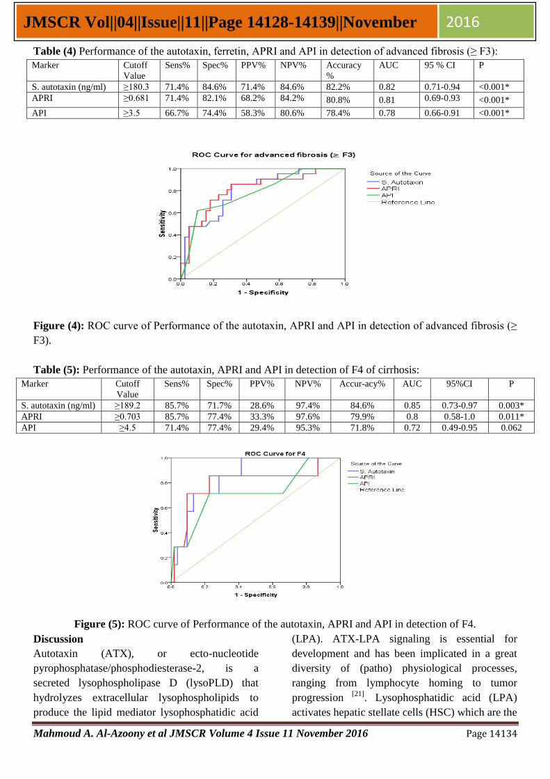
followed by APRI score (table 5, figure 5).

Mahmoud A. Al-Azoony et al JMSCR Volume 4 Issue 11 November 2016 Page 14132
JMSCR Vol||04||Issue||11||Page 14128-14139||November 2016
Table (1): Comparing the studied groups as regarding age, sex and laboratory investigation:
Variable HCV group
(N=60)
Controls
(N=20)
P value
Mean ± SD Mean ± SD
Age (years) 43.98 9.81 41.95 9.96 0.42
Hb (gm/dl) 13.56 1.794 14.28 1.355 0.1
RBCs 4.88 0.779 4.92 0.815 0.85
WBCs 6.20 2.029 5.37 0.715 0.08
PLTs 229.56 76.153 235.95 48.427 0.72
MPV 8.42 1.145 8.98 1.290 0.07
Fasting serum glucose (mg/dl) 101.96 29.215 96.65 15.725 0.44
Albumin (gm/dl) 4.11 0.56082 4.13 0.24979 0.61
PT (seconds) 14.56 1.76415 13.05 0.08272 <0.001*
INR 1.12 0.13689 1.03 0.06231 0.001*
T. bilirubin (mg/dl) 0.83 0.33828 0.75 0.18423 0.75
D. bilirubin (mg/dl) 0.28 0.14238 0.25 0.04767 0.73
AST(U/L) 44.63 25.59990 33.40 4.51197 0.16
ALT (U/L) 47.41 35.76064 30.60 6.12501 0.033*
ALP (U/L) 106.55 57.59779 80.90 11.21512 0.07
Urea (mg/dl) 30.25 6.00016 30.55 4.63936 0.82
Creatinine (mg/dl) 0.93 0.20839 0.92 0.11118 0.65
Median IQR Median IQR
AFP(ng/ml) 4.2 2.69-8.97 3.4 2.55-4.0 0.032*
Serum autotaxin (ng/ml) 162.9 96.2-261 36.8 29.2-45.7 <0.001*
APRI 0.49 0.28-0.84 0.35 0.28-0.43 0.017*
API 3.0 2.0-5.0 2.0 1.0-3.75 0.02*
Table (2) comparing males and females as regarding mean values of serum Autotaxin level in the studied
groups:
Variable HCV group
(N=60)
Controls
(N=20)
Test of sig.
Mean ±SD Mean ±SD
S. Autotaxin (ng/ml)
female
male
179.09
188.23
87.41210
135.22116
40.7
33.3
8.25
12.56
St. "t"=0.8
P value=0.75 P value=0.14
(a) (b)
0
1
2
3
4
5
0 200 400 600
F st
age
Serum Autotaxin
0
1
2
3
4
5
0 1 2 3
F st
age
APRI

Mahmoud A. Al-Azoony et al JMSCR Volume 4 Issue 11 November 2016 Page 14133
JMSCR Vol||04||Issue||11||Page 14128-14139||November 2016
(c)
Figure (1): Spearman’s correlation between the fibrosis stage and the three noninvasive markers:
(a)Spearman’s correlation between serum autotaxin (ng/ml) and the fibrosis stage.(b) Spearman’s correlation
between APRI and the fibrosis stage. (c)Spearman’s correlation between API and the fibrosis stage.
(a) (b)
Figure (2): Spearman’s correlation between serum autotoxin and other variables: (a) Spearman’s correlation
between serum autotaxin (ng/ml)and API.(b)Spearman’s correlation between serum autotaxin (ng/ml)and
AST (U/L).
Table (3) Performance of the autotaxin, APRI and API in detection of F0:
Marker Cutoff
Value
Sens% Spec% PPV% NPV% Accuracy % AUC 95%CI P
S. autotaxin
(ng/ml)
<115.8 83.3% 74.1% 26.3% 97.6% 79.2% 0.79
0.57-1.0 0.02*
APRI <0.42 100% 63% 23.1% 100% 78.1% 0.78 0.65-0.91 0.025*
API <2.5 83.3% 42.6% 13.9% 95.8% 73.1% 0.73 0.52-0.94 0.065
Figure (3): ROC curve of performance of the autotaxin, APRI and API in detection of F0.
0
100
200
300
400
500
0 2 4 6 8
Au
tota
xin
API
0
100
200
300
400
500
600
0 50 100 150
Au
tota
xin
AST
0
1
2
3
4
5
0 2 4 6 8 F
stag
e
API
Mahmoud A. Al-Azoony et al JMSCR Volume 4 Issue 11 November 2016 Page 14134
JMSCR Vol||04||Issue||11||Page 14128-14139||November 2016
Table (4) Performance of the autotaxin, ferretin, APRI and API in detection of advanced fibrosis (≥ F3):
Marker Cutoff
Value
Sens% Spec% PPV% NPV% Accuracy
%
AUC 95 % CI P
S. autotaxin (ng/ml) ≥180.3 71.4% 84.6% 71.4% 84.6% 82.2% 0.82 0.71-0.94 <0.001*
APRI ≥0.681 71.4% 82.1% 68.2% 84.2% 80.8% 0.81 0.69-0.93 <0.001*
API ≥3.5 66.7% 74.4% 58.3% 80.6% 78.4% 0.78 0.66-0.91 <0.001*
Figure (4): ROC curve of Performance of the autotaxin, APRI and API in detection of advanced fibrosis (≥
F3).
Table (5): Performance of the autotaxin, APRI and API in detection of F4 of cirrhosis:
Marker Cutoff
Value
Sens% Spec% PPV% NPV% Accur-acy% AUC 95%CI P
S. autotaxin (ng/ml) ≥189.2 85.7% 71.7% 28.6% 97.4% 84.6% 0.85 0.73-0.97 0.003*
APRI ≥0.703 85.7% 77.4% 33.3% 97.6% 79.9% 0.8 0.58-1.0 0.011*
API ≥4.5 71.4% 77.4% 29.4% 95.3% 71.8% 0.72 0.49-0.95 0.062
Figure (5): ROC curve of Performance of the autotaxin, APRI and API in detection of F4.
Discussion
Autotaxin (ATX), or ecto-nucleotide
pyrophosphatase/phosphodiesterase-2, is a
secreted lysophospholipase D (lysoPLD) that
hydrolyzes extracellular lysophospholipids to
produce the lipid mediator lysophosphatidic acid
(LPA). ATX-LPA signaling is essential for
development and has been implicated in a great
diversity of (patho) physiological processes,
ranging from lymphocyte homing to tumor
progression [21]
. Lysophosphatidic acid (LPA)
activates hepatic stellate cells (HSC) which are the

Mahmoud A. Al-Azoony et al JMSCR Volume 4 Issue 11 November 2016 Page 14135
JMSCR Vol||04||Issue||11||Page 14128-14139||November 2016
main cell type involved in development of liver
fibrosis, stimulates their contraction and inhibits
their apoptosis [11]
.
In the present study we found that serum ATX
levels were significantly increased in HCV group
versus control group. This camein agreement with
many studies [14,15,22,23]
. Recent evidence indicates
that ATX is rapidly cleared from the circulation
by liver sinusoidal endothelial cells [13]
.
HSC are an important factor in the development
of liver fibrosis and cirrhosis. On repeated or
persistent liver damage they transdifferentiate into
myofibroblasts [24]
. Progression of hepatic fibrosis
is associated with an increased number of HSC [25]
. Therefore, one can speculate that elevated
serum ATX levels are an indicator for activation
of HSC during development of fibrosis and
cirrhosis [26]
.
Portal hypertension is a result of enhanced
intrahepatic vascular resistance with activation of
HSC and endothelial dysfunction as well as
splanchnic vasodilatation [27]
. As ATX is cleared
by sinusoidal endothelial cells, dysfunctional
endothelium may be a factor for higher blood
concentrations of ATX in cirrhotic subjects [13]
. In
contrast to our results, Ezzat et al [28]
reported that
serum Autotaxin was not a diagnostic marker of
liver fibrosis.
In agreement with the results of Ezzat et al [28]
,
this study showed that there was no significant
difference between males and females as
regarding the mean values of serum autotaxin
level. On the contrary, Nakagawa and his
colleagues [23]
talked about gender bias of ATX
expression but they did not give explanation about
this bias.
Correlation study between the fibrotic stage and
the three noninvasive markers revealed highly
significant positive correlations between the
fibrosis stage and serum autotaxin, APRI and API
scores. This coincided with Fouad et al [29]
who
found a significant correlation between both APRI
and API scores not only with the stage of hepatic
fibrosis but also with the grade of activity. Also,
many other studies [14,15,22,23]
reported a significant
positive correlation between serum ATX and the
fibrosis stage.
Also, we found a significant negative correlation
between platelets and fibrosis stage. This result
agreed with Attallah et al [30]
who reported that
platelet count was correlated with progression of
fibrosis. Thrombocytopenia in patients with
advanced hepatic fibrosis may be explained by
reduction of thrombopoietin synthesis by liver,
also portal hypertension leads to pooling of
platelets in the enlarged spleen [31]
.
In the present study there was highly significant
positive correlation between AST and fibrosis
stage. Also, there was significant positive
correlation between ALT and fibrosis stge. These
results met the findings of Wai et al [18]
who found
gradual increase in serum AST level with
progression of fibrosis and explained this by the
fact that progression of liver fibrosis may reduce
the clearance of AST leading to increased serum
AST levels. In addition, advanced liver disease
may be associated with mitochondrial injury,
resulting in increased release of AST which is
present in mitochondria and cytoplasm.
Other correlations between the fibrotic stage and
the rest of the studied parameters revealed that
there was significant positive correlation between
PT, INR and fibrosis stage. This came in
agreement with Cadranel and Philippe[32]who
found that PT was an accurate, cheap and
reproducible serum marker for extensive fibrosis,
and they advised all clinicians to consider that PT
as a reliable marker of fibrosis progression. This is
attributed to decreased synthesis of coagulation
factors by the liver[33]
. On the other hand,
Coverdale et al [34]
reported that PT failed to
correlate with fibrosis progression.
We found asignificant negative correlation
between serum albumin and fibrosis stage.
Friedman et al [35]
also reported that
hypoalbuminemia was common in chronic liver
diseases that could be explained as albumin is the
most important plasma protein synthesized by the
liver and its synthesis is decreased in advanced
hepatic fibrosis.
Mahmoud A. Al-Azoony et al JMSCR Volume 4 Issue 11 November 2016 Page 14136
JMSCR Vol||04||Issue||11||Page 14128-14139||November 2016
Data obtained from this study revealed a
significant positive correlation between direct
biliubin and fibrosis stage. Hepatocyte damage as
in hepatitis C infection results in reduced
efficiency of bilirubin excretion into bile.
Conjugated bilirubin refluxes into the circulation
and is found in urine [36]
.
In accordance with Omran et al [37]
, we observed a
highly significant correlation between α
fetoprotein and fibrosis stage. Some hypotheses
attribute the high AFP level in hepatic damage to
the selective transcriptional activation of AFP
gene [38]
.
In the current study there was no significant
correlation between PCR results and fibrosis
stage. These results coincided with Ibrahim and
Mandour [2]
finding no significant correlation
between HCV RNA titers and the stage of liver
fibrosis. On the contrary, Adinolfi and his
colleagues [39]
concluded in their study that serum
HCV-RNA titer correlated with the severity of
liver damage, which can be accelerated by high
HCV load. Many factors may cause the
discrepancies between these studies. For example,
serum HCV load fluctuates, so it is unstable
parameter and cannot reflect the degree of liver
damage in a given subject [40]
. Also, HCV
replicates in extra-hepatic sites as well as within
the liver [41]
.
In consistency with Shahid et al [42]
ALP levels
correlated positively with the stage of liver
fibrosis. On the other hand, Ibrahim and Mandour [2]
reported that ALP levels were not correlated
with fibrosis stage.
On evaluating the performance of the three
noninvasive markers (serum autotaxin, APRI and
API) in prediction of the degree of liver fibrosis,
the results revealed that serum autotaxin level was
the best parameter among the three noninvasive
parameters in exclusion of fibrosis with AUC
(0.79).
All studied markers were considered fair tests in
detection of F1 and F2.
APRI was the best parameter in detection of
significant fibrosis (≥ F2) with AUC (0.754)
followed by serum autotaxin with AUC (0.751).
This came in agreement with Wai et al [18]
who
found that APRI was a good test in prediction of
significant fibrosis (F ≥ 2) at cutoff value > 1.5 the
AUC was 0.88. Also, Nakagawa et al [23]
found
that serum autotaxin level was the second best
parameter in both males and females for
prediction of significant fibrosis and had
sensitivity (70%), specificity (73.1%), AUC
(0.799) in male and sensitivity (86.5%),
specificity (70.6%), AUC (0.876) in female.
In the current study serum autotaxin level at a
cutoff value (≥180.3 ng/ml) was the best
parameter in detection of advanced fibrosis (≥ F3)
with AUC (0.82) and at a cutoff value (≥189.2
ng/ml) was the best parameter in detection of
cirrhosis with AUC (0.85). These results came in
accordance with Nakagawa and his colleagues [23]
who assessed the performance of serum autotaxin
level in prediction of liver fibrosis in comparison
with serum hyaluronic acid level and APRI and
found that serum autotaxinwas the best parameter
in male with sensitivity (82.4%), specificity
(74.4%), AUC (0.863) and the third-best
parameter in female with sensitivity (81.5%),
specificity (77.3%), AUC (0.872) for prediction of
cirrhosis.
In Conclusion
Serum autotaxin level is a valuable test for
exclusion of fibrosis and detection of advanced
fibrosis and cirrhosis in chronic HCV patients. As
it lacks discrimination of intermediate stages of
fibrosis, liver biopsy until now remains the gold
standard method for discrimination of different
stages of hepatic fibrosis. Further extended studies
should be done on larger numbers of chronic
hepatitis C patients using various combinations of
non invasive fibrosis markers to detect the
excellent test that can replace liver biopsy.
References
1. Palmeri ML, Wang MH, Rouze NC,
Abdelmalek MF, Guy CD, Moser
B, Diehl AM, et al. Noninvasive
evaluation of hepatic fibrosis using
acoustic radiation force-based shear
Mahmoud A. Al-Azoony et al JMSCR Volume 4 Issue 11 November 2016 Page 14137
JMSCR Vol||04||Issue||11||Page 14128-14139||November 2016
stiffness in patients with nonalcoholic fatty
liver disease. Journal of Hepatology 2011;
55(3): 666-672.
2. Ibrahim WS and Mandour EM.
Correlation of Liver Biopsy with Liver
Enzymes and PCR among Egyptian
Patients with Chronic Hepatitis C.
Academic Journal of Cancer Research
2014; 7 (2): 59-64.
3. Friedman SL. Liver fibrosis from bench to
bedside. J Hepatol2003; 38(1): 38-53.
4. Lauer GM and Walker BD. Hepatitis C
virus infection. N Engl J Med 2001;
345(1): 41-52.
5. Boursier J, Isselin G, Fouchard-Hubert I,
Oberti F, Dib N, Lebigot J, Bertrais S, et
al. Acoustic radiation force impulse: a new
ultrasonographic technology for the
widespread noninvasive diagnosis of liver
fibrosis. European journal of
gastroenterology &hepatology 2010;
22(9):1074-84.
6. Leroy V, Hilleret MN, Sturm N, Trocme
C, Renversez JC, Faure P, Morel F, et al.
Prospective comparison of six non-
invasive scores for the diagnosis of liver
fibrosis in chronic hepatitis C. J Hepatol
2007; 46(5): 775–782.
7. Nakanaga K, Hama K and Aoki J.
Autotaxin--an LPA producing enzyme
with diverse functions. Journal of
biochemistry 2010; 148(1):13-24.
8. Ikeda H and Yatomi Y. Autotaxin in liver
fibrosis.ClinChimActa 2012; 413(23-1
9. Koike S, Yutoh Y, Keino-Masu K, Noji
S, Masu M and Ohuchi H.Autotaxin is
required for the cranial neural tube closure
and establishment of the midbrain–
hindbrain boundary during mouse
development. DevDyn 2011; 240(2): 413-
421.
10. Brindley DN. Lipid phosphate phosph-
atases and related proteins: signaling
functions in development, cell division,
and cancer. J Cell Biochem 2004;
92(5):900-912.
11. Ikeda H, Nagashima K, Yanase M,
Tomiya T, Arai M, Inoue Y, Tejima K, et
al. Involvement of Rho/Rho kinase
pathway in regulation of apoptosis in rat
hepatic stellate cells. Am J Physiol
Gastrointest Liver Physiol 2003; 285(5):
880–886.
12. Tanaka M, Okudaira S, Kishi Y, Ohkawa
R, Iseki S, Ota M, Noji S, Yatomi Y, Aoki
J, Arai H. Autotaxin stabilizes blood
vessels and is required for embryonic
vasculature by producing lysophosphatidic
acid. J BiolChem 2006; 281(35): 25822–
25830.
13. Jansen S, Andries M, Vekemans K,
Vanbilloen H, Verbruggen A, Bollen
M.Rapid clearance of the circulating
metastatic factor autotaxin by the
scavenger receptors of liver sinusoidal
endothelial cells. Cancer Lett 2009; 284
(2): 216–221.
14. Watanabe N, Ikeda H, Nakamura K
, Ohkawa R, Kume Y, Aoki J, Hama K, et
al. Both plasma lysophosphatidic acid and
serum autotaxin levels are increased in
chronic hepatitis C. J ClinGastroenterol
2007; 41(6): 616–623.
15. Pleli T, Martin D, Kronenberger
B, Brunner F, Köberle V, Grammatikos
G, Farnik H, et al. Serum autotaxin is
a parameter for the severity of liver
cirrhosis and overall survival in patients
with liver cirrhosis--a prospective cohort
study. PLoS One 2014; 9(7): e103532.
16. Nustad K, Paus E, Kierulf B andBørmer
OP.Specificity and affinity of 30
monoclonal antibodies against alpha-
fetoprotein.TumourBiol 1998;19(4):293-
300.
17. Butler JE, Spradling JE, Suter M, Dierks
SE, Heyermann H, Peterman JH.The
immunochemistry of sandwich ELISAs--I.
The binding characteristics of
immunoglobulins to monoclonal and
polyclonal capture antibodies adsorbed on
plastic and their detection by symmetrical
Mahmoud A. Al-Azoony et al JMSCR Volume 4 Issue 11 November 2016 Page 14138
JMSCR Vol||04||Issue||11||Page 14128-14139||November 2016
and asymmetrical antibody-enzyme
conjugates. Molecular immunology 1986;
23(9): 971-82.
18. Wai CT, Greenson JK, Fontana
RJ,Kalbfleisch JD, Marrero JA,
Conjeevaram HS, Lok AS. A simple
noninvasive index can predict both
significant fibrosis and cirrhosis in patients
with chronic HCV. Hepatology 2003;
38(2): 518-526.
19. Poynard T and Bedossa P. Age and
platelet count: a simple index for
predicting the presence of histological
lesions in patients with antibodies to
hepatitis C virus. METAVIR and
CLINIVIR Cooperative Study Groups. J
Viral Hepat 1997; 4(3):199-208.
20. Bedossa P and Poynard T.An algorithm for
the grading of activity in chronic hepatitis
C. for the METAVIR Cooperative Study
Group.Hepatology 1996; 24: 289-293.
21. Perrakis A and Moolenaar WH.
Autotaxin: structure-function and
signaling. J Lipid Res 2014; 55(6): 1010–
1018.
22. Benesch MG, Ko YM, McMullen TPand
Brindley DN.Autotaxin in the crosshairs:
taking aim at cancer and other
inflammatory conditions. FEBS Lett 2014;
588(16): 2712-2727.
23. Nakagawa H, Ikeda H, Nakamura
K, Ohkawa R, Masuzaki R, Tateishi
R, Yoshida H, et al. Autotaxin as a novel
serum marker of liver fibrosis. Clinica
ChimicaActa 2011; 412(13-14):1201-
1206.
24. Rockey DC, Boyles JK, Gabbiani G and
Friedman SL. Rat hepatic lipocytes
express smooth muscle actin upon active-
tion in vivo and in culture. J Submicrosc
CytolPathol 1992; 24(2):193–203.
25. Yamaoka K, Nouchi T, Marumo F and
Satoc C. Alpha-smooth-muscle actin
expression in normal and fibrotic human
livers. Dig Dis Sci1993; 38(8): 1473–
1479.
26. Tanaka M, Okudaira S, Kishi Y, Ohkawa
R, Iseki S, Ota M, Noji S, et al.Autotaxin
stabilizes blood vessels and is required for
embryonic vasculature by producing
lysophosphatidic acid. J BiolChem2006;
281(35):25822–25830.
27. Garcı ´a-Paga´nJC, Gracia-SanchoJ and
BoschJ. Functional aspects on the
pathophysiology of portal hypertension in
cirrhosis.J Hepatol 2012; 57(2):458–461.
28. Ezzat WM,Ragab HM, El MaksoudNA,
Abdulla NA, Yasser A and Elhosary YA.
Validity of Autotaxin as a Novel
Diagnostic Marker for Liver Fibrosis in
Egyptian Chronic HCV Patients. Maced J
Med Sci2013; 6(4): 359-364.
29. Fouad SA, Esmat S, Omran D, Rashid L
and Kobaisi MH. Non-invasive assessment
of hepatic fibrosis in Egyptian patients
with chronic hepatitis C virus infection.
World J Gastroenterology 2012; 18(23):
2988-2994.
30. Attallah AM, El-Far M, Omran MM, Farid
K, Albannan MS and El-Dosoky I.
Noninvasive Diagnosis of Liver Fibrosis
and Cirrhosis in Chronic Hepatitis C
Patients. Journal of clinical laboratory
analysis 2013; 27(2): 121-129.
31. Lackner C, Struber G, Liegl B, Leibl
S, Ofner P, Bankuti C, Bauer B, et al.
Comparison and validation of simple
noninvasive tests for prediction of fibrosis
in chronic hepatitis C. Hepatology 2005;
41(6):1376-1382.
32. Cadranel JF and Philippe M.Prothrombin
index decrease: a useful and reliable
marker of extensive fibrosis. Eur J
Gastroenterol Hepatol 2002; 14(10):
1057-1059.
33. Mistry PK and Jain D.Haematological
disorders of the liver. In: Dooley JS, Lok
ASF, Burroughs AK and Heathcote
EJ(eds.).Sherlock’s Diseases of The Liver
and Biliary System. 12th ed. United
Kingdom: Blackwell Publishing Ltd, 2011,
p. 48-69.

Mahmoud A. Al-Azoony et al JMSCR Volume 4 Issue 11 November 2016 Page 14139
JMSCR Vol||04||Issue||11||Page 14128-14139||November 2016
34. Coverdale SA, Samarasinghe DA, Lin R,
Kench J, Byth K, Khan MH, Crewe E, et
al. Changes in antipyrine clearance and
platelet count, but not conventional liver
tests, correlate with fibrotic change in
chronic hepatitis C: value for predicting
fibrotic progression. Am. J. Gastroenterol
2003; 98(6): 1384-1390.
35. Friedman SL, Mantin P and Munoz SJ.
Laboratory methods for evalution of the
patients with liver disease. In: Zakim D,
Boyer TD(eds.).A textbook of liver
diseases. Philadelphia: saunder, 2002, p.
661-708.
36. Elias E. Jaundice and cholestasis. In:
Dooley JS, Lok ASF, Burroughs AK and
Heathcote EJ (eds.).Sherlock’s Diseases of
The Liver and Biliary System.12th ed.
Blackwell Publishing Ltd, 2011, p.234-
256.
37. Omran MM, Farid K, Emran TM and
Attallah AA. Fibro α-score as a simple and
useful non-invasive test for predicting
significant liver fibrosis in chronic
hepatitis C patients. Arab Journal of
Gastroenterology 2011; 12(2):74-79.
38. Taketa K. Alpha fetoprotein: reevaluation
in hepatology. Hepatology 1990; 12(6):
1420-1432.
39. Adinolfi LE, Utili R, Andreana A, Tripodi
MF, Marracino M, Gambardella M,
Giordano M, et al. Serum HCV RNA
levels correlate with histological liver
damage and concur with steatosis in
progression of chronic hepatitis C. Dig Dis
Sci. 2001; 46(8): 1677-1683.
40. Magrin S, Craxi A, Fabiano C, Simonetti
RG, Fiorentino G, Marino L, Diquattro O,
et al.Hepatitis C viremia in chronic liver
disease: relationship to interferon-alpha or
corticosteroid treatment. Hepatology 1994;
19(2): 273-279.
41. Nousbaum, JB, Pol S, Nalpas B, Landais
P, Berthelot P and Bréchot C. Hepatitis-C-
virus-type 1b (II) infection in France and
Italy. Collaborative Study Group.Ann.
intern. Med 1995; 122(3): 161-168.
42. Shahid M, Idrees M, Nasir B, Raja
AJ, Raza SM, Amin I, Rasul A, et al.
Correlation of biochemical markers and
HCV RNA titers with fibrosis stages and
grades in chronic HCV-3a patients. Eur J
Gastroenterol Hepatol 2014; 26(7): 788-
794.