evaluating the bioactivity of a hydroxyapatite
TRANSCRIPT
RESEARCH ARTICLE Open Access
Evaluating the bioactivity of ahydroxyapatite-incorporatedpolyetheretherketone biocompositeRui Ma1* and Dagang Guo2
Abstract
Background: Polyetheretherketone (PEEK) exhibits stable chemical properties, excellent biocompatibility, andrational mechanical properties that are similar to those of human cortical bone, but the lack of bioactivity impedesits clinical application.
Methods: In this study, hydroxyapatite (HA) was incorporated into PEEK to fabricate HA/PEEK biocomposite using acompounding and injection-molding technique. The tensile properties of the prepared HA/PEEK composites (HAcontent from 0 to 40 wt%) were tested to choose an optimal HA content. To evaluate the bioactivity of thecomposite, the cell attachment, proliferation, spreading and alkaline phosphatase (ALP) activity of MC3T3-E1 cells,and apatite formation after immersion in simulated body fluid (SBF), and osseointegration in a rabbit cranial defectmodel were investigated. The results were compared to those from ultra-high molecular weight polyethylene(UHMWPE) and pure PEEK.
Results: By evaluating the tensile properties and elastic moduli of PEEK composite samples/PEEK composites withdifferent HA contents, the 30 wt% HA/PEEK composite was chosen for use in the subsequent tests. The results ofthe cell tests demonstrated that PEEK composite samples/PEEK composite exhibited better cell attachment,proliferation, spreading, and higher ALP activity than those of UHMWPE and pure PEEK. Apatite islands formed onthe HA/PEEK composite after immersion in SBF for 7 days and grew continuously with longer time periods. Animaltests indicated that bone contact and new bone formation around the HA/PEEK composite were more obviousthan those around UHMWPE and pure PEEK.
Conclusions: The HA/PEEK biocomposite created by a compounding and injection-molding technique exhibitedenhanced osteogenesis and could be used as a candidate of orthopedic implants.
Keywords: Polyetheretherketone, Hydroxyapatite, Composite, Bioactivity, Osseointegration
BackgroundPolyaryletherketones (PAEKs) are a family ofhigh-temperature thermoplastic polymers. The most com-mon of these polymers that is used in the medical field ispolyetheretherketone (PEEK), which is often applied as amaterial for an interbody fusion cage [1]. The chemicalstructure of the aromatic backbone molecular chains ofPEEK confers exceptionally good chemical resistance andstability at high temperature or in the sterilization process
[2]. Furthermore, PEEK is biocompatible, wear-resistant,and radiolucent [3], and the mechanical properties of PEEKare similar to those of human cortical bone, which preventsthe stress-shielding effect that is frequently caused by metalimplants [4]. However, PEEK is biologically inert and doesnot directly bind to bone [4, 5], which prevents its completeintegration with the neighboring bone after implantation.Hydroxyapatite (HA), which has a chemical compos-
ition of Ca10(PO4)6(OH)2, is the closest pure syntheticequivalent to human bone mineral [6]. Synthetic HAmaterial is biocompatible and bioactive and clinicallyused as an important bone substitute [7]. HA possessesosteoconductive abilities as well as a remarkable ability
* Correspondence: [email protected] of Orthopedics, The Second Affiliated Hospital of Xi’an JiaotongUniversity, Xi’an 710004, Shaanxi, ChinaFull list of author information is available at the end of the article
© The Author(s). 2019 Open Access This article is distributed under the terms of the Creative Commons Attribution 4.0International License (http://creativecommons.org/licenses/by/4.0/), which permits unrestricted use, distribution, andreproduction in any medium, provided you give appropriate credit to the original author(s) and the source, provide a link tothe Creative Commons license, and indicate if changes were made. The Creative Commons Public Domain Dedication waiver(http://creativecommons.org/publicdomain/zero/1.0/) applies to the data made available in this article, unless otherwise stated.
Ma and Guo Journal of Orthopaedic Surgery and Research (2019) 14:32 https://doi.org/10.1186/s13018-019-1069-1
to bind directly to bone [8, 9]. Therefore, HA is com-monly coated onto a metal core or incorporated intopolymers to prepare composites. The first HA-reinfor-ced-polymer composite was micro-scale HAparticle-reinforced high-density polyethylene (HDPE) de-veloped by Bonfield and coworkers in the 1980s [10]. Thiscomposite containing 40 vol% HA was commercializedunder the trade name HAPEX™ for use innon-load-bearing otologic and maxillofacial implants [11].The strategy of adding HA fillers to make HA/PEEK
composite to improve the bioactivity of PEEK has beenproved to be feasible. HA has been incorporated intoPEEK to prepare HA/PEEK composites using varioustechniques. Zhang et al. [12] manufactured HA/PEEKcomposites via a selective laser sintering technique anddetermined that the HA/PEEK composite supported cellgrowth of osteoblasts; in addition, composites withhigher HA contents exhibited enhanced cell proliferationand osteogenic differentiation. Yu et al. [13] preparedHA/PEEK composite with 10, 20, 30, and 40 vol% HA bya mixing, compaction, and pressureless sintering processand evaluated the bioactivities of the HA/PEEK compos-ites using the simulated body fluid (SBF) immersing test.The growth rate of the apatite increased with the HAvolume fraction, suggesting that the bioactivity of theHA/PEEK composite increased with increasing HA con-tent in the composite [13]. Ma et al. [14, 15] preparedHA/PEEK composite via an in situ synthetic process,and they demonstrated that the composite exhibitedgood biocompatibility and satisfactory bioactivity.In the current study, micro-scale HA powders and
PEEK powders were utilized to fabricate a HA/PEEKcomposite using a compounding and injection-moldingtechnique. Theoretically speaking, higher HA contentmeans higher bioactivity; however, the mechanical prop-erties may be compromised with HA content increasing
[16]. Therefore, we firstly evaluated the tensile propertiesof the 0–40 wt% HA/PEEK composites to choose an op-timal HA content. Then, the bioactivity of the HA/PEEKcomposite was assessed and compared to that ofultra-high molecular weight polyethylene (UHMWPE)and pure PEEK by evaluating the osteoblast interactions,apatite-formation in vitro, and osseointegration in vivo.
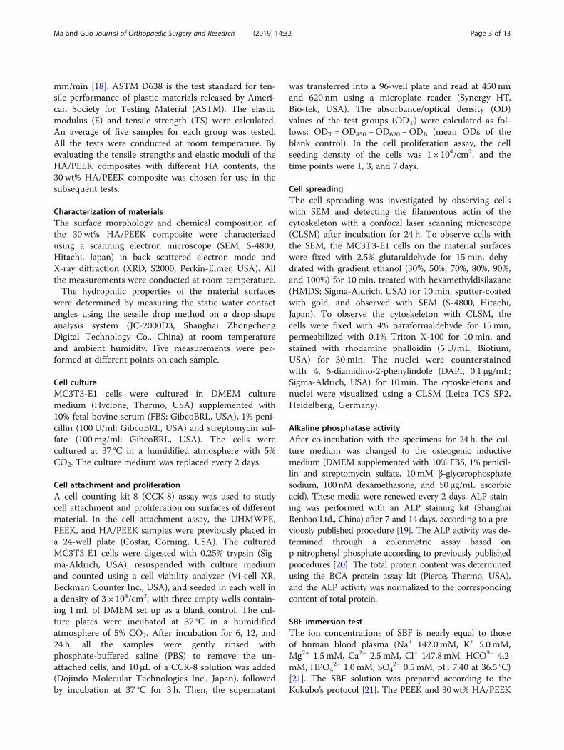
MethodsPreparation of materialsHA was prepared by a precipitation method using cal-cium hydroxide (Ca(OH)2, purity 99.0%; Sigma-Aldrich,USA) and phosphoric acid (H3PO4, purity 85.0%;Sigma-Aldrich, USA) [17]. PEEK powders with a meanparticle size of 20 μm were used (Victrex, South York-shire, UK). The HA/PEEK composite samples with HAcontents at 10, 20, 30, and 40 wt% were prepared via acompounding and injection-molding process (Fig. 1).The HA and PEEK powders were compounded in ahigh-speed ball mill (QM-3B, Nanjing T-Bota ScietechInstruments & Equipment Ltd., China) at a mixing speedof 500 rpm for 1 h, and then, the mixtures were dried at150 °C for 24 h. The HA/PEEK composite samples wereproduced using an injection molding machine (BA-300/050CD, Battenfeld, Belgium) with an injectingtemperature of 380 °C. In addition, the pure PEEK sam-ples were prepared with PEEK powders via an injectionand molding process. Ultra-high molecular weight poly-ethylene (UHMWPE; Roechling, Munich, Germany)with a molecular weight of two million served as thecontrol. All the samples were cut into 2-mm-thick diskswith a diameter of 15 mm.
Tensile propertiesThe tensile tests were performed in accordance withASTM D638 (ISO 527-1) with across head speed of 1.0
Fig. 1 Schematic diagram of the preparation and biological evaluation of the HA/PEEK biocomposite
Ma and Guo Journal of Orthopaedic Surgery and Research (2019) 14:32 Page 2 of 13
mm/min [18]. ASTM D638 is the test standard for ten-sile performance of plastic materials released by Ameri-can Society for Testing Material (ASTM). The elasticmodulus (E) and tensile strength (TS) were calculated.An average of five samples for each group was tested.All the tests were conducted at room temperature. Byevaluating the tensile strengths and elastic moduli of theHA/PEEK composites with different HA contents, the30 wt% HA/PEEK composite was chosen for use in thesubsequent tests.
Characterization of materialsThe surface morphology and chemical composition ofthe 30 wt% HA/PEEK composite were characterizedusing a scanning electron microscope (SEM; S-4800,Hitachi, Japan) in back scattered electron mode andX-ray diffraction (XRD, S2000, Perkin-Elmer, USA). Allthe measurements were conducted at room temperature.The hydrophilic properties of the material surfaces
were determined by measuring the static water contactangles using the sessile drop method on a drop-shapeanalysis system (JC-2000D3, Shanghai ZhongchengDigital Technology Co., China) at room temperatureand ambient humidity. Five measurements were per-formed at different points on each sample.
Cell cultureMC3T3-E1 cells were cultured in DMEM culturemedium (Hyclone, Thermo, USA) supplemented with10% fetal bovine serum (FBS; GibcoBRL, USA), 1% peni-cillin (100 U/ml; GibcoBRL, USA) and streptomycin sul-fate (100 mg/ml; GibcoBRL, USA). The cells werecultured at 37 °C in a humidified atmosphere with 5%CO2. The culture medium was replaced every 2 days.
Cell attachment and proliferationA cell counting kit-8 (CCK-8) assay was used to studycell attachment and proliferation on surfaces of differentmaterial. In the cell attachment assay, the UHMWPE,PEEK, and HA/PEEK samples were previously placed ina 24-well plate (Costar, Corning, USA). The culturedMC3T3-E1 cells were digested with 0.25% trypsin (Sig-ma-Aldrich, USA), resuspended with culture mediumand counted using a cell viability analyzer (Vi-cell XR,Beckman Counter Inc., USA), and seeded in each well ina density of 3 × 10⁴/cm2, with three empty wells contain-ing 1 mL of DMEM set up as a blank control. The cul-ture plates were incubated at 37 °C in a humidifiedatmosphere of 5% CO2. After incubation for 6, 12, and24 h, all the samples were gently rinsed withphosphate-buffered saline (PBS) to remove the un-attached cells, and 10 μL of a CCK-8 solution was added(Dojindo Molecular Technologies Inc., Japan), followedby incubation at 37 °C for 3 h. Then, the supernatant
was transferred into a 96-well plate and read at 450 nmand 620 nm using a microplate reader (Synergy HT,Bio-tek, USA). The absorbance/optical density (OD)values of the test groups (ODT) were calculated as fol-lows: ODT =OD450 −OD620 −ODB (mean ODs of theblank control). In the cell proliferation assay, the cellseeding density of the cells was 1 × 10⁴/cm2, and thetime points were 1, 3, and 7 days.
Cell spreadingThe cell spreading was investigated by observing cellswith SEM and detecting the filamentous actin of thecytoskeleton with a confocal laser scanning microscope(CLSM) after incubation for 24 h. To observe cells withthe SEM, the MC3T3-E1 cells on the material surfaceswere fixed with 2.5% glutaraldehyde for 15 min, dehy-drated with gradient ethanol (30%, 50%, 70%, 80%, 90%,and 100%) for 10 min, treated with hexamethyldisilazane(HMDS; Sigma-Aldrich, USA) for 10 min, sputter-coatedwith gold, and observed with SEM (S-4800, Hitachi,Japan). To observe the cytoskeleton with CLSM, thecells were fixed with 4% paraformaldehyde for 15 min,permeabilized with 0.1% Triton X-100 for 10 min, andstained with rhodamine phalloidin (5 U/mL; Biotium,USA) for 30 min. The nuclei were counterstainedwith 4, 6-diamidino-2-phenylindole (DAPI, 0.1 μg/mL;Sigma-Aldrich, USA) for 10min. The cytoskeletons andnuclei were visualized using a CLSM (Leica TCS SP2,Heidelberg, Germany).
Alkaline phosphatase activityAfter co-incubation with the specimens for 24 h, the cul-ture medium was changed to the osteogenic inductivemedium (DMEM supplemented with 10% FBS, 1% penicil-lin and streptomycin sulfate, 10mM β-glycerophosphatesodium, 100 nM dexamethasone, and 50 μg/mL ascorbicacid). These media were renewed every 2 days. ALP stain-ing was performed with an ALP staining kit (ShanghaiRenbao Ltd., China) after 7 and 14 days, according to a pre-viously published procedure [19]. The ALP activity was de-termined through a colorimetric assay based onp-nitrophenyl phosphate according to previously publishedprocedures [20]. The total protein content was determinedusing the BCA protein assay kit (Pierce, Thermo, USA),and the ALP activity was normalized to the correspondingcontent of total protein.
SBF immersion testThe ion concentrations of SBF is nearly equal to thoseof human blood plasma (Na+ 142.0 mM, K+ 5.0 mM,Mg2+ 1.5mM, Ca2+ 2.5mM, Cl− 147.8mM, HCO3− 4.2mM, HPO4
2− 1.0mM, SO42− 0.5mM, pH 7.40 at 36.5 °C)
[21]. The SBF solution was prepared according to theKokubo’s protocol [21]. The PEEK and 30wt% HA/PEEK
Ma and Guo Journal of Orthopaedic Surgery and Research (2019) 14:32 Page 3 of 13
samples were immersed in 40mL of SBF at 37 °C in a hu-midified atmosphere. After 7, 14, 21, and 28 days, the sam-ples were rinsed three times with distilled water and driedat 37 °C overnight. Then, the samples were observed andanalyzed with SEM and energy dispersive spectrometry(EDS; X-Max, Horiba, Japan). Furthermore, the concen-trations of Ca and P ions in the soaked SBF were mea-sured by inductively coupled plasma atomic emissionspectroscopy (ICP-AES; Varian, USA).
In vivo studyThirty mature female New Zealand white rabbits weight-ing 2.4 ± 0.2 kg were divided into three groups as fol-lows: group A (UHMWPE), group B (PEEK), and groupC (HA/PEEK). The operative procedures and animalcare were performed according to the ethical principlesof the Second Affiliated Hospital of Xi’an Jiaotong Uni-versity on animal experimentation (approval number:2016105). The anesthesia was induced by intramuscularinjection of 2% xylazine (12 mg/kg) and ketamine (80mg/kg). A longitudinal incision was made down to theperiosteum from the nasal bone to the occipital protu-berance at the parietal bone, and the periosteum wasundermined and lifted off the parietal skull. The skulldefect was drilled in the middle of the parietal bone withan outer diameter of 8.0 mm using a trephine (Med-XResearch Institute of Shanghai Jiaotong University,China). Finally, the samples with a diameter of 8 mmwere implanted into the defect. At 3 weeks, 2 weeks, and1 week prior to being sacrificed, the rabbits were intra-muscularly injected with alizarin red solution (30 mg/kg), calcein solution (20 mg/kg), and alizarin red solution(30 mg/kg), respectively. After feeding for 8 weeks, allthe rabbits were sacrificed, and the parietal bones wereretrieved, soaked into 75% ethanol for 1 week, and dehy-drated with gradient ethanol (80%, 95%, and 100%).The samples were embedded in methylmethacrylate
and cut into 150 μm sections with a microtome (SP1600, Leica, Germany). Then, the sections were then ad-hered to organic glass slides; compressed for 24 h;polished to 50 μm using P300, P800, and P1200 abrasivepaper; and then burnished with flannelette and abradumto 20–30 μm. At least three sections of each implantwere stained with picric acid/fuchsine. A light micro-scope (Leica Microsystems AG, Germany) was used forhistological evaluation. The fluorescence of the newbone formation labeled with alizarin red and calcein wasvisualized using a CLSM (TCS SP2, Leica Microsystems,Germany).
Statistical analysisAll the data are presented as the means ± standarddeviations. All the in vitro experiments were conductedin triplicate and repeated three times. Statistical
significances between different groups were analyzedusing a one-way analysis of variance (ANOVA) test, andmultiple comparisons were performed using the leastsignificant difference (LSD) test to determine any signifi-cant differences between each pair of groups. p < 0.05was considered statistically significant, and p < 0.01 wasconsidered highly statistically significant.
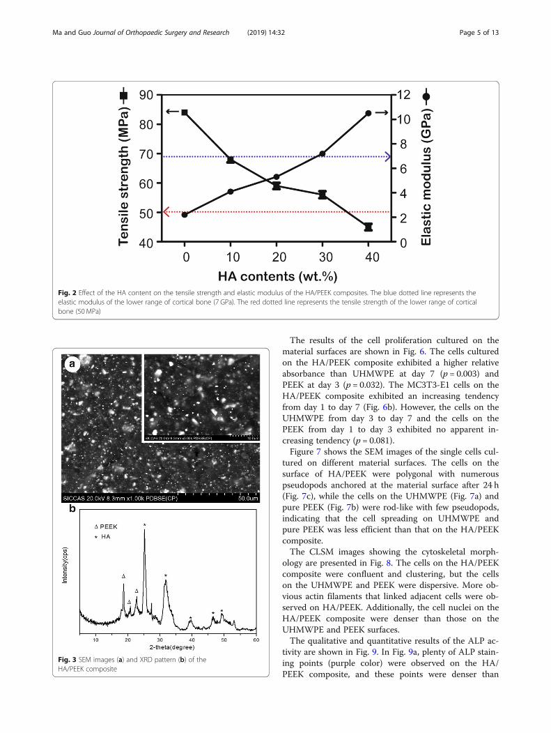
ResultsFigure 2 shows the tensile property results for the HA/PEEK composites. An increase in the amount of HA(from 0 to 40%) resulted in an increase in the elasticmodulus and a decrease in the tensile strength. In com-parison with PEEK, the elastic moduli of the HA/PEEKcomposites with HA contents of 10%, 20%, 30%, and40% increased by approximately 86%, 141%, 241%, and468%, respectively. The elastic moduli of the HA/PEEKcomposites (30% and 40%) were higher than that of thelower range of cortical bone (7 GPa) [22]. When the HAcontents were equal to or less than 30%, the tensilestrengths of the HA/PEEK composites were higher thanthat of the lower range of cortical bone (50MPa) [22].However, the tensile strength of the 40% HA/PEEK com-posite (45 ± 2.5MPa) was lower than 50MPa, which wasnot compatible with cortical bone. By evaluating the ten-sile properties and elastic moduli of the HA/PEEK com-posites with different HA contents, a HA content of 30wt% was chosen as an optimal HA content.Figure 3 shows the surface morphology (Fig. 3a) and
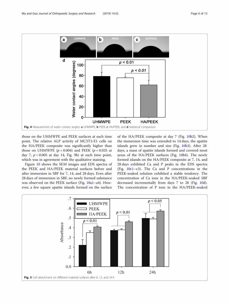
phase composition (Fig. 3b) of the HA/PEEK surface ob-served by SEM and XRD. Numerous HA particles (i.e.,white spots) were uniformly distributed on the materialsurface. The diffraction peaks of the composite at ap-proximately 2θ = 18.5°, 21°, and 22.5° corresponded tothe characteristic peaks of PEEK [23], and the peaks atapproximately 2θ = 25.5°, 32°, 40°, 46.5°, and 49.5° werethe characteristic peaks of HA [24]. The results indi-cated that the HA/PEEK composite was composed ofHA and PEEK.The hydrophilic properties of the material surfaces
were evaluated by measuring the water contact angles.The water contact angles of UHMWPE, pure PEEK, andHA/PEEK were 78.3 ± 8.1°, 75.6 ± 1.9°, and 61.9 ± 9.2°,respectively (Fig. 4). The water contact angle of the HA/PEEK composite was significantly lower than those ofUHMWPE and pure PEEK (p = 0.006; Fig. 4d), indicatingthat the HA/PEEK composite was more hydrophilic thanUHMWPE and pure PEEK.Figure 5 shows the cell attachment results from the
CCK-8 assay. The number of adhered cells on the HA/PEEK composite was significantly higher than that onUHMWPE at 6 h (p = 0.001), 12 h (p = 0.000), and 24 h(p= 0.024). Furthermore, more cells were adhered on HA/PEEK than on PEEK at 12 h (p= 0.000) and 24 h (p= 0.017).
Ma and Guo Journal of Orthopaedic Surgery and Research (2019) 14:32 Page 4 of 13
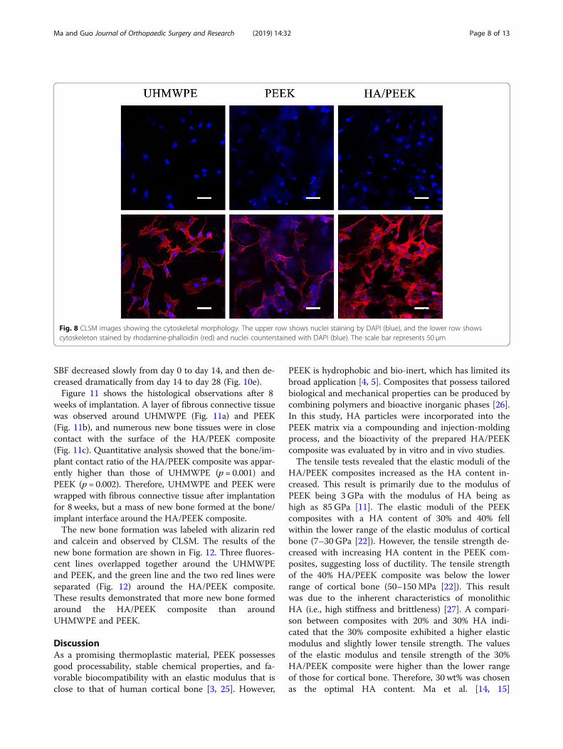
The results of the cell proliferation cultured on thematerial surfaces are shown in Fig. 6. The cells culturedon the HA/PEEK composite exhibited a higher relativeabsorbance than UHMWPE at day 7 (p = 0.003) andPEEK at day 3 (p = 0.032). The MC3T3-E1 cells on theHA/PEEK composite exhibited an increasing tendencyfrom day 1 to day 7 (Fig. 6b). However, the cells on theUHMWPE from day 3 to day 7 and the cells on thePEEK from day 1 to day 3 exhibited no apparent in-creasing tendency (p = 0.081).Figure 7 shows the SEM images of the single cells cul-
tured on different material surfaces. The cells on thesurface of HA/PEEK were polygonal with numerouspseudopods anchored at the material surface after 24 h(Fig. 7c), while the cells on the UHMWPE (Fig. 7a) andpure PEEK (Fig. 7b) were rod-like with few pseudopods,indicating that the cell spreading on UHMWPE andpure PEEK was less efficient than that on the HA/PEEKcomposite.The CLSM images showing the cytoskeletal morph-
ology are presented in Fig. 8. The cells on the HA/PEEKcomposite were confluent and clustering, but the cellson the UHMWPE and PEEK were dispersive. More ob-vious actin filaments that linked adjacent cells were ob-served on HA/PEEK. Additionally, the cell nuclei on theHA/PEEK composite were denser than those on theUHMWPE and PEEK surfaces.The qualitative and quantitative results of the ALP ac-
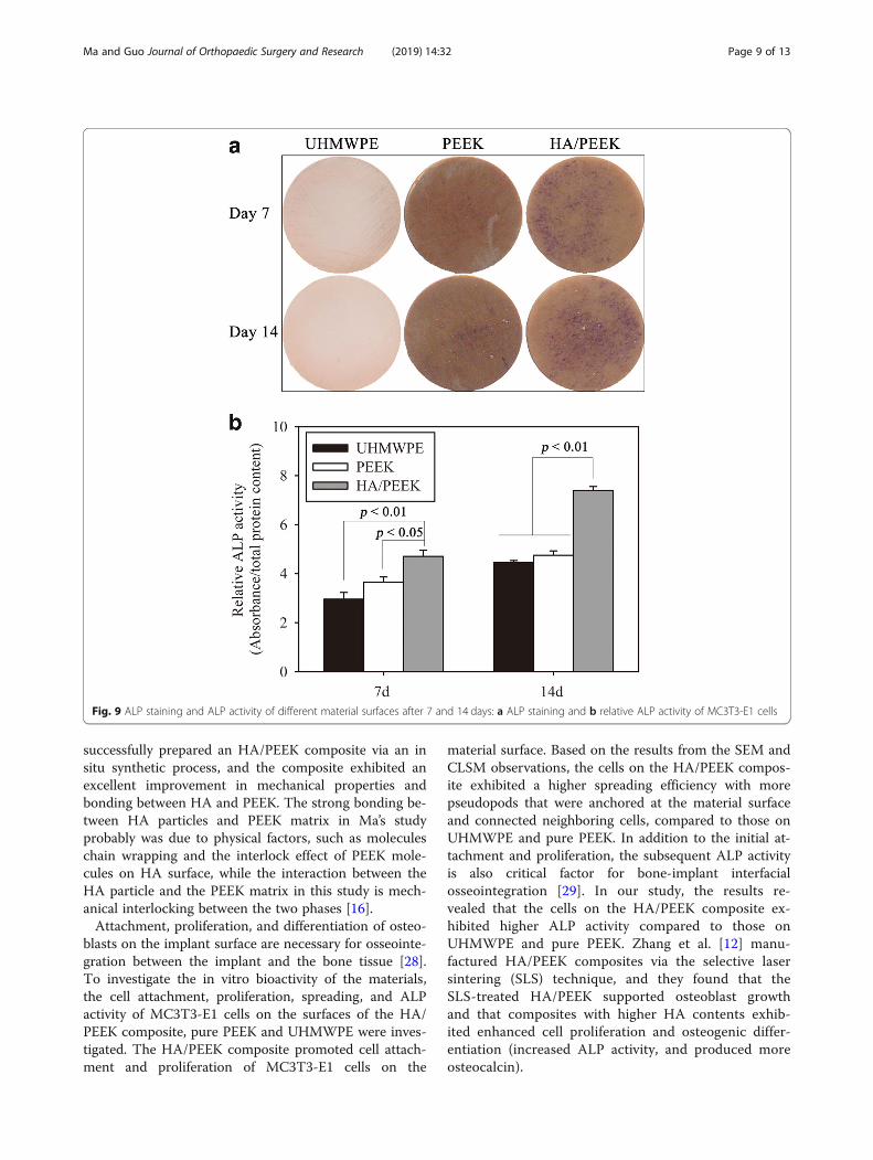
tivity are shown in Fig. 9. In Fig. 9a, plenty of ALP stain-ing points (purple color) were observed on the HA/PEEK composite, and these points were denser than
Fig. 3 SEM images (a) and XRD pattern (b) of theHA/PEEK composite
Fig. 2 Effect of the HA content on the tensile strength and elastic modulus of the HA/PEEK composites. The blue dotted line represents theelastic modulus of the lower range of cortical bone (7 GPa). The red dotted line represents the tensile strength of the lower range of corticalbone (50 MPa)
Ma and Guo Journal of Orthopaedic Surgery and Research (2019) 14:32 Page 5 of 13
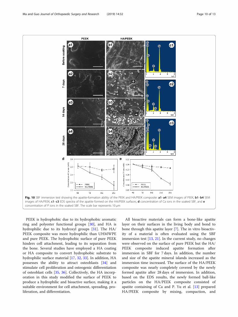
those on the UHMWPE and PEEK surfaces at each timepoint. The relative ALP activity of MC3T3-E1 cells onthe HA/PEEK composite was significantly higher thanthose on UHMWPE (p = 0.004) and PEEK (p = 0.035 atday 7, p = 0.005 at day 14, Fig. 9b) at each time point,which was in agreement with the qualitative staining.Figure 10 shows the SEM images and EDS spectra of
the PEEK and HA/PEEK material surfaces before andafter immersion in SBF for 7, 14, and 28 days. Even after28 days of immersion in SBF, no newly formed substancewas observed on the PEEK surface (Fig. 10a1–a4). How-ever, a few square apatite islands formed on the surface
of the HA/PEEK composite at day 7 (Fig. 10b2). Whenthe immersion time was extended to 14 days, the apatiteislands grew in number and size (Fig. 10b3). After 28days, a mass of apatite islands formed and covered mostareas of the HA/PEEK surfaces (Fig. 10b4). The newlyformed islands on the HA/PEEK composite at 7, 14, and28 days exhibited Ca and P peaks in the EDS spectra(Fig. 10c1–c3). The Ca and P concentrations in thePEEK-soaked solution exhibited a stable tendency. Theconcentration of Ca ions in the HA/PEEK-soaked SBFdecreased incrementally from days 7 to 28 (Fig. 10d).The concentration of P ions in the HA/PEEK-soaked
Fig. 4 Measurement of water contact angles: a UHMWPE, b PEEK, c HA/PEEK, and d statistical comparison
Fig. 5 Cell attachment on different material surfaces after 6, 12, and 24 h
Ma and Guo Journal of Orthopaedic Surgery and Research (2019) 14:32 Page 6 of 13
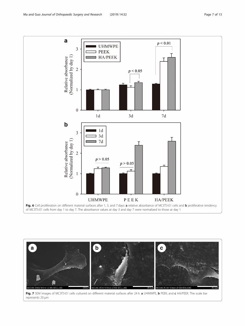
Fig. 6 Cell proliferation on different material surfaces after 1, 3, and 7 days: a relative absorbance of MC3T3-E1 cells and b proliferative tendencyof MC3T3-E1 cells from day 1 to day 7. The absorbance values at day 3 and day 7 were normalized to those at day 1
Fig. 7 SEM images of MC3T3-E1 cells cultured on different material surfaces after 24 h: a UHMWPE, b PEEK, and c HA/PEEK. The scale barrepresents 20 μm
Ma and Guo Journal of Orthopaedic Surgery and Research (2019) 14:32 Page 7 of 13
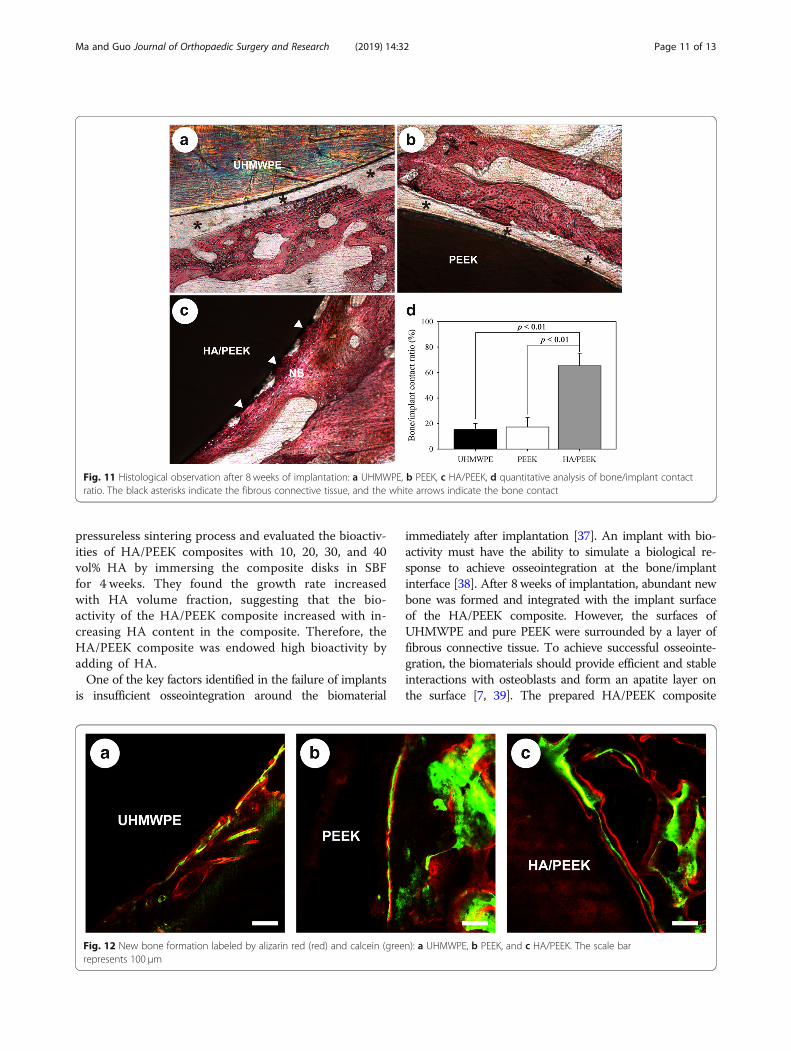
SBF decreased slowly from day 0 to day 14, and then de-creased dramatically from day 14 to day 28 (Fig. 10e).Figure 11 shows the histological observations after 8
weeks of implantation. A layer of fibrous connective tissuewas observed around UHMWPE (Fig. 11a) and PEEK(Fig. 11b), and numerous new bone tissues were in closecontact with the surface of the HA/PEEK composite(Fig. 11c). Quantitative analysis showed that the bone/im-plant contact ratio of the HA/PEEK composite was appar-ently higher than those of UHMWPE (p = 0.001) andPEEK (p = 0.002). Therefore, UHMWPE and PEEK werewrapped with fibrous connective tissue after implantationfor 8 weeks, but a mass of new bone formed at the bone/implant interface around the HA/PEEK composite.The new bone formation was labeled with alizarin red
and calcein and observed by CLSM. The results of thenew bone formation are shown in Fig. 12. Three fluores-cent lines overlapped together around the UHMWPEand PEEK, and the green line and the two red lines wereseparated (Fig. 12) around the HA/PEEK composite.These results demonstrated that more new bone formedaround the HA/PEEK composite than aroundUHMWPE and PEEK.
DiscussionAs a promising thermoplastic material, PEEK possessesgood processability, stable chemical properties, and fa-vorable biocompatibility with an elastic modulus that isclose to that of human cortical bone [3, 25]. However,
PEEK is hydrophobic and bio-inert, which has limited itsbroad application [4, 5]. Composites that possess tailoredbiological and mechanical properties can be produced bycombining polymers and bioactive inorganic phases [26].In this study, HA particles were incorporated into thePEEK matrix via a compounding and injection-moldingprocess, and the bioactivity of the prepared HA/PEEKcomposite was evaluated by in vitro and in vivo studies.The tensile tests revealed that the elastic moduli of the
HA/PEEK composites increased as the HA content in-creased. This result is primarily due to the modulus ofPEEK being 3 GPa with the modulus of HA being ashigh as 85 GPa [11]. The elastic moduli of the PEEKcomposites with a HA content of 30% and 40% fellwithin the lower range of the elastic modulus of corticalbone (7–30 GPa [22]). However, the tensile strength de-creased with increasing HA content in the PEEK com-posites, suggesting loss of ductility. The tensile strengthof the 40% HA/PEEK composite was below the lowerrange of cortical bone (50–150MPa [22]). This resultwas due to the inherent characteristics of monolithicHA (i.e., high stiffness and brittleness) [27]. A compari-son between composites with 20% and 30% HA indi-cated that the 30% composite exhibited a higher elasticmodulus and slightly lower tensile strength. The valuesof the elastic modulus and tensile strength of the 30%HA/PEEK composite were higher than the lower rangeof those for cortical bone. Therefore, 30 wt% was chosenas the optimal HA content. Ma et al. [14, 15]
Fig. 8 CLSM images showing the cytoskeletal morphology. The upper row shows nuclei staining by DAPI (blue), and the lower row showscytoskeleton stained by rhodamine-phalloidin (red) and nuclei counterstained with DAPI (blue). The scale bar represents 50 μm
Ma and Guo Journal of Orthopaedic Surgery and Research (2019) 14:32 Page 8 of 13
successfully prepared an HA/PEEK composite via an insitu synthetic process, and the composite exhibited anexcellent improvement in mechanical properties andbonding between HA and PEEK. The strong bonding be-tween HA particles and PEEK matrix in Ma’s studyprobably was due to physical factors, such as moleculeschain wrapping and the interlock effect of PEEK mole-cules on HA surface, while the interaction between theHA particle and the PEEK matrix in this study is mech-anical interlocking between the two phases [16].Attachment, proliferation, and differentiation of osteo-
blasts on the implant surface are necessary for osseointe-gration between the implant and the bone tissue [28].To investigate the in vitro bioactivity of the materials,the cell attachment, proliferation, spreading, and ALPactivity of MC3T3-E1 cells on the surfaces of the HA/PEEK composite, pure PEEK and UHMWPE were inves-tigated. The HA/PEEK composite promoted cell attach-ment and proliferation of MC3T3-E1 cells on the
material surface. Based on the results from the SEM andCLSM observations, the cells on the HA/PEEK compos-ite exhibited a higher spreading efficiency with morepseudopods that were anchored at the material surfaceand connected neighboring cells, compared to those onUHMWPE and pure PEEK. In addition to the initial at-tachment and proliferation, the subsequent ALP activityis also critical factor for bone-implant interfacialosseointegration [29]. In our study, the results re-vealed that the cells on the HA/PEEK composite ex-hibited higher ALP activity compared to those onUHMWPE and pure PEEK. Zhang et al. [12] manu-factured HA/PEEK composites via the selective lasersintering (SLS) technique, and they found that theSLS-treated HA/PEEK supported osteoblast growthand that composites with higher HA contents exhib-ited enhanced cell proliferation and osteogenic differ-entiation (increased ALP activity, and produced moreosteocalcin).
Fig. 9 ALP staining and ALP activity of different material surfaces after 7 and 14 days: a ALP staining and b relative ALP activity of MC3T3-E1 cells
Ma and Guo Journal of Orthopaedic Surgery and Research (2019) 14:32 Page 9 of 13
PEEK is hydrophobic due to its hydrophobic aromaticring and polyester functional groups [30], and HA ishydrophilic due to its hydroxyl groups [31]. The HA/PEEK composite was more hydrophilic than UHMWPEand pure PEEK. The hydrophobic surface of pure PEEKhinders cell attachment, leading to its separation fromthe bone. Several studies have employed a HA coatingor HA composite to convert hydrophobic substrate tohydrophilic surface material [17, 32, 33]. In addition, HApossesses the ability to attract osteoblasts [34] andstimulate cell proliferation and osteogenic differentiationof osteoblast cells [35, 36]. Collectively, the HA incorp-oration in this study modified the surface of PEEK toproduce a hydrophilic and bioactive surface, making it asuitable environment for cell attachment, spreading, pro-liferation, and differentiation.
All bioactive materials can form a bone-like apatitelayer on their surfaces in the living body and bond tobone through this apatite layer [7]. The in vitro bioactiv-ity of a material is often evaluated using the SBFimmersion test [13, 21]. In the current study, no changeswere observed on the surface of pure PEEK but the HA/PEEK composite induced apatite formation afterimmersion in SBF for 7 days. In addition, the numberand size of the apatite mineral islands increased as theimmersion time increased. The surface of the HA/PEEKcomposite was nearly completely covered by the newlyformed apatite after 28 days of immersion. In addition,based on the EDS results, the newly formed ball-likeparticles on the HA/PEEK composite consisted ofapatite containing of Ca and P. Yu et al. [13] preparedHA/PEEK composite by mixing, compaction, and
Fig. 10 SBF immersion test showing the apatite-formation ability of the PEEK and HA/PEEK composite: a1–a4 SEM images of PEEK, b1–b4 SEMimages of HA/PEEK, c1–c3 EDS spectra of the apatite formed on the HA/PEEK surfaces, d concentration of Ca ions in the soaked SBF, and econcentration of P ions in the soaked SBF. The scale bar represents 10 μm
Ma and Guo Journal of Orthopaedic Surgery and Research (2019) 14:32 Page 10 of 13
pressureless sintering process and evaluated the bioactiv-ities of HA/PEEK composites with 10, 20, 30, and 40vol% HA by immersing the composite disks in SBFfor 4 weeks. They found the growth rate increasedwith HA volume fraction, suggesting that the bio-activity of the HA/PEEK composite increased with in-creasing HA content in the composite. Therefore, theHA/PEEK composite was endowed high bioactivity byadding of HA.One of the key factors identified in the failure of implants
is insufficient osseointegration around the biomaterial
immediately after implantation [37]. An implant with bio-activity must have the ability to simulate a biological re-sponse to achieve osseointegration at the bone/implantinterface [38]. After 8 weeks of implantation, abundant newbone was formed and integrated with the implant surfaceof the HA/PEEK composite. However, the surfaces ofUHMWPE and pure PEEK were surrounded by a layer offibrous connective tissue. To achieve successful osseointe-gration, the biomaterials should provide efficient and stableinteractions with osteoblasts and form an apatite layer onthe surface [7, 39]. The prepared HA/PEEK composite
Fig. 11 Histological observation after 8 weeks of implantation: a UHMWPE, b PEEK, c HA/PEEK, d quantitative analysis of bone/implant contactratio. The black asterisks indicate the fibrous connective tissue, and the white arrows indicate the bone contact
Fig. 12 New bone formation labeled by alizarin red (red) and calcein (green): a UHMWPE, b PEEK, and c HA/PEEK. The scale barrepresents 100 μm
Ma and Guo Journal of Orthopaedic Surgery and Research (2019) 14:32 Page 11 of 13
promoted cell attachment, spreading, proliferation, andALP expression and induced apatite formation in vitro.Histological examinations in vivo indicated that the apatitelayer was formed on the ceramic surface early in the im-plantation period and the bone matrix integrated into theapatite [40]. After being implanted, the HA/PEEK compos-ite may induce the formation of an apatite layer, and theattracted osteoblasts attached onto the surface, proliferated,and differentiated to produce collagen and protein, whichmineralized to form new bone at the bone/implant inter-face. However, due to hydrophobic and bio-inert properties,UHMWPE and PEEK did not exhibit favorable osseointe-gration, and only fibrous connections eventually formed.At present, PEEK materials are most widely used as a
spinal fusion device in spinal surgery. The HA/PEEK bio-composite prepared in this study is a promising materialto be applied in the clinic as a material for spinal fusiondevice and bioactive artificial joint prosthesis. In addition,this study has some limitations. The connection betweenmicro-scale HA and PEEK in the composite is mainlyphysical bonding, so increase in the binding strength andlong-term stability of the interface needs to be appreci-ated. In addition, more animal model validation works areneeded to expand its clinical application value.
ConclusionTo improve the bioactivity of PEEK, HA particles wereincorporated into the PEEK matrix to prepare a HA/PEEK biocomposite using a compounding andinjection-molding technique. By evaluating the tensileproperties and elastic moduli of the HA/PEEK compos-ites with different HA contents (0–40 wt%), the 30 wt%HA was considered to be an optimal content. In vitrotests demonstrated that the HA/PEEK composite exhib-ited improved attachment, proliferation, and spreading,and higher ALP activity than those of UHMWPE andpure PEEK. Additionally, the HA/PEEK composite in-duced apatite formation after immersion in SBF. The invivo results indicated that the osseointegration efficiencyaround the HA/PEEK composite was higher than thataround UHMWPE and pure PEEK. All these resultsconfirmed that the bioactivity and osteogenesis of PEEKimproved substantially after incorporation of HA.
AbbreviationsALP: Alkaline phosphatase; CCK-8: Cell counting kit-8; CLSM: Confocal laserscanning microscope; DAPI: 4, 6-Diamidino-2-phenylindole; E: Elasticmodulus; EDS: Energy dispersive spectrometry; FBS: Fetal bovine serum;HA: Hydroxyapatite; HDPE: High-density polyethylene;HMDS: Hexamethyldisilazane; ICP-AES: Inductively coupled plasma atomicemission spectroscopy; OD: Optical density; PAEKs: Polyaryletherketones;PBS: Phosphate-buffered saline; PEEK: Polyetheretherketone; SBF: Simulatedbody fluid; SEM: Scanning electron microscope; TS: Tensile strength;UHMWPE: Ultra-high molecular weight polyethylene; XRD: X-ray diffraction
AcknowledgementsWe thank Prof. Tingting Tang and Jie Wei for experimental help.
FundingThis research was financially supported by the National Natural ScienceFoundation of China (No. 81702130) and the Fundamental Research Fundsfor the Central Universities.
Availability of data and materialsData sharing not applicable to this article as no datasets were generated oranalysed during the current study.
Authors’ contributionsRM performed the experiments. DG prepared the materials. RM wrote themanuscript. Both authors read and approved the final manuscript.
Ethics approval and consent to participateThe operative procedures and animal care obtained ethics approval andconsent of the ethical principles of the Second Affiliated Hospital of Xi’anJiaotong University on animal experimentation.
Consent for publicationNot applicable.
Competing interestsThe authors declare that they have no competing interests.
Publisher’s NoteSpringer Nature remains neutral with regard to jurisdictional claims inpublished maps and institutional affiliations.
Author details1Department of Orthopedics, The Second Affiliated Hospital of Xi’an JiaotongUniversity, Xi’an 710004, Shaanxi, China. 2State Key Laboratory for MechanicalBehavior of Materials, School of Materials Science and Engineering, Xi’anJiaotong University, Xi’an 710049, China.
Received: 5 September 2018 Accepted: 15 January 2019
References1. Kurtz SM, Devine JN. PEEK biomaterials in trauma, orthopedic, and spinal
implants. Biomaterials. 2007;28:4845–69.2. Jarman-Smith M. Evolving uses for implantable PEEK and PEEK based
compounds. Med Device Technol. 2008;19:12–5.3. Ma R, Tang T. Current strategies to improve the bioactivity of PEEK. Int J
Mol Sci. 2014;15:5426–45.4. Wang H, Xu M, Zhang W, Kwok DT, Jiang J, Wu Z, et al. Mechanical and
biological characteristics of diamond-like carbon coated poly aryl-ether-ether-ketone. Biomaterials. 2010;31:8181–7.
5. Briem D, Strametz S, Oder K, Meenen N, Lehmann W, Linhart W, et al.Response of primary fibroblasts and osteoblasts to plasma treatedpolyetheretherketone(PEEK) surfaces. J Mater Sci: Mater M. 2005;16:671–7.
6. Ryan KR, Gabriel LC, Robert JK, Weimin Y. Hydroxyapatite-reinforcedpolymer biocomposites for synthetic bone substitutes. JOM. 2008;60:38–45.
7. Kokubo T, Kim H-M, Kawashita M. Novel bioactive materials with differentmechanical properties. Biomaterials. 2003;24:2161–75.
8. Hollinger JO, Battistone GC. Biodegradable bone repair materials. Syntheticpolymers and ceramics. Clin Orthop Relat Res. 1986;207:290–305.
9. Hammerle CH, Olah AJ, Schmid J, Fluckiger L, Gogolewski S, Winkler JR, etal. The biological effect of natural bone mineral on bone neoformation onthe rabbit skull. Clin Oral Implants Res. 1997;8:198–207.
10. Bonfield W, Grynpas MD, Tully AE, Bowman J, Abram J. Hydroxyapatitereinforced polyethylene-a mechanically compatible implant material forbone replacement. Biomaterials. 1981;2:185–6.
11. Wang M, Porter D, Bonfield W. Processing, characterization, and evaluation ofhydroxyapatite reinforced polyethylene composites. Brit Ceram T. 1994;93:91–5.
12. Zhang Y, Hao L, Savalani MM, Harris RA, Di Silvio L, Tanner KE. In vitrobiocompatibility of hydroxyapatite-reinforced polymeric compositesmanufactured by selective laser sintering. J Biomed Mater Res A. 2009;91:1018–27.
13. Yu S, Hariram KP, Kumar R, Cheang P, Aik KK. In vitro apatite formation andits growth kinetics on hydroxyapatite/polyetheretherketone biocomposites.Biomaterials. 2005;26:2343–52.
Ma and Guo Journal of Orthopaedic Surgery and Research (2019) 14:32 Page 12 of 13
14. Ma R, Weng L, Bao X, Ni Z, Song S, Cai W. Characterization of in situsynthesized hydroxyapatite/polyetheretherketone composite materials.Mater Lett. 2012;71:117–9.
15. Ma R, Weng L, Bao X, Song S, Zhang Y. In vivo biocompatibility andbioactivity of in situ synthesized hydroxyapatite/polyetheretherketonecomposite materials. J Appl Polym Sci. 2013;127:2581–7.
16. Bakar MSA, Cheang P, Khor KA. Tensile properties and microstructuralanalysis of spheroidized hydroxyapatite/poly(etheretherketone)biocomposites. Mat Sci Eng A. 2003;345:55–63.
17. Lee JH, Jang HL, Lee KM, Baek HR, Jin K, Hong KS, et al. In vitro and in vivoevaluation of the bioactivity of hydroxyapatite-coated polyetheretherketonebiocomposites created by cold spray technology. Acta Biomater. 2013;9:6177-87.
18. D638-01 AS. Standard test method for tensile properties of plastics. WestConshohocken, PA, USA: American Society for Testing and Materials; 2001.
19. Fan Q, Tang T, Zhang X, Dai K. The role of CCAAT/enhancer binding protein(C/EBP)-alpha in osteogenesis of C3H10T1/2 cells induced by BMP-2. J CelMol Med. 2009;13:2489–505.
20. Sun H, Wu C, Dai K, Chang J, Tang T. Proliferation and osteoblasticdifferentiation of human bone marrow-derived stromal cells on akermanite-bioactive ceramics. Biomaterials. 2006;27:5651–7.
21. Kokubo T, Takadama H. How useful is SBF in predicting in vivo bonebioactivity? Biomaterials. 2006;27:2907–15.
22. Hench LL, Wilson J. In: Hench LL, Wilson J, editors. An introduction tobioceramics. Singapore: World Scientific; 1993. p. 12.
23. Han CM, Lee EJ, Kim HE, Koh YH, Kim KN, Ha Y, et al. The electron beamdeposition of titanium on polyetheretherketone (PEEK) and the resultingenhanced biological properties. Biomaterials. 2010;31:3465–70.
24. Santhosh S, Balasivanandha Prabu S. Thermal stability of nanohydroxyapatite synthesized from sea shells through wet chemical synthesis.Mater Lett. 2013;97:121–4.
25. Toth JM, Wang M, Estes BT, Scifert JL, Seim HB 3rd, Turner AS.Polyetheretherketone as a biomaterial for spinal applications. Biomaterials.2006;27:324–34.
26. Boccaccini AR, Blaker JJ. Bioactive composite materials for tissueengineering scaffolds. Expert Rev Med Devi. 2005;2:303–17.
27. Bakar MSA, Cheang P, Khor KA. Mechanical properties of injection moldedhydroxyapatite-polyetheretherketone biocomposites. Compos Sci Technol.2003;63:421–5.
28. Tan H, Guo S, Yang S, Xu X, Tang T. Physical characterization andosteogenic activity of the quaternized chitosan-loaded PMMA bonecement. Acta Biomater. 2012;8:2166–74.
29. Hu X, Neoh KG, Shi Z, Kang ET, Poh C, Wang W. An in vitro assessment oftitanium functionalized with polysaccharides conjugated with vascularendothelial growth factor for enhanced osseointegration and inhibition ofbacterial adhesion. Biomaterials. 2010;31:8854–63.
30. Noiset O, Schneider YJ, Marchand-Brynaert J. Fibronectin adsorption or/andcovalent grafting on chemically modified PEEK film surfaces. J Biomater SciPolym E. 1999;10:657–77.
31. Bodhak S, Bose S, Bandyopadhyay A. Role of surface charge and wettabilityonearly stage mineralization and bone cell-materials interactions ofpolarizedhydroxyapatite. Acta Biomater. 2009;5:2178–88.
32. Yang F, Wolke JGC, Jansen JA. Biomimetic calcium phosphate coatingonelectrospun poly(e-caprolactone) scaffolds for bone tissue engineering.Chem Eng J. 2008;137:154–61.
33. Ito Y, Hasuda H, Kamitakahara M, Ohtsuki C, Tanihara M, Kang IK.Acomposite of hydroxyapatite with electrospun biodegradable nanofibersas atissue engineering material. J Biosci Bioeng. 2005;100:43–9.
34. Thamaraiselvi TV, Rajeswari S. Biological evaluation of bioceramic materials-areview. Trends Biomater Artif Organs. 2004;18:9-17.
35. Ruan J, Grant MH. Biocompatibility evaluation in vitro. Part I: Morphologyexpression and proliferation of human and rat osteoblasts on thebiomaterials. J Cent South Univ T. 2001;8:1–8.
36. Yuan H, Kurashina K, de Bruijn JD, Li Y, de Groot K, Zhang X. A preliminarystudy on osteoinduction of two kinds of calcium phosphate ceramics.Biomaterials. 1999;20:1799–806.
37. Christenson EM, Anseth KS, van den Beucken JJ, Chan CK, Ercan B, Jansen JA,et al. Nanobiomaterial applications in orthopedics. J Orthop Res. 2007;25:11–22.
38. Hench LL, Splinter RJ, Allen W, Greenlee T. Bonding mechanisms at theinterface of ceramic prosthetic materials. J Biomed Mater Res. 1971;5:117–41.
39. Lee JH, Jang HL, Lee KM, Baek HR, Jin K, Hong KS, et al. In vitro and in vivoevaluation of the bioactivity of hydroxyapatite-coated polyetheretherketonebiocomposites created by cold spray technology. Acta Biomater. 2013;9:6177–87.
40. Ohtsuki C, Kushitani H, Kokubo T, Kotani S, Yamamuro T. Apatite formationon the surface of Ceravital-type glass-ceramic in the body. J Biomed MaterRes. 1991;25:1363–70.
Ma and Guo Journal of Orthopaedic Surgery and Research (2019) 14:32 Page 13 of 13