european trends in epilepsy surgery
TRANSCRIPT

ARTICLE
European trends in epilepsy surgeryMaxime O. Baud, MD, PhD, Thomas Perneger, MD, PhD, Attila Racz, MD, PhD, Max C. Pensel, MD,
Christian Elger, MD, PhD, FRCP, Bertil Rydenhag, MD, PhD, Kristina Malmgren, MD, PhD,
J. Helen Cross, MD, PhD, Grainne McKenna, BA, BChir, Martin Tisdall, MD, Herm J. Lamberink, MD,
Sylvain Rheims, MD, PhD, Philippe Ryvlin, MD, PhD, Jean Isnard, MD, PhD, François Mauguiere, MD, PhD,
Alexis Arzimanoglou, MD, PhD, Serdar Akkol, MD, Kaancan Deniz, MD, Cigdem Ozkara, MD,
Morten Lossius, MD, PhD, Ivan Rektor, MD, Reetta Kalviainen, MD, Lotta-Maria Vanhatalo, MS,
Petia Dimova, MD, Krassimir Minkin, MD, Anke Maren Staack, MD, Bernhard J. Steinhoff, MD,
Adam Kalina, MD, Pavel Krsek, MD, PhD, Petr Marusic, MD, PhD, Zsofia Jordan, MD, Daniel Fabo, PhD,
Evelien Carrette, PhD, Paul Boon, MD, PhD, Saulius Rocka, MD, R�uta Mameniskiene, MD, PhD,
Serge Vulliemoz, MD, PhD, Francesca Pittau, MD, PhD, Kees P.J. Braun, MD, PhD, and Margitta Seeck, MD
Neurology® 2018;0:e1-e11. doi:10.1212/WNL.0000000000005776
Correspondence
Dr. Seeck
AbstractObjectiveResective surgery is effective in treating drug-resistant focal epilepsy, but it remains unclearwhether improved diagnostics influence postsurgical outcomes. Here, we compared practiceand outcomes over 2 periods 15 years apart.
MethodsSixteen European centers retrospectively identified 2 cohorts of children and adults whounderwent epilepsy surgery in the period of 1997 to 1998 (n = 562) or 2012 to 2013 (n = 736).Data collected included patient (sex, age) and disease (duration, localization and diagnosis)characteristics, type of surgery, histopathology, Engel postsurgical outcome, and complications,as well as imaging and electrophysiologic tests performed for each case. Postsurgical outcomepredictors were included in a multivariate logistic regression to assess the strength of date ofsurgery as an independent predictor.
ResultsOver time, the number of operated cases per center increased from a median of 31 to 50 per2-year period (p = 0.02). Mean disease duration at surgery decreased by 5.2 years (p < 0.001).Overall seizure freedom (Engel class 1) increased from 66.7% to 70.9% (adjusted p = 0.04),despite an increase in complex surgeries (extratemporal and/or MRI negative). Surgeriesperformed during the later period were 1.34 times (adjusted odds ratio; 95% confidenceinterval 1.02–1.77) more likely to yield a favorable outcome (Engel class I) than earliersurgeries, and improvement was more marked in extratemporal and MRI-negative temporalepilepsy. The rate of persistent neurologic complications remained stable (4.6%–5.3%, p = 0.7).
ConclusionImprovements in European epilepsy surgery over time are modest but significant, includinghigher surgical volume, shorter disease duration, and improved postsurgical seizure outcomes.Early referral for evaluation is required to continue on this encouraging trend.
RELATED ARTICLE
EditorialThe changing landscape ofepilepsy surgery: No longerthe “last resort”
Page 55
From the Department of Neurology (M.O.B., S.V., F.P., M.S.), and Center for Clinical Research (T.P.), University Hospital Geneva; Department of Neurology (M.O.B.), University HospitalBern; Wyss Center for Bio- and Neuro-Engineering (M.O.B.), Geneva, Switzerland; Klinik und Poliklinik fur Epileptologie (A.R., M.C.P., C.E.), Universitatsklinikum Bonn, Germany; Sahl-grenska University Hospital and Sahlgrenska Academy at the University of Gothenburg (B.R., K. Malmgren), Sweden; UCL Great Ormond Street Hospital (J.H.C., G.M., M.T.), London, UK;Department of Child Neurology (H.J.L., K.P.J.B.), Brain Center Rudolf Magnus, University Medical Center Utrecht, the Netherlands; Department of Functional Neurology and Epileptology(S. Rheims, J.I., P.R., F.M.) and Department of Clinical Epileptology, Sleep Disorders, and Functional Neurology in Children (A.A., P.R.), Hospices Civils de Lyon andUniversity of Lyon, France;Department of Neurology (P.R.), University Hospital Lausanne, Switzerland; Department of Neurology (S.A., K.D., C.O.), Cerrahpasa Medical Faculty, Istanbul University, Turkey; Clinic forNeuroscience (M.L.), National Center for Epilepsy, Oslo University Hospital, Norway; Epilepsy Centre, (I.R.), Masaryk University, Hospital Ste Anne, and CEITEC–Neuroscience Centre, Brno,Czech Republic; KuopioUniversity Hospital andUniversity of Eastern Finland (R.K., L.-M.V.); St. Ivan Rilski University Hospital (P.D., K.Minkin), Bulgaria; EpilepsiezentrumKork (A.M.S., B.J.S.),Germany; Second Faculty of Medicine (A.K., P.K.,, P.M.), Charles University, Motol University Hospital, Prague, Czech Republic; Juhasz Pal Epilepsy Centrum (Z.J., D.F.), National Institute ofClinical Neurosciences, Hungary; Reference Center for Refractory Epilepsy (E.C., P.B.), GhentUniversity Hospital, Belgium; andDepartment ofNeurology andNeurosurgery (S. Rocka, R.M.),Vilnius University, Lithuania.
Go to Neurology.org/N for full disclosures. Funding information and disclosures deemed relevant by the authors, if any, are provided at the end of the article.
Copyright © 2018 American Academy of Neurology e1
Copyright ª 2018 American Academy of Neurology. Unauthorized reproduction of this article is prohibited.
Published Ahead of Print on June 13, 2018 as 10.1212/WNL.0000000000005776

Comprehensive European epilepsy programs began toemerge in the 1970s that were aimed at achieving tailoredepilepsy surgeries.1 Over the years, this approach has provenhighly effective for the treatment of drug-resistant focalepilepsies in children and adults with confirmation by 2randomized controlled trials.2,3 Given these results, theAmerican Academy of Neurology issued a recommendationin 2003 advocating lenient early referral to tertiary centersthat offer comprehensive evaluation and, if indicated, sur-gery.4 To help address the knowledge base of when to refer,the International League Against Epilepsy redefined drug-resistant epilepsy as 2 failed appropriate antiepileptic drugtrials.5 Recent data suggest that earlier surgery leads tobetter seizure outcome6–8 and improved quality of life3 andmay mitigate mortality related to sudden unexpected deathin epilepsy (SUDEP).9 However, despite the evidence andofficial recommendations, observational studies evaluatingtrends over time reveal that referrals remain delayed10 andthat the number of surgeries is stagnant in the UnitedStates11–13 and individual centers in Germany14,15 or evendecreasing in the United Kingdom,16 whereas it has beenshown to be increasing in children in the Netherlands.8 Thishighlights regional differences and potentially differenttrends in pediatric and adult epilepsy surgery.
Over the last 20 years, there have been major advances inepilepsy knowledge, diagnostic methods, and their applicationto identify suitable surgical candidates. For example, thespectrum of autoimmune epilepsy has been established17 thatregroups patients who may be poor surgical candidates. Onthe other hand, patients with MRI-negative epilepsy withoutan immunologic cause but with a focus well identified onfunctional imaging (e.g., PET, source imaging) may be bettersurgical candidates than hitherto suggested.18 Thus, strongerfield MRIs and voxel-based morphometry,19 molecular18 andfunctional imaging,19 electric or magnetic source localiza-tion,20 and multimodal coregistration19 may yield improvedguidance of surgeries and thus improved outcomes.
Recent meta-analyses report postsurgical seizure freedomrates in the range of 50% to 60%21,22 but have includedoperations performed in the 1990s, possibly underestimatingthe true contemporary surgical outcomes. Single-centerstudies8,15 are likely influenced by local policies, availableequipment, and referral bias, which could all be attenuatedwhen data are pooled across centers.
The present study included 16 European centers and aimedto determine whether postsurgical seizure outcome hasimproved in the 21st century, taking into account the
evolving practice in the field.13 As the primary hypotheses,we tested whether recommendations and continuous ed-ucation have led to earlier referrals and therefore decreaseddisease duration and whether European epilepsy surgeryoutcomes have improved in the recent years compared tothe 1990s. Secondary outcomes included the total numberand types of surgeries, surgical complications rankedby severity, and the number and type of diagnostic testsperformed.
MethodsStudy designWe designed a retrospective cohort study done in 2 waveswith the goal of comparing 2-year epochs 15 years apart(1997–1998 vs 2012–2013 inclusive), hereafter referred to asthe 1990s and 2010s, respectively. This delay was chosenbecause it comprises an era of major technical and knowledgedevelopment and the publication of official guidelines but isshort enough that each contributing center was able to re-trieve detailed information about individual cases. We col-lected the following primary study outcomes: disease durationfrom epilepsy onset to surgery (in years) and 2-year post-surgical seizure outcome as the Engel class. We also collectedsecondary study outcomes: number and type of diagnostictests performed, number and type of surgeries, number andseverity of surgical complications, and the individual center’s“surgical ratio” (see below). The target study size was 1,000patients (500 for each period) to even out the effect of an-ticipated practice variability across sites and to include enoughpatients in predefined surgical subgroups.
Epilepsy center inclusionWe invited 27 European epilepsy programs to participate.Inclusion criteria were at least 10 operations per year; surgeryon adults, children, or both; access to patient data covering the2 studied time periods; patients with a follow-up of ≥2 years;and the willingness or possibility to contribute to our study.Sixteen epilepsy centers met the inclusion criteria, includingthe Swedish National Epilepsy Surgery Register (alphabeti-cally: Bonn, Germany; Brno, Czech Republic; Budapest,Hungary; Geneva, Switzerland; Gent, Belgium; Istanbul,Turkey; Kuopio, Finland; Kork, Germany; London, UnitedKingdom; Lyon, France; Oslo, Norway; Prague, Czech Re-public; Utrecht, the Netherlands; Sofia, Bulgaria; and Vilnius,Lithuania, see also author affiliations for exact center details).Three centers fulfilled inclusion criteria only in the 2010s(Prague, Sofia, Vilnius) because of low surgical volumes in the1990s but were nevertheless included because staff, moni-toring, and imaging equipment were comparable to the other
GlossaryETLE = extratemporal lobectomy; FDG = fluorodeoxyglucose; HS = hippocampal sclerosis; SUDEP = sudden unexpecteddeath in epilepsy; TLE = temporal lobectomy.
e2 Neurology | Volume �, Number � | Month 0, 2018 Neurology.org/N
Copyright ª 2018 American Academy of Neurology. Unauthorized reproduction of this article is prohibited.

centers. All centers met regularly at European meetings toexchange views and to discuss clinical cases.
Standard protocol approvals, registrations,and patient consentsEach contributing epilepsy center received approval from anethical standards committee on human experimentation (in-stitutional or regional) for any experiments using humanparticipants. When indicated by the said committee, eachcenter received written informed consent from all patients (orguardians of patients) participating in the study (consent forresearch).
Data collectionData sources included medical records at local hospitals orpreexisting site-specific research databases. On the basis oflocal data, each center filled in a pre-established templatespreadsheet including a description of each variable to becollected and coded as numerical values (e.g., 0 = no, 1 =yes). Individual data were systematically checked for anyincongruence and validated through clarification with thecenter of origin before information was merged into 1common dataset. All cases that did not have follow-up afterat least 24 months were discarded (50 [7.8%] and 35[4.3%] patients in the 1990s and 2010s, respectively). Ifonly a longer outcome was available (e.g., 3 years), then itwas assumed that the 2-year outcome was the same. Palli-ative epilepsy surgeries (resection significantly limited byeloquent cortex, bitemporal epilepsy, or disconnectivesurgeries, including callosotomies and multiple subpialtransections) were excluded from the analysis because theydo not aim at seizure freedom (figure e-1, links.lww.com/WNL/A569). Laser thermo-ablation and ablation ofhamartomas were not included because they representcategories that were not available in the 1990s. Surgeries formalignant tumor were also not included because they arenot considered epilepsy surgery.
VariablesAmong descriptive variables, pediatric status was defined asage <16 years at the time of surgery. We collected informationon known effect modifiers, including the site of surgery,presurgical diagnosis as defined by MRI (lesional vs MRInegative), and postsurgical histopathology. The site of surgerywas divided into temporal lobectomy (TLE; temporal loberesection of any size, including or excluding mesial struc-tures), extratemporal lobectomy (ETLE; frontal, insular, pa-rietal, or occipital), multilobectomy (>1 lobe, includingposterior disconnections), and hemispherectomy (anatomicor functional). Histopathologic categories included hippo-campal sclerosis (HS), focal cortical dysplasia (I and II, III ifwithout HS), normal tissue, not available, and other pathologysubdivided into benign tumors, gliosis, parenchymal (e.g.,polymicrogyria) or vascular malformation, Rasmussen en-cephalitis, leukomalacia, or dual pathology (defined as HSplus any other MRI epileptogenic lesion for both periods).Missing data on quantitative variables were ignored during the
calculation of means and considered a category of their ownfor histopathology.
The postsurgical seizure outcome was defined according tothe Engel classification with additional quantitative criteriafor seizure improvement: Engel I (seizure freedom): com-plete absence of seizures, presence of seizures without im-pairment of awareness, or seizures of any type limited to theimmediate postoperative period or after discontinuation ofantiepileptic drugs; and Engel II, III, and IV: almost seizure-free (i.e., ≥90% reduction in seizure frequency), worthwhileimprovement (i.e., ≥50% reduction in seizure frequency),and no worthwhile improvement (i.e., <50% reduction inseizure frequency), respectively. Nine cases of death within 2years of surgery were included as Engel class IV: 6 in the1990s (2 accidents related to seizures, 1 SUDEP, 1 suicide,and 1 unknown) and 3 in the 2010s (2 SUDEP and 1perioperative).
Diagnostic tests were coded as binary variables (done/notdone) and included invasive EEG, Wada test, neuropsychol-ogy, ictal SPECT, interictal fluorodeoxyglucose (FDG)-PET,fMRI (of any modality: language, motor, sensory), and sourceimaging done with EEG, magnetoencephalography, or com-bined EEG-fMRI. Invasive EEG, when performed, was sub-divided into foramen ovale, subdural grid stereo-EEGelectrodes, or a combination.
Complications were divided into the following categoriesinspired from recent work23: no complication; transientneurologic (focal deficit of any kind, headaches) or neuro-surgical (minor: temporalis muscle atrophy, wound leak, orany intracranial finding that was managed conservatively;major: intracranial or wound infection, intracranial hemor-rhages, and hydrocephalus requiring surgery) complicationsthat had completely resolved at the 2-year follow-up; orpersistent new or worsened neurologic deficit at the 2-yearfollow-up (dysarthria, facial paresis, sensory loss, diplopia,behavioral syndrome, any memory deficit, persistent aphasia,hemiparesis, hemianopsia). Quadrantanopia in TLE, visualloss in posterior disconnections, and aggravated hemiparesisin hemispherectomy were considered expected adverse eventsand therefore not included in complications. Systemic com-plications were beyond the focus of the present study andtherefore not included. The individual center’s surgical ratiowas defined as the number of surgeries performed over thetotal number of patients admitted (>3 days) for epilepsy lo-calization over the defined 2-year periods.
StatisticsUnless otherwise specified, continuous variables are presentedas mean ± SD and categorical variables as percentage (withcorresponding total number). Statistical tests were done withSPSS and Matlab (MathWorks, Natick, MA). We tested thefirst main hypothesis, decreased disease duration, using a t teston the mean. We tested the second main hypothesis, increasedseizure freedom, using a multivariate logistic regression model
Neurology.org/N Neurology | Volume �, Number � | Month 0, 2018 e3
Copyright ª 2018 American Academy of Neurology. Unauthorized reproduction of this article is prohibited.

and presented predictors as odds ratio and 95% confidenceinterval. The dependent variable was favorable postsurgicalseizure outcome (Engel class I), and the main fixed predictorwas the period in which the surgery occurred (2010s vs 1990s).We included potential categorical and continuous confoundersthat were significant on univariate testing with the χ2 or t test,respectively. We also included known effect modifiers formultivariate statistics (surgery type, lesional vs MRI negative,histopathology). To account for within-center clustering, weused a mixed-effect logistic regression model with the above
variables as fixed effects and the individual centers as randomeffect. The 3 centers that contributed data only for the 2010swere taken into account in the random effect. Among sec-ondary outcomes, we tested the distribution of diagnostic tests,surgery type, and complications using a χ2 test and the totalnumber of surgeries and the median surgical ratio using a Wil-coxon rank test. The type and number of diagnostic tests werenot included as potential confounders of postsurgical outcomesbecause they specifically contribute to changes in practice overtime that was evaluated here.
Figure 1 Changing landscape of European epilepsy surgery over 2 decades
(A) Geographic distribution of all included cases (children and adults) operated on in the 1990s (1997–1998) and 2010s (2012–2013). Above the flag of eachcountry, we indicate a “representativeness index” as the number of included over total number of structured epilepsy surgery programs in the country. Beloweach flag, we indicate the year when each epilepsy surgery program started systematic recording of its data. Individual centers (n = 16; for visualization, 2centers merged for Germany and the Czech Republic) showed an increased number of treated cases on average. Note that the number of treated patientsdecreased in 1 German center because a local health system reorganization between the 2 periods. (B) Total number of cases included in the study. Agecategorization reveals an increased proportion of children in the 2010s cohort. (C) Distribution of age at onset of disease was similar during the 2 periodswithpeaks in infancy and adolescence (3-year steps up to 15 years of age, then 5-year steps). (D) Age at surgery shifted to earlier surgeries in 2010s. (E) Average ageat onset (empty circles, SD as dotted line) was not different, but age at surgery (solid circles, SD as solid line) was 5.2 years younger (p < 0.001) in the 2010scompared to the 1990s. Disease duration (width of rectangle and numbers therein) was therefore shorter. NS = not significant.
e4 Neurology | Volume �, Number � | Month 0, 2018 Neurology.org/N
Copyright ª 2018 American Academy of Neurology. Unauthorized reproduction of this article is prohibited.

Table 1 Patient and epilepsy characteristics
1990s 2010s p Value Missing, %
Total no. of patients 562 736
Male, % (n) 52.8 (297) 53.8 (396) 0.7 0
Children (age <16 y), % (n) 20.6 (116) 38.7 (285) <0.001 0.2
Age at onset, y
Children 3.3 ± 3.7 3.1 ± 3.5 0.5 0.7
Adults 12.4 ± 9.6 16.7 ± 12.1 <0.001 2.3
All 10.5 ± 9.4 11.5 ± 11.8 0.09 2.0
Age at surgery, y
Children 9.2 ± 4.6 7.7 ± 4.6 0.002 0
Adults 33.2 ± 10.6 34.4 ± 12.4 0.1 0
All 28.3 ± 13.7 24.0 ± 16.4 <0.001 0
Disease duration, y
Children 5.9 ± 4.2 4.8 ± 3.6 0.005 0.7
Adults 20.9 ± 12.0 17.7 ± 12.0 <0.001 2.3
All 17.8 ± 12.4 12.6 ± 11.5 <0.001 2.0
Excluding 2 centers 18.7 ± 12.5 15.2 ± 12.1 <0.001 2.2
Epilepsy characteristics, % (n)
MRI negative 13.5 (68) 11.8 (82) 0.4 0
Left lateralization 50.5 (283) 45.1 (332) 0.06 0.2
Localization
Temporal 76.5 (429) 56.4 (415)
<0.001 0Multilobar 3.9 (23) 8.3 (61)
Hemispheric 5.0 (28) 9.2 (68)
Extratemporal 14.6 (82) 26.1 (192)
Frontal or insular 9.1 (51) 19.4 (143)
Parietal 3.0 (17) 4.9 (36)
Occipital 2.3 (13) 2.0 (15)
Pathology, % (n)
Hippocampal Sclerosis 38.7 (217) 19.6 (144)
<0.001 0
Focal Cortical Dysplasia (type I to III) 5.3 (30) 20.0 (147)
Normal 4.5 (25) 4.4 (32)
Not available 12.5 (71) 3.9 (29)
Other 39.0 (204) 52.2 (377)
Tumor 19.0 (107) 24.0 (176)
Gliosis 6.2 (35) 8.6 (63)
Malformation 3.0 (17) 8.1 (60)
Vascular 3.9 (23) 4.1 (29)
Dual 0.9 (5) 3.8 (28)
Continued
Neurology.org/N Neurology | Volume �, Number � | Month 0, 2018 e5
Copyright ª 2018 American Academy of Neurology. Unauthorized reproduction of this article is prohibited.
Data availabilityTabulated and deidentified data can be made available onreasonable request to the corresponding author and afterconsultation with the other originally contributing authors.
ResultsWe included 16 individual epilepsy surgery centers that rep-resented a median of 42% of all centers in 14 Europeancountries (range of representativeness index 1/1–1/14, figure1A). By 1990, the majority of these centers had startedstructured epilepsy surgery programs with systematic patientregistry (median 1993, range 1949–2006). We examined1,484 epilepsy surgery cases pooled from these centers,retained 1,298 cases meeting our inclusion criteria (seeflowchart, figure e-1, links.lww.com/WNL/A569.), and in-cluded 1,270 cases (2% missing data) in the main analysis.
The total number of surgeries performed over 2 years in-creased for individual centers from the 1990s to the 2010sfrom a median of 31 (range 0–197) to 50 (range 14–125, p =0.019, Wilcoxon rank test, no missing data, figure 1A). Onenotable exception is the Bonn center in Germany where thenumber of surgeries decreased from 183 in the 1990s to 49 in2010s because of local changes in the health care organization.The estimated surgical ratio (proportion of surgeries pernumber of inpatient EEG monitoring) remained stable acrosscenters with medians of 26.5% and 24.5%, respectively (p =0.34, Wilcoxon rank test, missing data for 3 centers). Table 1summarizes the patients’ clinical variables. The proportion ofpediatric cases increased from 20.6% to 38.7% (figure 1B).While the distribution of age at disease onset was similar forthe 2 periods, showing bimodal peaks in infancy and adoles-cence (figure 1C), age at surgery peaked in adulthood in the1990s but shifted to a younger age in the 2010s (figure 1D).
Table 1 Patient and epilepsy characteristics (continued)
1990s 2010s p Value Missing, %
Leukomalacia 1.6 (9) 2.2 (16)
Rasmussen 1.4 (8) 0.7 (5)
Disease duration was calculated for all centers and excluding the 2 centers in London and Utrecht because these centers specialized in pediatric epilepsysurgery.Missing values are indicated for each variable. Type III FCDwithHS is classified as dual pathology.Malformation included periventricular heterotopia,polymicrogyria, tuberous sclerosis, and megalencephaly.
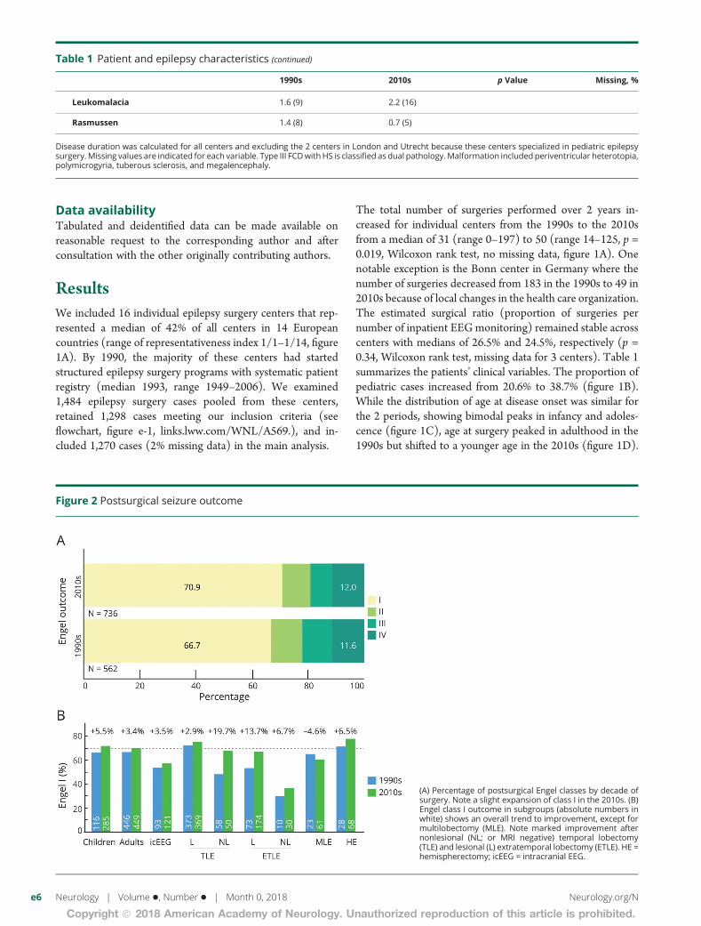
Figure 2 Postsurgical seizure outcome
(A) Percentage of postsurgical Engel classes by decade ofsurgery. Note a slight expansion of class I in the 2010s. (B)Engel class I outcome in subgroups (absolute numbers inwhite) shows an overall trend to improvement, except formultilobectomy (MLE). Note marked improvement afternonlesional (NL; or MRI negative) temporal lobectomy(TLE) and lesional (L) extratemporal lobectomy (ETLE). HE =hemispherectomy; icEEG = intracranial EEG.
e6 Neurology | Volume �, Number � | Month 0, 2018 Neurology.org/N
Copyright ª 2018 American Academy of Neurology. Unauthorized reproduction of this article is prohibited.

Overall, delay to surgery decreased by 5.2 years from the 1990sto the 2010s (p < 0.001, figure 1E and table 1). These effectswere driven in part by the 2 pediatric centers, but even whenexcluding these centers, we found similar age distributions anda significantly decreased delay to surgery of 3.5 years (p < 0.001,table 1). Note that some patients with disease onset in child-hood are now operated on as children, whereas they would have
been operated on as adults in the 1990s. This is reflected in theolder age at onset in the adult category. Yet, overall diseaseduration is shorter across age categories.
Extratemporal surgeries were carried out more frequentlyover the more recent period. However, the proportion ofMRI-negative cases remained stable over time (table 1).
Table 2 Factors associated with postsurgical Engel class I outcome at 2 years
Engel class I, % (n/N) Percent change Multivariate OR (95% CI) p Value
2010s 70.9 (522/736) 4.2 1.34 (1.02–1.77)a 0.038
1990s 66.7 (375/562)
Children 70.4 (283/402) 1.9 1.21 (0.85–1.75) 0.28
Adults 68.5 (614/896)
Lesional 71.4 (820/1,148) 20.1 1.65 (1.09–2.49)a 0.017
MRI negative 51.3 (77/150)
Surgery type
Temporal lobectomy 72.2 (609/844) 1 (Referent)
Extratemporal lobectomy 59.5 (163/274) −12.7 0.53 (0.37–0.76)a <0.001
Multilobectomy 61.9 (52/84) −10.3 0.53 (0.31–0.89)a 0.016
Hemispherectomy 76.0 (73/96) 3.8 0.92 (0.52–1.62) 0.77
Pathology
Hippocampal Sclerosis 72.9 (263/361) 1 (Referent)
Focal cortical dysplasia 62.2 (110/177) −10.7 0.80 (0.49–1.30) 0.37
Other 71.8 (433/603) −1.1 1.02 (0.72–1.44) 0.91
Normal 43.9 (25/57) −29.0 0.51 (0.27–0.97)a 0.039
Not available 66.0 (66/100) −6.9 0.87 (0.51–1.47) 0.6
Disease duration (per year) 1.00 (0.99–1.01) 0.81
Abbreviations: CI = confidence interval; OR = odds ratio.Note that 1,270 who underwent curative surgery were included for multivariate statistics (2.0% missing due to missing age at disease onset). All arecategorical variables except for disease duration.a Independent predictors showing statistically significant OR.
Figure 3 Complication rates and severity by decade of surgery
Note the relative stability of categories over time. Patientsare represented only once; therefore, a patient with persis-tent and other transient complications appears only in thepersistent category. See table 3 for type of complications.
Neurology.org/N Neurology | Volume �, Number � | Month 0, 2018 e7
Copyright ª 2018 American Academy of Neurology. Unauthorized reproduction of this article is prohibited.

Concordant with these observations, a decrease in the num-ber of cases of HS and an increase in focal cortical dysplasia,benign tumors, and dual pathology were noted (table 1).
Figure 2 shows Engel postsurgical seizure outcomes for the1990s and 2010s. Comparing all curative surgery cases in the2010s to the 1990s without adjustment identified a trendtoward improved Engel seizure outcome after surgery per-formed in the 2010s (p = 0.07, univariate logistic regression,no missing data, figure 2A). Seizure freedom (Engel class I) at2 years after surgery increased from 66.7% to 70.9%, i.e., a4.2% difference. The difference became statistically significantwhen confounders and effect modifiers (e.g., more extra-temporal surgeries in 2010s) were included in a multivariatelogistic regression model (p = 0.04). In the 2010s, the odds ofachieving an Engle class I outcome for any curative epilepsysurgery at 2 years was 1.34 higher (95% confidence interval1.02–1.77, table 2) compared to the 1990s. For both periods,we observed a modifying effect of lesional category (in-creasing the odds by ≈2), type of surgery (extratemporal andmultilobar decreasing the odds by ≈2), and postsurgicalnormal histopathology (decreasing the odds by ≈2), but therewas no additional effect of age at onset, disease duration, orpresence of a focal cortical dysplasia. We did not find anysignificant interaction between the decade and the type ofsurgery or the age group (adult vs children), suggesting thatoutcomes modestly improved over time similarly in children
and adults and for all types of surgery. Figure 2B shows detailsof the main effect for surgical subgroups; all, except multilobarresections, showed higher rates of seizure-free patients. Thestrongest improvements noted in MRI-negative TLE andlesional ETLE likely drove the overall effect.
Rate and severity of surgical complications were similar in the1990s and 2010s, despite more complex surgery (e.g., extra-temporal, p = 0.71, χ2, missing data for 2 patients, figure 3).Table 3 shows the distribution of specific types of persistentneurologic deficits and transient neurosurgical complications.
As expected, diagnostics evolved over time, and the number oftests per patient increased significantly from 2.0 to 2.2 on average(table 4). Source imaging of spikes (including magneto-encephalography, EEG, and EEG-fMRI) became more prevalentin the 2010s, as did FDG-PET. In contrast, the use of ictal SPECTdecreased. The use of the Wada test decreased considerably, thelikely result of increased availability of fMRI. The proportion ofinvasive EEG procedures remained stable; however, the pro-portion of stereotactic over subdural studies increased in the2010s. Foramen ovale electrodes were nearly abandoned.
DiscussionHere, we surveyed 16 European tertiary centers that provideepilepsy surgery to a mixed population of pediatric and adult
Table 3 Major and persistent complication rates
1990s(n = 562),% (n)
2010s(n = 736),% (n)
Percentchange,%
Neurosurgicalcomplication
Intracranialhemorrhage
2.0 (11) 0.7 (5) −1.3
Intracranialinfection
1.1 (6) 0.4 (3) −0.7
Hydrocephalus 1.1 (6) 1.1 (8) ≈0
Death 0.0 (0) 0.1 (1) ≈0
Persistentneurologic deficit
Hemiparesis/monoparesis
1.2 (7) 2.2 (16) 1.0
Aphasia 0.9 (5) 1.1 (8) ≈0
Memory deficit 1.6 (9) 0.5 (4) −1.1
Behavioral syndrome 0.7 (4) 0.1 (1) ≈0
Hemianopsia 0.5 (3) 0.4 (3) ≈0
Cranial nerve deficit 0.9 (5) 0.1 (1) −0.8
Number of transient major neurosurgical complications and persistentdeficits (a given patient can have >1). Right column shows signed differencefrom the 1990s to the 2010s approximated by 0 difference when absolutevalue was <0.7%. No missing data.
Table 4 Use of diagnostic tests, including intracranial EEG
Diagnostic test1990s(n = 562)
2010s(n = 736) p Value
Tests, n 2.0 ± 1.1 2.2 ± 1.4 <0.001a
Neuropsychology, % (n) 87.2 (490) 89.0 (653) 0.32
Source imaging, % (n) 10.9 (61) 28.5 (210) <0.001a
FDG-PET, % (n) 26.6 (149) 39.8 (288) <0.001a
Ictal SPECT, % (n) 15.9 (89) 10.6 (77) 0.006a
fMRI, % (n) 0.7 (4) 29.2 (210) <0.001a
Wada, % (n) 31.0 (174) 8.5 (64) <0.001a
icEEG, % (n) 30.8 (173) 29.5 (213) 0.59
Subtype icEEG, n 173 213
Depth, % (n) 32.4 (56) 46.0 (98)
Subdural, % (n) 37.6 (65) 34.6 (74) 0.002a
Combination, % (n) 22.0 (38) 18.0 (39)
Foramen ovale, % (n) 8.1 (14) 1.4 (3)
Abbreviations: FDG-PET = fluorodeoxyglucose PET; icEEG = intracranial EEG.First row shows average total number of test performed per patient ± SD.Other rows show the percentage of patients who had the test. The last 4rows show the distribution of specific techniques in patients who receivedintracranial investigations. Structural MRI and EEGmonitoring was used forall patients in the 2 decades and are therefore not included. Nomissing data.a Significant.
e8 Neurology | Volume �, Number � | Month 0, 2018 Neurology.org/N
Copyright ª 2018 American Academy of Neurology. Unauthorized reproduction of this article is prohibited.
patients and found that overall postsurgical seizure-freedomimproved from 66.7% in the 1990s to 70.9% in the 2010s(+4.2%). This modest improvement is significant only afteradjustment for the fact that surgeries performed in the2010s became more complex (e.g., increased extratemporalprocedures). It translates into a number needed to treat of 24patients to gain 1 Engel class I outcome in the 2010s comparedto 1990s. Disease duration decreased by 5.2 years on average,with significant shortening in both children and adults.
Limitations of this study of 2 sequential cohorts in time includeits retrospective nature with a potential bias toward randomimprecisions for data from the 1990s. The report of compli-cations may be sensitive to better documentation in the 2010s,potentially obscuring a significant improvement in the 2010scompared to the 1990s. Despite a higher inclusion number thantargeted, statistical power was lacking for post hoc analysis insurgical subgroups. For this reason, we performed statisticalanalysis on the overall effect and report illustrative subgroupfigures without statistics. Our inclusion criterion for centerswith >10 surgeries per year could have obscured possible trendsof an increasing number of nonuniversity smaller centers,similar to those observed in nationwide US surveys.11,12
However, within the European countries included here, the vastmajority of epilepsy surgeries are performed at academic cen-ters. Our survey was not inclusive of all European hospitalsperforming epilepsy surgery but sampled from nationwidesystems (Norway, Sweden, pediatric surgeries from theNetherlands), all to one-quarter of the centers in small coun-tries (e.g., Belgium, Lithuania, Switzerland) and down to 1/15in larger countries like France, and thus, our data are blinded tolocal transfer or sharing of surgical volume among centers,potentially leading to an underestimation of the actual increasein surgical volume. We chose the 2-year Engel outcome, whichmay not be definitive but has been shown to be predictive oflonger-term seizure outcome (>10 years).24 Finally, we ac-knowledge the fact that the pharmacologic arsenal has in-creased between study periods. However, this is not thought toincrease the rate of drug-responsive patients.25
Contrary to nationwide UK16 and US10–12,26,27 studies andsingle-center European studies,8,14,15 our study shows thatsurgery is probably more widely used and yields improvedpostoperative seizure control and that presurgery disease du-ration is shorter. We hypothesize that European practitionerstend to refer potential candidates earlier, likely reflecting officialrecommendations to curtail years of disabling seizures.4 Indeed,we observed an important shift from adult to pediatric de-mography and a decrease in disease duration in both groups.Pediatric programs have largely expanded in Europe8 and theUnited States28 to afford the need to mitigate long-term cog-nitive disabilities.29,30 In spite of these encouraging trends, itremains puzzling that in the 21st century the delay from epi-lepsy onset to surgery still exceeds 10 years on average, inparticular in those who undergo surgery in adulthood. Reasonsmay include temporary seizure remission with new drugs,overestimation of surgical risks, underestimation of seizure-
related mortality and morbidity, and lack of access to healthcare resources.31 Patients treated by physicians who lack in-formation on the surgical possibility or do not appreciate thepotential of a comprehensive evaluation may suffer fromdelayed referrals. However, the surgical volume in individualcenters increased on average, whereas the estimated surgicalratio to total number of admitted patients for epilepsy moni-toring remained stable, contrary to US surveys.11,12 This mayindicate more lenient referrals and greater acceptance of epi-lepsy surgery as a therapeutic option.
Similar to previous observations,12 the practice of epilepsysurgery has evolved over the decade: we observed a shift tomore noninvasive imaging (e.g., FDG-PET) and from in-vasive functional mapping (Wada test and subdural electro-des) to noninvasive techniques (fMRI). However, the use ofinvasive brain recordings remained unchanged, encompassingone-third of all cases, with an increasing proportion of stereo-EEG to target deep and extratemporal foci. That said, stereo-EEGwas already used in >50% of invasive studies in the 1990sin Europe, sharply contrasting with the more recent spread ofthe technique in the United States.32 Regarding the un-derlying pathologic substrate, resections for HS decreased byhalf, whereas extratemporal lobe resections for focal corticaldysplasia increased by 4 times in the 2010s.
Overall, the odds of receiving a successful surgery of any type inthe 2010s are modestly improved compared to the 1990s.21
Improvement was most pronounced for surgical subgroups, in-cluding MRI-negative TLE (from 48% to 68%, i.e., 20% im-provement) and lesional ETLE (from 54% to 68%, i.e., 14%improvement), approaching figures established for morestraightforward surgeries such as lesional TLE.7 This could resultfrom the increased use of multimodal diagnostic tests and theexpansion of knowledge of these conditions. For example, de-lineation of focal cortical dysplasias improves with the use of 3TMRIs with voxel-based morphometry19 or with MRI-PET cor-egistration. In addition, the increased use of FDG-PET scans inthe 2010s likely identified surgically remediable MRI-negativetemporal lobe epilepsy18 after exclusion of an autoimmune17 orgenetic etiology (e.g., focal-onset seizures in ring chromosome 20epilepsy33). On the other hand, surgical outcomes after lesionalTLE were stagnant over time, suggesting that advances in diag-nostics or disease understandingwere not sufficient for improvedsurgical success in that category. Given that TLE represents 65%of surgeries across the 2 cohorts, the overall statistical effectsreported here were influenced by these stagnant figures.
Despite the advent of noninvasive source imaging,20 only 3% ofour cohort underwent surgery for MRI-negative extratemporallobe epilepsy, yielding ≈30% to 40% seizure freedom, inagreement with previous studies.34 This likely reflects a re-luctance to attempt surgery in this difficult scenario. Prospectivestudies may help determine whether consistent and compre-hensive application of modern diagnostic techniques couldincrease surgical success in nonlesional extratemporal lobeepilepsy. Despite increasing surgical complexity, the overall
Neurology.org/N Neurology | Volume �, Number � | Month 0, 2018 e9
Copyright ª 2018 American Academy of Neurology. Unauthorized reproduction of this article is prohibited.

complication rate remained stable at ≈5%, in line with a recentprospective study23 and meta-analysis.35 Of note, our cohortencompassed only 1 perisurgical death but 5 seizure-relateddeaths in patients who were not seizure-free after surgery,highlighting that the surgical risk has to be balanced with thecumulative lifetime risk of continued seizures.9
We strived to pool data from several centers with the aim ofobtaining a balanced representation of epilepsy surgery practicein Europe. There are undoubtedly epidemiologic, health sys-tem, and medical practice differences within Europe and withother countries. Results presented here remain intimatelylinked to the practice of individual centers that has evolved overtime, possibly at differing paces. However, we believe that thefigures shown here reflect knowledge shared and disseminatedat national and international conferences, leading to a moreharmonized approach in epilepsy care, and may, in that sense,reflect an encouraging global trend to follow guidelines andrecommendations. Early referral to specialized centers is crucialto globally reduce years of suffering and unproductivity linkedto epilepsy and potentially to improve ultimate cognitive out-comes. As epilepsy knowledge accumulates, current statisticson postsurgical results are required to allow appropriatecounseling of prospective surgical candidates. At the turn of themillennium, the postsurgical seizure outcome has improvedmodestly despite the increasing complexity of procedures andwithout compromising patient safety. These encouragingtrends suggest that future efforts will contribute to optimizingthe surgical care of patients with epilepsy.
Author contributionsM.S. and K.P.J.B. designed the study. All authors contributedto data collection. M.O.B., M.S., and T.P. analyzed the data.M.O.B. and M.S. drafted the manuscript. All authors con-tributed to editing the manuscript.
AcknowledgmentThe authors thank Mrs. Birgitta Esser for facilitating datacollection.
Study fundingM.S. was funded by the Special Program in University Med-icine grant from the Swiss National Science Foundation140332 and 163398.
DisclosureM. Baud is a part-time employee at the Wyss Center for Bio-and Neuro-Engineering. T. Perneger, A. Racz, and M. Penselreport no disclosures relevant to the manuscript. C. Elgerreports personal fees in the form of honoraria from UCB,Desitin, BIAL, and Eisai. C.E.E. has also received grants fromthe Deutsche Forschungsgemeinschaft, the Bundesministe-rium fur Bildung und Forschung, and the Marga and WalterBoll Stiftung. C.E.E. also served as medical director for theLife & Brain Institute until 2015 and as platform leader forCognitive Neuroscience until 2017. B. Rydenhag and K.Malmgren report no disclosures relevant to the manuscript.
H. Cross has received remuneration to her department asa clinical investigator for Vitaflo, GW Pharma, and Zogenix.She has participated in advisory boards for GSK, UCB,Zogenix, GW Pharma, Nutricia, and Eisai and as speaker forShire, Nutricia, Zogenix, and GW Pharma, again for whichremuneration was made to her department. She holds grantsfrom the European Union, National Institute for Health andResearch, Action Medical Research, Great Ormond StreetHospital Charity, and SPARKS. G. McKenna, M. Tisdall, andH. Lamberink report no disclosures relevant to the manu-script. S. Rheims received consultant and/or speaker feesfrom UCB Pharma, EISAI, Livanova, and GW Pharma. P.Ryvlin, J. Isnard, and F. Mauguiere report no disclosuresrelevant to the manuscript. A. Arzimanoglou occasionallyserves on scientific advisory boards for Biomarin, Eisai, GWPharma, Shire, Takeda, and UCB, as well as on Data SafetyMonitoring boards for UCB. He received funding for travel orspeaker honoraria from Eisai, GW Pharma, Shire, and UCB.Since 2004, he has served as editor-in-chief of the educationaljournal of the International League Against Epilepsy, EpilepticDisorders (John Libbey Eurotext editions), and since 2010, hehas served as associated editor of the European Journal ofPaediatric Neurology (Elsevier). He received research supportfrom the European Commission, the Caixa Bank Foundation,and UCB. He serves as visiting professor at the Universitat deBarcelona, Spain, and as coordinator of research for the Pe-diatric Epilepsy Unit of the children’s University Hospital SanJuan de Deu, Barcelona, Spain. S. Akkol, K. Deniz, and C.Ozkara report no disclosures relevant to the manuscript. M.Lossius serves on the advisory boards for Esai and UCB, hasreceived speaker honoraria at meetings for physicians spon-sored by UCB and Eisai, and has served as an editorial advi-sory board member at Epilepsy Research and Treatment,Hindawi, from 2015 until it ceased publication in 2017. I.Rektor, R. Kalviainen, L. Vanhatalo, P. Dimova, K. Minkin, A.Staack, B. Steinhoff, A. Kalina, P. Krsek, P. Marusic, Z. Jordan,and D. Fabo report no disclosures relevant to the manuscript.E. Carrette was supported by Elekta Neuromag to cover traveland/or registration costs to give presentations at internationalconferences. P. Boon, S. Rocka, R. Mameniskiene, S. Vul-liemoz, F. Pittau, and K. Braun report no disclosures relevantto the manuscript. M. Seeck reports shares for Epilog, EEGdiagnostic technology. Go to Neurology.org/N for fulldisclosures.
Received November 26, 2017. Accepted in final form April 13, 2018.
References1. Schijns OEMG, Hoogland G, Kubben PL, Koehler PJ. The start and development of
epilepsy surgery in Europe: a historical review. Neurosurg Rev 2015;38:447–461.2. Wiebe S, Blume WT, Girvin JP. A randomized, controlled trial of surgery for
temporal-lobe epilepsy. N Engl J 2001;345:311–318.3. Engel J, McDermott MP, Wiebe S, et al. Early surgical therapy for drug-resistant
temporal lobe epilepsy: a randomized trial. JAMA 2012;307:922–930.4. Engel J, Wiebe S, French J, et al. Practice parameter: temporal lobe and localized neo-
cortical resections for epilepsy: report of the quality standards subcommittee of theAmerican Academy of Neurology, in association with the American Epilepsy Society andthe American Association of Neurological Surgeons. Neurology 2003;60:538–547.
5. Kwan P, Arzimanoglou A, Berg AT, et al. Definition of drug resistant epilepsy:consensus proposal by the ad hoc task force of the ILAE Commission on TherapeuticStrategies. Epilepsia 2010;51:1069–1077.
e10 Neurology | Volume �, Number � | Month 0, 2018 Neurology.org/N
Copyright ª 2018 American Academy of Neurology. Unauthorized reproduction of this article is prohibited.

6. Simasathien T, Vadera S, Najm I, Gupta A, Bingaman W, Jehi L. Improved outcomeswith earlier surgery for intractable frontal lobe epilepsy. Ann Neurol 2013;73:646–654.
7. Janszky J, Janszky I, Schulz R, et al. Temporal lobe epilepsy with hippocampal scle-rosis: predictors for long-term surgical outcome. Brain 2005;128:395–404.
8. Lamberink HJ, Boshuisen K, van Rijen PC, Gosselaar PH, Braun KPJ; Dutch Col-laborative Epilepsy Surgery Program (DCESP). Changing profiles of pediatric epi-lepsy surgery candidates over time: a nationwide single-center experience from 1990to 2011. Epilepsia 2015;56:717–725.
9. Tomson T, Nashef L, Ryvlin P. Sudden unexpected death in epilepsy: currentknowledge and future directions. Lancet Neurol 2008;7:1021–1031.
10. Haneef Z, Stern J, Dewar S, Engel J Jr. Referral pattern for epilepsy surgery afterevidence-based recommendations: a retrospective study. Neurol Am Acad Neurol2010;75:699–704.
11. Englot DJ, Ouyang D, Garcia PA, Barbaro NM, Chang EF. Epilepsy surgery trends inthe United States, 1990-2008. Neurology 2012;78:1200–1206.
12. Kaiboriboon K, Malkhachroum AM, Zrik A, et al. Epilepsy surgery in the UnitedStates: analysis of data from the National Association of Epilepsy Centers. EpilepsyRes 2015;116:105–109.
13. Jehi L, Friedman D, Carlson C, et al. The evolution of epilepsy surgery between 1991and 2011 in nine major epilepsy centers across the United States, Germany, andAustralia. Epilepsia 2015;56:1526–1533.
14. Cloppenborg T, May TW, Blumcke I, et al. Trends in epilepsy surgery: stable surgicalnumbers despite increasing presurgical volumes. J Neurol Neurosurg Psychiatr 2016;87:2016–313831.
15. Bien CG, Raabe AL, Schramm J, Becker A, Urbach H, Elger CE. Trends in presurgicalevaluation and surgical treatment of epilepsy at one centre from 1988-2009. J NeurolNeurosurg Psychiatr 2013;84:54–61.
16. Neligan A, Haliasos N, Pettorini B, Harkness WFJ, Solomon JK. A survey of adult andpediatric epilepsy surgery in the United Kingdom. Epilepsia 2013;54:e62–e65.
17. Wright S, Vincent A. Progress in autoimmune epileptic encephalitis. Curr OpinNeurol 2016;29:151–157.
18. Muhlhofer W, Tan YL, Mueller SG, Knowlton R. MRI-negative temporal lobe epi-lepsy: what do we know? Epilepsia 2017;51:1256.
19. Duncan JS, Winston GP, Koepp MJ, Ourselin S. Brain imaging in the assessment forepilepsy surgery. Lancet Neurol 2016;15:420–433.
20. Brodbeck V, Spinelli L, Lascano AM, et al. Electroencephalographic source im-aging: a prospective study of 152 operated epileptic patients. Brain 2011;134:2887–2897.
21. Jobst BC, Cascino GD. Resective epilepsy surgery for drug-resistant focal epilepsy:a review. JAMA 2015;313:285–293.
22. Englot DJ, Rolston JD, Wang DD, Sun PP, Chang EF, Auguste KI. Seizure outcomesafter temporal lobectomy in pediatric patients. J Neurosurg Pediatr 2013;12:134–141.
23. Bjellvi J, Flink R, Rydenhag B, Malmgren K. Complications of epilepsy surgery inSweden 1996–2010: a prospective, population-based study. J Neurosurg 2015;122:519–525.
24. de Tisi J, Bell GS, Peacock JL, et al. The long-term outcome of adult epilepsy surgery,patterns of seizure remission, and relapse: a cohort study. Lancet 2011;378:1388–1395.
25. Tang F, Hartz AMS, Bauer B. Drug-resistant epilepsy: multiple hypotheses, fewanswers.Front Neurol 2017;8:1005.
26. Choi H, Carlino R, Heiman G, Hauser WA, Gilliam FG. Evaluation of duration ofepilepsy prior to temporal lobe epilepsy surgery during the past two decades. EpilepsyRes 2009;86:224–227.
27. Schiltz NK, Koroukian SM, Lhatoo SD, Kaiboriboon K. Temporal trends in pre-surgical evaluations and epilepsy surgery in the U.S. from 1998 to 2009. Epilepsy Res2013;103:270–278.
28. Pestana Knight EM, Schiltz NK, Bakaki PM, Koroukian SM, Lhatoo SD, KaiboriboonK. Increasing utilization of pediatric epilepsy surgery in the United States between1997 and 2009. Epilepsia 2015;56:375–381.
29. Jenny B, Smoll N, Hassani El Y, et al. Pediatric epilepsy surgery: could age bea predictor of outcomes? J Neurosurg Pediatr 2016;18:235–241.
30. Skirrow C, Cross JH, Cormack F, Harkness W, Vargha-Khadem F, Baldeweg T. Long-term intellectual outcome after temporal lobe surgery in childhood. Neurology 2011;76:1330–1337.
31. Jette N, Sander JW, Keezer MR. Surgical treatment for epilepsy: the potential gapbetween evidence and practice. Lancet Neurol 2016;15:982–994.
32. Gonzalez Martinez J, Bulacio J, Alexopoulos A, Jehi L, Bingaman W, Najm I. Ster-eoelectroencephalography in the “difficult to localize” refractory focal epilepsy: earlyexperience from a North American epilepsy center. Epilepsia 2013;54:323–330.
33. Vignoli A, Bisulli F, Darra F, et al. Epilepsy in ring chromosome 20 syndrome.Epilepsy Res 2016;128:83–93.
34. Jeha LE, Najm I, Bingaman W, Dinner D, Widdess-Walsh P, Luders H. Surgicaloutcome and prognostic factors of frontal lobe epilepsy surgery. Brain 2007;130:574–584.
35. Hader WJ, Tellez-Zenteno J, Metcalfe A, et al. Complications of epilepsy surgery:a systematic review of focal surgical resections and invasive EEGmonitoring. Epilepsia2013;54:840–847.
Neurology.org/N Neurology | Volume �, Number � | Month 0, 2018 e11
Copyright ª 2018 American Academy of Neurology. Unauthorized reproduction of this article is prohibited.

DOI 10.1212/WNL.0000000000005776 published online June 13, 2018Neurology
Maxime O. Baud, Thomas Perneger, Attila Rácz, et al. European trends in epilepsy surgery
This information is current as of June 13, 2018
ServicesUpdated Information &
776.full.htmlhttp://n.neurology.org/content/early/2018/06/13/WNL.0000000000005including high resolution figures, can be found at:
Subspecialty Collections
http://n.neurology.org//cgi/collection/outcome_researchOutcome research
http://n.neurology.org//cgi/collection/epilepsy_surgery_Epilepsy surgery
http://n.neurology.org//cgi/collection/cohort_studiesCohort studies
http://n.neurology.org//cgi/collection/class_iiiClass III
http://n.neurology.org//cgi/collection/all_epilepsy_seizuresAll Epilepsy/Seizuresfollowing collection(s): This article, along with others on similar topics, appears in the
Permissions & Licensing
http://n.neurology.org/misc/about.xhtml#permissionsits entirety can be found online at:Information about reproducing this article in parts (figures,tables) or in
Reprints
http://n.neurology.org/misc/addir.xhtml#reprintsusInformation about ordering reprints can be found online:
rights reserved. Print ISSN: 0028-3878. Online ISSN: 1526-632X.1951, it is now a weekly with 48 issues per year. Copyright © 2018 American Academy of Neurology. All
® is the official journal of the American Academy of Neurology. Published continuously sinceNeurology