epstein-barr-virus-positiveb-celllymphomaof ... · lymph node revealed the diagnosis of hodgkin...
TRANSCRIPT
Hindawi Publishing CorporationCase Reports in TransplantationVolume 2012, Article ID 164824, 5 pagesdoi:10.1155/2012/164824
Case Report
Epstein-Barr-Virus-Positive B-Cell Lymphoma ofRecipient Origin Despite of the Elimination of ClonallyEBV-Infected T Cells by Allogeneic Stem Cell Transplantation ina Patient with Chronic Active EBV Infection
Masayuki Nagasawa
Department of Pediatrics and Developmental Biology, Graduate School of Medical and Dental sciences,Tokyo Medical and Dental University, 5-45, Yushima 1-chome, Bunkyo-ku, Tokyo 113-8519, Japan
Correspondence should be addressed to Masayuki Nagasawa, [email protected]
Received 17 December 2011; Accepted 14 February 2012
Academic Editors: C. F. Classen, C. Costa, and H. J. Jeong
Copyright © 2012 Masayuki Nagasawa. This is an open access article distributed under the Creative Commons Attribution License,which permits unrestricted use, distribution, and reproduction in any medium, provided the original work is properly cited.
A 20-year-old patient with chronic active EBV infection (CAEBV) received peripheral blood stem cell transplantation (PBSCT)from HLA-one-locus-mismatched mother. Although EB-virus-infected T cells were eliminated after PBSCT, she developed EB-virus-positive B-cell lymphoma of recipient origin in the brain. By reducing the immunosuppressive therapy, the initial lesiondisappeared. However, another lesion in the opposite lateral brain appeared later and was resistant to further reduction ofimmunosuppressive therapy. EBV-DNA was persistently negative after PBSCT in the peripheral blood. This case is suggestivein management of EBV reactivation after SCT and understanding alloimmune response to EBV.
1. Introduction
Epstein-Barr virus (EBV) is a ubiquitously spread herpesfamily virus, which infects more than 90% of adults [1].Primary EBV infection often is asymptomatic and sometimespresents as acute febrile mononucleosis. EBV usually infectsB cells and latently infected B cells are regulated by potentT cells [2]. In an immunodeficient state, these B cells occa-sionally induce lymphoproliferative disease, which is some-times life threatening [3–5].
Chronic active EBV infection (CAEBV) is another EBV-related lymphoproliferative disease in which EBV infects Tor NK cells [6, 7]. Although the pathogenesis is not wellunderstood, the prognosis is poor in the long run andallogeneic stem cell transplantation (allo-HSCT) is recom-mended to eradicate infected T or NK cells [8]. With anincreased transplantation-related mortality by conventionalmyeloablative conditioning regimen (MAC), reduced inten-sity conditioning regimen (RIC) has been applied, resultingin an improved 3-year overall survival of 95.0± 4.9% in RIC
compared to 54.5± 15.0% in MAC [9]. Recently, the authorhas reported that level of serum granulysin is useful fordiscriminating NK cell type CAEBV from T cell type [10].
The author has experienced a patient with CAEBV whodeveloped EBV-positive B-cell lymphoma of recipient originafter allogeneic stem cell transplantation. The clinical courseof this patient is suggestive to understand the significance ofallo-immune response against EBV-associated lymphoprolif-erative diseases (EBV-LPD) in the setting of allo-HSCT.
2. Case Report
The patient presented with mosquito hypersensitivity at theage of five. Thereafter, she had recurrent cervical lymphad-enopathy with fever, which resolved spontaneously in acouple of weeks. When she was 14 years old, systemic lym-phadenopathy with fever developed. Biopsy of cervicallymph node revealed the diagnosis of Hodgkin lymphoma.She received chemotherapy of COPP/ABVD and modifiedEPOCH for two years. However, febrile lymphadenopathy

2 Case Reports in Transplantation
N P P. C.
∗
Figure 1: Infection status of EBV was monoclonal. DNA from the patient’s peripheral blood mononuclear cells was digested by EcoRI, andsouthern blot hybridization was performed with EBV BamHI W probe. An arrow points at the monoclonal bands. Asterisk shows the non-specific band. N: normal control; P: patient; P.C.: positive control (Raji cells).
Day 230 Day 280
Day 294 Day 378
Figure 2: Brain MRI of the patient. Initial frontal tumor that was surrounded by T2 high edematous area disappeared completely on the294th day, but right paraventricular tumor grew progressively.
Case Reports in Transplantation 3
CD79a
(a)
CD3
(b)
LMP-1
(c)
HE
(d)
PAS
(e) (f)
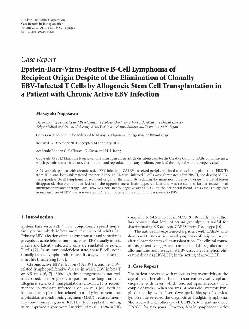
Figure 3: Histology of the brain tumor. Infiltrating cells in the brain tumor were CD79a+, CD3−, and LMP-1+. Arrow indicates the rightparaventricular tumor.
resumed after chemotherapy. Then, she was diagnosed aschronic active EBV infection from the persistent presence ofincreased EBV-DNA (104–105 copies/mL) in the peripheralblood and high titer of anti-EB virus antibody. Analysis oflymphocyte subsets revealed that EBV infected both αβT andγδT cells and they were expanded clonally (Figure 1).
She was referred to our hospital at the age of 20. Shereceived peripheral blood stem cell transplantation (PBSCT)from HLA-one-locus-mismatched mother at the age of 21.Conditioning regimen included fludarabine (25 mg/m2 × 5),melphalan (70 mg/m2 × 2), and antithymoglobulin (Lym-phoglobuline; 10 mg/kg× 2). GVHD prophylaxis was tacrol-imus with short-term MTX. Engraftment was on the 9th day,and grade III GVHD (skin: stage 3, liver: stage 2) occurredon the 14th day, for which prednisolone and mycophenolatemofetil (MMF) were started. GVHD was intractable andgrade II GVHD (skin) persisted. On the 198th day, she wasdischarged for follow-up of chronic GVHD on medication oftacrolimus (0.1 mg/kg/day), prednisolone (0.5 mg/kg/day),and MMF (50 mg/kg/day). EBV-DNA in the peripheralblood was turned negative on the 10th day. On the 40th day,it became transiently positive but was consistently negativeafter the 52nd day.
On the 227th day, she was transferred to our hospitalbecause of convulsion. MRI revealed a mass with diameterof 20 mm in the left frontal lobe (Figure 2). EBV, CMV,VZV, and HHV-1, 2, 6, 7, 8 were all negative in both of theperipheral blood and spinal fluid by PCR method. Con-sidering the EBV-associated lymphoma or toxoplasmosis,immunosuppressive drugs were reduced and antitoxoplasmadrug (pyrimethamine + sulfadiazine) was started. Stage 2skin GVHD developed, and left temporal mass was graduallyregressed. However, MRI on the 280th day disclosed anothermass in the right paraventricular area (Figure 1). EBV-DNAwas detected in the spinal fluid on the 373rd day, althoughstill negative in the peripheral blood. The mass was progres-sively enlarged in spite of stopping tacrolimus and MMF. Sheand her family were reluctant to further intensive therapyincluding radiotherapy. She died from brain stem herniationon the 417th day after PBSCT.
Autopsy revealed EBV-positive diffuse large cell lympho-ma positive for CD20 and CD79a (Figure 3). Analysis of IgHand EBV terminal repeat presented monoclonal proliferationand STR (short tandem repeat) analysis showed the recipientorigin (Figure 4). In the left hemisphere, ghost cells wereaggregated where the first mass was detected. Ghost cells were

4 Case Reports in Transplantation
250 260 270 280 290
Chimerism
Lymphoma cells
(a)
250 260 270 280 290
Donor
(b)
250 260 270 280 290
Recipient
(c)
Figure 4: STR (short tandem repeat) analysis of the lymphomacells.
positive for CD20 and LMP-1 (latent-membrane-protein-1)and considered to be apoptotic lymphoma cells. There wasno evidence of toxoplasmosis histologically.
3. Discussion
It has been reported that EBV reactivation occurs in 0.6 to26% after allo-HSCT, with this being higher in the context ofT-cell depletion or prolonged immunosuppressive treatmentfor intractable GVHD. EBV-LPD typically occurs withinthe first 6 months after allo-HSCT, and mortality rates canrange between 50 and 80% [11]. In most of the cases, EBV-LPD arises from donor lymphocytes in allo-HSCT contraryto our case. To avoid the development of EBV-LPD, weeklyscreening of EBV-DNA for at least 3 months and early pre-emptive intervention with rituximab is recommended whenEBV-DNA exceeds 103 copies/105 cells in the peripheralblood for high-risk patients [12].
It is quite interesting that EBV-DNA was persistentlynegative in the peripheral blood after PBSCT in this patient,and it finally turned positive only in the spinal fluid at thelater stage. This observation suggests that serial monitoringof EBV-DNA in the peripheral blood could not be sufficientenough for the detection and evaluation of EBV-LPD in thecentral nervous system (CNS). Isolated CNS EBV-LPD isextremely rare, and only five cases have been reported inthe medical literature so far. In one case, EBV-DNA was alsonegative in the spinal fluid by PCR method as our case [13].
In this patient, EBV-infected αβT and γδT cells wereeliminated after allo-HSCT, which was confirmed by thedisappearance of EBV-DNA from the peripheral blood.However, EBV-positive B cell lymphoma was induced underimmunosuppressive treatment for chronic GVHD.
In this case, allogeneic immune response was consideredenough to eliminate EBV-infected monoclonal T cells but not
EBV-infected monoclonal B cells. One possibility is that thecentral nervous system was completely separated from allo-geneic immune response. However, with the fact that initialbrain tumor regressed after reducing immune suppressivedrugs, this scenario seems not enough to explain the diseaseprogress. Although not investigated precisely, the initiallesion in the left hemisphere could be biologically differentfrom that in the right brain because the former regressed byreducing the immunosuppressive drugs. Recently, it has beenreported that loss of alloantigen is associated with the resis-tance to immune surveillance system in malignant hema-tological disease [14]. Another possibility is that the latterlymphoma cells were more aggressively transformed to beresistant to apoptotic stimuli.
Although we could not investigate the underliningmolecular mechanism in the different resistance for eachlymphoproliferative disease to alloimmunity in detail, thiscase seems suggestive to consider the efficacy and limit of cel-lular immune response to EBV-related lymphoproliferativedisease.
Acknowledgment
The authors wish to thank the staff of Cell Therapy Centre atTokyo Medical and Dental University for the comprehensivevirus examination for the patient’s sample.
References
[1] G. Miller, “Epstein-Barr virus. Biology, pathogenesis, andmedical aspects,” in Virology, B. N. Fields and D. M. Knipe,Eds., chapter 68, Raven Press, New York, NY, USA, 2ndedition, 1990.
[2] C. Alfieri, M. Birkenbach, and E. Kieff, “Early events in Ep-stein-Barr virus infection of human B lymphocytes,” Virology,vol. 181, no. 2, pp. 595–608, 1991.
[3] D. L. Birx, R. R. Redfield, and G. Tosato, “Defective regulationof Epstein-Barr virus infection in patients with acquiredimmunodeficiency syndrome (AIDS) or AIDS-related disor-ders,” The New England Journal of Medicine, vol. 314, no. 14,pp. 874–879, 1986.
[4] P. S. Randhawa, R. Jaffe, A. J. Demetris et al., “Expression ofEpstein-Barr virus-encoded small RNA (by the EBER-1 gene)in liver specimens from transplant recipients with post-transplantation lymphoproliferative disease,” The New Eng-land Journal of Medicine, vol. 327, no. 24, pp. 1710–1714, 1992.
[5] R. S. Shapiro, K. McCLain, G. Frizzera et al., “Epstein-Barrvirus associated B cell lymphoproliferative disorders followingbone marrow transplantation,” Blood, vol. 71, no. 5, pp. 1234–1243, 1988.
[6] H. Kanegane, K. Nomura, T. Miyawaki, and G. Tosato, “Bio-logical aspects of Epstein-Barr virus (EBV)-infected lympho-cytes in chronic active EBV infection and associated malignan-cies,” Critical Reviews in Oncology/Hematology, vol. 44, no. 3,pp. 239–249, 2002.
[7] M. Okano, K. Kawa, H. Kimura et al., “Proposed guidelinesfor diagnosing chronic active Epstein-Barr virus infection,”American Journal of Hematology, vol. 80, no. 1, pp. 64–69,2005.
[8] E. Sato, S. Ohga, H. Kuroda et al., “Allogeneic hematopoieticstem cell transplantation for Epstein-Barr virus-associated

Case Reports in Transplantation 5
T/natural killer-cell lymphoproliferative disease in Japan,”American Journal of Hematology, vol. 83, no. 9, pp. 721–727,2008.
[9] K. Kawa, A. Sawada, M. Sato et al., “Excellent outcome of allo-geneic hematopoietic SCT with reduced-intensity condition-ing for the treatment of chronic active EBV infection,” BoneMarrow Transplantation, vol. 46, no. 1, pp. 77–83, 2010.
[10] M. Nagasawa, K. Ogawa, K. Nagata, and N. Shimizu, “Serumgranulysin as a possible biomarker of natural killer cell neo-plasms,” British Journal of Haematology, vol. 148, no. 5, pp.812–814, 2010.
[11] C. V. Paya, J. J. Fung, M. A. Nalesnik et al., “Epstein-barr virus-induced posttransplant lymphoproliferative disorders,” Trans-plantation, vol. 68, no. 10, pp. 1517–1525, 1999.
[12] H. E. Heslop, “How I treat EBV lymphoproliferation,” Blood,vol. 114, no. 19, pp. 4002–4008, 2009.
[13] M. Hamadani, L. K. Martin, D. M. Benson, E. A. Copelan,S. M. Devine, and C. C. Hofmeister, “Central nervous systempost-transplant lymphoproliferative disorder despite negativeserum and spinal fluid Epstein-Barr virus DNA PCR,” BoneMarrow Transplantation, vol. 39, no. 4, pp. 249–251, 2007.
[14] T. Isoda, A. M. Ford, D. Tomizawa et al., “Immunologicallysilent cancer clone transmission from mother to offspring,”Proceedings of the National Academy of Sciences of the UnitedStates of America, vol. 106, no. 42, pp. 17882–17885, 2009.

Submit your manuscripts athttp://www.hindawi.com
Stem CellsInternational
Hindawi Publishing Corporationhttp://www.hindawi.com Volume 2014
Hindawi Publishing Corporationhttp://www.hindawi.com Volume 2014
MEDIATORSINFLAMMATION
of
Hindawi Publishing Corporationhttp://www.hindawi.com Volume 2014
Behavioural Neurology
EndocrinologyInternational Journal of
Hindawi Publishing Corporationhttp://www.hindawi.com Volume 2014
Hindawi Publishing Corporationhttp://www.hindawi.com Volume 2014
Disease Markers
Hindawi Publishing Corporationhttp://www.hindawi.com Volume 2014
BioMed Research International
OncologyJournal of
Hindawi Publishing Corporationhttp://www.hindawi.com Volume 2014
Hindawi Publishing Corporationhttp://www.hindawi.com Volume 2014
Oxidative Medicine and Cellular Longevity
Hindawi Publishing Corporationhttp://www.hindawi.com Volume 2014
PPAR Research
The Scientific World JournalHindawi Publishing Corporation http://www.hindawi.com Volume 2014
Immunology ResearchHindawi Publishing Corporationhttp://www.hindawi.com Volume 2014
Journal of
ObesityJournal of
Hindawi Publishing Corporationhttp://www.hindawi.com Volume 2014
Hindawi Publishing Corporationhttp://www.hindawi.com Volume 2014
Computational and Mathematical Methods in Medicine
OphthalmologyJournal of
Hindawi Publishing Corporationhttp://www.hindawi.com Volume 2014
Diabetes ResearchJournal of
Hindawi Publishing Corporationhttp://www.hindawi.com Volume 2014
Hindawi Publishing Corporationhttp://www.hindawi.com Volume 2014
Research and TreatmentAIDS
Hindawi Publishing Corporationhttp://www.hindawi.com Volume 2014
Gastroenterology Research and Practice
Hindawi Publishing Corporationhttp://www.hindawi.com Volume 2014
Parkinson’s Disease
Evidence-Based Complementary and Alternative Medicine
Volume 2014Hindawi Publishing Corporationhttp://www.hindawi.com