human herpesviruses 6a and 6b (hhv-6a and hhv-6b)...
TRANSCRIPT

Human Herpesviruses 6A and 6B (HHV-6A and HHV-6B) cause alterations in
E2F1/Rb pathways, E2F1 localization, as well as cell cycle arrest in infected T
cells.
Guy Mlechkovich and Niza Frenkel*
The S. Daniel Abraham Institute for Molecular Virology, and the Department of Cell
Research and Immunology, Tel Aviv University. Israel.
Running title: HHV-6 alters E2F1/Rb pathways and arrests cell cycle
Word count for the abstract: 248.
Word count for the text: 8,306
Corresponding author: Niza Frenkel, PhD
Dept. of Cell Research and Immunology
The S. Daniel Abraham Institute for Molecular Virology
George S. Wise Faculty of Life Sciences
Britannia Building 203
Tel Aviv University, 69978 Tel Aviv, Israel
Phone: 972-3-6407166
Fax: 972-3-640-7165
E-mail: [email protected]
ACCEPTED
Copyright © 2007, American Society for Microbiology and/or the Listed Authors/Institutions. All Rights Reserved.J. Virol. doi:10.1128/JVI.01496-07 JVI Accepts, published online ahead of print on 3 October 2007
on May 23, 2018 by guest
http://jvi.asm.org/
Dow
nloaded from
1
ABSTRACT
E2F transcription factors play pivotal roles in controlling the expression of genes involved in cell
viability as well as genes involved in cell death. E2F1 is an important constituent of this protein
family, thus far containing 8 members. The interaction of E2F1 with its major regulator
retinoblastoma protein (Rb) has been studied extensively in the past two decades, concentrating on
E2F1 role in transcriptional regulation and the role of Rb in cell replication and cancer formation.
Additionally, the effect of viral infections on E2F1/Rb interactions has been analyzed, for different
viruses, concentrating on cell division, essential for viral replication. In the present study we
followed E2F1/Rb interactions, during Human Herpesviruses 6A and 6B (HHV-6A and HHV-6B)
infections of SupT1 T cells. The results have shown dramatic alterations in E2F1-Rb pathways,
compared to parallel mock infected control cultures. Specifically: (i) The E2F1 levels were
elevated during viral infections. (ii) The cellular localization of E2F1 was dramatically altered and
it was found to accumulate both in the cytoplasmc and nuclear fractions, as opposed to strict
nuclear localization in the mock infected cells. (iii) Although E2F1 expression was elevated, two
exemplary target genes, Cyclin E and MCM5, were not upregulated. (iv) The Rb protein was
dephosphorylated early post infection, a trait that occurred also with UV-inactivated virus. (v)
Infection was associated with significant reduction of E2F1/Rb complexing. (vi) HHV-6 infections
were accompanied by cell cycle arrest. The altered E2F1/Rb interactions and functions, might
contribute to the observed cell cycle arrest.
ACCEPTED
on May 23, 2018 by guest
http://jvi.asm.org/
Dow
nloaded from
2
INTRODUCTION
Human herpesviruses 6A (HHV-6A) and HHV-6B were initially isolated from peripheral blood
mononuclear cells (PBMC) of immunologically deprived AIDS patients and patients with
lymphoproliferative disorders (42, 58). They were recognized as distinct viruses, employing
restriction enzyme analyses, antigenicity and epidemiology (1, 60). Together with HHV-7 they
constitute the group of roseolovirus, a subgroup of the Betaherpesvirus subfamily, possessing
collinear gene arrangement (54, 64). HHV-6A, HHV-6B and HHV-7 association with diseases
have been recently reviewed (24, 37). Both HHV-6A and HHV-6B variants use CD46 as a
receptor (59) to gain entry into varied types of cells. They replicate productively in cultured CD4+
T cells, but are known to have also central nervous system (CNS) involvement, as reviewed in (24,
37). HHV-6B infects the majority of children by the age of 2, causing either asymptomatic
infections or Roseola Infantum, characterized by high fever and skin rash, with isolates from
PBMC (78, 79). In more rare cases, HHV-6 and HHV-7 infections were reported to cause seizures,
convulsions and encephalopathy (6, 49, 66, 70, 77, 78, 80, 82, 84). Furthermore, HHV-6B strains
were found to be activated from latency, in bone marrow, kidney, liver and other transplantations
(9, 25, 41, 55, 65, 74, 81, 83-88). Of interest is our study (55), whereby HHV-6B reactivation was
prophylactically inhibited by ganciclovir treatment at the onset of transplantation. There is no acute
disease currently known to be caused by HHV-6A. Viral isolates, were implicated in the
aggravation of symptoms of Chronique Fatigue Syndrome (14, 34) and also in a subset of
Relapsing-Remitting Multiple Sclerosis (RRMS) exacerbations. Recent studies have been reported
testing these associations, e.g., (2-4, 23, 44, 56, 76).
In the present paper, we describe the interaction of HHV-6A and HHV-6B with the host cell,
concentrating on changes in E2F1/Rb pathways in infected SupT1 T cells. There are 8 known
ACCEPTED
on May 23, 2018 by guest
http://jvi.asm.org/
Dow
nloaded from
3
members of the E2F family, which function to activate or repress the transcription of different
combinations of genes, promoting or inhibiting cell cycle progression (recent review by (18). The
E2F1 transcription factor, originally identified as the cellular protein capable of binding to the
adenovirus E2 gene promoter (36), is an extensively studied member of the family (18, 48). The
activation of the factor induces rapid response to intracellular signaling pathways, resulting in gene
expression vs. repression of selected target genes. The retinoblastoma (Rb) protein was documented
to have growth suppression activity, through its interaction with E2F1 (28, 39, 69). The ability of
Rb to bind and inactivate E2F1 is an important downstream function of the protein, sequestering
E2F1 from affecting cell proliferation through transcription of genes involved in the cell cycle, as
well as inducing apoptosis, either by p53 dependant transcription or by alternative pathways (11,
12, 18, 46, 69, 75). This interaction was first recognized by the binding of the adenovirus E1A
protein to Rb, releasing E2F1 from the inhibitory complex, so as to start active transcription (7, 16,
27, 32, 62). Proteins encoded by different viruses were found to interact with Rb, including E7 of
the human papillomavirus type 16 (HPV16), the large T antigen of SV40 and the E1A protein of
adenovirus 12. These oncoproteins, as well as other RB-binding proteins encoded by plant and
additional animal viruses, use a conserved LXCXE motif to interact with RB (15, 20, 38). The
activation of E2F1 by viral proteins may promote cell cycle arrest at the S phase (26), facilitating
the replication of small viruses that depend heavily on cellular DNA replication machinery. A
known mechanism, which releases E2F1 from the Rb inhibitory complex, functions by Rb
phosphorylation, eliminating its ability to inhibit E2F1, as shown for HCMV (31, 67). For HSV-1
the formation of E2F1/Rb complexes inhibited cell cycle progression at the G1 phase (21, 29). In
this paper we describe HHV-6A and HHV-6B induced alterations in E2F1/Rb interactions,
including protein level, localization, phosphorylation, and the expression of exemplary target genes.
ACCEPTED
on May 23, 2018 by guest
http://jvi.asm.org/
Dow
nloaded from

4
MATERIALS AND METHODS
Cells and viruses. The Human T cell line SupT1 was cultured in RPMI 1640 medium, 10 % fetal
calf serum (FCS). For virus propagation HHV-6A (U1102) and HHV-6B (Z29) infected cells were
incubated with uninfected cells, until they reached maximal CPE (cell-to-cell passaging). Cell free
virus was concentrated from infected cell medium by centrifugation at 21,000 rpm for 2.25 hours.
The pellet was suspended in small amount of medium, frozen and used for infection. For the
experiments the infections were routinely done using cell free virus stocks of titers 1-3 x 10⁶
TCID50 to infect 4 x 10⁶
cells. Following 2 hrs of virus adsorption in minimal volumes, the cells
were incubated in growth medium at the concentration of 2 x 10⁵ cells per ml, to allow optimal
culturing, devoid of cell stress due to excessive cell accumulation per ml.
Transfection. The SupT1 cells were electroporated, using a microporator (Digital Bio Technology)
in the following parameters: 1300V, 2 pulse and 20ms, according to the manufacturer's instructions
Plasmids. E2F1 expressing plasmid was a gift of Professor Yoel Kloog, Tel Aviv University,
Israel.
Antibodies. Rabbit polyclonal antibodies E2F1 (C-20), Rb (C-15), pRb (ser-759), eIF2α (FL-315),
MCM5 (H-300), γTubulin (H-300) as well as Mouse Cyclin E (clone HE-12) monoclonal antibody
were purchased from Santa Cruz Biotechnology. Mouse MAb 9A5, reactive to the HHV-6 P41
processivity factor, encoded by the U27 gene, was a kind gift of Dr. Bala Chandran. Secondary
antibody (goat anti rabbit - 111-035-144 and goat anti mouse -115-035-146), HRP conjugated, were
purchased from Jackson ImmunoResearch laboratories.
ACCEPTED
on May 23, 2018 by guest
http://jvi.asm.org/
Dow
nloaded from

5
Immunofluorescence. After a PBS rinse, concentrated cells were placed on glass slides coated
with poly-L-lysine (1mg/ml), fixed with 4% paraformaldehyde for 3 hr, rinsed with PBS and
incubated with 0.7% TritonX-100 for 15 min. After PBS rinse, and blocking by incubation for 30
min. in PBS, 20% FCS, the cells were incubated for 1 hr. RT with the primary antibodies, rinsed 3
times with PBS at RT for 10 min. Secondary antibody, (1:200 Goat anti-mouse IgG Cye2/5) was
added for 30 min. and rinsed 3 times for 10 minutes in PBS. Sytox orange was used (1:100,000) to
dye the nucleus, and rinsed 3 times in PBS. Galvanol mounting reagent (Calbiochem, LaJolla, CA)
was added and covered with cover slip. The cells were viewed in an Axiovert 135M confocal
microscope (Carl Zeiss, NY) equipped with an argon-krypton laser, using a 100X objective lens.
Immunoprecipitation. The cells were lysed for 1 h at 4°C in 10mM TRIS, 150mM NaCl, 2mM
EDTA, 0.5% NP40, and 1/20 protease inhibitor (Roche), followed by spinning at 14,000 rpm.
Lysate supernatants were incubated with anti-E2F1 at 4°C overnight and protein A-Agarose beads
(Santa Cruz sc-2001) added and shaken for 1 hour at 4°C. The beads were rinsed twice with cold
lysis buffer, resuspended in Laemmli reducing sample buffer (LRSB) (50 mM Tris (pH 6.8), 2%
SDS, 10% glycerol, 50 mM dithiothreitol, 0.5% bromophenol blue), and boiled for 5 min. The
immunoprecipitants were analyzed by SDS-polyacrylamide gel electrophoresis (SDS-PAGE; 10%
polyacrylamide).
Western blots. Cells were lysed in whole cell lysis buffer (50mM Tris (PH=7.5), 280mM NaCl,
0.5% NP-40, 0.2mM EDTA, 2mM EGTA, 1mM DDT, 10mM NaF, 10mM β-glycerophosphate,
0.1mM sodium orthovanadate, 1mM PMSF in isopropanol, 1/20 protease inhibitor cocktail) for 30
min at 4°C. Extracts were spun at 14,000 rpm and samples were loaded onto SDS-polyacrylamide
gels, transferred to polyvinylidene difluoride membranes (Bio-Rad) for Western blotting. The blots
were blocked with 3% BSA in Tris-buffered saline (TBS), containing 0.02% Tween 20 (TTBS) for
ACCEPTED
on May 23, 2018 by guest
http://jvi.asm.org/
Dow
nloaded from

6
1 hr at RT and incubated for 2 hr at 4°C with primary antibody in TTBS containing 3% BSA and
0.05% sodium azid. Following TTBS rinsing, they were incubated with secondary antibody,
conjugated with horseradish peroxidase in TTBS containing 3% skim milk, rinsed with TTBS, and
developed using an ECL detection kit (PIERCE), according to the manufacturer's instructions. For
the analyses of cytoplasmic and nuclear proteins, samples were first incubated in ice, for 10 min in
Cyto lysis buffer (10mM Hepes, PH 7.5, 10mM KCL, 3mM MgCL2, 0.05% NP-40, 1mM EDTA,
1mM DTT, 10mM NaF, 10mM β-glycerophosphate, 0.1mM sodium orthovanadate, 1mM PMSF
1/20 protease inhibitor cocktail). They were then centrifuged at 500×g for 5 min at 4oC. The
supernatant, cytoplasmic extracts, were frozen at -80oC. The nuclear pellets were rinsed once in
Cyto lysis and lysed with nuclear lysis buffer (5OmM Hepes, PH 7.9, 250mM KCL, 1% NP-40,
5% glycerol, 0.1mM EDTA, 1mM DTT, 10mM NaF, 10mM β-glycerophosphate, 0.1mM sodium
Orthovandate, 1mM PMSF, 1/20 protease inhibitor cocktail). The samples were frozen and thawed
three times and incubated in ice for 30 minutes. The insoluble material was pelleted at 14,000 rpm
for 10 min. at 4oC. The supernatant fluids with the nuclear extract were frozen at -80
oC, prior
Western blot analyses.
FACS analyses. Each sample contained 1-2X10⁶ cells which were fixed in cold methanol and
stored at -20°C. For nuclear labeling with antibody the methanol fixed cells were rinsed in FACS
medium (cell medium +5% serum), incubated in 50µl with primary antibody for 1 hour at 4°C, then
rinsed 3 times with FACS medium and incubated with secondary antibody in total volume of 50µl,
4°C for 30 min. Cells were washed 3 times with FACS medium and resuspended in 500µl PBS and
0.01% azide before FACS analyses.
ACCEPTED
on May 23, 2018 by guest
http://jvi.asm.org/
Dow
nloaded from

7
RESULTS
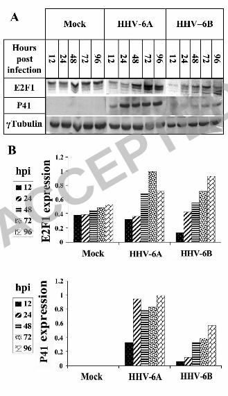
E2F1 levels increase in HHV-6 infected cells.
Due to its pivotal role in cell cycle progression and in gene regulation, it was of interest to examine
E2F1 regulation during viral infections. Earlier studies by microarray analyses revealed that E2F1
mRNA increased during HHV-6 infections of T cells (45, 71). To verify and extend this
information, cultures of SupT1 T cells were mock infected or infected with HHV-6A (U1102) and
HHV-6B (Z29). Protein extracts prepared 12, 24, 48, 72 and 96 hours post infection (hpi), were
tested in Western blots, employing the E2F1, P41 and γ-tubulin antibodies (Fig. 1A). The 9A5
MAb, produced by Bala Chandran and coworkers (8, 13) was chosen to follow infection
progression; it recognizes the HHV-6 41-kDa phosphoprotein (P41), encoded by the U27 gene,
which functions as a DNA polymerase accessory protein. The protein is and is highly conserved in
HHV-6A and HHV-6B strains (8, 13). As shown in Fig. 1A and quantified in Fig, 1B, the E2F1
levels increased in both HHV-6A (U102) and HHV-6B (Z29) infections, paralleling the
accumulation of P41. To test whether the E2F1 increase took place in the infected cells, or in
neighboring uninfected cells, SupT1 cells were infected with HHV-6B (Z29) and tested at 24, 48
and 72 hpi for reactivity with the E2F1 and the 9A5 antibodies. The nuclei were stained with
Cytox Orange. The triple staining showed that the elevated levels of E2F1 coincided with the
HHV-6 P41 staining, starting 48 hpi (Fig. 2). Thus, the E2F1 levels increased specifically in the
infected cells.
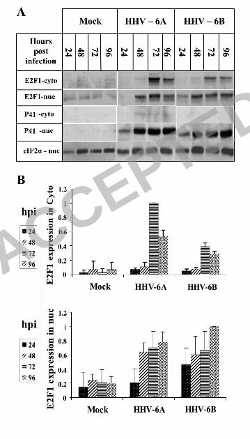
E2F1 cytoplasmic and nuclear localization is altered in the infected cells.
ACCEPTED
on May 23, 2018 by guest
http://jvi.asm.org/
Dow
nloaded from
8
The functions of the E2F transcription factors depend on their cellular localization and alterations
in protein localization were found to play a role in their trasncriptional activity (5, 40). While some
members of the E2F family can be found in the cytoplsamic as well as in the nuclear fractions,
E2F1 is regularly confined to the nucleus. To examine whether E2F1 undergoes alterations in its
localization, proteins were prepared from uninfected and HHV-6 infected cells, using cytoplasmic
and nuclear lysis. Since P41 used to follow the infection is a nuclear viral protein, it could also
serve as a control for nuclear contaminations of the cytoplasmic fractions. The eIF2α and γ-Tubulin
proteins were used to equalize protein loading. The results (Fig 3) revealed the following: (i) The
P41 protein was recovered from the infected cells by 24 hpi, revealing the expected progress of the
infection. Furthermore, it was recovered solely from the nuclei, in accordance with its function in
viral DNA replication and it could be concluded that there was no nuclear contamination of the
cytoplasmic fraction and no nuclear protein spillage, due to non-specific virally induced nuclear
breakage. (ii) In the mock infected cells E2F1 which has a nuclear localization signal (NLS) (43,
47, 75), was recovered mostly in the nuclear fraction, as expected. Strikingly, in the infected cells it
was found in both cytoplasmic and nuclear fractions, starting at 72 hpi (Fig. 3). (iii) All differences
in E2F1 nuclear and cytoplasmic localizations in the mock and infected cells, starting from 48 hpi
were statistically significant, as determined by t-test. The finding that the P41 protein was
recovered solely in the nuclear fraction, whereas E2F1 was recovered in both nuclear and
cytoplasmic fractions post infection, attested to the specificity of the E2F1 cytoplasmic and nuclear
recovery in the infected cultures.
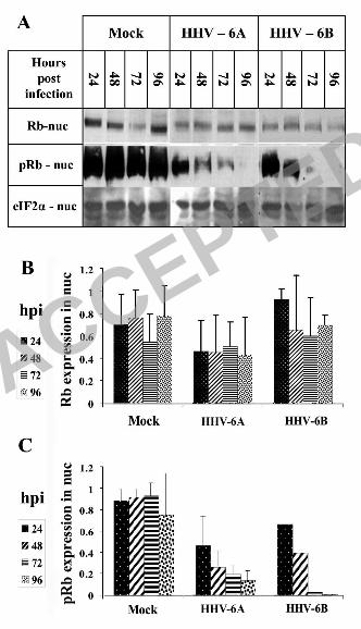
The total amount of the Rb protein and its localization did not change during the infection,
but the protein was dephosphorylated.
Following the finding that E2F1 was upregulated and could be recovered from the cytoplasmic
fraction of infected cells, we tested the presence of Rb, known to be a major regulator of E2F1.
ACCEPTED
on May 23, 2018 by guest
http://jvi.asm.org/
Dow
nloaded from
9
Both E2F1 and its regulator Rb are known to act in the nuclear fraction. Rb levels, Rb
phosphorylation and cellular localization were of interest, inasmuch as hypophosphorylated Rb was
previously shown to bind E2F1, affecting its transcriptional activity (48). The cytoplasmic and
nuclear lysates of 12×106 mock infected and HHV-6A infected cells were tested by Western
blotting, employing antibodies to Rb and phosphorylated Rb (pRb) (Fig. 4A). The data were
corrected for loading variations, employing the eIF2α protein. The quantitative results (Fig. 4B and
4C) can be summarized as follows: (i) The levels of Rb recovered from the cytoplasmic fractions
were similar in the mock infected, HHV-6A and HHV-6B infected cells. Furthermore, pRb was not
detected in the cytoplasmic fractions (data not shown). (ii) Similar levels of the Rb protein were
detected in the nuclear fractions of the mock infected and infected cells. (iii) Most significantly,
when compared to the mock infected cells, the levels of pRb were greatly reduced in the nuclear
fractions of the HHV-6A and 6B infections, starting at 24 hpi. (iv) Starting from 48 hpi, the
differences in pRb levels between mock and infected cells were statistically significant, as
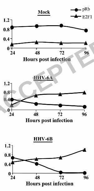
determined by the t-test. Fig. 5 summarizes the alterations observed in E2F1 and pRb nuclear
recovery.
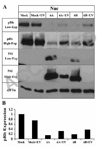
Rb dephosphorylation occurs also in cells infected with UV inactivated virus.
Following the finding that Rb was de-phosphorylated during HHV-6A and HHV-6B infections, it
was of interest to test whether the effect required viral gene expression post infection. To evaluate
this question 4x10⁶ cells were either: (i) mock infected, (ii) treated with medium which was UV-
irradiated, (iii) infected with cell free HHV-6A and HHV-6B, (iv) treated with equal quantity of
cell free virus which was UV-irradiated to inactivate the virus. Protein extracts prepared at 72 hpi
were tested in Western blots, employing the pRb, P41 and eIF2α antibodies. The results (Fig. 6)
are shown with low and high exposures of the Western blots and can be summarized as follows: (i)
Significant Rb de-phosphorylation was observed in the cells infected with live, untreated viruses, as
ACCEPTED
on May 23, 2018 by guest
http://jvi.asm.org/
Dow
nloaded from

10
described above. (ii) Significant Rb dephosphorylation was apparent also with the UV irradiated
viruses, when compared to the mock infected cells, although this was to a lower extent than in the
cells receiving the equivalent live virus infections. The quantitative analyses (Fig. 6B) revealed
that the infections with live HHV-6A and HHV-6B reduced Rb phosphorylation to 5.3% and 3.7%
respectively, of their levels in the mock infected cells. The UV irradiated HHV-6A and HHV-6B
reduced the Rb phosphorylation to 10.3% and 22.3% respectively, compared to the mock infected
cells, treated with the UV irradiated medium. (iii) Comparison of P41 expression in cells infected
with the live HHV-6A and 6B vs. the UV irradiated virus showed that viral inactivation in the
HHV-6A UV treated samples was incomplete, and there were still low levels of P41 protein
expression (high exposure lane, Fig. 6). Taken together these results indicated that Rb de-
phosphorylation in HHV-6A and HHV-6B infections might be caused by protein(s) brought into
the cells within the structural particles or might result from viral absorption or entry into the cells.
There was no requirement for elaborate viral gene expression and replication.
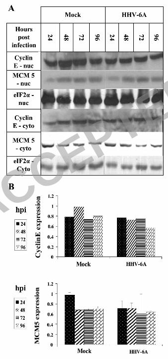
E2F1 accumulation does not result in higher levels of Cyclin E and MCM5 – two known E2F1
transcribed target genes.
E2F1 acts as a transcription factor for a large number of genes initiated in late G1, when E2F1
accumulates promoting cell cycle progression by various mechanisms (33). To test whether the
E2F1 targeted genes also increased during viral infections two target genes were chosen, including
Cyclin E and MCM5, both transcribed by E2F1 (30, 35, 50, 51). Cyclin E is an evolutionarily
conserved protein, which plays an essential role in promoting cell cycle transition from the G1 to
the S phase. It binds to and activates the Cyclin-dependent kinase Cdk2, with consequent
effects on
cell cycle progression (63). In this state it is able to phosphorylate Rb, which is first
phosphorylated by Cyclin D. Altogether, the phosphorylation of Rb results in its release from
E2F1. MCM5 is one of seven Minichromosome Maintenance (MCM) family members, highly
ACCEPTED
on May 23, 2018 by guest
http://jvi.asm.org/
Dow
nloaded from

11
conserved in eukaryotes. They function as DNA helicases, forming heterohexamer complexes that
bind to the DNA replication origins, moving along with the DNA polymerase, during DNA
replication (57). To determine the level of Cyclin E and MCM5, 12×106 SupT1 cells were mock
infected or infected with HHV-6A (U1102). The eIF2α was employed to correct for equal protein
loading. As shown in Fig. 7, although E2F1 levels increased during infection, neither Cyclin E nor
MCM5 levels changed, when compared to the uninfected cells. Furthermore, there were no
changes in their cytoplasmic and nuclear localizations.
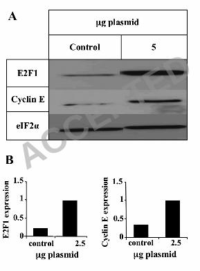
E2F1 activation pathway is intact in the SupT1 cells.
To test whether E2F1 accumulation can cause upregulation of its known target genes in the cell line
used in this work, SupT1 cells were transfected by electroporation with an E2F1 expression
plasmid driven by the HCMV promoter. Extracts prepared one day post transfection were tested in
Western blots using E2F1, Cyclin E and eIF2α antibodies. As seen in Fig 8, after transfection with
5µg of the E2F1 plasmid, the E2F1 levels were found to increase 5 fold, in a similar manner to the
increase observed following the HHV-6A and HHV-6B infections (Fig 3). In contrast to the
infected cells, where no upregulation of E2F1 target genes was found, 5µg of the E2F1 plasmid
caused upregulation of Cyclin E. These results confirm that E2F1 can induce the expression of
cyclin E in SupT1 cells, suggesting a different route for E2F1 in the infection.
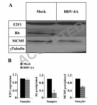
Rb and MCM5 have reduced complexing with E2F1 in the infected cells.
Following the finding that E2F1 was upregulated in the infected cells, it was of interest to examine
whether E2F1 was in its free form, or complexed to Rb. Additionally, MCM5 is known to interact
with E2F1 through Rb and to play critical roles in the formation of complexes functioning in host
DNA replication, cell cycle regulation and chromosomal stabilization (10, 22, 61, 68). Disruption
ACCEPTED
on May 23, 2018 by guest
http://jvi.asm.org/
Dow
nloaded from
12
of the complexing upon viral replication might be related to the inhibition of cell DNA replication,
as we have previously shown (19). To address these questions, protein lysates prepared from mock
infected or HHV-6A infected SupT1 cells were immunoprecipitated, employing E2F1 antibody,
coupled to protein A beads. Resultant precipitates were subjected to Western blotting employing
Rb, MCM5 and γ-tubulin antibodies. The results revealed the following (Fig. 9): (i) As expected,
γ-Tubulin, which is not known to interact with E2F1, was not recovered in the immunoprecipitate.
(ii) In the mock infected samples both Rb and MCM5 proteins were recovered in the
immunoprecipitated E2F1 samples. (iii) Based on two independent experiments there were lower
levels of Rb (35%) and MCM5 protein (60%) recovered in the E2F1 precipitates compared to their
recoveries in the mock infected samples. (iv) The differences in the Rb and MCM5 co-
precipitations were statistically significant, as determined by t-test. We conclude that the
upregulated E2F1 in the infected cells was mostly uncoupled with its main regulator Rb, and there
were somewhat lower quantities of MCM5 containing E2F1complexes.
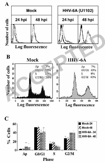
HHV-6 infection in SupT1 cells causes cell cycle arrest.
E2F1 and Rb play significant roles in the regulation of the cell cycle. Free E2F1 promotes cell
cycle progression past G1 into the S phase, whereas E2F1 bound to Rb promotes cell cycle arrest at
G1. The increased E2F1 levels post infection predicted progression into the S phase, but the E2F1
target genes cyclin E and MCM5 were unaffected during the infection. Moreover, there was
reduced coupling of Rb and E2F1. To test whether these alterations induced changes in cell cycle
progression, 4X10⁶ cells were mock infected or infected with HHV-6A (U11O2). At 24 and 48 hpi
prior the appearance of pronounced CPE, the cultures were fixed and subjected to FACS analysis:
2X10⁶ cells were incubated with the anti P41 9A5 MAb, and a secondary antibody - FITS goat anti
mouse, in order to mark and determine the percentage of the infected cells. The remaining 2X10⁶
cells were subjected to cell cycle analysis, using PI. Prior to cell cycle analysis by the FACS the
ACCEPTED
on May 23, 2018 by guest
http://jvi.asm.org/
Dow
nloaded from
13
cells were marked with the 9A5 MAb and a secondary FITC goat anti mouse antibody, analyzing
only the infected cells, by gating for FITC in the FACS cell cycle assay (Fig 10B). The choice of
the time points 24 and 48 hpi, prior to appearance of pronounced CPE was dictated by the inability
to adequately sort cell cultures containing the large synsytial cells late post infection. The results
revealed the following: (i) More then 95% of the cells were infected by 48 hpi, and contained the
P41 protein, marked by the 9A5 antibody (Fig. 10A). (ii) Cell cycle arrest was evident at the G2
phase, starting at 24 hpi (Fig. 10B). There were reduced numbers of cells in the G1 and S phases
and increased fraction of cells in the G2/M phase. (iii) The differences in the ratios of the G2/M
phases between the mock and infected cells, at 24 and 48 hpi, were statistically significant, as
determined by t-test, based on the FACS analyses of three experiments.
ACCEPTED
on May 23, 2018 by guest
http://jvi.asm.org/
Dow
nloaded from

14
DISCUSSION
E2F1 and its main repressor Rb, serve as major regulators for the transcription of genes that
function in cell cycle progression, viability as well as apoptosis. The studies described in this paper
revealed significant alterations in the E2F1/Rb pathways, at different points of viral infection.
Specifically: (i) E2F1 increased during HHV-6A and HHV-6B infections and could be recovered
in both the cytoplasmic and nuclear fractions, unlike its strict nuclear localization in the uninfected
cells. The cytoplasmic recovery of E2F1, starting at 72 hpi was intriguing in its specificity, unlike
the nuclear recovery of the P41 viral protein, indicating the absence of random nuclear leakage. It
is noteworthy that the p53 protein, also possessing a NLS was reported to have cytoplasmic
recovery in HHV-6 infected cells, attributed to its interaction with the U14 viral protein and its
encapsidation within structural virions (52, 53, 72, 73). Since high levels of E2F1 promote
apoptosis, the diversion of excess E2F1 into the cytoplasm might avoid early apoptosis, securing
cell survival for viral replication. Further studies are needed to determine the reason(s) for the
increased cytoplasmic E2F1. (ii) In the infected cells there was significant reduction in Rb
complexing with the E2F1 protein, and also some reduction in the MCM5 protein co precipitating
with E2F1 antibodies. Both reductions were statistically significant by t-tests. The release of E2F1
from Rb is known to result from Rb phosphorylation, or following binding of viral proteins to Rb,
disrupting the E2F1/Rb complexes. In the HHV-6 infected cells, the total levels of Rb did not
change, but its phosphorylation was significantly reduced. Moreover, Rb dephosphorylation
occurred also with UV irradiated virus, indicating that it was either mediated by virion protein(s),
incoming into the cells with the infecting virus, or by viral attachment, and/or interaction with viral
receptor at the cell surface. Considering the kinetics of E2F1, Rb and pRb during the infection, the
lower recovery of E2F1/Rb complexes most likely reflected the upregulation of E2F1 along with
the steady non-increased levels of Rb, resulting in free E2F1. (iii) The increased free E2F1 did not
ACCEPTED
on May 23, 2018 by guest
http://jvi.asm.org/
Dow
nloaded from
15
induce higher accumulation of Cyclin E and MCM 5, known to be induced by E2F1 accumulation,
although such increase was possible in the SupT1 infected cells. (iv) In the HHV-6A (U1102)
infected cells there was an arrest in cell cycle progression. Similar cell cycle arrest was observed
also in HHV-6B (Z29) infected cells (data not shown). Different viruses were found to affect cell
replication and cell cycle arrest was previously reported to occur in certain herpesvirus infections,
including HSV-1 and HCMV, which were reported to undergo arrest at the G1/S phase.
Furthermore, earlier reports have documented arrest in HHV-6 infected cells, at different levels of
the cell cycle. De-Bolle and coworkers reported cell cycle arrest, at the G2 phase, during HHV-6A
infection of cord blood cells (17). Hollsberg and coworkers have shown that HHV-6B infection of
MOLT 3 cells caused cell cycle arrest at the G1 phase, concomitantly with p53 phosphorylation
and accumulation (52). More recently, they have shown that the arrest at the G1/S phase involved a
pathway independent of p53 (53). In the present study we have found arrest at the G2 phase as
early as 24/48 hpi, prior the appearance of pronounced CPE. The differences in cell cycle profiles
of the mock and infected cells were reproduced in 3 separate experiments and were statistically
significant by t-test (fig. 10). The arrest might have resulted from both p53 and the presence of
complexes of E2F1/Rb early post infection, upon Rb dephosphorylation. Cell cycle arrest, at early
parts of the infection may be advantageous for virus replication in dividing lymphocytes, inasmuch
as with this slowly replicating virus, arresting cell replication might enable the formation of
synsytia and more efficient viral spread. At later times the infection was accompanied by total shut
off of host DNA replication, as shown in previous studies quantitating P32
phosphate incorporation
into viral and host DNA in infected PHA activated Peripheral Blood Lymphocytes (PBLs) at 65-86
hpi (19). Eventually, the infection might lead to apoptosis and/or necrosis. Overall, the data
presented in this study shed new light on HHV6 interaction with the infected cells, suggesting
different manipulation of key factors relevent to cell proliferation and death.
ACCEPTED
on May 23, 2018 by guest
http://jvi.asm.org/
Dow
nloaded from

16
ACKNOWLEDGEMENTS
We thank Dr. Ester Michael and Dr. Shulamit Grinshtein for valuable technical assistance. Our
research was supported by the S.Daniel Abraham Institute for Molecular Virology and the S.
Daniel Abraham Chair for Molecular Virology and Gene Therapy. We thank Dr Bala Chandran
from Chicago Medical School for his kind contribution of the 9A5 MAb.
ACCEPTED
on May 23, 2018 by guest
http://jvi.asm.org/
Dow
nloaded from
17
REFERENCES
1. Ablashi, A. H., Berneman Z, et al. 1993. Human herpesvirus-6 strain groups: A
nomenclature. Arch Virol 129:363-366.
2. Alvarez-Lafuente, R., V. De Las Heras, M. Bartolome, M. Garcia-Montojo, and R.
Arroyo. 2006. Human herpesvirus 6 and multiple sclerosis: a one-year follow-up study.
Brain Pathol 16:20-7.
3. Alvarez-Lafuente, R., V. de Las Heras, M. Garcia-Montojo, M. Bartolome, and R.
Arroyo. 2007. Human herpesvirus-6 and multiple sclerosis: relapsing-remitting versus
secondary progressive. Mult Scler 13:578-83.
4. Alvarez-Lafuente, R., M. Garcia-Montojo, V. De las Heras, M. Bartolome, and R.
Arroyo. 2006. Clinical parameters and HHV-6 active replication in relapsing-remitting
multiple sclerosis patients. J Clin Virol 37 Suppl 1:S24-6.
5. Apostolova, M. D., I. A. Ivanova, C. Dagnino, S. J. D'Souza, and L. Dagnino. 2002.
Active nuclear import and export pathways regulate E2F-5 subcellular localization. J
Biol Chem 277:34471-9.
6. Asano, Y., T. Yoshikawa, Y. Kajita, R. Ogura, S. Suga, T. Yazaki, T. Nakashima,
A. Yamada, and T. Kurata. 1992. Fatal encephalitis/encephalopathy in primary human
herpesvirus-6 infection. Arch Dis Child 67:1484-5.
7. Bagchi, S., R. Weinmann, and P. Raychaudhuri. 1991. The retinoblastoma protein
copurifies with E2F-I, an E1A-regulated inhibitor of the transcription factor E2F. Cell
65:1063-72.
8. Balachandran, N., S. Tirawatnapong, B. Pfeiffer, D. V. Ablashi, and S. Z.
Salahuddin. 1991. Electrophoretic analysis of human herpesvirus 6 polypeptides
immunoprecipitated from infected cells with human sera. J Infect Dis 163:29-34.
9. Boeckh, M., V. Erard, D. Zerr, and J. Englund. 2005. Emerging viral infections after
hematopoietic cell transplantation. Pediatr Transplant 9 Suppl 7:48-54.
10. Bosco, G., W. Du, and T. L. Orr-Weaver. 2001. DNA replication control through
interaction of E2F-RB and the origin recognition complex. Nat Cell Biol 3:289-95.
11. Bracken, A. P., M. Ciro, A. Cocito, and K. Helin. 2004. E2F target genes: unraveling
the biology. Trends Biochem Sci 29:409-17.
12. Cam, H., and B. D. Dynlacht. 2003. Emerging roles for E2F: beyond the G1/S
transition and DNA replication. Cancer Cell 3:311-6.
13. Chang, C. K., and N. Balachandran. 1991. Identification, characterization, and
sequence analysis of a cDNA encoding a phosphoprotein of human herpesvirus 6. J
Virol 65:2884-94.
14. Chapenko, S., A. Millers, Z. Nora, I. Logina, R. Kukaine, and M. Murovska. 2003.
Correlation between HHV-6 reactivation and multiple sclerosis disease activity. J Med
Virol 69:111-7.
15. Chau, B. N., C. W. Pan, and J. Y. Wang. 2006. Separation of Anti-Proliferation and
Anti-Apoptotic Functions of Retinoblastoma Protein through Targeted Mutations of Its
A/B Domain. PLoS ONE 1:e82.
16. Chellappan, S. P., S. Hiebert, M. Mudryj, J. M. Horowitz, and J. R. Nevins. 1991.
The E2F transcription factor is a cellular target for the RB protein. Cell 65:1053-61.
17. De Bolle, L., S. Hatse, E. Verbeken, E. De Clercq, and L. Naesens. 2004. Human
herpesvirus 6 infection arrests cord blood mononuclear cells in G(2) phase of the cell
cycle. FEBS Lett 560:25-9.
ACCEPTED
on May 23, 2018 by guest
http://jvi.asm.org/
Dow
nloaded from

18
18. DeGregori, J., and D. G. Johnson. 2006. Distinct and Overlapping Roles for E2F
Family Members in Transcription, Proliferation and Apoptosis. Curr Mol Med 6:739-
48.
19. Di Luca, D., G. Katsafanas, E. C. Schirmer, N. Balachandran, and N. Frenkel.
1990. The replication of viral and cellular DNA in human herpesvirus 6-infected cells.
Virology 175:199-210.
20. Dick, F. A., E. Sailhamer, and N. J. Dyson. 2000. Mutagenesis of the pRB pocket
reveals that cell cycle arrest functions are separable from binding to viral oncoproteins.
Mol Cell Biol 20:3715-27.
21. Ehmann, G. L., T. I. McLean, and S. L. Bachenheimer. 2000. Herpes simplex virus
type 1 infection imposes a G(1)/S block in asynchronously growing cells and prevents
G(1) entry in quiescent cells. Virology 267:335-49.
22. Forsburg, S. L. 2004. Eukaryotic MCM proteins: beyond replication initiation.
Microbiol Mol Biol Rev 68:109-31.
23. Fotheringham, J., and S. Jacobson. 2005. Human herpesvirus 6 and multiple
sclerosis: potential mechanisms for virus-induced disease. Herpes 12:4-9.
24. Frenkel, N., and R. Borenstein. 2006. Characterization of the lymphotropic
Amplicons-6 and Tamplicon-7 vectors derived from HHV-6 and HHV-7. Curr Gene
Ther 6:399-420.
25. Frenkel, N., G. C. Katsafanas, L. S. Wyatt, T. Yoshikawa, and Y. Asano. 1994.
Bone marrow transplant recipients harbor the B variant of human herpesvirus 6. Bone
Marrow Transplant 14:839-43.
26. Grand, R. J., A. P. Ibrahim, A. M. Taylor, A. E. Milner, C. D. Gregory, P. H.
Gallimore, and A. S. Turnell. 1998. Human cells arrest in S phase in response to
adenovirus 12 E1A. Virology 244:330-42.
27. Helin, K., J. A. Lees, M. Vidal, N. Dyson, E. Harlow, and A. Fattaey. 1992. A cDNA
encoding a pRB-binding protein with properties of the transcription factor E2F. Cell
70:337-50.
28. Helin, K., C. L. Wu, A. R. Fattaey, J. A. Lees, B. D. Dynlacht, C. Ngwu, and E.
Harlow. 1993. Heterodimerization of the transcription factors E2F-1 and DP-1 leads to
cooperative trans-activation. Genes Dev 7:1850-61.
29. Hilton, M. J., D. Mounghane, T. McLean, N. V. Contractor, J. O'Neil, K.
Carpenter, and S. L. Bachenheimer. 1995. Induction by herpes simplex virus of free
and heteromeric forms of E2F transcription factor. Virology 213:624-38.
30. Hwang, H. C., and B. E. Clurman. 2005. Cyclin E in normal and neoplastic cell
cycles. Oncogene 24:2776-86.
31. Jault, F. M., J. M. Jault, F. Ruchti, E. A. Fortunato, C. Clark, J. Corbeil, D. D.
Richman, and D. H. Spector. 1995. Cytomegalovirus infection induces high levels of
cyclins, phosphorylated Rb, and p53, leading to cell cycle arrest. J Virol 69:6697-704.
32. Kaelin, W. G., Jr., W. Krek, W. R. Sellers, J. A. DeCaprio, F. Ajchenbaum, C. S.
Fuchs, T. Chittenden, Y. Li, P. J. Farnham, M. A. Blanar, and et al. 1992.
Expression cloning of a cDNA encoding a retinoblastoma-binding protein with E2F-like
properties. Cell 70:351-64.
33. Kel, A. E., O. V. Kel-Margoulis, P. J. Farnham, S. M. Bartley, E. Wingender, and
M. Q. Zhang. 2001. Computer-assisted identification of cell cycle-related genes: new
targets for E2F transcription factors. J Mol Biol 309:99-120.
34. Komaroff, A. L. 2006. Is human herpesvirus-6 a trigger for chronic fatigue syndrome?
J Clin Virol 37 Suppl 1:S39-46.
35. Koutsami, M. K., P. K. Tsantoulis, M. Kouloukoussa, K. Apostolopoulou, I. S.
Pateras, Z. Spartinou, A. Drougou, K. Evangelou, C. Kittas, J. Bartkova, J. Bartek,
ACCEPTED
on May 23, 2018 by guest
http://jvi.asm.org/
Dow
nloaded from
19
and V. G. Gorgoulis. 2006. Centrosome abnormalities are frequently observed in non-
small-cell lung cancer and are associated with aneuploidy and cyclin E overexpression. J
Pathol 209:512-21.
36. Kovesdi, I., R. Reichel, and J. R. Nevins. 1986. Identification of a cellular
transcription factor involved in E1A trans-activation. Cell 45:219-28.
37. Krueger G, a. D. V. A. 2006. Human Herpesvirus-6, General Virology, Epidemiology
and Clinical Pathology (Perspectives in Medical Virology), vol. 12.
38. Lee, C., and Y. Cho. 2002. Interactions of SV40 large T antigen and other viral
proteins with retinoblastoma tumour suppressor. Rev Med Virol 12:81-92.
39. Lees, J. A., M. Saito, M. Vidal, M. Valentine, T. Look, E. Harlow, N. Dyson, and K.
Helin. 1993. The retinoblastoma protein binds to a family of E2F transcription factors.
Mol Cell Biol 13:7813-25.
40. Lindeman, G. J., S. Gaubatz, D. M. Livingston, and D. Ginsberg. 1997. The
subcellular localization of E2F-4 is cell-cycle dependent. Proc Natl Acad Sci U S A
94:5095-100.
41. Ljungman, P., and N. Singh. 2006. Human herpesvirus-6 infection in solid organ and
stem cell transplant recipients. J Clin Virol 37 Suppl 1:S87-91.
42. Lopez, C., P. Pellett, J. Stewart, C. Goldsmith, K. Sanderlin, J. Black, D. Warfield,
and P. Feorino. 1988. Characteristics of human herpesvirus-6. J Infect Dis 157:1271-3.
43. Magae, J., C. L. Wu, S. Illenye, E. Harlow, and N. H. Heintz. 1996. Nuclear
localization of DP and E2F transcription factors by heterodimeric partners and
retinoblastoma protein family members. J Cell Sci 109 (Pt 7):1717-26.
44. Mameli, G., V. Astone, G. Arru, S. Marconi, L. Lovato, C. Serra, S. Sotgiu, B.
Bonetti, and A. Dolei. 2007. Brains and peripheral blood mononuclear cells of multiple
sclerosis (MS) patients hyperexpress MS-associated retrovirus/HERV-W endogenous
retrovirus, but not Human herpesvirus 6. J Gen Virol 88:264-74.
45. Mayne, M., C. Cheadle, S. S. Soldan, C. Cermelli, Y. Yamano, N. Akhyani, J. E.
Nagel, D. D. Taub, K. G. Becker, and S. Jacobson. 2001. Gene expression profile of
herpesvirus-infected T cells obtained using immunomicroarrays: induction of
proinflammatory mechanisms. J Virol 75:11641-50.
46. Muller, H., A. P. Bracken, R. Vernell, M. C. Moroni, F. Christians, E. Grassilli, E.
Prosperini, E. Vigo, J. D. Oliner, and K. Helin. 2001. E2Fs regulate the expression of
genes involved in differentiation, development, proliferation, and apoptosis. Genes Dev
15:267-85.
47. Muller, H., M. C. Moroni, E. Vigo, B. O. Petersen, J. Bartek, and K. Helin. 1997.
Induction of S-phase entry by E2F transcription factors depends on their nuclear
localization. Mol Cell Biol 17:5508-20.
48. Mundle, S. D., and G. Saberwal. 2003. Evolving intricacies and implications of E2F1
regulation. Faseb J 17:569-74.
49. Nagasawa, T., I. Kimura, Y. Abe, and A. Oka. 2007. HHV-6 encephalopathy with
cluster of convulsions during eruptive stage. Pediatr Neurol 36:61-3.
50. Ohtani, K., J. DeGregori, and J. R. Nevins. 1995. Regulation of the cyclin E gene by
transcription factor E2F1. Proc Natl Acad Sci U S A 92:12146-50.
51. Ohtani, K., R. Iwanaga, M. Nakamura, M. Ikeda, N. Yabuta, H. Tsuruga, and H.
Nojima. 1999. Cell growth-regulated expression of mammalian MCM5 and MCM6
genes mediated by the transcription factor E2F. Oncogene 18:2299-309.
52. Oster, B., B. Bundgaard, and P. Hollsberg. 2005. Human herpesvirus 6B induces cell
cycle arrest concomitant with p53 phosphorylation and accumulation in T cells. J Virol
79:1961-5.
ACCEPTED
on May 23, 2018 by guest
http://jvi.asm.org/
Dow
nloaded from
20
53. Oster, B., M. D. Kaspersen, E. Kofod-Olsen, B. Bundgaard, and P. Hollsberg. 2006.
Human herpesvirus 6B inhibits cell proliferation by a p53-independent pathway. J Clin
Virol 37 Suppl 1:S63-8.
54. Pellett, D. a. 2002. Human Herpesviruses 6A, 6B, and 7 and Their Replication, p. pg
2769 - 2784, Fields VIROLOGY, Fourth Edition ed, vol. Volume 2, Chapter 80.
Lippincott - Raven publishers.
55. Rapaport, D., D. Engelhard, G. Tagger, R. Or, and N. Frenkel. 2002. Antiviral
prophylaxis may prevent human herpesvirus-6 reactivation in bone marrow transplant
recipients. Transpl Infect Dis 4:10-6.
56. Rotola, A., I. Merlotti, L. Caniatti, E. Caselli, E. Granieri, M. R. Tola, D. Di Luca,
and E. Cassai. 2004. Human herpesvirus 6 infects the central nervous system of
multiple sclerosis patients in the early stages of the disease. Mult Scler 10:348-54.
57. Ryu, S., J. Holzschuh, S. Erhardt, A. K. Ettl, and W. Driever. 2005. Depletion of
minichromosome maintenance protein 5 in the zebrafish retina causes cell-cycle defect
and apoptosis. Proc Natl Acad Sci U S A 102:18467-72.
58. Salahuddin, S. Z., D. V. Ablashi, P. D. Markham, S. F. Josephs, S. Sturzenegger,
M. Kaplan, G. Halligan, P. Biberfeld, F. Wong-Staal, B. Kramarsky, and et al. 1986. Isolation of a new virus, HBLV, in patients with lymphoproliferative disorders.
234:596-601.
59. Santoro, F., P. E. Kennedy, G. Locatelli, M. S. Malnati, E. A. Berger, and P. Lusso.
1999. CD46 is a cellular receptor for human herpesvirus 6. Cell 99:817-27.
60. Schirmer, E. C., L. S. Wyatt, K. Yamanishi, W. J. Rodriguez, and N. Frenkel.
1991. Differentiation between two distinct classes of viruses now classified as human
herpesvirus 6. Proc Natl Acad Sci U S A 88:5922-6.
61. Schwed, G., N. May, Y. Pechersky, and B. R. Calvi. 2002. Drosophila
minichromosome maintenance 6 is required for chorion gene amplification and genomic
replication. Mol Biol Cell 13:607-20.
62. Shan, B., X. Zhu, P. L. Chen, T. Durfee, Y. Yang, D. Sharp, and W. H. Lee. 1992.
Molecular cloning of cellular genes encoding retinoblastoma-associated proteins:
identification of a gene with properties of the transcription factor E2F. Mol Cell Biol
12:5620-31.
63. Singer, J. D., M. Gurian-West, B. Clurman, and J. M. Roberts. 1999. Cullin-3
targets cyclin E for ubiquitination and controls S phase in mammalian cells. Genes Dev
13:2375-87.
64. Singer, O., and N. Frenkel. 1997. Human herpesvirus 7 (HHV-7) DNA: analyses of
clones spanning the entire genome. Arch Virol 142:287-303.
65. Smith, J. M., and R. A. McDonald. 2006. Emerging viral infections in transplantation.
Pediatr Transplant 10:838-43.
66. Soldan Samantha S., G. A. D., Jacobson Steven. 2006. HHV-6 and the Central
Nervous System, 2 ed, vol. Human Herpesvirus-6: General virology, Epidemiolgy and
Clinical Pathology. Elsevier.
67. Song, Y. J., and M. F. Stinski. 2002. Effect of the human cytomegalovirus IE86
protein on expression of E2F-responsive genes: a DNA microarray analysis. Proc Natl
Acad Sci U S A 99:2836-41.
68. Stagljar, I., U. Hubscher, and A. Barberis. 1999. Activation of DNA replication in
yeast by recruitment of the RNA polymerase II transcription complex. Biol Chem
380:525-30.
69. Stanelle, J., and B. M. Putzer. 2006. E2F1-induced apoptosis: turning killers into
therapeutics. Trends Mol Med 12:177-85.
ACCEPTED
on May 23, 2018 by guest
http://jvi.asm.org/
Dow
nloaded from

21
70. Suga, S., K. Suzuki, M. Ihira, T. Yoshikawa, Y. Kajita, T. Ozaki, K. Iida, Y. Saito,
and Y. Asano. 2000. Clinical characteristics of febrile convulsions during primary
HHV-6 infection. Arch Dis Child 82:62-6.
71. Takaku, T., J. H. Ohyashiki, Y. Zhang, and K. Ohyashiki. 2005. Estimating
immunoregulatory gene networks in human herpesvirus type 6-infected T cells.
Biochem Biophys Res Commun 336:469-77.
72. Takemoto, M., M. Koike, Y. Mori, S. Yonemoto, Y. Sasamoto, K. Kondo, Y.
Uchiyama, and K. Yamanishi. 2005. Human herpesvirus 6 open reading frame U14
protein and cellular p53 interact with each other and are contained in the virion. J Virol
79:13037-46.
73. Takemoto, M., Y. Mori, K. Ueda, K. Kondo, and K. Yamanishi. 2004. Productive
human herpesvirus 6 infection causes aberrant accumulation of p53 and prevents
apoptosis. J Gen Virol 85:869-79.
74. Tomonari, A., S. Takahashi, J. Ooi, T. Iseki, K. Takasugi, M. Uchiyama, T.
Konuma, M. Futami, N. Ohno, K. Uchimaru, A. Tojo, and S. Asano. 2005. Human
herpesvirus 6 variant B infection in adult patients after unrelated cord blood
transplantation. Int J Hematol 81:352-5.
75. Trimarchi, J. M., and J. A. Lees. 2002. Sibling rivalry in the E2F family. Nat Rev Mol
Cell Biol 3:11-20.
76. Tuke, P. W., S. Hawke, P. D. Griffiths, and D. A. Clark. 2004. Distribution and
quantification of human herpesvirus 6 in multiple sclerosis and control brains. Mult
Scler 10:355-9.
77. Ward, K. N., N. J. Andrews, C. M. Verity, E. Miller, and E. M. Ross. 2005. Human
herpesviruses-6 and -7 each cause significant neurological morbidity in Britain and
Ireland. Arch Dis Child 90:619-23.
78. Yamanishi, K. 2002. Human Herpesvirus 6 and Human Herpesvirus 7, p. pg 2785 -
2801, Fields VIROLOGY, Fourth Edition ed, vol. Volume 2, Chapter 81. Lippincott -
Raven publishers.
79. Yamanishi, K., T. Okuno, K. Shiraki, M. Takahashi, T. Kondo, Y. Asano, and T.
Kurata. 1988. Identification of human herpesvirus-6 as a causal agent for exanthem
subitum. Lancet 1:1065-7.
80. Yamashita, N., and T. Morishima. 2005. HHV-6 and seizures. Herpes 12:46-9.
81. Yao, K., N. Akyani, D. Donati, N. Sengamalay, J. Fotheringham, E. Ghedin, M.
Bishop, J. Barrett, F. Kashanchi, and S. Jacobson. 2006. Detection of HHV-6B in
post-mortem central nervous system tissue of a post-bone marrow transplant recipient: a
multi-virus array analysis. J Clin Virol 37 Suppl 1:S57-62.
82. Yoshikawa, T., and Y. Asano. 2000. Central nervous system complications in human
herpesvirus-6 infection. Brain Dev 22:307-14.
83. Yoshikawa, T., Y. Asano, M. Ihira, K. Suzuki, M. Ohashi, S. Suga, K. Kudo, K.
Horibe, S. Kojima, K. Kato, T. Matsuyama, and Y. Nishiyama. 2002. Human
herpesvirus 6 viremia in bone marrow transplant recipients: clinical features and risk
factors. J Infect Dis 185:847-53.
84. Yoshikawa, T., T. Nakashima, S. Suga, Y. Asano, T. Yazaki, H. Kimura, T.
Morishima, K. Kondo, and K. Yamanishi. 1992. Human herpesvirus-6 DNA in
cerebrospinal fluid of a child with exanthem subitum and meningoencephalitis.
Pediatrics 89:888-90.
85. Yoshikawa, T., S. Suga, Y. Asano, T. Nakashima, T. Yazaki, Y. Ono, T. Fujita, K.
Tsuzuki, S. Sugiyama, and S. Oshima. 1992. A prospective study of human
herpesvirus-6 infection in renal transplantation. Transplantation 54:879-83.
ACCEPTED
on May 23, 2018 by guest
http://jvi.asm.org/
Dow
nloaded from
22
86. Yoshikawa, T., S. Suga, Y. Asano, T. Nakashima, T. Yazaki, R. Sobue, M. Hirano,
M. Fukuda, S. Kojima, and T. Matsuyama. 1991. Human herpesvirus-6 infection in
bone marrow transplantation. Blood 78:1381-4.
87. Zerr, D. M. 2006. Human herpesvirus 6 and central nervous system disease in
hematopoietic cell transplantation. J Clin Virol 37 Suppl 1:S52-6.
88. Zerr, D. M. 2006. Human herpesvirus 6: a clinical update. Herpes 13:20-4.
ACCEPTED
on May 23, 2018 by guest
http://jvi.asm.org/
Dow
nloaded from
23
FIGURES;
Fig. 1: E2F1 expression during HHV-6A (U11O2) and HHV-6B (Z29) infections. A. Western
blot analyses of proteins from mock infected and infected cells, employing antibodies to E2F1, P41
and γ Tubulin. The antibody used for P41 is the MAb 9A5 of Bala Chandran (8, 13). B.
Quantitative representation of the results. Shown are the E2F1 and P41 protein accumulation,
relative to protein level in the highest point of the HHV-6A samples, at 72 hpi. The P41 and E2F1
were corrected for loading variations by the intensity of the gamma tubulin staining.
Fig. 2: EE2F1 is increased in the infected cells. Cultures of 4×10⁶
cells were infected with
HHV-6B (Z29) and reacted with primary antibodies for E2F1 (rabbit) and P41 (mouse), as well as
with cytox orange, for nuclear localization. Cye2 (anti rabbit) and Cye5 (anti mouse) secondary
antibodies were used to mark the E2F1 in green and the P41 in purple. Each time frame contains:
(i) Cye 5 field (9A5), (ii) Cye 2 field (E2F1), (iii) cytox orange field, (iv) Bright field and (v)
Merging of fields 1-3.
Fig. 3: Alterations in E2F1 cytoplasmic and nuclear localization. (A) Recovery of E2F1 and
P41 proteins in the cytoplasmic or nuclear fractions (B) Quantitative representation of the results
shown relative to highest protein expression in each figure. The E2F1-nuc analyses represented
averages of four experiments, and the E2F1 cytoplasmic data represented the averages of 2
experiments. All differences were statistically significant by t-test.
Fig. 4: Rb hypophosphorylation in HHV-6A and HHV-6B infected SupT1 cells. (A) Western
blots of mock infected and infected cell lysates were reacted with antibodies to Rb and
phosphorylated Rb proteins. (B) Graphic representation of the results, relative to the highest Rb
ACCEPTED
on May 23, 2018 by guest
http://jvi.asm.org/
Dow
nloaded from
24
and pRb protein in each figure. The intensities of Rb and pRb were corrected for loading variations
by the intensity of eIF2α. The measurements for the Rb were based on two experiments. The
comparison of the pRb in the mock infection vs. HHV-6B (Z29) infection was based on a single
experiment. pRb in the cytoplasmic fractions of mock and infected cells was not detected (data not
shown). The quantitation of pRb in the mock and HHV-6A (U1102) infections were based on three
separate experiments
Fig. 5: Alterations of pRb and E2F1 in infected and mock infected cells. Graphic
representations of E2F1 and pRb in the nuclei of mock and infected cells. pRb and E2F1 values are
shown relative to the highest protein level detected: E2F1 in the 96 hpi sample of HHV-6B
infections and pRb at 72 hpi in the mock infected cells.
Fig. 6: The de-phosphorylation of Rb in cells infected with inactivated virus. (A) The viruses
were either irradiated by UV or not and Western blots from mock infected and infected cells lysates
were reacted with antibodies to the pRb and P41 proteins (9A5 MAb). Two exposures of the blots
(low and high exp) are shown for the pRb and the P41 proteins. (B) Graphic representation of the
results, relative to the highest pRb levels (mock infected cells). The intensities of pRb were
corrected for loading variations by the intensity of eIF2α.
Fig. 7: The levels of Cyclin E and MCM5 proteins in HHV-6A (U11O2) SupT1 infected cells.
(A) Western blots of proteins prepared from the cytoplasmic and nuclear fractions of mock infected
and HHV-6A infected cell, tested with Cyclin E, MCM5 and eIF2α antibodies (B) Quantitative
representation of the results for the nuclear fractions, relative to the highest protein expression. The
ACCEPTED
on May 23, 2018 by guest
http://jvi.asm.org/
Dow
nloaded from
25
data are based on a single experiment for Cyclin E, and the averages of two experiments for
MCM5.
Fig. 8: E2F1 can initiate Cyclin E expression in SupT1 cells. (A) Western blots tested with
E2F1, Cyclin E and eIF2α antibodies after transfection of SupT1 cells with an E2F1 expressing
plasmid. (B) Graphic representation of the results, relative to the highest protein expression in each
figure
Fig. 9: Reduced Rb and MCM5 complexes with E2F1 in the infected cells. (A) E2F1 was
immunoprecipitated with anti-E2F1 antibody, starting with 500µg lysate. Proteins, which were
coprecipitated, were identified using antibodies to Rb, MCM5 and γ-tubulin. (B) Graphic
representation of the results, relative to the highest protein expression in each figure. The
quantitative analyses were based on two independent experiments. The differences of Rb and
MCM5 between mock and infected cells were statistically significant, as determined by t-test.
Fig. 10: The effect of the infection on the cell cycle. (A) FACS analyses of mock and infected
cells, prepared at 24 and 48 hpi, labeled with the P41 9A5 MAb (gray line). (B) Cell cycle analyses
at 48 hpi. (C) Graphic representations of the FACS results based on 3 separate experiments. The
differences in the ratios G2/M phases between mock and infected, at 24 and 48 hrs, were
statistically significant as determined by t-test
ACCEPTED
on May 23, 2018 by guest
http://jvi.asm.org/
Dow
nloaded from