epilepsy, female reproductive health and neurodevelopment...
TRANSCRIPT
DEPARTMENT OF NEUROLOGY SERIES OF REPORTS NO 87, 2007
KATRIINA VIINIKAINEN
Epilepsy, Female Reproductive Health and
Neurodevelopment of the Offspring
Doctoral dissertation
To be presented with assent of the Medical Faculty of the University of Kuopio
for public examination in Auditorium, Mediteknia building, University of Kuopio,
on Friday 20th April 2007, at 12 noon
Department of Neurology Department of Obstetrics and Gynecology
University of Kuopio and Kuopio University Hospital
Distributor: Department of Neurology University of Kuopio P.O. Box 1627 FI-70211 Kuopio FINLAND Tel. +358 17 162 682 Fax +358 17 162 048 Author’s address: Social and Welfare Health Center of Äänekoski Terveyskatu 10 FIN – 44100 Äänekoski FINLAND Tel. +358 50 307 3150 E-mail: [email protected] Supervisors: Docent Reetta Kälviäinen, M.D., Ph.D. Kuopio Epilepsy Center
Department of Neurology Kuopio University Hospital Professor Seppo Heinonen, M.D., Ph.D. Department of Obstetrics and Gynecology Kuopio University Hospital Docent Kai Eriksson, M.D., Ph.D. Pediatric Neurology Unit
Pediatric Research Centre Tampere University Hospital Reviewers: Docent Eija Gaily, M.D., Ph.D. Department of Pediatric Neurology Hospital for Children and Adolescents Helsinki University Hospital Docent Jukka Uotila, M.D., Ph.D. Department of Obstetrics and Gynecology Tampere University Hospital Opponent: Docent Tapani Keränen, M.D., Ph.D. Department of Neurology Tampere University Hospital
ISBN 978-951-781-379-2 ISBN 978-951-27-0217-6 (PDF) ISSN 0357-6043
Kopijyvä Kuopio 2007 Finland

Viinikainen, Katriina. Epilepsy, female reproductive health and neurodevelopment of the offspring. Series of Reports, No 87, Department of Neurology, University of Kuopio 2007. 85 p. ISBN 978-951-781-379-2 ISBN 978-951-27-0217-6 (PDF) ISSN 0357-6043 ABSTRACT
One of the most common neurological diseases in women of childbearing age is epilepsy, affecting approximately 0.8 percent of the women. An increased risk for both fetal and maternal complications, has been reported in women with epilepsy due to epilepsy itself, epilepsy related co-morbidities and antiepileptic drugs. Some of the frequent concerns are reproductive problems, pregnancy complications, congenital malformations and developmental problems in the offspring.
The purpose of this study was to evaluate the fertility and reproductive health in women with active epilepsy living in the Kuopio University Hospital area. The study was also extended to evaluate the pregnancy outcome of these women and to the assessment of the cognitive and neurological performance of the children exposed to valproate and carbamazepine monotherapy during pregnancy.
We found no difference between women with epilepsy and control women in their reproductive health and also the overall rate of women having children was the same, if we exclude the infertility caused by a higher proportion of severe co-morbid factors, which differentiate the women with active epilepsy from the general population. We followed the pregnant women throughout the pregnancy with a pre-decided protocol. The course of pregnancy was uncomplicated in the majority of the women with epilepsy. Congenital malformations were observed in 4.8 % of the live-births in women with epilepsy and the rate of small-for-date infants as well as the rate of admissions to a neonatal intensive care unit, were higher in the infants of the women with epilepsy. Women using valproate for epilepsy had a lower intelligence quotient than women using carbamazepine or women without antiepileptic drugs and also their level of education was lower, reflecting perhaps the nature of the epilepsies responding specifically to valproate. Children exposed to valproate had also lower mean intelligence scores, though the difference did not reach statistical significance. They also scored lower values in the neuropsychological tests and had received more educational support than children exposed to carbamazepine or children without drug exposure. In 62 % of the children exposed to valproate, one or more minor dysmorphic features were observed compared to 15% in other children. Children with carbamazepine exposure did not differ from controls.
In conclusion, women with active epilepsy represent a particularly challenging population for neurologists and other health care professionals. However, in our population with the pre-decided protocol used for the follow-up of pregnancies and well controlled epilepsy, the majority of the women with epilepsy have uncomplicated pregnancies. The risk for congenital malformations is nearly two-fold in the offspring of women with epilepsy exposed to antiepileptic drugs compared with the results of the national malformation registry. The cognitive outcome of children exposed to carbamazepine does not differ from controls, which is in line with previous reports. Our findings suggest that valproate may have a negative impact on neurocognitive development of the exposed offspring, though many confounding factors, including the type of epilepsy and level of schooling of the mother, may explain some of this result. National Library of Medicine Classification: QV 85, WL 385, WQ 210, WQ 240 Medical Subject Headings: Carbamazepine/adverse effects; Epilepsy; Finland; Infant, Newborn; Maternal Exposure; Pregnancy; Pregnancy Complications; Pregnancy Outcome; Pregnant Women; Prenatal Exposure Delayed Effects; Risk; Uterus/drug effects; Valproic Acid/adverse effects


To my family


ACKNOWLEDGEMENTS
The present study was carried out in the Department of Neurology and in the Department of
Obstetrics and Gynecology, Kuopio University Hospital and University of Kuopio during the
years 2001-2007.
I express my deepest gratitude to my principal supervisor, Docent Reetta Kälviäinen, who
introduced me to this field and who provided me with the opportunity to carry out this
scientific work. Her enthusiasm, expertise and constant support during these years has been
extremely valuable for me and has provided a warm and friendly atmosphere in which to
complete my work.
I am most grateful to my second supervisor, Professor Seppo Heinonen, for his guidance in
the field of obstetrics and gynecology. His attitude has been encouraging and optimistic and
he has always been available for comments and advice to help me forward with my work.
I express my warmest thanks to my third supervisor, Docent Kai Eriksson. His expertise in
Pediatric Neurology has been essential for this study. His warm and calm attitude and
understanding have made the collaboration easy.
I wish to express my thanks to Professor Hilkka Soininen, Head of the Department of
Neurology, for providing the facilities which allowed me to carry on this work.
I am very grateful to the official reviewers of this thesis, Docent Eija Gaily and Docent Jukka
Uotila, for their constructive criticism and suggestions for improving my thesis and to Dr.
Ewen MacDonald for revising the English language of this thesis.
I also want to thank warmly my co-authors and collaborators whose help and expertise have
been important and valuable during these years. My special thanks go to Marja Äikiä, Iiris
Sorri, Anne Mönkkönen and Pirkko Nieminen.

I express my sincere thanks to the personnel of the Department of Neurology, Kuopio
Epilepsy Center and Department of Obstetrics and Gynecology for their assistance and
guidance throughout the work. I especially want to thank Virpi Savolainen and Pirjo Lavi
from the Kuopio Epilepsy Center and Olavi Kauhanen from the Department of Obstetrics and
Gynecology for his statistical assistance.
I warmly want to thank all my friends. With you I have been able to enjoy many memorable
moments in my life.
I dedicate my dearest thanks to my parents Tuula and Hannu Viinikainen and to my brother,
Tuomas Viinikainen. Your love and encouragement have carried me forward in my life.
Finally I wish to express my love and thanks to Ari for his loving support and understanding.
This study was financially supported by the grants from Päivikki and Sakari Sohlberg
Foundation, Vaajasalo Foundation, Maire Taponen Foundation, The Finnish Cultural
Foundation of Northern Savo, The Research Foundation of Orion Group and by the Scientific
Grant from the Social and Welfare Health Center of Äänekoski.
Jyväskylä, March 2007
Katriina Viinikainen

ABBREVIATIONS
AED antiepileptic drug
CBZ carbamazepine
CI confidence interval
CTRS Conners’ Teacher Rating Scale
FACS fetal anticonvulsant syndrome
FIQ full intelligence quotient
IBE International Bureau for Epilepsy
IQ intelligence quotient
ILAE International League Against Epilepsy
KUH Kuopio University Hospital
LTG lamotrigine
MCM major congenital malformation
MRI magnetic resonance imaging
NEPSY developmental neuropsychological assessment
NI neurological impairment
NS non-significant
OR odds ratio
OXC oxcarbazepine
PB phenobarbital
PCO polycystic ovaries
PCOS polycystic ovary syndrome
PHT phenytoin
PGE primary generalized epilepsy
PIQ performance intelligence quotient
SD standard deviation
SE status epilepticus
SGA small for gestational age
SHBG sex-hormone binding globuline
SMR standardised mortality ratio
SUDEP sudden, unexpected death in epilepsy

TLE temporal lobe epilepsy
VGB vigabatrin
VIQ verbal intelligence quotient
VPA valproate, valproic acid
WAIS Wechsler Adult Intelligence Scale
WISC Wechsler Intelligence Scale for children
WWAE women with active epilepsy
LIST OF ORIGINAL PUBLICATIONS
This thesis is based on the following articles, which are referred to in the text by the Roman
numerals (I-V).
I Viinikainen K, Heinonen S, Eriksson K, Kälviäinen R. Fertility in women with
epilepsy. Submitted
II Viinikainen K, Heinonen S, Eriksson KJ, Kälviäinen R. Community-based,
prospective, controlled study of obstetric and neonatal outcome of 179
pregnancies in women with epilepsy. Epilepsia 2006;47(1):186-92
III Sorri I, Herrgård E, Viinikainen K, Pääkkönen A, Heinonen S, Kälviäinen R.
Ophthalmologic and neurologic findings in two children exposed to vigabatrin
in utero. Epilepsy Research 2005;65:117-120
IV Eriksson K, Viinikainen K, Mönkkönen A, Äikiä M, Nieminen P, Heinonen S,
Kälviäinen R. Children exposed to valproate in utero – Population based
evaluation of risks and confounding factors for long-term neurocognitive
development. Epilepsy Research 2005;65:189-200
V Viinikainen K, Eriksson K, Mönkkönen A, Äikiä M, Nieminen P, Heinonen S,
Kälviäinen R. The effects of valproate exposure in utero on behaviour and the
need for educational support in school-aged children. Epilepsy & Behavior
2006;9(4):636-40

CONTENTS
1. INTRODUCTION 15
2. REVIEW OF THE LITERATURE 17 2.1. Epilepsy – definition 17 2.2. Epidemiology – prevalence, incidence and aetiology 17 2.3. Classification of epilepsies 18 2.4. The impact of epilepsy for an individual 19 2.5. Antiepileptic drug treatment 22 2.6. Epilepsy and female reproductive health 24 2.7. Epilepsy and pregnancy 26
2.7.1. Maternal outcome 26 2.7.2. Fetal outcome 28
2.7.2.1. Major malformations 30 2.7.2.2. Minor malformations and fetal anticonvulsant syndrome 34
2.7.3. Cognitive and behavioural outcome 36 2.8. Pregnancy management, recommendations and birth registers 39
3. AIMS OF THE STUDY 44
4. SUBJECTS AND METHODS 45 4.1. Subjects 45
4.1.1. Study I 45 4.1.2. Study II 46 4.1.3. Study III 47 4.1.4. Studies IV-V 47
4.2. Methods 47 4.2.1. Kuopio Birth register and register data used for
fertility assessments (Studies I-II) 47 4.2.2. Assessment of the AED exposed children 49
4.2.2.1. Study III 49 4.2.2.2. Studies IV-V 49
4.2.2.2.1. Neurological evaluation 50 4.2.2.2.2. Neuropsychological evaluation 50 4.2.2.2.3. Behavioural assessment 51
4.2.3. Statistical analysis 51 4.2.4. Ethics 52
5. RESULTS 53 5.1. Epilepsy and female fertility (Study I) 53 5.2. Epilepsy and pregnancy (Study II) 54 5.3. Children and mothers exposed to vigabatrin (Study III) 56

5.4. Neurological assessment of the exposed children at school-age (Studies IV-V) 57
5.5. Neuropsychological assessment of the exposed children (Studies IV-V) 58
5.6. Behavioural assessment (Study V) 59 5.7. Dysmorphic features and the overall neurocognitive
development (Study V) 60
6. DISCUSSION 61 6.1. Fertility 61 6.2. Pregnancy outcome 61 6.3. Ophthalmologic outcome of the offspring exposed to VGB 63 6.4. Neurodevelopmental outcome and dysmorphic features 64
7. CONCLUSIONS 67
REFERENCES 68 APPENDIX: ORIGINAL PUBLICATIONS (I-V)
15
1. INTRODUCTION
Epilepsy is a common neurological disorder, affecting approximately 0.5-1.0 percent of the
population. It is characterized by recurrent epileptic seizures caused by abnormal excessive or
synchronous neuronal activity in the brain. The incidence of epilepsy is highest in children
and in elderly people. Epilepsy is not a specific disease or a single syndrome but rather a
broad category of symptom complexes arising from disordered brain functions that
themselves may be secondary to a variety of pathological processes. Recurrent epileptic
seizures are a cause of severe morbidity and even mortality. In addition to social restrictions,
seizures may cause injuries and even lead to death. Also, recurrent seizures may have a
negative effect on a patient’s cognitive abilities. Therefore the active and effective treatment
of epilepsy is important. Freedom from seizures is the ultimate goal in treating patients with
epilepsy.
In the majority of patients, epilepsy is well controlled with antiepileptic drugs (AEDs). Two
thirds of the patients achieve seizure freedom with current antiepileptic medication. However,
up to 30% of all epilepsy patients develop intractable epilepsy. The goal of the AED treatment is
to achieve seizure control without causing any adverse effects to the patient. Nowadays over
20 different AEDs are available and an individualized treatment for every patient should be
used. Some of intractable epilepsy patients can be also helped by epilepsy surgery.
Women of reproductive age with epilepsy represent a unique group of epilepsy patients due to
the challenges of epilepsy treatment. Some of the aspects which need to be taken into
consideration are the effects of AEDs on the female endocrine system, the impact of
pregnancy on seizure control, the reported maternal and fetal complications and the risk not
only of epilepsy but also of AED treatment to the developing child and also the long-term
neurodevelopment of the offspring.
The present study was conducted to provide an overview of the women with epilepsy in
Kuopio University Hospital area. This study addresses the issue of fertility and hormonal
factors in women with epilepsy as well as pregnancy complications and the pregnancy

16
outcome. We also studied the long-term effect of AEDs on neurodevelopment in those
school-aged children who had been exposed to AEDs in utero.

17
2. REVIEW OF THE LITERATURE
2.1. Epilepsy - definition
According to definitions proposed by the International League Against Epilepsy (ILAE) and
the International Bureau for Epilepsy (IBE), an epileptic seizure is a transient occurrence of
signs and/or symptoms due to abnormal excessive or synchronous neuronal activity in the
brain (Fisher et al. 2005). Epilepsy is a disorder of the brain characterized by an enduring
predisposition to generate epileptic seizures and by the neurobiologic, cognitive,
psychological, and social consequences of this condition. The definition of epilepsy requires
the occurrence of at least one epileptic seizure. Epilepsy is not a specific disease or a single
syndrome but rather a broad category of symptom complexes arising from disordered brain
functions that themselves may be secondary to a variety of pathologic processes.
2.2. Epidemiology – prevalence, incidence and aetiology
In the Nordic countries, the prevalence rates of epilepsy vary between 3.6-5.3/1000 in
children and 5.5-6.3/1000 in adults (Keränen et al. 1989; Forsgren 1992 and 2004, Eriksson
and Koivikko 1997). Similar results have been reported in other developed countries (Hauser
et al. 1991). In the less developed countries, the reported prevalence rates have a higher
variation; in the reports from South and Central America, the prevalence rates tend to be
higher than those found in the developed countries (Forsgren 2004).
It is still debatable whether epilepsy is more common in men than in women (Forsgren 2004).
Keränen et al. (1989) and Forsgren (1992) found that the prevalence of epilepsy among men
was slightly higher than in women. However, the opposite information is also available
(Hauser et al. 1991). Overall, epilepsy is the most common serious neurological disorder
affecting approximately 0.5 to 1.0 percent of the population with a slightly higher prevalence
observed in men (Keränen et al. 1989; Forsgren 1992).
18
The annual incidence rates of epilepsy vary around 50/100 000 according to the studies from
Sweden and United States (Hauser et al. 1993; Forsgren et al. 1996). In different age-groups,
the incidence is highest in young children and in the elderly and lowest during young
adulthood (Hauser et al. 1993; Forsgren et al. 1996; Sillanpää et al. 2006). In recent years, the
incidence of epilepsy in young children has declined and correspondingly, an increase has
been observed in the elderly (Forsgren 2004; Sillanpää et al. 2006). Incidence rates have also
been reported to be higher in men than in women (Keränen et al. 1989; Hauser et al. 1993).
The cause of epilepsy is unknown in the majority of patients (Forsgren 1992; Hauser et al.
1993). The most common identified aetiology for epilepsy is stroke, accounting for
approximately 11 % of epilepsy cases (Forsgren 1992; Hauser et al. 1993). Other common
causes are head trauma 5-7 %, tumour 4-5 % and infection 2-3 %. Neurological deficits
coexistent with epilepsy have been observed in 7-8% of patients with epilepsy (Forsgren
1992; Hauser et al. 1993). These epilepsies with a known cause are said to be ‘remote
symptomatic epilepsies’. It is evident that the more extensive the investigation, the more
likely it is that etiological factors will be identified. Brain magnetic resonance imaging (MRI)
identifies a high rate of abnormal findings in hospital based surveys (Li et al. 1995). However,
no population-based epidemiological study with modern neuroimaging has been reported.
Therefore, it is likely that the true incidence of symptomatic epilepsies is higher than that
reported in previous studies, and that MRI will have an important impact on the diagnosis of
previously undetectable structural abnormalities such as cortical dysplasias underlying
epilepsy.
2.3. Classification of epilepsies
Epilepsy (or epileptic syndrome) affects all age-groups, has different type of aetiologies and
manifestations. Therefore epilepsies can be subdivided into groups of characteristic clinical
features related to e.g. family history of epilepsy, age at the onset of seizures, seizure type and
associated neurological symptoms and signs. The epileptic syndromes can be divided into
localization-related (or focal) and generalized epilepsies. According to this aetiology, these
can be further categorized into idiopathic, symptomatic and probably symptomatic epilepsies.
Idiopathic epilepsies are presumed to be genetic in origin, symptomatic epilepsies have

19
usually a known cause and probably symptomatic epilepsies are presumed to be symptomatic,
even though no aetiology has been identified (Engel 2001; Dodson 2004).
Epileptic seizures are classified into two categories; partial seizures with or without secondary
generalization in which the seizure originates from a focal region and generalized seizures, in
which the epileptiform activity is present in both hemispheres at the onset of the seizure. The
International League Against Epilepsy (ILAE) updates the classification of seizure types and
epileptic syndromes according to the current knowledge (Engel 2001).
2.4. The impact of epilepsy for an individual
Epilepsy is a common, serious neurological disorder. It is described as a disorder of the
anatomical and functional neuronal network of the brain and is characterised by an enduring
predisposition to generate epileptic seizures. These seizures can vary in severity e.g. from
mild subjective symptoms without any impairment of consciousness to automatisms with
impaired consciousness and on to generalized tonic-clonic seizures with total loss of
consciousness. Seizures, since they are the most prominent feature of epilepsy, have a serious
impact on quality of life.
For the individual patient with epilepsy, epileptic seizures are not only socially restricting, but
they also increase the risk of morbidity and mortality. Accidents and injuries have been
reported more often in epileptic patients than in the general population (Tomson et al. 2004d).
These injuries can take many forms e.g. fractures, burns, head traumas and drowning. In a
recent review by Tomson (2004d) some of the injuries were related more commonly to
recurrent seizures, especially to generalized tonic-clonic seizures.
An increased risk of unexpected death in patients with epilepsy has been reported in many
studies (Olafsson et al. 1998c; Lindsten et al. 2000; Mohanraj et al. 2006). Standardised
mortality ratios (SMR, the difference between observed and expected deaths) are found to be
2-3 times higher in patients with epilepsy compared to the general population (Lindsten et al.
2000; Duncan et al. 2006; Mohanraj et al. 2006). In the population-based study by Lindsten et
al. (2000), an increased mortality rate was observed both in men and in women with epilepsy
20
and it was associated with both partial and generalized seizures. During a 30-year follow-up
period, the overall survivorship of patients with epilepsy was decreased compared with the
general population and the SMR was especially high in patients with remote symptomatic
epilepsy (Olafsson et al. 1998c). In the recent study of patients with newly diagnosed epilepsy
reported by Mohanraj et al. (2006), the SMR for all patients was 1.42 and for patients not
responding to treatment 2.54 whereas it was normal, i.e 0.95, for those patients who achieved
remission emphasizing the importance of seizure control on mortality.
The excess mortality may be due to the underlying aetiology leading to epilepsy, but an
estimated proportion of 20% is thought to be due to epilepsy itself (Forsgren 2004). These
epilepsy related deaths are most commonly associated with epileptic seizures e.g. status
epilepticus and sudden unexpected death in epilepsy (SUDEP). SUDEP, where an otherwise
healthy person with epilepsy dies unexpectedly with no cause found at autopsy, is the most
important group of epilepsy related deaths (Nashef and Langan 2004; Tomson et al. 2004d).
In patients with chronic epilepsy, the rate of SUDEP is higher than in patients with newly
diagnosed epilepsy (Mohanraj et al. 2006). The increased risk of SUDEP is associated with
high seizure frequency, early-onset epilepsy and concomitant use of multiple AEDs according
to the study of Nilsson et al. (1999).
Status epilepticus (SE) is a neurological emergency, in which the epileptic seizure and
epileptiform activity in the brain persists for over 30 minutes. Convulsive SE is extremely
harmful to the patient and a recent survey from the United Kingdom (UK) reported that
mortality during the first SE can be as high as 16% (Rossetti et al. 2006).
The possible effect of epilepsy on the cognitive abilities of the patient has also been
addressed. People with epilepsy as a group show impaired intellectual performance compared
with healthy subjects matched for age and education (Perrine and Kiolbasa 1999). Most of the
studies of cognitive functioning in epilepsy are of patients with chronic epilepsy, but also
newly diagnosed epilepsy patients have been shown to perform more poorly than control
subjects in a number of cognitive tasks (Kälviäinen et al. 1992; Prevey et al. 1998; Pulliainen
et al. 2000; Äikiä et al. 2001). A retrospective analysis of patients with epilepsy, who had
undergone two evaluations of cognition over a time span of more than 10 years, was reported
21
by Thompson and Duncan (2005). A clear, cognitive decline was seen between the two
evaluations, and this was associated with a high seizure frequency and the duration of
epilepsy (Thompson and Duncan 2005).
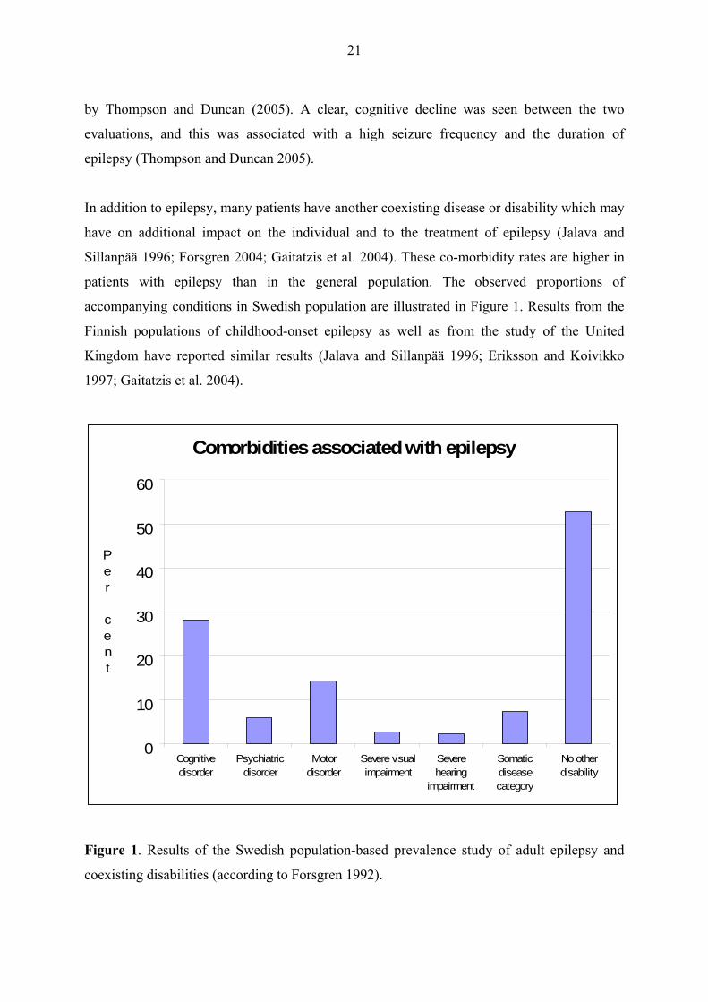
In addition to epilepsy, many patients have another coexisting disease or disability which may
have on additional impact on the individual and to the treatment of epilepsy (Jalava and
Sillanpää 1996; Forsgren 2004; Gaitatzis et al. 2004). These co-morbidity rates are higher in
patients with epilepsy than in the general population. The observed proportions of
accompanying conditions in Swedish population are illustrated in Figure 1. Results from the
Finnish populations of childhood-onset epilepsy as well as from the study of the United
Kingdom have reported similar results (Jalava and Sillanpää 1996; Eriksson and Koivikko
1997; Gaitatzis et al. 2004).
Comorbidities associated with epilepsy
0
10
20
30
40
50
60
Cognitivedisorder
Psychiatricdisorder
Motordisorder
Severe visualimpairment
Severehearing
impairment
Somaticdiseasecategory
No otherdisability
Per cent
Figure 1. Results of the Swedish population-based prevalence study of adult epilepsy and
coexisting disabilities (according to Forsgren 1992).
22
2.5. Antiepileptic drug treatment
The need for effective AED treatment is evident due to the detrimental effects of the epileptic
seizures and their impact on the individual patient. The recent review of epilepsy-related
injuries and mortality by Tomson et al. (2004d), concluded that effective treatment of
epilepsy appears to decrease the injuries and mortality related to epilepsy, which further
emphasizes the importance of effective epilepsy treatment. Also patients with epilepsy report
seizure-freedom as one of the most important factors affecting their quality of life (Birbeck et
al. 2002).
The first AED (potassium bromide) was introduced in 1857 and since that time the treatment
of epilepsy as well as knowledge of the effects of AEDs have advanced significantly.
Nowadays over 20 different AEDs are available. Two of the most commonly used AEDs are
carbamazepine (CBZ) for partial epilepsies and valproate (VPA) for generalized epilepsies,
which are described in detail in Table 1 below. The aim of the AED treatment is to achieve
optimal seizure control without eliciting any adverse effects to the patient (Duncan et al.
2006). According to the current findings, seizure remission can be achieved in approximately
two thirds of all patients. However, up to 30% of all epilepsy patients develop intractable
epilepsy (Hauser and Hesdorffer 2001). Despite optimal treatment, these patients continue to
experience seizures or other symptoms of epileptic syndrome, restricting their ability to lead a
full life (Hauser and Hesdorffer 2001). Due to the different underlying aetiologies for epilepsy
and individual variations of the patients, an individualised approach to the treatment of
epilepsy is recommended (Duncan et al. 2006). Women with epilepsy represent one unique
group of epilepsy patients whose treatment is challenging since so many aspects need to be
considered.
23
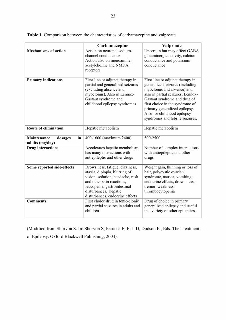
Table 1. Comparison between the characteristics of carbamazepine and valproate Carbamazepine Valproate Mechanisms of action Action on neuronal sodium-
channel conductance Action also on monoamine, acetylcholine and NMDA receptors
Uncertain but may affect GABA glutaminergic activity, calcium conductance and potassium conductance
Primary indications First-line or adjunct therapy in partial and generalized seizures (excluding absence and myoclonus). Also in Lennox-Gastaut syndrome and childhood epilepsy syndromes
First-line or adjunct therapy in generalized seizures (including myoclonus and absence) and also in partial seizures, Lennox-Gastaut syndrome and drug of first choice in the syndrome of primary generalized epilepsy. Also for childhood epilepsy syndromes and febrile seizures.
Route of elimination Hepatic metabolism Hepatic metabolism
Maintenance dosages in adults (mg/day)
400-1600 (maximum 2400) 500-2500
Drug interactions Accelerates hepatic metabolism, has many interactions with antiepileptic and other drugs
Number of complex interactions with antiepileptic and other drugs
Some reported side-effects Drowsiness, fatigue, dizziness, ataxia, diplopia, blurring of vision, sedation, headache, rash and other skin reactions, leucopenia, gastrointestinal disturbances, hepatic disturbances, endocrine effects
Weight gain, thinning or loss of hair, polycystic ovarian syndrome, nausea, vomiting, endocrine effects, drowsiness, tremor, weakness, thrombocytopenia
Comments First choice drug in tonic-clonic and partial seizures in adults and children
Drug of choice in primary generalized epilepsy and useful in a variety of other epilepsies
(Modified from Shorvon S. In: Shorvon S, Perucca E, Fish D, Dodson E , Eds. The Treatment
of Epilepsy. Oxford:Blackwell Publishing, 2004).
24
2.6. Epilepsy and female reproductive health
It has been estimated that 0.5 % of the childbearing population suffer from epilepsy and
therefore it is one of the major neurological concerns also to be taken into consideration in
fertile-aged women (Richmond et al. 2004). Many studies suggest that the fertility in women
with epilepsy is decreased compared to the general population (Dansky et al. 1980; Webber et
al. 1986; Artama et al. 2004). In the United States, in women with epilepsy the live birth rate
was 0.85 of that expected during a 40-year period of time (Webber et al. 1986). The birth rate
was lowest in the 1940’s and has increased decade by decade so that by the 1970’s no
reduction was noted (Webber et al. 1986). In the United Kingdom, the fertility rate was found
by Wallace et al. (1998) to be 33% lower in women with epilepsy compared to controls.
However, in a population-based study from Iceland, no reduction was noted in the fertility of
women with epilepsy compared with controls (Olafsson et al. 1998b). In that study, 30% of
women with epilepsy did not have children compared to 25% in the general population and
only patients with remote symptomatic epilepsy and a severe co-morbidity (e.g. cerebral palsy
or mental retardation) were childless more often than controls (odds ratio 22) (Olafsson et al.
1998b). In the study of Dansky et al. (1980), 58% of women with epilepsy had at least one
pregnancy and 85% of married women had children, but the marriage rate was reduced in
women with epilepsy compared to unaffected women. Schupf et al. (1996) reported that
fertility was reduced in patients with epilepsy after the onset of epilepsy but not before its
appearance. Jalava et al. (1997) reported that women with epilepsy may be disadvantaged in
terms of marrying but when they are married, their fertility or pregnancies do not differ from
controls according to a population-based follow-up study of patients with childhood-onset
epilepsy.
A higher frequency of menstrual disturbances (e.g. anovulatory cycles, irregular menstrual
cycles) have been reported in women with epilepsy than in unaffected women (Cummings et
al. 1995; Svalheim et al. 2003; Herzog 2006) and this has been thought to be one of the most
important factors explaining their reduced fertility. These changes have been associated with
epilepsy itself and to the use of AEDs. In the retrospective cohort study of Svalheim et al.
(2003), menstrual disturbances were associated with frequent seizures but not with the
25
epilepsy type. The association of epilepsy type and menstrual disturbances was studied by
Cummings et al. (1995). They reported that 35.5 % of women with temporal-lobe epilepsy
(TLE) had anovulatory cycles but this disturbance was not found in any of the women with
primary generalized epilepsy (PGE). Irregular menstrual cycles and anovulation may also
occur in association with polycystic ovarian syndrome (PCOS). In some studies, PCOS has, in
addition to AEDs, been associated with epilepsy itself (Herzog 2006).
Of the individual AEDs, especially VPA has been associated with a high frequency of
menstrual disturbances (Isojärvi et al. 1993 and 1996; Svalheim et al. 2003). Several
mechanisms have been proposed e.g. changes in sex hormone levels and a higher prevalence
of polycystic ovaries (PCO) and hyperandrogenism in women using VPA for epilepsy.
Isojärvi et al. (1996) reported that PCO and/or hyperandrogenism were found in 64% of
women using VPA compared to 19% in controls. These women had also a higher level of
serum testosterone, dehydroepiandrosterone (DHEAS) and insulin levels and lower serum
levels of sex-hormone binding globuline (SHBG). Accordingly, Rättyä et al. (2001) showed
that after one month of VPA medication, the levels of testosterone, luteinizing hormone (LH)
and follicle-stimulating hormone (FSH) increased. However, data in this area can be
conflicting (Morrow and Craig 2003). Bauer et al. (2000) found no difference in the
prevalence of PCOS in women using VPA or CBZ for epilepsy compared with untreated
epileptic women.
Obesity is also more common in women using VPA (59 %) and obesity-associated PCOS has
been thought to have an important role as a source of the menstrual disturbances in women
using VPA (Isojärvi et al. 1993 and 1996). In the study of Isojärvi et al. (1993), the effects of
VPA were more common in women who had received the AED medication before the age of
20.
Epilepsy, epileptic seizures and AEDs may have an effect on the hormones involved in
endocrine regulation (Morrel 1998; Herzog 2006). These include an increase in prolactin
levels after tonic-clonic seizures and direct effects on the hypothalamic-pituitary axis e.g.
altered secretion of gonadotrophins. Conversely, these hormones may have an effect on the
seizures – generally it has been considered that estrogens are proconvulsants whereas

26
progesterone has anticonvulsant properties (Tettenborn et al. 2002). This has been associated
with a specific type of epilepsy, catamenial epilepsy, in which the seizure frequency seems to
vary according to the cyclical hormonal changes during the menstrual cycle (Tettenborn et al.
2002).
CBZ and other enzyme-inducing AEDs (e.g. phenytoin (PHT) and oxcarbazepine (OXC))
which induce hepatic microsomal enzymes of the P-450 system, can evoke interactions with
endogenous and exogenous hormones (Zupanc 2006). These AEDs increase steroid
metabolism and affect protein binding and therefore by accelerating the metabolism of ethinyl
estradiol, reduce the effectiveness of hormonal contraception. CBZ has also been reported to
increase the level of SHBG in women with epilepsy which decreases the free progestin levels
in the plasma (Isojärvi et al. 1996; Rättyä et al. 2001). Therefore the risk of contraceptive
failure and breakthrough bleeding in women with epilepsy using AEDs is more common than
in the general population and this needs to be taken into consideration when treating women
with epilepsy (Tomson 2004a; Zupanc 2006).
The effects of epilepsy and AEDs on sexual functioning, have been studied in recent years. In
these studies, women with epilepsy have reported sexual dysfunction more often than controls
(Morrell et al. 2005). This is especially prevalent in women with localization related epilepsy
(Morrell et al. 2005). In the study by Herzog et al. (2003), sexual dysfunction was more often
present in women with TLE and especially right-sided TLE whereas no difference was found
between women treated with and without AED treatment indicating an independent role of
epilepsy in this respect. In terms of the specific AEDs, enzyme-inducers including PHT had a
negative impact on sexual functioning (Morrel et al. 2005).
2.7. Epilepsy and pregnancy
2.7.1. Maternal outcome
Though the majority of the women with epilepsy enjoy uncomplicated pregnancies (Tomson
and Battino 2005), women with epilepsy have been reported to have a greater risk of
pregnancy complications than control women (Yerby et al. 1985; Sabers et al. 1998; Pilo et al.
27
2006). Some of the most frequently reported complications include toxaemia, bleeding in
pregnancy, placental abruption; both induced and prolonged labour and an increase in the rate
of caesarean sections. However, conflicting data concerning the adverse effects of epilepsy
and AEDs on pregnancy outcome have been published, and in many of the later studies very
few differences have been noted between women with epilepsy and controls with respect to
pregnancy complications (Hiilesmaa et al. 1985; Yerby 1991; Morrow et al. 2003). In a
Finnish study of pregnant women with epilepsy in an outpatient clinic material, pregnancy
complications occurred as frequently in women with epilepsy as in controls (Hiilesmaa et al.
1985). In a recent study the rate of complications in pregnancy was increased only with
respect to hypertension (not associated with pre-eclampsia) and induced delivery (Richmond
et al. 2004).
Pregnancy may also have an effect on epilepsy since it may cause a change in seizure
frequency, a decline in serum AED levels and changes in AED pharmacokinetics. In a recent
report by the EURAP Study Group (2006) 58% of the women were seizure-free during
pregnancy and the seizure frequency was unchanged in 63 % of the women. An increase in
the number of seizures was observed in 17% of the women and the occurrence of seizures was
independently associated with AED polytherapy and localization-related epilepsy. A similar
increase in seizure frequency has been observed independently in a Swedish (Tomson et al.
1994) and an Italian study (Tanganelli and Regesta 1992), in which the increase in the seizure
frequency was also associated with focal epilepsy and women with a higher frequency of
seizures also in the pre-pregnancy period. In a review prepared by Tomson (1997a) it was
noted that approximately 5% of women with epilepsy experience seizures during labour,
delivery or immediately thereafter. Status epilepticus (SE) is of special concern at any time,
but especially during pregnancy. However, SE does not occur more frequently during
pregnancy and it is observed in less than one percent of pregnancies (Tomson 1997a).
Pregnancy alters many of the mechanisms associated with the pharmacokinetics of the AEDs.
The serum levels of AEDs tend to decrease, most likely due to decreased protein binding,
changes in the blood volume or changes in drug metabolism (Tomson 2004a). In some
patients this decline of AED levels may affect the seizure frequency during pregnancy. In the
study of Yerby et al. (1992), the total concentrations of several AEDs (CBZ, PHT, PB, VPA)
28
declined significantly during pregnancy, but the free concentrations of the drugs remained
unchanged or even increased (VPA). Accordingly, Tomson et al. (1994) reported that though
total plasma levels of CBZ decline during pregnancy, the free plasma levels stay the same or
even increase due to decreased protein binding. The same study showed no association
between plasma AED levels and seizure control during pregnancy (Tomson et al. 1994). A
pronounced increase in the clearance of lamotrigine (LTG) during pregnancy has been
demonstrated (de Haan et al. 2004). The fall in LTG plasma levels during pregnancy is
considerably greater than that reported for other AEDs, and could result in an increase in
seizure frequency thus necessitating dose adjustment. The active monohydroxy derivative of
oxcarbazepine (OXC), which is mainly responsible for the drug’s pharmacological effect,
shares with LTG a primary route of elimination via glucuronidation and it seems to also
undergo the same kind of pharmacokinetic alterations during pregnancy that are observed for
LTG (Mazzucchelli et al. 2006).
In some women, the increased seizure frequency may be due to lack of compliance during
pregnancy, usually due to fear of the teratogenic effects of AEDs (Tanganelli and Regesta
1992; Tomson 2004a). Although the absolute risk is low, maternal death has been estimated
to be ten times higher for women with epilepsy than those in the general population (Adab et
al. 2004b). Case histories suggest that these fatalities are a result of the seizures that are often
associated with abrupt withdrawal of AEDs or with poor compliance. Also environmental
factors e.g. sleep deprivation and anxiety during pregnancy, have been suggested as reasons
for the increased seizure frequency. Adequate pre-pregnancy counseling is essential to
increase the likelihood of maintained seizure control: a lack of such counseling has been
identified as a major risk factor for an increase in seizure frequency during pregnancy (Lopes-
Cendes et al. 1992).
2.7.2. Fetal outcome
Some of the reported fetal complications associated with maternal epilepsy are spontaneous
abortion, intrauterine growth retardation, asphyxia, low Apgar scores and increased perinatal
mortality, but data are often conflicting and complication rates vary between different studies
(Yerby et al. 1985; Sabers 1997). In the prospective, multicenter study of Battino et al. (1999)
29
the overall proportion of children with low birth weight was not increased. However, the
Danish prospective, cohort study of Hvas et al. (2000) reported that children exposed to AEDs
were small for gestational age (SGA) and they had reduced body length and head
circumference compared with unexposed children. A low birth weight was also reported by
Yerby et al. (1985) in a large, population-based retrospective study. In the Swedish
prospective study of Wide et al. (2000) a slight decrease in body weight was observed in
infants exposed to polytherapy, in particular the head circumference was reduced in infants
exposed to CBZ. Battino et al. (1999) reported that a small head circumference was associated
with polytherapy and the use of PB and primidone. This smaller head circumference in a
combination therapy of PB and PTH has also been reported in a retrospective analysis of
Dessens et al. (2000). In the Finnish population- based prospective study published by Gaily
et al. (1990a), a reduction in the head circumference was shown in children exposed to CBZ
and a combination therapy of barbiturates with the difference persisting for up to five years.
However, the paternal head circumference was also below the average in these two groups
and the difference in the children disappeared after adjusting for paternal head circumference
(Gaily et al. 1990a). Thus genetics may contribute, at least partially, to the head
circumference of the AED exposed children. Pilo et al. (2006) reported that children exposed
to AEDs suffer a higher frequency of respiratory distress syndrome.
Maternal seizures during pregnancy may have an adverse effect on the fetus. A decrease in
fetal heart rate has been observed after maternal tonic-clonic seizures, and this has been
considered to be due to changes in the blood circulation, transient lactic acidosis and asphyxia
(Hiilesmaa et al. 1985). In addition, trauma caused by tonic-clonic seizure may harm the
fetus. However, in the recent prospective study by Kaaja et al. (2003), maternal seizures
during the first trimester were not associated with congenital malformations.
It has been thought previously that infants exposed to enzyme-inducing AEDs in utero are at a
risk of having complications due to neonatal bleeding. This may be due to a deficiency of
vitamin K1 in the fetus induced by the AEDs. Therefore many guidelines have recommended
vitamin K1 supplementation (American Academy of Neurology 1998; Morrow and Craig
2003). However, in the first epidemiological study assessing the occurrence of bleeding
complications in newborns, no difference was noted between controls and infants exposed to
30
AEDs (Kaaja et al. 2002). In a logistic regression analysis, the bleeding was associated with
premature birth (<32 weeks) and alcohol abuse, but not to enzyme-inducing AEDs. Similarly,
in another study from the United States, no haemorrhagic disease was observed in newborns
exposed to AEDs (Choulika et al. 2004).
Perinatal mortality has been observed to be two to three times higher in infants of mothers
with epilepsy compared to controls (Sabers 1997). In the study of Hiilesmaa et al. (1985), the
rate of perinatal deaths was 5/150 (three stillbirths and two deaths during the first week of
life). However, in the report of Annegers et al. (1988) no association was found between
maternal epilepsy and the use of AEDs with any recognized foetal loss.
2.7.2.1 Major malformations
The increased risk in the rate of major congenital malformations (MCM) in children of
mothers with epilepsy is well established (Samrén et al. 1997; Wide et al. 2004; Wyszynski et
al. 2005). It has been related to the use of AEDs, especially to AED polytherapy during
pregnancy (Kaneko et al. 1999; Richmond et al. 2004). No increase in the rate of
malformations has been observed in children of mothers with epilepsy but without AED
medication (Holmes et al. 2001). Also the type of maternal epilepsy has not been associated
with different rates of malformations (Kaneko et al. 1988). In some AED regimens, a dose-
dependent mechanism of teratogenesis has also been observed (Samrén et al. 1997).
In the prospective, multicenter study by Kaneko et al. (1999) MCMs were observed in 9.0 %
of the AED exposed children compared to 3.1% of those without drug exposure. Pooled risks
for MCMs in children exposed to AEDs vary from two to fourfold in different studies (Dravet
et al. 1992; Olafsson et al. 1998; Canger et al. 1999; Artama et al. 2005). With respect to the
individual drugs, PB, primidone and VPA, have been reported to have the highest association
with malformations according to Kaneko et al. (1999). In a recent prospective Finnish study,
the rate of MCMs was 3.8% compared to 0.8% in control population and the risk was
independently associated with the use of CBZ, VPA, OXC, low serum folate concentration
and a low maternal level of education (Kaaja et al. 2003).
31
Of the currently used AEDs, VPA has most often been associated with a higher frequency of
malformations than other AEDs (Samrén et al. 1997; Canger et al. 1999; Wide et al. 2004).
This risk of MCMs was four-fold compared to other AEDs according to the North American
pregnancy registry and over 7- fold to women without AEDs (Wyszynski et al. 2005). The
risk of MCMs was increased in women receiving VPA at doses of >1000mg/day compared
with women receiving VPA <600mg/day indicating a dose-dependent teratogenic effect
(Samrén et al. 1997). This teratogenic effect at high VPA doses (>1100 mg/day) has also been
reported in the preliminary data of the Australian Pregnancy Registry (Vajda et al. 2006).
However, data from the UK Register suggest that even low doses of VPA may be no worse in
this respect than >200 mg/day doses of lamotrigine (Morrow et al. 2006) and suggest a dose-
dependent effect of CBZ, LTG and VPA. A dose-dependent effect has been suggested also for
PB in a retrospective analysis of birth register information (Samrén et al. 1999).
In a meta-analysis of the teratogenicity of CBZ, the malformation rate was 6.8 % compared to
2.34 % in controls in patients with CBZ monotherapy and this was increased up to 18.8% in
polytherapy pregnancies (Matalon et al. 2002). This especially harmful effect of polytherapy
has been reported in many studies (Kaneko et al. 1988; Holmes et al. 2001). In the
prospective study of Kaneko et al. (1988), the frequency of MCMs was highest in the
combination therapy of VPA and CBZ and/or PHT.
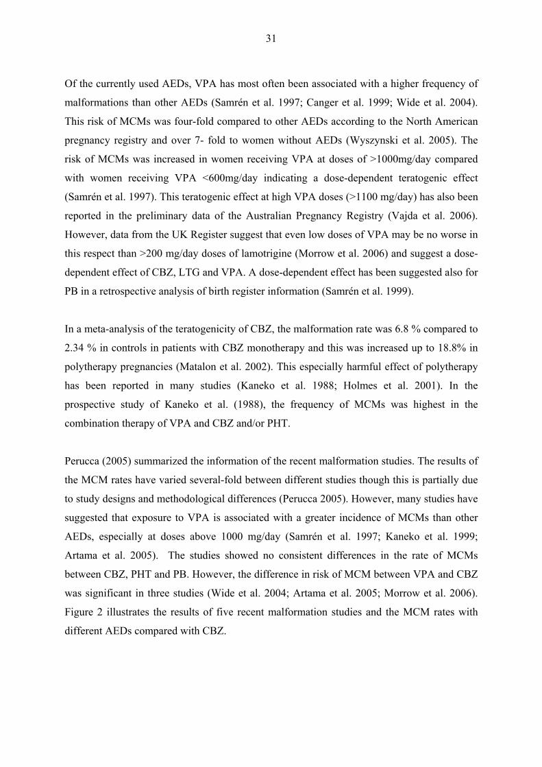
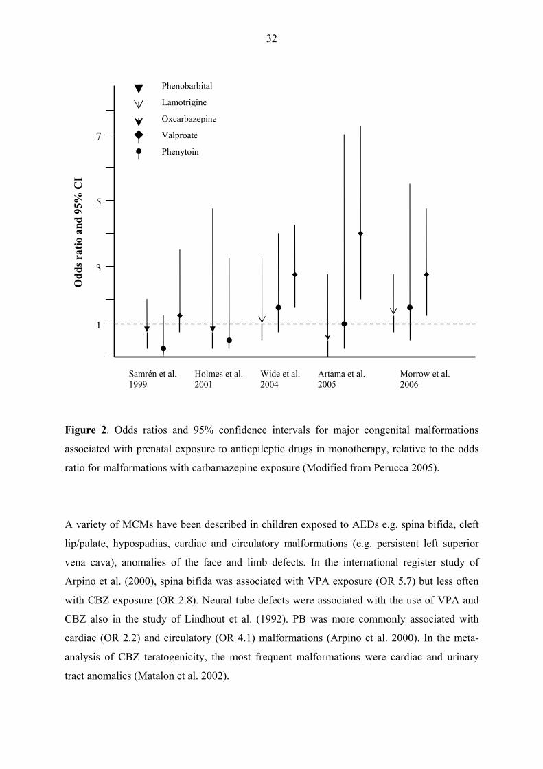
Perucca (2005) summarized the information of the recent malformation studies. The results of
the MCM rates have varied several-fold between different studies though this is partially due
to study designs and methodological differences (Perucca 2005). However, many studies have
suggested that exposure to VPA is associated with a greater incidence of MCMs than other
AEDs, especially at doses above 1000 mg/day (Samrén et al. 1997; Kaneko et al. 1999;
Artama et al. 2005). The studies showed no consistent differences in the rate of MCMs
between CBZ, PHT and PB. However, the difference in risk of MCM between VPA and CBZ
was significant in three studies (Wide et al. 2004; Artama et al. 2005; Morrow et al. 2006).
Figure 2 illustrates the results of five recent malformation studies and the MCM rates with
different AEDs compared with CBZ.
32
Figure 2. Odds ratios and 95% confidence intervals for major congenital malformations
associated with prenatal exposure to antiepileptic drugs in monotherapy, relative to the odds
ratio for malformations with carbamazepine exposure (Modified from Perucca 2005).
A variety of MCMs have been described in children exposed to AEDs e.g. spina bifida, cleft
lip/palate, hypospadias, cardiac and circulatory malformations (e.g. persistent left superior
vena cava), anomalies of the face and limb defects. In the international register study of
Arpino et al. (2000), spina bifida was associated with VPA exposure (OR 5.7) but less often
with CBZ exposure (OR 2.8). Neural tube defects were associated with the use of VPA and
CBZ also in the study of Lindhout et al. (1992). PB was more commonly associated with
cardiac (OR 2.2) and circulatory (OR 4.1) malformations (Arpino et al. 2000). In the meta-
analysis of CBZ teratogenicity, the most frequent malformations were cardiac and urinary
tract anomalies (Matalon et al. 2002).
Samrén et al. 1999
Holmes et al. 2001
Wide et al. 2004
Artama et al. 2005
Morrow et al.2006
Phenobarbital
Lamotrigine
Oxcarbazepine
Valproate
Phenytoin
1
3
5
7
Odd
s rat
io a
nd 9
5% C
I

33
As emphasized previously, neural tube defects e.g. spina bifida have been observed more
commonly in women using AEDs (especially VPA and CBZ) compared to reference
populations. A reduction in the rate of neural tube defects was noted after administration of
folic acid supplementation preconceptually in the general population (Wald and Sneddon
1991). Also in normal pregnancies, without AEDs, the serum folate levels decline and in
pregnant women with epilepsy the decline is even steeper (Dansky et al. 1992). Therefore, it
has been recommended that women with epilepsy should receive adequate folic acid
supplementation preconceptually until the end of the first trimester, though no clear
association has been shown to support the belief that this strategy decreases the number of
neural tube defects in infants of mothers with epilepsy (Morrow and Craig 2003; Tomson
2004b). It has also been suggested that high-dose folic acid substitution may even be seizure-
provoking or may have an association with an increased risk of miscarriages but this
information is still needing to be confirmed (Tomson 2004b). In a Swedish study reported by
George et al. (2002), an association of low plasma folate levels and an increased risk of early
spontaneous abortion was established but no association was found with high levels of folate.
When one wishes to assess the risk of MCMs in the children of epileptic mothers, AEDs
(dosages and combinations) are not the only contributing factors. Genetic factors may also
play a role in malformations. Malm et al. (2002) published a case report in which three sets of
siblings born to mothers with epilepsy and exposed to VPA in utero were examined. All of
the children in these three families exhibited fetal valproate syndrome, which strongly
suggests that there is a hereditary susceptibility to valproate. Also it is likely that
pharmacogenetics has an impact on the teratogenicity of AEDs. Finnell et al. (1992) studied
phenytoin-induced teratogenesis. During the metabolism of PHT, oxidative metabolites are
produced e.g. epoxides. Those children exposed to PHT and who developed a fetal hydantoin
syndrome had lower epoxide hydrolase levels than children with PHT exposure but without
malformations, emphasizing that individual genetic factors may be involved in the
teratogenicity e.g. determining the activity of specific enzymes.
34
2.7.2.2. Minor malformations and fetal anticonvulsant syndrome
In addition to major malformations, minor malformations/anomalies (usually defined as
unusual morphological features with no serious medical or cosmetic consequences to the
patient) which become recognizable later in life have been found to be more common in AED
exposed children (Yerby et al. 1992; Wide et al. 2000). In a prospective study by Koch et al.
(1992) infants exposed to AEDs had a higher number of anomalies than children of mothers
with epilepsy but without AED exposure (5.03 vs. 1.91). In children exposed to VPA, this
value was even higher (8.0). However, children of fathers with epilepsy did not show more
anomalies than control children (Koch et al. 1992). Also in a Swedish population-based
prospective study, the number of minor anomalies was increased in children exposed to AEDs
in utero, but the children had rarely more than one anomaly and no specific pattern of
anomalies could be identified (Wide et al. 2000).
In a large retrospective analysis of Adab et al. (2004a), dysmorphic features were observed in
44 % of children exposed to VPA compared to 9% in children exposed to CBZ. Also in the
retrospective study of Kini et al. (2006), children exposed to VPA exhibited a higher
proportion of dysmorphic features compared with children exposed to CBZ and PHT.
However, also 45% of the children with no AED exposure were found to have dysmorphic
features indicating that single minor anomalies are common also in the general population
(Kini et al. 2006). Dysmorphic features in AED exposed children have been associated with
the cognitive outcome (Adab et al. 2004a; Holmes et al. 2005). In the studies of Adab et al.
(2004a) and Kini et al. (2006), children with dysmorphic features and VPA exposure had
lower VIQ scores than children without dysmorphic features.
In a population-based prospective study by Gaily et al. (1988a), the minor anomalies in AED
exposed children were studied. The anomalies were subdivided into typical anomalies
(epicanthus, hypertelorism, typical nose, long philtrum, abnormal ears, low hairline, nail
hypoplasia, distal phalangeal hypoplasia, and three or more dermal arches) and other
anomalies; no excess of the other anomalies were observed in these children (Gaily et al.
1988a). Some of the anomalies may also be genetically determined as they reported that also
the mothers had more minor anomalies than control women. Of the many reported anomalies,
35
only the association between distal phalangeal hypoplasia and phenytoin has been well
established (Gaily et al. 1990c).
Fetal anticonvulsant syndrome (FACS) term is often used when referring to children who
have suffered adverse teratogenic effects due to AED exposure. FACS refers to a group of
disorders in which malformations and developmental disorders occur in association with a
characteristic facial appearance. Characteristic dysmorphic features as well as malformations
have been described in children exposed to PHT, VPA and CBZ. However, the extent of
developmental problems in these children is less known.
The term of fetal hydantoin syndrome was first introduced by Hanson and Smith (1975). It
consisted of typical minor anomalies (e.g. epicanthus, hypertelorism and long philtrum) and
included also mental deficiency, growth retardation and microcephaly. Also specific facial
features have been associated with VPA exposure and a fetal valproate syndrome was
described in 1984 by DiLiberti et al. In the retrospective cohort study of Kini et al. (2006), a
large variety of dysmorphic features was recognized both in AED exposed children as well as
in control children indicating that some of the typical FACS features are common also in the
general population. However, features noted more often in valproate exposed children were
medial deficiency of eyebrows, infraorbital grooves, broad nasal bridge, anteverted nose,
abnormal philtrum and a thin upper lip (Kini et al. 2006). In CBZ exposed children, full
cheeks with a small chin and an everted lower lip were more common (Kini et al. 2006).
Moore et al. (2000) studied 57 children with FACS who were identified through the FACS
Association and found that 33% of the exposed children had glue ear and up to 70% had a
type of joint laxity. He also reported a similar facial dysmorphology in the exposed children
as that described by Kini et al. (2006) though significant overlapping does seem to occur
between different exposure groups and facial features. The study also reported a high
frequency of autistic type behaviours and hyperactivity in children with FACS (Moore et al.
2000). Kini et al. (2006) reported also a correlation between valproate exposure, dysmorphic
features and low verbal IQ in children with FACS. However, prospective population based
studies have not revealed similar results. In the study of Gaily et al. (1988b), the majority of
the AED exposed children were exposed to PHT, and no children with typical fetal hydantoin
syndrome features could be identified.
36
Abnormal ophthalmologic findings have been reported to be more common in children with
FACS (Glover et al. 2002). Of the 46 children with FACS 67% had ocular abnormalities,
most commonly errors of refraction (41%). In particular myopia (50%) was common in VPA
exposed children.
2.7.3. Cognitive and behavioural outcome
After the studies were published describing the immediate teratogenic effects of AEDs, the
question of the long term effects of AED exposure in utero, was raised. The first systematic
studies concerning the long term cognitive effects of AED exposure were conducted in the
1970´s and 1980´s. The prospective study by Gaily et al. (1988b) concluded that the children
of mothers with epilepsy, most of whom had been exposed to AEDs in utero, had a lower
mean intelligence quotient (IQ) than control children. Mental deficiency was observed in
1.4% of the children and borderline intelligence in 1.7% of the children (Gaily et al. 1988b).
Overall, however, the prevalence of mental deficiency was the same or slightly increased in
children of epileptic mothers compared with unexposed children (Gaily et al. 1988b). Koch al
al. (1999) reported the results of a longitudinal prospective study. They also found that the
intelligence scores were lower in AED exposed children. This deficit was associated mainly
with polytherapy with primidone but the socio-economic status of the family also had an
effect on the cognitive outcome of the children (Koch et al. 1999).
The effects of specific AED regimens have also been studied. In the prospective, population-
based study of Gaily et al. (1988b) the majority of the children were exposed to PHT (103
children), which was also the case in the cohort study of Scolnik et al. (1994) (34 children).
Gaily et al. (1988) found no association between low IQ and phenytoin exposure. Also the
occurrence of brief maternal convulsions during pregnancy did not affect the cognitive
outcome. In the smaller, prospective cohort study of Scolnik et al. (1994), children exposed to
PHT had a lower global IQ as well as lower language developmental scores than unexposed
controls. In the prospective, population-based study from Sweden, preschool-aged children
with PHT exposure received lower scores in their locomotor development than control
37
children (Wide et al. 2004) although the number of children in PHT exposure group was
rather small (N=15).
In addition to PHT, the effects of CBZ exposure on the cognitive development of the children
have been studied. Gaily et al. (2004) reported the results of a large population-based
prospective study of the cognitive development of CBZ exposed children at preschool- and
school-age. Children with CBZ exposure did not differ from controls and they were reported
to have normal intelligence. This normal intelligence in CBZ exposed children has also been
reported in other studies (Gaily et al. 1988b; Scolnik et al. 1994). Wide et al. (2000) studied
the AED exposed children at the age of 9 months and at that time point no difference was
noted in the development of the AED exposed children (of which the majority had been
exposed to CBZ) compared with unexposed controls. At preschool age the neurodevelopment
of the CBZ exposed children was also reported to be normal (Wide et al. 2004).
During recent years, interest has mainly been focussed on the effects of prenatal VPA
exposure on the cognitive development of the exposed children. In particular exposure to
VPA was associated especially with a lower VIQ compared with other monotherapy
exposures in the prospective study of Gaily et al. (2004) and also in the retrospective study of
Adab et al. (2004a). A negative correlation of the VPA dose and VIQ was observed, but
neither the type of maternal epilepsy nor the occurrence of generalized seizures during
pregnancy, were associated with this deficiency (Gaily et al. 2004). Also in children exposed
to VPA, even though the individual variation in performance has been great, a large
proportion of these children have been reported to exhibit an especially low IQ (Adab et al.
2004a).
An increased risk of poorer cognitive performance has been observed in children exposed to
polytherapy compared to monotherapy exposure (Koch et al. 1999; Gaily et al. 2004; Adab et
al. 2004a). In the study of Koch et al. (1999), both the PIQ and the VIQ were lower in
children exposed to polytherapy but in addition to AEDs, the socio-economic status of the
family was also predictive of the cognitive outcome.
38
In addition to the effect of AEDs, many confounding factors affect the cognitive outcome of
the children. Predictive factors for the low VIQ in children exposed to VPA were maternal IQ
and frequent tonic-clonic seizures (Adab et al. 2004a). According to Gaily et al. (1990b)
several factors were associated with the cognitive dysfunction observed in children exposed to
AEDs, i.e. maternal seizures during pregnancy, partial seizures and paternal level of
education. In the study of Holmes et al. (2005), a lower IQ was observed in children with
AED exposure and microcephaly compared to children with AED exposure but normal head
circumference. The IQ deficit was also more commonly seen in children with midface
hypoplasia. The socio-economic status of the family has also been reported to affect the
cognitive outcome of the children (Koch et al. 1999).
The cognitive and behavioural problems observed in children exposed to AEDs have been
thought to lead to problems at school. Though the majority of the children with AED
exposure attend mainstream school, a need for educational support and special arrangements
regarding schooling have been reported (Adab et al. 2001). This reported risk has been
especially prevalent in children with VPA exposure (Adab et al. 2001 and 2004a). Autism
and related disorders as well as behavioural problems are also more common in children with
AED exposure (VPA, CBZ and polytherapy) than in unexposed children (Dean et al. 2002).
In the population-based, retrospective study from UK, disorders of the autistic spectrum or
Asperger syndrome were diagnosed in 4.6 % (12/260) of the AED exposed children (Rasalam
et al. 2005). These disorders were most common among VPA exposed children 8.9% (5/56).
Dessens et al. (2000) found that adults who had been exposed to PB and PHT in utero
exhibited more learning problems and mental retardation than controls. However, in this area,
no true prospective, population-based studies have been conducted. The studies of Adab et al.
(2001 and 2004a), Dean (2002) and Rasalam (2005) had a retrospective design and in the
study of Dessens et al. (2000) the recruitment of subjects was retrospective with a subsequent
prospective follow-up. These methodological shortcomings may have biased the reliability of
the results.

39
2.8. Pregnancy management, recommendations and birth registers
Many guidelines have been given concerning the management of fertile-aged women with
epilepsy. These recommendations have attempted to highlight the special concerns and
provide answers to practitioners (ACOB 1997; Morrell 1998; Crawford 1999). The need of
adequate pre-conception counselling for women with epilepsy is evident and emphasized also
in the guidelines (Morrell 1998; Tomson 2004b). However, a prospective population based
study analyzing the care of women with epilepsy reported that only 38% of the women had
received pre-conceptional counselling and less than 50% of the women had planned their
pregnancies (Fairgrieve et al. 2000). Therefore much work still needs to be done in order to
optimize the treatment of women with epilepsy.
In order to enable rational decisions, women with epilepsy and their partners need to be
counselled about conception, pregnancy, breast-feeding and caring for the infant. The
avoidance of unplanned pregnancy requires the use of effective contraception. During
counselling before conception, it is important to confirm the diagnosis of active epilepsy and
reconsider the need for AED treatment (Kälviäinen and Tomson 2006). A woman who wishes
to become pregnant and who has been seizure-free for two or more years may attempt to
discontinue AEDs under supervision by her physician. The decision to withdraw medication
gradually should follow the generally accepted principles for the treatment of adults with
epilepsy. It will depend mainly on the level of risk that the individual patient is willing to
accept. The risks include worsening of seizures, perhaps seizures that result in physical harm
to the mother (and the fetus once the woman becomes pregnant). Although the risks are low,
the possibility of status epilepticus and SUDEP should be discussed realistically before
attempting drug withdrawal (Morrow and Craig 2003). If treatment is continued, the woman
should use the most effective AED for her epilepsy type at the lowest possible dose to control
seizures following the general principles for the treatment of epilepsy. The classification of
the seizure and epilepsy syndrome should be re-evaluated and the appropriate medication
adjusted accordingly (Kälviäinen and Tomson 2006).

40
The re-evaluation should be performed carefully for all patients, including those with
difficult-to-treat epilepsies, to assess the possibility of improved seizure control with fewer
AEDs. It is clear that some patients will still need polytherapy, but this should be limited to as
few AEDs as possible. Epilepsy surgery may be an option in selected cases, e.g. in temporal
lobe epilepsy, and this may eventually result in seizure control without drugs before the
patient becomes pregnant (Kälviäinen and Tomson 2006). The treatment of new-onset
epilepsy during pregnancy should follow the same principles as used in the non-pregnant
woman.
Low folate concentrations within maternal erythrocytes have been correlated with neural tube
defects in infants (anencephaly, spina bifida, and encephalocele) (Fishman 2000). Therefore,
it has been recommended that women with epilepsy should have a daily intake of 0.4mg of
folate from the time they commence trying to become pregnant until the twelfth gestational
week (Tomson et al. 1997b). If the woman has previously given birth to a child with a neural
tube defect, the recommended daily folate supplement is 4 mg for the above-mentioned period
(Tomson et al. 1997b). Many guidelines recommend higher doses of folate (4-5mg/day) also
for women being treated with valproate or carbamazepine (AAN guidelines 1998). However,
in this population, the documentation for any benefit of high-dose folate supplementation is
lacking and unknown adverse events of high-dose folate supplementation cannot be excluded.
In Finland, therefore, 1 mg supplementation has been used, which is also convenient for
patients, as they only have to take one tablet per day and because the duration of
supplementation may last for as long as several years in some patients.
Ideally, drug levels should be obtained before pregnancy for comparison with the
concentrations to be monitored during gestation. For highly protein bound AEDs, such as
VPA and PHT, unbound levels are preferred. Drug level monitoring is most important for
AEDs that may undergo major alterations in their active concentrations, e.g. PHT and in
particular LTG, and for AEDs with unknown effects of pregnancy. Whether a decline in drug
concentrations per se should prompt dosage adjustments is still controversial, but this kind of
strategy appears to be justified, at least for LTG (Kälviäinen and Tomson 2006). Otherwise
dose adjustments are made according to the level of seizure control.
41
All women with epilepsy who become pregnant should be referred to a specialist maternity
unit and at least structural ultrasound should be performed. Most women will have a normal
uncomplicated vaginal delivery, however, tonic-clonic seizures may result in fetal hypoxia.
Therefore it is generally recommended that delivery takes place in a unit equipped with
facilities for both maternal and neonatal resuscitation (Morrow and Craig 2003).
AED levels quickly revert to pre-pregnancy levels after the delivery. Hence, if the dose of an
AED has been increased during pregnancy because of falling AED-levels or seizures during
pregnancy, it may be useful to measure plasma levels at 2-4 weeks after the delivery and
make any necessary dosage adjustments. All women with epilepsy should be encouraged to
breastfeed their babies (Crawford 1999; Morrow and Craig 2003). The AED concentration
profiled in breast milk follows the plasma concentration curve. The total amount of drug
transferred to infants via breast milk is usually much smaller than the amount transferred via
the placenta during pregnancy.
In Kuopio University Hospital, maternity and epilepsy clinics are jointly following up all
women with epilepsy during their pregnancy with a pre-decided protocol (Table 2).
42
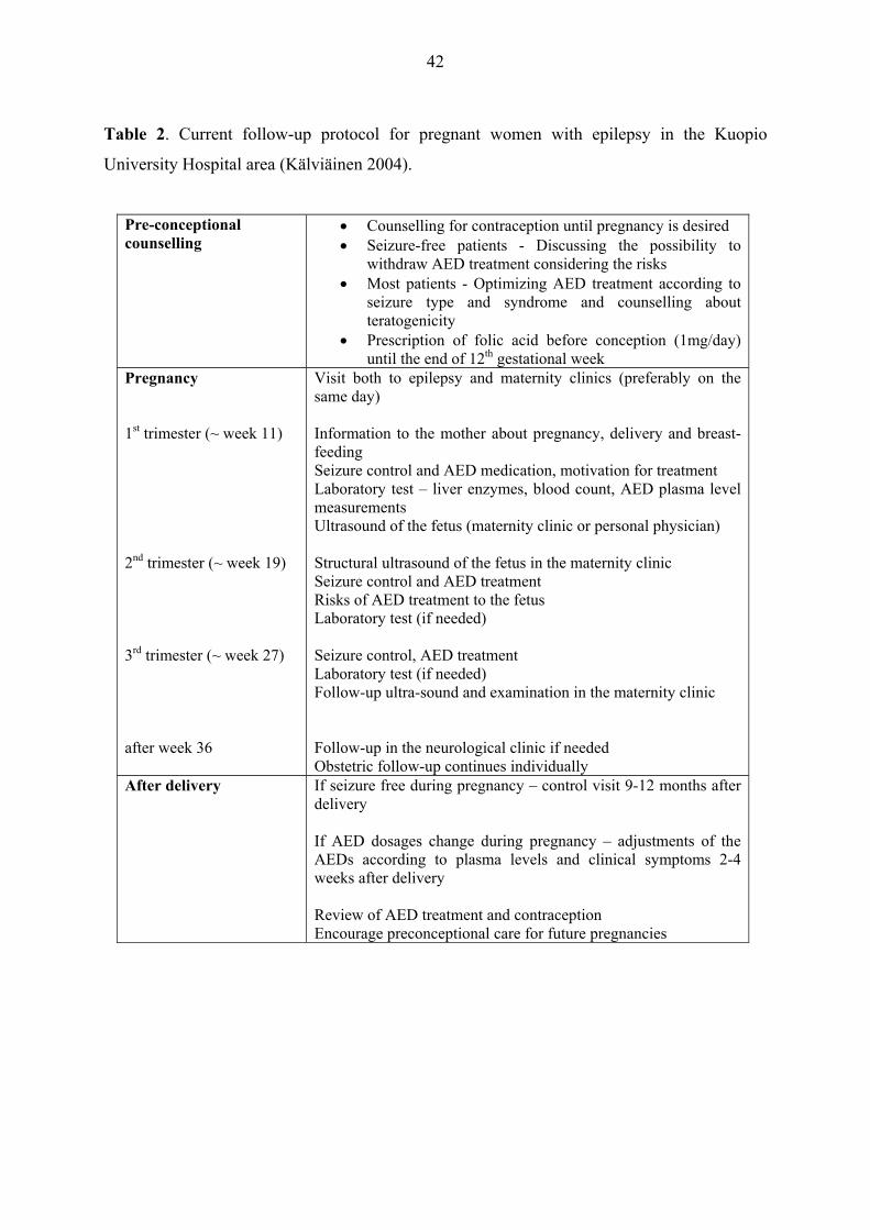
Table 2. Current follow-up protocol for pregnant women with epilepsy in the Kuopio
University Hospital area (Kälviäinen 2004).
Pre-conceptional counselling
• Counselling for contraception until pregnancy is desired • Seizure-free patients - Discussing the possibility to
withdraw AED treatment considering the risks • Most patients - Optimizing AED treatment according to
seizure type and syndrome and counselling about teratogenicity
• Prescription of folic acid before conception (1mg/day) until the end of 12th gestational week
Pregnancy 1st trimester (~ week 11) 2nd trimester (~ week 19) 3rd trimester (~ week 27) after week 36
Visit both to epilepsy and maternity clinics (preferably on the same day) Information to the mother about pregnancy, delivery and breast-feeding Seizure control and AED medication, motivation for treatment Laboratory test – liver enzymes, blood count, AED plasma level measurements Ultrasound of the fetus (maternity clinic or personal physician) Structural ultrasound of the fetus in the maternity clinic Seizure control and AED treatment Risks of AED treatment to the fetus Laboratory test (if needed) Seizure control, AED treatment Laboratory test (if needed) Follow-up ultra-sound and examination in the maternity clinic Follow-up in the neurological clinic if needed Obstetric follow-up continues individually
After delivery If seizure free during pregnancy – control visit 9-12 months after delivery If AED dosages change during pregnancy – adjustments of the AEDs according to plasma levels and clinical symptoms 2-4 weeks after delivery Review of AED treatment and contraception Encourage preconceptional care for future pregnancies
43
Optimal AED treatment is essential not only during pregnancy in view of the recognized
adverse effects of AEDs but also the threat of seizures, if left untreated. The need for
information on the effects of the older and newer AEDs is essential. To achieve this purpose
many international birth registries have been established to gather details of the effects of
AEDs (Beghi et al. 2001). Current registries include for example EURAP, the UK Epilepsy
and Pregnancy Register, Australian Pregnancy Registry and North American Antiepileptic
Drug Pregnancy Registry (Holmes et al. 2004; Russel et al. 2004; Tomson et al. 2004c; Vajda
et al. 2004). In the majority of these registries, information has been gathered prospectively,
which hopefully will provide a source of unbiased information on the adverse effects of AED
treatment and will help to improve the future treatment of women with epilepsy.

44
3. AIMS OF THE STUDY
3.1. To determine the proportion of WWAE having children and to investigate factors related
to fertility in women using VPA and CBZ
3.2. To compare the course of pregnancy, delivery and pregnancy outcome in WWAE and in
the general pregnant population
3.3. To evaluate the possible effect of vigabatrin (VGB) treatment in utero to the visual
function in the offspring
3.4. To investigate the cognitive performance of school-aged children exposed to VPA in
utero compared to children exposed to CBZ as well as to children without AED exposure
3.5. To assess the neurological development, behavioural aspects and additional educational
needs in children exposed to VPA in utero compared to children exposed to CBZ as well as to
children without AED exposure.
45
4. SUBJECTS AND METHODS
4.1. Subjects
4.1.1. Study I
All pregnancies in the Kuopio University Hospital (KUH) area (population 250 000) since
1989 have been prospectively registered in the Birth Register of the Department of Obstetrics
and Gynaecology. The term pregnancy is used when the gestational age is over 22 weeks or
the birth weight over 500 gr. From this register, we retrieved all women who had a diagnosis
of epilepsy in the data of the pregnancy registry and had been residents in the KUH area. Our
study period was from January 1989 to October 2000 and during this period of time a total of
85 women with active epilepsy (WWAE, women who had epilepsy and had used AED
medication during conception and throughout the pregnancy) had a singleton pregnancy (127
pregnancies) and 38 women who had a history of epilepsy but needed no AED medication
during pregnancy (52 pregnancies). At the same time, 16505 women without epilepsy (24778
pregnancies) had given birth. In the study group of 85 WWAE, 20 subjects had used VPA
monotherapy during their pregnancy and 49 had received CBZ monotherapy. When multiple
pregnancies were included, the numbers were 87 WWAE and 18146 control women.
During same time period (1989-2000), 130 fertile-aged (18-40 years) women who had used
VPA monotherapy and 316 women who had used CBZ monotherapy and were residents of
the KUH area had visited the outpatient clinic of the Department of Neurology. To assess the
fertility aspect more thoroughly, we reviewed the medical records of these 130 women with
VPA monotherapy and selected 130 women with closest birth date and CBZ monotherapy as
controls.

46
4.1.2. Study II
In study II we examined the pregnancy outcome of the 85 WWAE compared to the outcome
of the 38 women with a history of epilepsy but without AEDs and also compared these to the
general population. The WWAE had given birth to 127 children and the other 38 women to
52 children. Multiple pregnancies and major chromosomal anomalies were excluded from the
pregnancy outcome analysis. As a control group we used all the pregnant women who had
been registered to the Birth Register but who did not have epilepsy and had a singleton
pregnancy (16505 women and 24778 pregnancies). See Figure 3 for study populations.
Figure 3. Study populations
Birth Register: (1989-10/2000) 24957 pregnancies
Singleton pregnancies of WWAE N = 127
Pregnancies in women with a history of epilepsy N = 52
Unaffected control pregnancies N = 24778
Children with VPA exposure N = 28
Children with CBZ exposure N = 72
School-aged and first-born children with informed consent N = 13
Controls with same age and gender N = 13
Controls with same age and gender N = 13 Studies
IV-V
Study II
47
4.1.3. Study III
From the Birth Register we were able to identify three women who had used VGB during
pregnancy and four children who had been exposed to the drug. Of the four children exposed,
we were able to examine two at school age to search for possible structural malformations and
visual dysfunction.
4.1.4. Studies IV-V
All women who had epilepsy and had used VPA monotherapy during pregnancy were
identified from the Birth Register. To investigate the effect of VPA exposure on the children,
we examined the children at school age (≥ 6 years old). Out of the 28 VPA exposed children,
16 were school-aged and first born during the study year (2003). Thirteen children
participated in this controlled study, and also their mothers were examined. The controls were
selected from the Birth Register children with the closest birth date and the same gender as
the VPA exposed children. Thirteen control children were exposed to CBZ and 13 children
had no AED exposure, but their mothers had a history of epilepsy.
4.2. Methods
4.2.1. Kuopio University Hospital Birth Register and register data used for fertility
assessment (Studies I-II)
The Kuopio University Hospital Birth Register contains comprehensive information regarding
the background of the mother, risk factors, course of the pregnancy and pregnancy outcome.
This information has been gathered to the register by questionnaires, which are sent to every
mother during their pregnancy. These questionnaires are returned to the maternity clinic of
KUH before the delivery and supplemented with information from the follow-up of the
pregnancy, delivery and pregnancy outcome at the delivery unit of KUH. From this register,
we obtained demographic data of the mothers and the information about the management and
course of the pregnancy as well as the data of the outcome of the pregnancy. Also the medical
records of the WWAE were reviewed and Birth Register information was supplemented with
48
the neurological data retrieved from medical records (e.g. the type of epilepsy, onset of
epilepsy, frequency of seizures, AED medication). The WWAE had been followed up
according to the pre-decided protocol (Table 2) jointly by the epilepsy and maternity clinics in
KUH.
To assess the overall fertility among women with epilepsy, we reviewed the medical records
of our Neurology outpatient clinic between years 1989 and 2000. We reviewed the medical
records of the 130 women with VPA monotherapy and of 130 women with CBZ monotherapy
with the closest birth date. From the medical records, we obtained information concerning the
number of children, hormonal disturbances (if any noted) and co-morbidities. We also looked
for any available reasons for not having children. No structured interview for the women was
used.
In addition to information obtained from the Birth Register and medical records of the women
studied, we retrieved epidemiological information from the Statistics Finland and from
STAKES (National Research and Development Centre for Welfare and Health). According to
Statistics Finland, 23% of Finnish women did not have children in the year 2000. To confirm
the number of women having active epilepsy and using AED medication in KUH area, we
assessed information obtained from the Social Insurance Institution of Finland (KELA).
Overall, 286 women (16-39 years) in KUH area had active epilepsy in year 2000. The registry
of STAKES provided information of fertile-aged women (16-39 years) (N = 36261) living in
the same hospital area at the same time (in the year 2000).
Premature delivery was defined as delivery before gestational week 37, and prolonged
pregnancy as lasting longer than week 42. Small-for-gestational-age newborns were below the
tenth percentile according to the normal tables of our population when adjusted for the
gestational age and sex. Infants with birth weight less than 2500g were considered to have
low birth weight. Fetal distress term was used when fetal venous pH was <7.15 and/or Apgar
scores at 5 minutes were <7 after birth. Major malformations were defined as structural
abnormalities with surgical, medical or cosmetic importance identified by a pediatrician at
birth and at discharge from hospital.

49
4.2.2. Assessment of the AED exposed children
4.2.2.1 Study III
The medical records of the mother and children exposed to VGB were reviewed with respect
to medication, seizures and neonatal information. A paediatric neurologist performed a
thorough examination of the children and an ophthalmologist examined both the mothers and
the children for any visual dysfunction. Visual acuity as well as visual fields were examined.
A standard ophthalmologic evaluation was performed to exclude other eye diseases. The
ophthalmologic examination consisted of a history of visual symptoms, testing the best
corrected visual acuity, phorias and tropias, biomicroscopy, and indirect ophthalmoscopy with
the aid of tropicamide 0.8% + phenylephrine 5% drops. Intraocular tension was measured
with the Goldmann applanation tonometer. Visual fields were examined with the kinetic
Goldmann perimeter.
4.2.2.2 Studies IV-V
The 13 children exposed to VPA in utero and their age- and gender matched controls were
examined and interviewed in the facilities of Clinical Research Center of Kuopio University.
They were first examined by a pediatric neurologist and then underwent a detailed evaluation
of neuropsychological functioning supervised by a psychologist lasting for 2 hours. Both the
pediatric neurologist and psychologist were blinded to the medical history and the drug
exposure of the children and mothers. The medical records of the children and their mothers
were reviewed and all relevant information concerning e.g. pregnancy and delivery, the type
of epilepsy and the use of AEDs and/or other medications, seizures before and during the
pregnancy, the pre-, peri- and postnatal details of growth and possible problems in
neurological development of the child were all recorded. The medical information was
combined with the information from the Birth Register and with the data from the clinical and
neuropsychological examinations.
50
4.2.2.2.1 Neurological evaluation
The neurological functioning of the children was assessed by using Touwen’s test (Touwen
1979). This standardized examination was used to detect any possible minor neurological
dysfunctions of the child and to exclude other neurological diseases that could influence the
results of the cognitive and neuropsychological evaluation. The results of the examination in
Touwen’s test are represented in six subsystems: sensorimotor function, posture, balance,
physical coordination, fine-finger dexterity and dyskinesia. Mild neurological dysfunction is
determined to be present if there are deficits in one or two of the categories and severe
(neurological impairment) if deficits are seen in over two categories. Minor dysmorphic
features were evaluated during the neurological evaluation from a checklist for dysmorphic
features and the results were based on blinded, but individual, non-controlled clinical
evaluation by a paediatric neurologist. Dysmorphic features were defined as unusual
morphological features with no medical importance of cosmetic consequence to the patient.
We individually sought facial anomalies (epicanthal folds, hypertelorism, flat nasal bridge,
long philtrum, midface hypoplasia, cleft lip or palate and other noted abnormalities of the
face) and abnormalities of the hand (finger and nail hypoplasia). Other observed anomalies
e.g. of the skin and joints, were also recorded if noted.
4.2.2.2.2 Neuropsychological evaluation
The general cognitive function of all mothers was evaluated with the Wechsler Adult
Intelligence Scale (WAIS, Wechsler 1955) or WAIS- Revised (Wechsler 1981). Full scale
intelligence Quotient (FIQ), verbal IQ (VIQ) and performance IQ (PIQ) were estimated on the
basis of six subtests: Information, Similarities, Digit Span, Digit Symbol, Picture Completion
and Block Design.
The children underwent individual neuropsychological assessments using the Wechsler
Intelligence Scale for Children III (WISC-III; Wechsler 1991) and the NEPSY, a
Developmental Neuropsychological Assessment (Korkman, Kirk and Kemp, 1998). Scores
for FIQ, for VIQ and for PIQ were estimated in the same way as WAIS. Low intelligence was
determined to be present if the FIQ score was less than 80 and exceptionally low intelligence
51
if the score was <70. Eight subtests of the NEPSY evaluating five cognitive domains were
administered: the Tower from Attention and Executive functions; Comprehension of
Instructions and Verbal Fluency from the Language domain; Visuomotor Precision from the
Sensorimotor domain, and Arrows from the Visuospatial domain. In addition, from the
Memory and Learning subtests, Memory for Faces, Narrative Memory and List Learning
were administered.
4.2.2.2.3 Behavioural assessment
The Conners’ Teacher Rating Scale – questionnaire (CTRS) was sent to every family and
after it was completed by teachers it was sent back for a further analysis of the behavior of the
child. The rating scale consists of (56) individual questions scored from zero (definitely not)
to three (definitely true). These individual questions were further categorized into 13 different
domains according to the Conners’ manual. These categories are Oppositional, Cognitive
Problems/Inattention, Hyperactivity, Anxious-Shy, Perfectionism, Social Problems, Conners’
ADHD Index, Conners’ Global Index: Restless-Impulsive, Conners’ Global Index: Total,
DSM-IV: Inattentive, DSM-IV: Hyperactive-Impulsive and DSM-IV: Total. The raw scores
of the rating scale were then transformed into standard points, which permitted a comparison
between children of different ages and sexes.
4.2.3. Statistical analysis
The SPSS statistical package (Ver. 9.0-13.0) was used for statistical analysis. In all of the
studies, t-test was used for normally distributed continuous variables and chi-square for
dichotomous variables. Fischer’s exact test was applied when the expected count was less
than five. Confidence intervals of 95% were used and P-values <0.05 were considered as
being statistically significant. Multiple logistic regression analysis, BMDP, was used to
evaluate the effect of maternal risk factors on the pregnancy outcome in Study II. The
pregnancy outcome variables were adjusted for all significant or nearly significant maternal
risk factors (p<0.1). In study IV for the comparison of the cognitive level of mother-child
pairs, the IQs were classified into categories of cognitive ability levels according to standard

52
deviation units above and below the mean. In studies IV-V, Kruskall-Wallis test was used for
continuous variables.
4.2.4. Ethics
This study was approved by the ethics committee of the Kuopio University Hospital and
Kuopio University. Informed consent was obtained in study III-V from the mothers and those
children who could understand the meaning of consent.
53
5. RESULTS
5.1. Epilepsy and female fertility (Study I)
The average number of children in WWAE was 2.1 ± 1.3 and in healthy pregnant population
2.2 ± 1.2 (p=0.6, NS). In KUH area, 4.7 per 1000 pregnancies (0.47%) involved a mother
with active epilepsy. At the same time, 0.79 percent of the fertile-aged population in KUH
area had active epilepsy and according to these numbers (0.47/0.79) 60% of women with
active epilepsy had given birth to a child. In Finland, 77 % of women have children according
to Statistics Finland (2005).
The menstruation cycle was regular in 85 % of controls and in 83 % of WWAE. No
difference was noted in the time needed to become pregnant between WWAE and controls
(4.7 months vs. 5.4 months, median 2 months in both groups). Overall 7.6 % of the control
women had used some form of fertility treatment, in WWAE, the equivalent figure was 4.7%
(NS) and none of the women in valproate group had used treatment for infertility.
Of the 118 women with VPA monotherapy attending the neurology outpatient clinic, 61
women (52 %) had given birth to a child with the respective figures for those with CBZ
medication being 66 and 58 %. Figure 4 shows the stated reasons for not having children for
WWAE with the most frequently used monotherapies (data for CBZ and VPA patients
combined). No significant differences were noted between women using VPA and CBZ in the
studied variables.
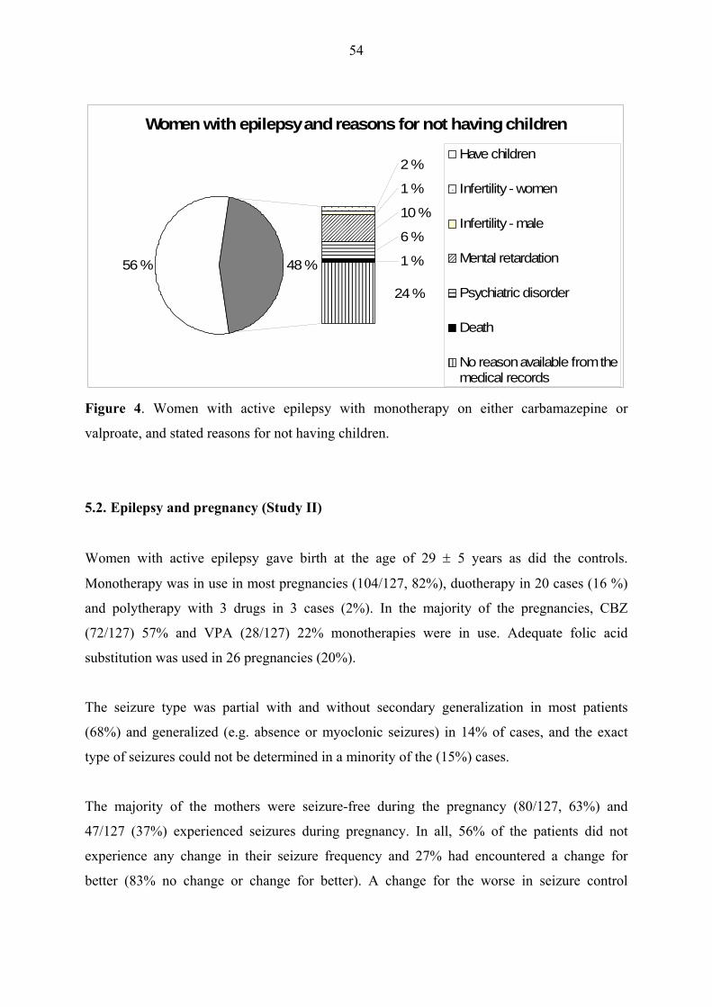
54
Women with epilepsy and reasons for not having children
56 %
2 %
1 %
10 %
6 %
1 %
24 %
48 %
Have children
Infertility - women
Infertility - male
Mental retardation
Psychiatric disorder
Death
No reason available from themedical records
Figure 4. Women with active epilepsy with monotherapy on either carbamazepine or
valproate, and stated reasons for not having children.
5.2. Epilepsy and pregnancy (Study II)
Women with active epilepsy gave birth at the age of 29 ± 5 years as did the controls.
Monotherapy was in use in most pregnancies (104/127, 82%), duotherapy in 20 cases (16 %)
and polytherapy with 3 drugs in 3 cases (2%). In the majority of the pregnancies, CBZ
(72/127) 57% and VPA (28/127) 22% monotherapies were in use. Adequate folic acid
substitution was used in 26 pregnancies (20%).
The seizure type was partial with and without secondary generalization in most patients
(68%) and generalized (e.g. absence or myoclonic seizures) in 14% of cases, and the exact
type of seizures could not be determined in a minority of the (15%) cases.
The majority of the mothers were seizure-free during the pregnancy (80/127, 63%) and
47/127 (37%) experienced seizures during pregnancy. In all, 56% of the patients did not
experience any change in their seizure frequency and 27% had encountered a change for
better (83% no change or change for better). A change for the worse in seizure control
55
occurred in 18% of the pregnancies but no specific reason, which could explain this
exacerbation of seizures, was found. One woman had a status epilepticus (SE) during both her
pregnancies and another woman experienced a flurry of seizures and a cesarean section was
performed during the premonitory phase of SE.
The study populations were similar with regard to the maternal risk factors and pregnancy
complications. There were no statistically significant difference in the duration of the
pregnancy in WWAE 280 ± 14 days, in women with non-active epilepsy 280 ± 13 days and in
the control group 278 ± 16 days.
The odds ratios for selected pregnancy outcome variables between study groups are presented
in Figure 5. A significant number of the children of WWAE were found to be small for
gestational age (SGA), but no differences were found in the number of newborns with low
birth weight. No difference was noted in the AED medication or seizure frequency during
pregnancy in the mothers of SGA children. However, the mothers with SGA children seemed
to be shorter than mothers without SGA children (161.4 ± 6.4 cm vs. 164.9 ± 5.7 cm, p<0.05).
Apgar scores at 1 minute in children of WWAE were low, but no longer at the age of 5
minutes. Accordingly, the admissions for neonatal intensive unit or to neonatal ward were
significantly (p=0.01) more prevalent in children of WWAE (N=17, OR 1.98, 95% CI 1.17-
3.34).
In the VPA group, 11/20 (55%) deliveries were induced compared to 16.7 % of the control
women, p<0.0001. The CBZ group did not differ from controls in this respect. Identified
reasons for the induction were postmaturity (3 pregnancies), pre-eclampsia (2), abnormal fetal
cardiotocography (2), absent or uncoordinated contractions (2) and social reasons (2).
The frequency of major malformations was 4.8% (95% CI -0.6-10.2) in the children of
WWAE, while no major malformations were found in the children of women with a history of
epilepsy but who were not using AEDs during pregnancy. The malformations are described in
detail in study II. All children with the malformations had been exposed to polytherapy or
high-dose monotherapy with VPA or CBZ.

56
Pregnancy outcome in WWAE
0
1
2
3
4
5
Pretermdelivery
Admission toa neonatal
unit
Apgar score< 7at 1minute
Apgar score< 7at 5minutes
Low birthweight
Small forgestational
age
AbnormalFHR
Fetaldistress
Odds ratio
High CILow CIOR
Figure 5. Pregnancy outcome variables of women with active epilepsy (adjusted for maternal
age >35 years, pregravid body mass index >25kg/m2 and unemployment of the mother).
(FHR = fetal heart rate, CI = confidence interval, OR = odds ratio)
5.3. Children and mothers exposed to vigabatrin (Study III)
There were two mothers who had used polytherapy (CBZ and CBZ + VPA) with VGB during
pregnancy. The total dose of VGB in the first mother 190g was during the pregnancy and
370g in the second mother. In the two exposed children, no ophthalmologic structural
abnormalities were detected. Visual field testing was unreliable and therefore visual
dysfunction could not be assessed.
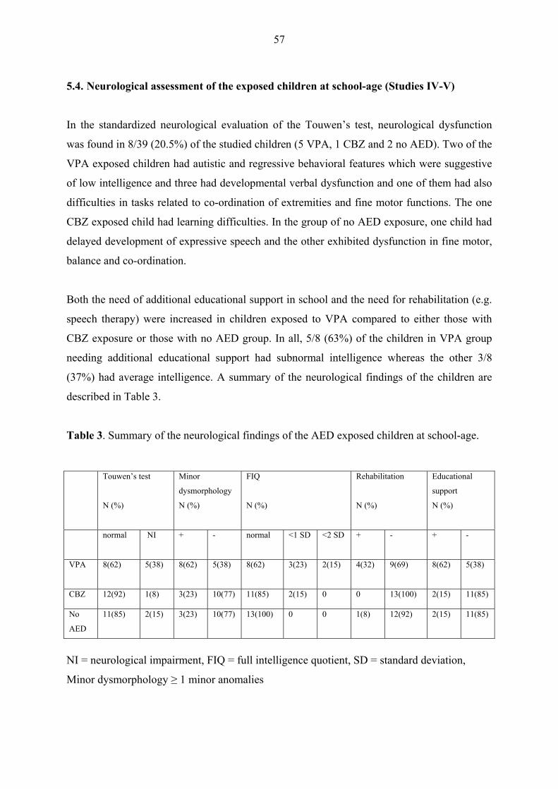
57
5.4. Neurological assessment of the exposed children at school-age (Studies IV-V)
In the standardized neurological evaluation of the Touwen’s test, neurological dysfunction
was found in 8/39 (20.5%) of the studied children (5 VPA, 1 CBZ and 2 no AED). Two of the
VPA exposed children had autistic and regressive behavioral features which were suggestive
of low intelligence and three had developmental verbal dysfunction and one of them had also
difficulties in tasks related to co-ordination of extremities and fine motor functions. The one
CBZ exposed child had learning difficulties. In the group of no AED exposure, one child had
delayed development of expressive speech and the other exhibited dysfunction in fine motor,
balance and co-ordination.
Both the need of additional educational support in school and the need for rehabilitation (e.g.
speech therapy) were increased in children exposed to VPA compared to either those with
CBZ exposure or those with no AED group. In all, 5/8 (63%) of the children in VPA group
needing additional educational support had subnormal intelligence whereas the other 3/8
(37%) had average intelligence. A summary of the neurological findings of the children are
described in Table 3.
Table 3. Summary of the neurological findings of the AED exposed children at school-age.
NI = neurological impairment, FIQ = full intelligence quotient, SD = standard deviation,
Minor dysmorphology ≥ 1 minor anomalies
Touwen’s test
N (%)
Minor
dysmorphology
N (%)
FIQ
N (%)
Rehabilitation
N (%)
Educational
support
N (%)
normal NI + - normal <1 SD
<2 SD + - + -
VPA 8(62)
5(38)
8(62) 5(38) 8(62) 3(23) 2(15) 4(32) 9(69) 8(62) 5(38)
CBZ 12(92) 1(8) 3(23) 10(77) 11(85) 2(15) 0 0 13(100) 2(15) 11(85)
No
AED
11(85) 2(15) 3(23) 10(77) 13(100) 0 0 1(8) 12(92) 2(15) 11(85)
58
5.5. Neuropsychological assessment of the exposed children (Studies IV-V)
In the neuropsychological evaluation of the children, all children with no AED exposure and
almost all of the children exposed to CBZ could perform adequately in the subtest of NEPSY
and scored within one S.D. from the mean. Two to four children of the VPA group could not
perform all of the NEPSY subtests. Those children who could perform adequately scored
statistically significantly lower in the subtest of Memory for Faces (p= 0.016 compared to
CBZ) and in the List Learning subtest (p= 0.008 compared to no AED exposure group). The
scores of the neuropsychological tests are reported in study IV.
Table 4 shows the results of the WISC scores in children and WAIS scores of the mothers.
Mothers with VPA had a significantly lower IQ and educational level than women using CBZ
and women without AED exposure. Also children exposed to VPA had a trend towards lower
IQ scores than the other children, though this difference did not reach statistical significance.
Also in the different WISC subtests, the VPA exposed children scored lower than the other
children. Two of the children with VPA exposure had exceptionally low intelligence
(FIQ<80) while their mothers had average intelligence. Children with CBZ exposure did not
differ from controls and had normal intelligence. Also the mothers using CBZ had IQ scores
similar to controls. The values of the WISC subtest scores are reported in detail in study IV.

59
Table 4. Intelligence quotients of the mothers and children not exposed to antiepileptic drugs
(No AED), exposed to carbamazepine (CBZ monotherapy) and exposed to valproate (VPA
monotherapy) and level of education (years) in mothers
AED groups No AED
(N=13)
CBZ
monotherapy
(N=13)
VPA
monotherapy
(N=13)
All
( N = 39)
p-values
WAIS IQs, education Mean maternal FIQ (95% CI)
Mean maternal VIQ (95% CI)
Mean maternal PIQ (95% CI)
Maternal education years
(S.D.)
AED exposure groups
WISC-III IQs
Mean FIQ children (95% CI)
Mean VIQ children (95% CI)
Mean PIQ children (95% CI)
108.8 (105.0-112.5)
107.5 (101.3-113.8)
109.5 (105.1-113.9)
14.2 (2.3)
No AED (N=13)
99.6 (95.0-104.3)
98.2 (91.2 -105.2)
102.1 (94.1-110.0)
109.2 (102.0-116.4)
105.1 (96.8-113.4)
112.5 (105.1-119.9)
14.8 (2.2)
CBZ (N = 13)
98.9 (91.2-106.5)
96.5 (86.9-106.0)
102.5 (93.7-11.4)
95.6* (89.5-101.8)
92.9* (85.8-100.1)
99.5* (92.4-106.7)
12.5* (1.9)
VPA (N = 13)
84.5 (69.4-99.7)
85.1 (70.2-99.9)
84.7 (68.4-101.0)
104.5 (100.8-108.2)
101.1 (97.5-106.2)
107.2 (103.3-111.0)
13.8 (2.3)
All ( N = 39)
94.3 (88.6-100.1)
93.7 (87.2-99.3)
96.4 (89.8-103.1)
0.003**
0.012*
0.022*
0.035*
p-values
0.157 NS
0.269 NS
0.071 NS
5.6. Behavioural assessment (Study V)
Conners’ Teacher Rating Scale was used to detect possible behavioural problems in these
school-aged children. Children exposed to VPA received generally higher scores in all of the
analysed domains but due to small number of patients, these differences did not reach
statistical significance. However, in two of the categories (social problems, p=0.07 and
cognitive problems/inattention, p=0.09) the VPA-exposed children showed a tendency
towards more behavioural problems. Children with CBZ and no AED exposure had very
similar scores and no statistical differences were noted between these two groups.
60
5.7. Dysmorphic features and the overall neurocognitive development
Dysmorphic features were seen in 8 children (62%) exposed to VPA and in three children
(23%) in both the CBZ and the no exposure group (p = 0.085). Four of the thirteen VPA
exposed children (31%) had 2 or more of these features. All the children in CBZ group had
only one minor dysmorphic feature and one child in the no exposure group had two features.
If the AED exposed children were summed together (VPA + CBZ) 11/26 (42 %) had
dysmorphic features compared to 23 % (3/13) in children with no AED exposure. The most
frequently observed features were flat nasal bridge, long philtrum and thin upper lip,
epicanthal folds and short fingers.
The overall performance of the AED exposed children was evaluated to obtain a
comprehensive view of their neurodevelopment. The results of the Touwen’s test, FIQ,
dysmorphic features, additional educational support, need for therapies and Conners’ Global
Index scores were grouped together. If all of the results fell in the normal range, the children
were categorized as having normal neurodevelopment, minor dysfunction if performance in
one or two of the categories was abnormal and as neurologically impaired if problems were
seen in more than two categories. All of the children exposed to VPA had minor or major
neurological/cognitive problems. The majority of the children exposed to CBZ and without
AED exposure had normal performance or only minor problems.
61
6. DISCUSSION
This study consists of five individual scientific papers which focus on (1) the fertility of
WWAE in the area of Kuopio University Hospital and the effects of co-morbidities and AEDs
on fertility, (2) the management and outcome of the pregnancies of WWAE, (3) the effect of
VGB exposure on visual function, (4) the long-term cognitive and (5) behavioral development
of the AED exposed children. Information of WWAE was obtained from the Birth Register of
KUH and from the records of the Neurology Outpatient Clinic covering the hospital district
providing a population-based, comprehensive data of the pregnancies. This prospective,
population-based study of the AED exposed children has provided valuable information for
the monitoring of the cognitive effects of AEDs.
6.1. Fertility
In our population, 60% of women with epilepsy had children compared to 77 % of unaffected
women. We believe that the most important factor explaining this 22% difference is the
higher number of co-morbidities (mental retardation and psychiatric disorders) observed in
women with epilepsy; they accounted for 15 to 18 percent in our study in monotherapy
population with well-controlled epilepsy and up to 29 % in the study of Forsgren (1992). In
our study, approximately 20% of women did not have any recognized or reported reason for
not having children. This number was identified from the medical records and therefore may
include a bias in that the women may not have told the physician the real reason for not
having children. However, we believe that this bias is not significant because these women
were mainly seen in our hospital at regular intervals and any infertility problems would be
treated mainly at KUH. Also the proportion of women not having children is fairly close to
that found in the general population (23% of women not having children) when the influence
of co-morbidities is excluded.
The majority of the studies concerning fertility in women with epilepsy have reported a
decreased fertility (Webber et al. 1986; Wallace et al. 1998). In these studies however, the
higher prevalence of severe co-morbidities in women with epilepsy compared to controls was
not taken into consideration. Severe co-morbidities e.g. mental retardation will independently

62
affect the patient’s social environment and her possibility to have children. In the population-
based study by Olafsson et al. (1998b), the fertility rate was the same in women with epilepsy
and controls and the fertility was reduced only in women with epilepsy together with a severe
co-morbidity e.g. cerebral palsy or mental retardation. These co-morbidity rates have been
studied by Forsgren (1992) showing that in both women and men with epilepsy, the
percentage of coexisting diseases/disabilities is as high as 47%, with contribution of mental
retardation being 23% and severe psychiatric condition being 6%.
Our findings are in concordance with the results of Olafsson et al. (1998b) and the research of
Artama et al. (2004) where the fertility rate in women with epilepsy was 0.88 of the reference
population. In addition, we found no difference in the time needed to become pregnant or in
the need for fertility treatments between WWAE and controls. No significant changes were
observed in the regularity of menstrual cycles, in the marriage rate or in the mean number of
infants. Also, no significant differences were noted between women using VPA those using
CBZ in terms of fertility or menstrual disturbances. Tettenborn et al. (2002) also concluded
that there are misconceptions in relation to fertility in women with epilepsy and that these
women do not have markedly reduced fertility compared to controls.
6.2. Pregnancy outcome
In our carefully monitored population with its pre-decided follow-up protocol, no significant
differences were found between WWAE and controls in the frequently reported pregnancy
complications. This result was consistent with the prospective results of Hiilesmaa et al.
(1985) and with the recent obstetric study of Richmond et al. (2004). However, the rate of
small for gestational age (SGA) infants was significantly higher in WWAE than in the
reference group, as was also observed in the prospective study by Hvas et al. (2000). This
increase in the number of small-for-date infants could not be explained by preterm delivery or
low birth weight because their number was not increased. Furthermore, neither the epilepsy
type nor the AED medication differed between mothers with SGA children and those giving
birth to normal weight infants. Also no increase was noted in the number of SGA children in
mothers with epilepsy but without AED medication. The mothers with SGA children,
however, were shorter which partially could have contributed to the outcome.
63
Apgar scores at 1 minute after birth were lower in infants of WWAE but no longer reduced at
the age of 5 minutes and accordingly the need of neonatal care after birth was increased when
compared to controls. Over half (55%) of the deliveries of women using VPA had to be
induced. This need for induced delivery has been observed before in women with epilepsy but
not associated so clearly with women using VPA (Yerby et al. 1985). There were many
different reasons for induction; no dominating reason could be identified. This finding needs
to be confirmed in further studies.
The overall rate of major malformations is 4.8% in our study. The malformations were
identified by a pediatrician at birth and at discharge from hospital. No reference group
examined with a similar method for identifying malformations was available. However,
according to information from STAKES, the mean malformation rate is 2.9% (1993-2000)
among all newborns, indicating an approximately two-fold increase in the rate of
malformations seen here, which is in concordance with previous studies (Olafsson et al. 1998;
Canger et al. 1999; Artama et al. 2005). The malformations occurred in children exposed to
either polytherapy or high-dose monotherapy (either CBZ or VPA) and were similar to those
reported in literature. No association between specific AEDs and malformations could be
drawn from this material. In previous prospective studies, the higher rate of malformations
has also been associated with polytherapy or high-dose monotherapy, especially with VPA
(Kaneko et al. 1988; Wide et al. 2004).
These findings underline the importance of careful pregnancy monitoring in WWAE.
Furthermore, the deliveries should take place in a hospital equipped and prepared for neonatal
intensive care.
6.3. Ophthalmologic findings of the offspring exposed to vigabatrin
In our case-report of the effects of AED polytherapy with VGB, no structural abnormalities
were found in the two VGB exposed children. However, visual fields could not be measured
reliably and no conclusions of the possible visual dysfunction can be drawn. It is, however,
well established that patients using VGB are at an increased risk of having bilateral, absolute

64
concentric constriction of the visual field, with varying degrees of severity (Kälviäinen and
Nousiainen 2001). Also in the first, controlled and systematic evaluation of the ocular
outcome in children exposed to AEDs during pregnancy, no serious defects were associated
with AED exposure (Fahnehjelm et al. 1999). However, none of the children in the study of
Fahnehjelm et al. (1999) had been exposed to VGB and therefore it is important to gather
information from the possible adverse effects also from these case-report studies.
6.4. Neurodevelopmental outcome and dysmorphic features
We evaluated the neurodevelopment of AED exposed children in a prospective, population-
based study. Only a few studies with prospective and population-based design have been
conducted previously (Gaily et al. 1988; Wide et al. 2002). In the VPA exposed children, the
scores of FIQ, VIQ and PIQ were lower than in other groups, though due to the small group
size, the difference did not reach statistical significance. In the VPA group the prevalence of
low intelligence (FIQ <80) was 38% and exceptionally low (FIQ <70) intelligence was
detected in 15% of the children. In previous population-based studies, VPA exposed children
have been reported to have lower VIQ than control children (Gaily et al. 2004). This specific
influence on verbal performance was not observed in our study. In the retrospective study of
Adab et al. (2004a), the lower VIQ in VPA exposed children was independently associated
with maternal IQ, VPA exposure and five or more tonic-clonic seizures during pregnancy. In
our study, the mothers using valproate scored significantly lower than women with CBZ and
those without AED medication in FIQ, VIQ and PIQ tests, though most of the results fell
within the normal range. Also their level of education, measured in years, was significantly
lower than that of the controls. However, only one mother in each group experienced tonic-
clonic seizures during pregnancy and each had only one seizure. Therefore it is evident that
many confounding factors can influence the cognitive outcome of the AED exposed children.
In our study, the lower level of schooling and cognitive performance of the mothers using
VPA could have contributed to the unfavourable neurocognitive outcome of their children.
Also in the study of Gaily et al. (2004), the mothers of children with VPA exposure had had
lower maternal education.
65
In the study by Gaily et al. (2004), a negative trend of VPA dose and VIQ score was found.
Adab et al. (2004a) reported also a dose-dependent relationship of VPA and VIQ – children
exposed to VPA doses >800mg/day during the first trimester had lower VIQ but no
relationship was found in doses <800mg/day. In our study, the average dose of VPA was
1182 mg/day and the women with CBZ medication were taking 751mg /day. Due to the small
sample size, no association could be detected between the VPA dose and possible cognitive
effects derived from this drug.
The children with CBZ exposure did not differ from control children and had average
intelligence. This finding is in agreement with the previous population-based studies (Gaily et
al. 2004; Wide et al. 2004), in which normal intelligence has been observed in children
prenatally exposed to CBZ.
The scores of the IQ tests of the mothers and the children cannot be compared directly as the
tests used are different (WAIS and WISC, respectively) and the tests have been standardized
for different kinds of populations and age-groups. The mean FIQ scores were higher in
mothers (WAIS) compared with the FIQ scores of WISC in children, reflecting perhaps this
difference between the IQ tests.
When assessing the AED exposed children at school age, 42% exhibited dysmorphic features;
this value rose to 62% if only children exposed to VPA were taken into account. These
children had one or more dysmorphic features. These have been associated with intelligence
scores in the retrospective study of Adab et al. (2004a). In our study, 14% (2/14) of children
with dysmorphic features had also subnormal intelligence. However, single dysmorphic
features are common also in the general population (Kini et al. 2006) and therefore the
significance of these features in AED exposed children needs to be interpreted cautiously. In
this respect, our numbers are similar to those reported by Wide et al. (2000).
Eight (62%) children with VPA exposure and 15 % of children with CBZ or those without
AED exposure needed additional educational support. The need was clearly increased in VPA
exposed children compared to that in general Finnish population (appr. 18%) (Michelsson and
Stenman 2001). This is in concordance with the results of Adab et al. (2001). These findings
66
indicate that the cognitive problems observed in these children in the neuropsychological test
have clinical significance and accordingly, their need for educational support is elevated. No
other prospective population-based studies examining the need for additional educational
support have been reported.
Children exposed to VPA scored also higher than the other children in Conners’ Teacher
Rating Scale indicating behavioural problems, although this difference did not reach statistical
significance. The high scores seem to focus on areas of social problems and cognitive
problems/inattention. The increased need of special education reported by Adab et al. (2001)
could reflect this increase in behavioural problems. The possible behavioural problems in
AED exposed children, is a field which has rarely been studied since it is difficult to study
objectively and it has a susceptibility to bias. These findings are preliminary and need to be
confirmed in further prospective, large-scale studies.
When the performance of each child studied at school-age was analysed, it showed that all of
the children exposed to VPA had at least minor and some of them exhibited major
neurological or cognitive problems. This finding is of special concern. Although the study
population is small, it is population-based, prospective and comprehensive. However, many
significant confounding factors, e.g. genetic traits, environmental variables, control of
mothers; epilepsy after delivery, and parenting habits and abilities are very difficult to control
in this kind of study setting and probably have also influenced the outcome.
67
7. CONCLUSIONS
1. There is no significant difference between the fertility rates in WWAE and the general
population when severe co-morbid factors are excluded. Clinically relevant hormonal
disturbances or treatments for fertility problems did not seem to be more common in
WWAE compared with controls.
2. The majority of women with epilepsy enjoy uncomplicated pregnancies and good
obstetric outcome, when a pre-decided protocol is used for both obstetrical and
neurological follow-up in antenatal care. However, the increased number of small for
gestational age infants as well as the need for neonatal intensive care underline the
importance of careful pregnancy and delivery monitoring. Major congenital
malformations were observed in 4.8 % of the pregnancies.
3. In the ophthalmologic examination, no structural abnormalities were found in the two
children exposed to VGB in utero.
4. Though many confounding factors influence the cognitive outcome of the AED exposed
children, an unfavourable impact of VPA exposure on cognition cannot be excluded.
However, children exposed to CBZ do seem to have normal intelligence.
5. All of the children exposed to VPA had at least minor and some of them suffered from
major neurological or cognitive problems. The frequency of behavioural problems and the
need for additional educational support were increased in VPA exposed children.
68
REFERENCES
Adab N, Kini U, Vinten J et al. The longer term outcome of children born to mothers with
epilepsy. J Neurol Neurosurg Psychiatry 2004a;75(11):1575-83
Adab N, Jacoby A, Smith D, Chadwick D. Additional educational needs in children born
to mothers with epilepsy. J Neurol Neurosurg Psychiatry 2001; 70(1):15-21
Adab N, Tudur SC, Vinten J, Williamson P, Winterbottom J. Common antiepileptic drugs
in pregnancy in women with epilepsy. Cochrane Database Syst Rev. 2004b;(3):CD004848
American Academy of Neurology. Practice Parameter: management issues for women
with epilepsy (summary statement): report of the quality standards subcommittee.
Neurology 1998;51:944–948
Annegers JF, Baumgartner KB, Hauser WA, Kurland LT. Epilepsy, antiepileptic drugs,
and the risk of spontaneous abortion. Epilepsia 1988;29(4):451-458
Arpino C, Brescianini S, Robert E et al. Teratogenic effects of antiepileptic drugs: use of
an international database on malformations and drug exposure (MADRE). Epilepsia
2000;41:1436–1443
Artama M, Auvinen A, Raudaskoski T, Isojärvi I, Isojärvi JI. Antiepileptic drug use of
women with epilepsy and congenital malformations in offspring. Neurology
2005;64:1874-1878
Artama M, Isojarvi JI, Raitanen J, Auvinen A. Birth rate among patients with epilepsy: a
nationwide population-based cohort study in Finland. Am J Epidemiol 2004;1159:1057-
63
69
Battino D, Kaneko S, Andermann E et al. Intrauterine growth in the offspring of epileptic
women: a prospective multicenter study. Epilepsy Res 1999;36: 53-60
Bauer J, Isojärvi JI, Herzog AG et al. Reproductive dysfunction in women with epilepsy:
recommendations for evaluation and management. J Neurol Neurosur Psych
2002;73:121–125
Bauer J, Jarred A, Klingmuller D, Elger CE. Polycystic ovary syndrome in patients with
focal epilepsy: a study in 93 women. Epilepsy Res 2000;41:163-167
Beghi E, Annegers JF for the Collaborative Group for the Pregnancy Registries in
Epilepsy. Pregnancy Registries in Epilepsy. Epilepsia 2001; 42(11):1422-1425
Birbeck GL, Hays RD, Cui X et al. Seizure reduction and quality of life improvements in
people with epilepsy. Epilepsia 2002;43:535-538
Bouchard TJ Jr, Lykken DT, McGue M., Segal NL, Tellegen A. Sources of human
psychological differences: The Minnesota study of children reared apart. Science
1990;250:223–228
Canger R, Battino D, Canevini MP et al. Malformation in offspring of women with
epilepsy : A prospective study. Epilepsia 1999;40(9):1231-1236
Choulika S, Grabowski E, Holmes LB. Is antenatal vitamin K prophylaxis needed for
pregnant women taking anticonvulsants? Am J Obstet Gynecol 2004;190(4):882-883
Commission on Classification and terminology of the International League Against
Epilepsy. Proposal for Revised Classification of Epilepsies and Epileptic Syndromes.
Epilepsia 1989;30:389-399
70
Commission on Genetics, Pregnancy, and the Child: International League Against
Epilepsy. Guidelines for the care of women of childbearing age with epilepsy. Epilepsia
1993;34:588–589
Committee on Educational Bulletins of the American College of Obstetricians and
Gynecologists. Seizure disorders in pregnancy. Int J Gynecol Obstet 1997;56:279–286
Crawford P, Appleton R, Betts T, Duncan J, Guthrie E, Morrow J, The Women with
Epilepsy Guidelines Development Group. Best practice guidelines for the management of
women with epilepsy. Seizure 1999;8:201–217
Cummings LN, Giudice L, Morrell MJ. Ovulatory Function in Epilepsy. Epilepsia
1995;36(4):355-359
Dansky LV, Andermann E, Andermann F. Marriage and fertility in epileptic patients.
Epilepsia 1980;21:261-71
Dansky LV, Rosenblatt DS, Anderman E. Mechanisms of teratogenesis: Folic acid and
antiepileptic therapy. Neurology 1992;42(Suppl 5):32-42
Dean JCS, Hailey H, Moore SJ, Lloyd DJ, Turnpenny PD, Little J. Long term health and
neurodevelopment in children exposed to antiepileptic drugs before birth. J Med Genet
2002;9:251-259
Delgado-Escueta AV, Janz D. Consensus guidelines: preconception counselling,
management and care of the pregnant woman with epilepsy. Neurology 1992;42(suppl
5):149–160
Dessens AB, Cohen-Kettenis PT, Mellenbergh GJ, Koppe JG, van De Poll NE, Boer K.
Association of prenatal phenobarbital and phenytoin exposure with small head size at
birth and with learning problems. Acta Paediatr 2000;89(5):505-507
71
Devlin B, Daniels M, Roeder K. The hereditability of IQ. Nature 1997;388:468-470
DiLiberty JH, Farndon PA, Dennis NR, Curry CJ. The fetal valproate syndrome. Am J
Med Genet 1984;19(3):473-81
Dodson W.E. Definitions and classification of epilepsy. In S. Shorvon, E. Perucca, D.
Fish, E. Dodson, eds. The Treatment of Epilepsy. Oxford: Blackwell Publishing, 2004:3-
20
Dravet C, Julian D, Legras D et al. Epilepsy, antiepileptic drugs, and malformations in
children of women with epilepsy: A French prospective cohort study. Neurology
1992;42(Suppl 5):75-82
Duncan JS, Sander JW, Sisodiya SM, Walker MC. Adult epilepsy. The Lancet
2006;367:1087-1100
Engel J Jr, International League Against Epilepsy (ILAE). A proposed diagnostic scheme
for people with epileptic seizures and with epilepsy: report of the ILAE Task Force on
Classification and Terminology. Epilepsia 2001;42:796–803
Eriksson K, Koivikko M. Prevalence, classification, and severity of epilepsy and epileptic
syndromes in children. Epilepsia 1997;38(12):1275-1282
The EURAP Study Group. Seizure control and treatment in pregnancy. Observations from
the EURAP Epilepsy Pregnancy Registry. Neurology 2006;66:354-360
Fahnehjelm KT, Wide K, Ygge J et al. Visual and ocular outcome in children after
prenatal exposure to antiepileptic drugs. Acta Ophthalmol Scand 1999;77:530-535
Fairgrieve SD, Jackson M, Jonas P et al. Population based, prospective study of the care
of women with epilepsy in pregnancy. BMJ 2000;321:674–675

72
Finnell RS, Dansky LV. Parental epilepsy, anticonvulsant drugs, and reproductive
outcome: epidemiological and experimental findings spanning three decades; 1: animal
studies. Reprod Toxicol 1991;5:281-299
Finnell RH, Buehler BA, Kerr BM, Ager PL, Levy RH. Clinical and experimental studies
linking oxidative metabolism to phenytoin-indued teratogenesis. Neurology
1992;42(suppl 5):25-31
Fisher RS, van Emde Boas W, Blume W et al. Epileptic seizures and epilepsy: Definitions
proposed by the International League Against Epilepsy (ILAE) and the International
Bureau for Epilepsy (IBE). Epilepsia 2005;46(4):470-472
Fishman MA. Birth defects and supplemental vitamins. Curr Treat Options Neurol.
2000;2(2):117-122
Forsgren L. Prevalence of epilepsy in adults in northern Sweden. Epilepsia
1992;33(3):450-8
Forsgren L. Epidemiology and prognosis of epilepsy and its treatment. In S. Shorvon, E.
Perucca, D. Fish, E. Dodson, eds. The Treatment of Epilepsy. Oxford: Blackwell
Publishing, 2004:21-42
Forsgren L, Bucht G, Eriksson S, Bergmark L. Incidence and clinical characterization of
unprovoked seizures in adults: a prospective population-based study. Epilepsia
1996;37:224-229
Gaily E. Distal phalangeal hypoplasia in children with prenatal phenytoin exposure:
results of a controlled anthropometric study. Am J Med Genet 1990c;35(4):574-8
Gaily E, Granström ML, Hiilesmaa V, Bardy A. Minor anomalies in offspring of epileptic
mothers. J Pediatr 1988a;112:520-9
73
Gaily E, Granström ML, Hiilesmaa V, Bardy A. Head circumference in children of
epileptic mothers: contributions of drug exposure and genetic background. Epilepsy Res
1990a;5:217-222
Gaily E, Kantola-Sorsa E, Granström ML. Intelligence of children of epileptic mothers. J
Pediatr 1988b;113:677-684
Gaily E, Kantola-Sorsa E, Granström ML. Specific cognitive dysfunction in children with
epileptic mothers. Developmental Medicine and Child Neurology 1990b;32:403-414
Gaily E, Kantola-Sorsa E, Hiilesmaa V et al. Normal intelligence in children with prenatal
exposure to carbamazepine. Neurology 2004;62:28-32
Gaitatzis A, Carroll K, Majeed A, Sander LW. The epidemiology of the comorbidity of
epilepsy in the general population. Epilepsia 2004;45:1613-1622
George L, Mills JL, Johansson ALV, Nordmark A, Olander B, Granath F, Cnattingius S.
Plasma folate levels and risk of spontaneus abortion. JAMA 2002;288:1867-1873
Gillberg C. Clinical child neuropsychiatry. Cambridge: Cambridge University Press,
1995.
Glover SJ, Quinn AG, Barter P et al. Ophthalmic findings in fetal anticonvulsant
syndrome(s). Ophthalmology 2002;109:942-947
de Haan GJ, Edelbroek P, Segers J, et al. Gestation-induced changes in lamotrigine
pharmacokinetics: a monotherapy study. Neurology 2004;63:571-3.
Hanson JW, Smith DW. The fetal hydantoin syndrome. J Pediatr 1975;87:285-290
Hauser WA, Annegers JF, Kurland LT. Prevalence of epilepsy in Rochester, Minnesota:
1940-1980. Epilepsia 1991;32(4):429-45
74
Hauser WA, Annegers JF, Kurland LT. Incidence of epileptic syndromes in Rochester,
Minnesota: 1940-1980. Epilepsia 1993;34:453-468
Hauser WA, Hesdorffer DC. Epilepsy: frequency, causes and consequences. New York:
Demos, 1990
Hauser WA, Hesdorffer DC. Epidemiology of intractable epilepsy. In: Epilepsy Surgery.
Eds. Luders HO, Comair YG. Lippincott Williams and Wilkins, Philadelphia, 2001;55-61.
Herzog AG, Coleman AE, Jacobs AR et al. Relationship of sexual dysfunction to epilepsy
laterality and reproductive hormone levels in women. Epilepsy & Behavior 2003;4:407-
413
Herzog AG. Menstrual disorders in women with epilepsy. Neurology 2006;66(Suppl
3):S23-S28
Hiilesmaa VK, Bardy A, Teramo K. Obstetric outcome in women with epilepsy. Am J
Obstet Gynecol 1985;152:499-504
Holmes LB, Coull BA, Dorfman J, Rosenberger PB. The correlation of deficits in IQ with
midface and digit hypoplasia in children exposed in utero to anticonvulsant drugs. J
Pediatrics 2005;1:118-122
Holmes LB, Harvey EA, Coull BA et al. The teratogenicity of anticonvulsant drugs. N
Engl J Med 2001; 344(15):1132-8.
Holmes LB, Wyszynski DF. North American Antiepileptic Drug Pregnancy Registry.
Epilepsia 2004;45:1465
Horwood LJ, Darlow BA, Mogridge N. Breast milk feeding and cognitive ability at 7-8
years. Arch Dis Child Fetal Neonat Ed. 2001;84(1):F23-7.
75
Hvas CL, Henriksen TB, Östergaard JR, Dam M. Epilepsy and pregnancy: effect of
antiepileptic drugs and lifestyle on birthweight. British Journal of Obstetrics and
Gynaecology 2000;107:896-902
Isojärvi JIT, Laatikainen TJ, Knip M, Pakarinen AJ, Juntunen KT, Myllylä VV. Obesity
and endocrine disorders in women taking valproate for epilepsy. Ann Neurol
1996;39:579-584
Isojärvi JIT, Laatikainen TJ, Pakarinen AJ, Juntunen KTS, Myllylä VV. Polycystic
ovaries and hyperandrogenism in women taking valproate for epilepsy. N Engl J Med
1993;329:1383-8
Jacobs N, Van Gestel S, Derom C et al. Hereditability estimates of intelligence in twins:
effect of chorion type. Behavior Genetics 2001;31(2):209-217
Jalava M, Sillanpää M. Concurrent illnesses in adults with childhood-onset epilepsy: A
population-based 35-year follow-up study. Epilepsia 1996;37(12):1155-1163
Jalava M, Sillanpää M. Reproductive activity and offspring health of young adults with
childhood-onset epilepsy: a controlled study. Epilepsia 1997;38:532-540
Kaaja E, Kaaja R, Matila R, Hiilesmaa V. Enzyme-inducing antiepileptic drugs in
pregnancy and the risk of bleeding in the neonate. Neurology 2002;58:549-553
Kaaja E, Kaaja R, Hiilesmaa V. Major malformations in offspring of women with
epilepsy. Neurology 2003;60:575-579
Kaneko S, Otani K, Fukushima Y et al. Teratogenicity of Antiepileptic Drugs. Analysis of
Possible Risk Factors. Epilepsia 1988;29(4):459-466
76
Kaneko S, Battino D, Andermann E, et al. Congenital malformations due to antiepileptic
drugs. Epilepsy Res 1999;33:145–158
Keränen T, Riekkinen PJ, Sillanpää M. Incidence and prevalence of epilepsy in adults in
eastern Finland. Epilepsia 1989;30:413-421
Kini U, Adab N, Vinten J, Fryer A, Clayton-Smith J on behalf of the Liverpool and
Manchester Neurodevelopmental Study Group. Dysmorphic features: an important clue to
the diagnosis and severity of fetal anticonvulsant syndromes. Arch Dis Child Fetal
Neonatal Ed 2006;92:F90-F95
Koch S, Titze K, Zimmermann R.B, Schröder M, Lehmkuhl U, Rauh H. Long-term
neuropsychological consequences of maternal epilepsy and anticonvulsant treatment
during pregnancy for school-age children and adolescents. Epilepsia 1999;40(9):1237-
1243
Koch S, Lösche G, Jager-romän E et al. Major and minor birth malformation and
antiepileptic drugs. Neurology 1992;42(suppl 5):83-88
Korkman M., Kirk U. and Kemp, S. NEPSY: A Developmental Neuropsychological
Assessment. San Antonio, TX: The Psychological Corporation, 1998
Kälviäinen R, Nousiainen I. Visual field defects with vigabatrin – Epidemiology and
Therapeutic Implications. CNS Drugs 2001;15(3):217-230
Kälviäinen R and Tomson T. Optimizing treatment of epilepsy during pregnancy.
Neurology 2006;67(Suppl 4):S59-S63.
Kälviäinen R, Äikiä M, Helkala EL, Mervaala E, Riekkinen PJ. Memory and attention in
newly diagnosed epileptic seizure disorder. Seizure 1992; 1: 255-262
77
Kälviäinen R. Raskaana olevan epilepsiaa sairastavan naisen seuranta neurologian
poliklinikalla. Kuopion Yliopistollisen sairaalan laatukäsikirja, 2004.
Lewis BA, Singer LT, Short EJ et al. Four-year language outcomes of children exposed to
cocaine in utero. Neurotoxicology and Teratology 2004;26:617-627
Li LM, Fish DR, Sisodiya SM, Shorvon SD, Alsanjari N, Stevens JM. High resolution
magnetic resonance imaging in adults with partial or secondary generalised epilepsy
attending a tertiary referral unit. J Neurol Neurosurg Psychiatry 1995;59(4):384-7
Lindhout D, Omtzigt J, Cornel M. Spectrum of neural-tube defects in 34 infants prenatally
exposed to antiepileptic drugs. Neurology 1992;42(suppl 5):111-118
Lindsten H, Nyström L, Forsgren L. Mortality Risk in an Adult Cohort with a Newly
Diagnosed Unprovoked Epileptic Seizure: A Population-Based Study. Epilepsia
2000;41(11):1469-1473
Lopes-Cendes IE, Andermann E, Cendes L, Dansky L, Andermann F. Risk factors for
changes in seizure frequency during pregnancy of epileptic women: a cohort study.
Epilepsia 1992;33(Suppl 3):57
Lyytinen H, Ahonen T, Eklund K et al. Developmental pathways of children with and
without familial risk for dyslexia during the first years of life. Developmental
Neuropsychology 2001;20(2):539–558
Malm H, Kajantie E, Kivirikko S, Kääriäinen H, Peippo M, Somer M. Valproate
embryopathy in three sets of siblings: Further proof of hereditary susceptibility.
Neurology 2002;59:630-633
Matalon S, Schechtman S, Goldzweig G, Ornoy A. The teratogenic effect of
carbamazepine: a meta-analysis of 1255 exposures. Reproductive Toxicology 2002;16:9-
17

78
Mazzucchelli I, Onat FY, Ozkara C et al. Changes in the disposition of oxcarbazepine and
its metabolites during pregnancy and the puerperium. Epilepsia 2006;47(3):504-9
McGue M. The democracy of genes. Nature 1997;388:417-418
Meador KJ. Neurodevelopmental effects of antiepilepsic drugs. Curr Neur Neurosc Report
2002;4:373-378
Michelsson, K. Stenman S. (eds.) The many faces of attention-deficit/hyperactivity
disorder, pp. 139-149. Helsinki: The Signe and Arne Gyllenberg Foundation, 2001
Mohanraj R, Norrie J, Stephen LJ, Kelly K, Hiritis N, Brodie MJ. Mortality in adults with
newly diagnosed and chronic epilepsy: a retrospective comparative study. Lancet Neurol
2006;5:481-87
Moore SJ, Turnpenny P, Quinn A et al. A clinical study of 57 children with fetal
anticonvulsant syndromes. J Med Genet 2000;37:489-497
Morrel MJ. Effects of epilepsy on women’s reproductive health. Epilepsia 1998;39(Suppl
8):S32-S37
Morrell MJ. Guidelines for the care of women with epilepsy. Neurology 1998;51(Suppl
4):S21-S27
Morrel MJ, Flynn KL, Doñe S, Flaste E, Kalayjian L, Pack AM. Sexual dysfunction, sex
steroid hormone abnormalities, and depression in women with epilepsy treated with
antiepileptic drugs. Epilepsy & Behavior 2005;3:360-365
Morrow JI, Craig JJ. Anti-epileptic drugs in pregnancy: current safety and other issues.
Expert Opin Pharmacother 2003;4(4):445-456

79
Morrow JL, Russell A, Guthrie E et al. Malformation risks of antiepileptic drugs in
pregnancy: a prospective study from the UK Epilepsy and Pregnancy Register. J Neurol
Neurosurg Psych 2006;77:193-198
Nashef L, Langan Y. Sudden Death in Epilepsy. In: Shorvon S, Perucca E, Fish D,
Dodson D, eds. The Treatment of Epilepsy. Oxford: Blackwell Publishing, 2004:43-49
Nilsson L, Farahmand BY, Persson P-G, Thiblin I, Tomson T. Risk factors for sudden
unexpected death in epilepsy: a case-control study. Lancet 1999;353:888-893
Olafsson E, Hallgrimsson JT, Hauser WA, Ludvigsson P, Gudmundsson G. Pregnancies
of women with epilepsy: a population-based study in Iceland. Epilepsia 1998a;39(8):887-
892
Olafsson E, Hauser WA, Gudmundsson G. Fertility in patients with epilepsy: a
population-based study. Neurology 1998b;51:71-3
Olafsson E, Hauser WA, Gudmunsson G. Long-Term Survival of People with
Unprovoked Seizures: A Population-Based Study. Epilepsia 1998c;39(1):89-92
Perrine K, Kiolbasa T. Cognitive deficits in epilepsy and contribution to psychopathology.
Neurology 1999;53(Suppl 2): S39-S48
Perucca E. Birth defects after prenatal exposure to antiepileptic drugs. Lancet Neurol
2005;4:781-786
Pilo C, Wide K, Winbladh B. Pregnancy, delivery and neonatal complications after
treatment with antiepileptic drugs. Acta Obstetricia et Gynecologica 2006;85:643-646
Polifka JE, Friedman JM. Medical genetics, 1: clinical teratology in the age of genomics.
CMAJ 2002;167:265–273
80
Prevey ML, Delaney RC, Cramer JA, Mattson RH, and VA Epilepsy Cooperative Study
264 Group. Complex partial and secondarily generalized seizure patients: cognitive
functioning prior to treatment with antiepileptic medication. Epilepsy Res 1998;30:1-9
Pulliainen V, Kuikka P, Jokelainen M. Motor and cognitive functions in newly diagnosed
adult seizure patients before antiepileptic medication. Acta Neurol Scand 2000;101:73-78.
Rasalam AD, Hailey H, Williams JHG, Moore SJ, Turnpenny PD, Lloyd DJ, Dean JCS.
Characteristics of fetal anticonvulsant syndrome associated autistic disorders.
Developmental Medicine & Child Neurology 2005;47:551-555
Russell AJC, Craig JJ, Morrison P, et al. UK epilepsy and pregnancy group. Epilepsia
2004;45:1467
Rättyä J, Pakarinen AJ, Knip M, Repo-Outakoski M, Myllylä VV, Isojärvi JIT. Early
hormonal changes during valproate or carbamazepine treatment. A 3-month study.
Neurology 2001;57:440-444
Richmond JR, Krishnamoorthy P, Andermann E, Benjamin A. Epilepsy and pregnancy:
An obstetric perspective. Am J Obst Gynecol 2004;190:371-379
Rossetti AO, Hurwitz S, Logroscino G, Bromfield EB. Prognosis of status epilepticus:
role of aetiology, age, and consciousness impairment at presentation. J Neurol Neurosurg
Psych 2006;77:611-615
Sabers A. Complications during pregnancy and delivery. In: Tomson T, Gram L,
Sillanpää M, Johannessen SI, eds. Epilepsy and pregnancy. Petersfield: Wrightson
Biomedical Publishing Ltd, 1997: 105-111
Sabers A, a´Rogvi-Hansen B, Dam M et al. Pregnancy and epilepsy: a retrospective study
of 151 pregnancies. Acta Neurol Scand 1998:97:164-170
81
Samrén EB, van Duijn CM, Koch S et al. Maternal use of antiepileptic drugs and the risk
of major congenital malformations: a Joint European Prospective Study of Human
Teratogenesis Associated with Maternal Epilepsy. Epilepsia 1997;38(9):981-990
Samrén EB, van Duijn CM, Christianens ML, Hofman A, Lindhout D. Antiepileptic drug
regimens and major congenital abnormalities in the offspring. Ann Neurol 1999;46:739-
746
Schupf N, Ottman R. Reproduction among individuals with idiopathic/cryptogenic
epilepsy: risk factors for reduced fertility in marriage. Epilepsia 1996;37:833-40
Scolnik D, Nulman I, Rovet J et al. Neurodevelopment of children exposed in utero to
phenytoin and carbamazepine monotherapy. JAMA 1994;271(10):767-770
Seale CG, Morrell MJ, Nelson L, et al. Analysis of prenatal and gestational care given to
women with epilepsy. Neurology 1998;51:1039–1045
Shorvon SD. Longer term outcome of children born to mothers with epilepsy. J Neurol
Neurosurg Psych 2004;75:1517-1518
Shorvon S, Perucca E, Fish D, Dodson E, Eds. The treatment of epilepsy. Second edition.
Oxford:Blackwell Publishing, 2004.
Sillanpää M, Kälviäinen R, Klaukka T, Helenius H, Shinnar S. Temporal changes in the
incidence of epilepsy in Finland: Nationwide study. Epilepsy Res 2006;71:206-215
Svalheim S, Tauböll E, Björnenak T et al. Do women with epilepsy have increased
frequency of menstrual disturbances? Seizure 2003;12:529-533
Tanganelli P, Regesta G. Epilepsy, pregnancy and major birth anomalies: an Italian
prospective, controlled study. Neurology 1992;42(suppl 5):89-93

82
Tettenborn B, Genton P, Polson D. Epilepsy and women’s issued: An update. Epileptic
Disord 2002;4(Suppl 2):S23-S31
Thompson PJ, Duncan JS. Cognitive decline in severe intractable epilepsy. Epilepsia
2005;46(11):1780-1787
Tomson T. Seizure control during pregnancy and delivery. In: Tomson T, Gram L,
Sillanpää M, Johannessen SI, eds. Epilepsy and Pregnancy. Petersfield: Wrightson
Biomedical Publishing Ltd, 1997a:113-123
Tomson T. Reproductive Aspects of Epilepsy Treatment. In: Shorvon S, Perucca E, Fish
D, Dodson E, eds. The Treatment of Epilepsy. Oxford: Blackwell Publishing, 2004a:277-
289.
Tomson T, Perucca E, Battino D. Navigating toward fetal and maternal health: the
challenge of treating epilepsy in pregnancy. Epilepsia 2004b; 45(10):1171-1175
Tomson T, Gram L, Sillanpää M, et al. Recommendations for the management and care of
pregnant women with epilepsy. In: Tomson T, Gram L, Sillanpää M, Johannessen S. eds.,
Epilepsy and pregnancy. Petersfield: Wrightson Biomedical Publishing Ltd, 1997b:201–
208.
Tomson T, Battino D. Teratogenicity of antiepileptic drugs: state of the art. Curr Opin
Neurol 2005;18:135-140
Tomson T, Battino D, Bonizzoni E, et al, on behalf of the collaborative EURAP study
group. EURAP: an international registry of antiepileptic drugs and pregnancy. Epilepsia
2004c;45:1463–1464
Tomson T, Beghi E, Sundqvist A, Johannessen SI. Medical risks in epilepsy: a review
with focus on physical injuries, mortality, traffic accidents and their prevention. Epilepsy
Res 2004d;60:1-16

83
Tomson T, Lindbom U, Ekqvist B, Sundqvist A. Epilepsy and Pregnancy: A prospective
study of seizure control in relation to free and total plasma concentrations of
carbamazepine and phenytoin. Epilepsia 1994;35(1):122-130
Touwen BCL. Examination of the child with minor neurological dysfunction. 2nd edition.
Clinics in Developmental Medicine No 71. London, Heinemann, 1979
Vajda F, Lander C, O’Brien T, et al. Australian Pregnancy Registry of Women Taking
Antiepileptic Drugs. Epilepsia 2004;45:1466
Vajda FJ, Hitchcock A, Graham J et al. Foetal malformations and seizure control: 52
months data of the Australian Pregnancy Registry. Eur J Neurol 2006;13(6):645-654
Vinten J, Adab N, Kini U, Gorry J, Gregg J, Baker GA. Neuropsychological effects of
exposure to anticonvulsant medication in utero. Neurology 2005;64:949-954
Wald N, Sneddon J. Prevention of neural tube defects: Results of the Medical Research
Council Vitamin Study. The Lancet 1991;338:131-37
Wallace H, Shorvon S, Tallis R. Age-specific incidence and prevalence rates of treated
epilepsy in an unselected population of 2052922 and age-specific fertility rates of women
with epilepsy. The Lancet 1998;352(9145):1970-1973
Webber MP, Hauser WA, Ottman R, Annegers JF. Fertility in persons with
epilepsy:1935-1974. Epilepsia 1986;27(6):746-752
Wechsler D. (1955) WAIS manual. New York: The Psychological Corporation.
Wechsler D. (1981) WAIS-R manual. New York: The Psychological Corporation.
84
Wechsler D. (1991) Wechsler intelligence scale for children (3rd ed.) San Antonio, TX:
The Psychological Corporation
Wechsler D. Adult Intelligence Scale - Revised. Psykologien Kustannus Oy, Helsinki
1992
Wechsler D. Wechsler Intelligence Scale for Children –III. Psykologien Kustannus Oy,
Helsinki 1999
Wide K, Henning E, Tomson T, Winbladh B. Psychomotor development in preschool
children exposed to antiepileptic drugs in utero. Acta Paediatr 2002; 91: 409–414
Wide K, Winbladh B, Källén B. Major malformations in infants exposed to antiepileptic
drugs in utero, with emphasis on carbamazepine and valproic acid: a nation-wide
population-based register study. Acta Paediatr 2004;93:174-176
Wide K, Winbladh B, Tomson T, Sars-Zimmer K, Berggren E. Psychomotor development
and minor anomalies in children exposed to antiepileptic drugs in utero: a prospective
population–based study. Developmental Medicine and Child Neurology 2000;42:87-92
Wide K, Winbladh B, Tomson T, Källén B. Body dimensions of infants exposed to
antiepileptic drugs in utero: observations spanning 25 years. Epilepsia 2000;41:854–861
Wyszynski DF, Nambisan M, Surve T, Alsdorf RM, Smith CR, Holmes LB for the
Antiepileptic Drug Pregnancy Registry. Increased rate of major malformation in offspring
exposed to valproate during pregnancy. Neurology 2005;64:961-965
Yerby MS. Pregnancy and Epilepsy. Epilepsia 1991;32(Suppl 6):S51-S59
Yerby MS, Friel PN, McCormick K. Antiepileptic drug disposition during pregnancy.
Neurology 1992;42(Suppl 5):12-16
85
Yerby M, Koepsell T, Daling J. Pregnancy complications and outcomes in a cohort of
women with epilepsy. Epilepsia 1985;26(6):631-5
Yerby MS, Leavitt A, Erickson DM et al. Antiepileptics and the development of
congenital anomalies. Neurology 1992;42(Suppl 5):132-140
Zupanc ML. Antiepileptic drugs and hormonal contraceptives in adolescent women with
epilepsy. Neurology 2006;(Suppl 3):S37-S45
Äikiä M, Salmenperä T, Partanen K, Kälviäinen R. Verbal memory in newly diagnosed
patients and patients with chronic left temporal lobe epilepsy. Epilepsy Behav 2001;2:20-
27


APPENDIX: ORIGINAL PUBLICATIONS (I-V)

I
Fertility in women with active epilepsy
Viinikainen K, Heinonen S, Eriksson K, Kälviäinen R
Submitted for publication
II
Community-based, prospective, controlled study of obstetric and neonatal outcome of
179 pregnancies in women with epilepsy.
Viinikainen K, Heinonen S, Eriksson KJ, Kälviäinen R.
Epilepsia 2006;47(1):186-192
Reprinted with permission from Blackwell Publishing.
III
Ophthalmologic and neurologic findings in two children exposed to vigabatrin in utero.
Sorri I, Herrgård E, Viinikainen K, Pääkkönen A, Heinonen S, Kälviäinen R.
Epilepsy Research 2005;65:117-120
Reprinted with permission from Elsevier.
IV
Children exposed to valproate in utero – Population based evaluation of risks and
confounding factors for long-term neurocognitive development.
Eriksson K, Viinikainen K, Mönkkönen A, Äikiä M, Nieminen P, Heinonen S, Kälviäinen R.
Epilepsy Research 2005;65:189-200
Reprinted with permission from Elsevier.
V
The effects of valproate exposure in utero on behaviour and need for educational
support in school-aged children.
Viinikainen K, Eriksson K, Mönkkönen A, Äikiä M, Nieminen P, Heinonen S, Kälviäinen R.
Epilepsy & Behavior 2006;9(4):636-40
Reprinted with permission from Elsevier.

PUBLICATIONS SERIES OF REPORTS, DEPARTMENT OF NEUROLOGY
1. Juhani Partanen (1978): Time-locked phenomena of human motor unit potentials. An
electromyographic study of satellites and doubles. 2. Eeva Leino (1981): Clinical and biochemical studies on progressive myoclonus epilepsy. 3. Hilkka Soininen (1981): Senile dementia. A clinical, neurochemical and etiological study. 4. Rolf Danner (1982): Adverse effects of anticonvulsive treatment on peripheral nerve conduction and
posterior dominant EEG rhythm. 5. Markku Saksa (1982): The autonomic nervous system in experimental allergic neuritis. A functional,
morphological and biochemical study. 6. Juhani Sivenius (1982): Studies on the rehabilitation, epidemiology and clinical features of stroke in
East Central Finland. 7. Asla Pitkänen (1987): Somatostatin in epilepsy. An experimental and clinical study. 8. Esa Mervaala (1987): Evoked potential in human epilepsy. A neurophysiological study. 9. Kari Reinikainen (1988): Neurotransmitters in Alzheimer’s disease. 10. Tapani Keränen (1988): Epilepsy in adults. An epidemiologic study in Eastern Finland. 11. Jukka Jolkkonen (1988): Vasopressin in the central nervous system. A study based on cerebrospinal
fluid measurements. 12. Jouni Sirviö (1989): The cholinergic system in ageing and dementia. With special reference to
acetylcholinesterase. 13. Hannu Koponen (1989): Delirium in the elderly. A clinical, neurochemical, neuropsychological and
neuroradiological study. 14. Asla Pitkänen (1989): Somatostatin in experimental and human epilepsy. 15. Eeva-Liisa Helkala (1990): Memory in patients with Alzheimer’s disease and demented patients with
Parkinson’s disease. 16. - 17. Paavo Riekkinen Jr (1990): Animal models of age-related degeneration of subcortical regulatory
systems. With special reference to cholinergic, noradrenergic and serotonergic systems. 18. Toivo Halonen (1990): Neurotransmitter amino acids in epileptic convulsions and during vigabatrin
treatment. 19. Ulla Lepola (1990): Panic disorder. A clinical, neurochemical, neuropsychological, and
neuroradiological study. 20. Kari Murros (1991): Stress reactions of brain infarction. A prospective study on 105 patients with
acute ischemic brain infarction of internal carotid artery territory. 21. Aarne Ylinen (1991): Hippocampal reactions and their pharmacotherapy in experimental epilepsy. 22. Antti Valjakka (1992): The subcortical deafferentation of the hippocampus and noradrenergic lesions
as experimental models of dementia. Hippocampal electrophysiology. 23. Aimo Rissanen (1992): Cerebrovascular disease in the Jyväskylä region, Central Finland. 24. Reetta Kälviäinen (1992): Newly diagnosed epileptic seizure disorder in adults. A prospective follow-
up study on 100 patients. 25. Maria Mazurkiewicz (1992): The effects of the enhanced GABAergic transmission on cognitive
functions: An experimental study. 26. Pekka Jäkälä (1992): Modulation of attention and working memory by noradrenergic, serotonergic and
cholinergic systems. An experimental neuropsychopharmacological study. 27. Kari Alhainen (1992): Anticholinesterase drug, tacrine (THA), in Alzheimer’s disease. Discrimination
of responders and nonresponders. 28. Riitta Miettinen (1993): Inhibitory circuits and subcortical innervation of the rat hippocampus:
Implications for normal function and pathophysiological processes. 29. Hannele Lahtinen (1993): Hippocampus in experimental models of temporal lobe epilepsy. Amino
acid-mediated neurotransmission and nerve cell injury following the transection of fimbria-fornix and the electrical stimulation of perforant pathway in rat.
30. Päivi Hartikainen (1994): Normal ageing. A neurochemical, neurophysiological and neuropsychological study with special reference to Alzheimer’s disease, Parkinson’s disease and amyotrophic lateral sclerosis.
31. Outi Heinonen (1994): Neuropathologic and peripheral markers of Alzheimer’s disease with special emphasis on β-amyloid accumulation.

32. Minna Riekkinen (1994): The interactions between cholinergic and serotonergic systems in the modulation of spatial navigation and passive avoidance behavior. An experimental neuropsychopharmacological study.
33. Keijo Koivisto (1995): Population-based dementia screening program in the city of Kuopio, Eastern Finland: Evaluation of screening methods, prevalence of dementia and dementia subtypes.
34. Arja Tuunainen (1995): Evaluation of epileptic patients for temporal lobe surgery and postoperative follow-up. An electrophysiological study with neuropsychological, psychiatric and clinical correlates.
35. Mervi Pitkänen (1995): The role and pharmacological modulation of the NMDA receptor/channel on hippocampal synaptic transmission and behavior.
36. Olli Kosunen (1996): A neuropathologic study on Alzheimer’s disease with a special emphasis on diagnostic accuracy.
37. Mikko Laakso (1996): MRI of hippocampus in incipient Alzheimer’s disease. 38. Maarit Lehtovirta (1996): Familial Alzheimer’s disease. A clinical and molecular genetic study. 39. Tuomo Hänninen (1996): Age-associated memory impairment. A neuropsychological and
epidemiological study. 40. Vesa Savander (1997): Organization of intrinsic connections in the rat amygdaloid complex with
special emphasis on the lateral, basal and accessory basal nuclei. 41. Heikki Sorvari (1997): Neurons containing calcium-binding proteins in the human amygdaloid
complex. 42. Tiina Kotti (1997): Excitotoxicity-induced neuropathological changes in the rodent hippocampus.
Possible functional consequences and drug treatments. 43. Sirja Ruotsalainen (1997): Serotonergic system and its interactions with cholinergic receptor mediated
mechanisms in the modulation of working memory. An experimental study. 44. Seppo Helisalmi (1998): Molecular genetics of Alzheimer’s disease with special emphasis on
presenilin, amyloid beta precursor protein and apolipoprotein E genes. 45. Merja Hallikainen (1998): Age-associated memory impairment, and apolipoprotein E. A population-
based clinical, neuropsychological, neurophysiological and neuroimaging study. 46. Matti Vanhanen (1998): Cognitive function in glucose intolerance in the elderly: the role of
hyperinsulinemia. 47. Kirsi Juottonen (1998): MRI-volumes of the entorhinal, perirhinal and temporopolar cortices in
normal aging and in Alzheimer´s disease. 48. Raimo Pussinen (1999): An experimental study on the role of α1-adrenoceptors and putrescine in the
modulation of hippocampal plasticity and memory encoding - interactions with NMDA receptors. 49. Tarja Puumala (1999): Monoamines in the modulation of attention and response inhibition:
development of a new animal model of attention deficit and impulsivity. 50. Mia Mikkonen (1999): The human entorhinal cortex. Anatomic organization and its alteration in
Alzheimer's disease and temporal lobe epilepsy. 51. Jukka Puoliväli (2000): An experimental study on the cholinergic modulation of cortical arousal and
cognitive functions. With special emphasis on apolipoprotein E. 52. Kauko Pitkänen (2000): Stroke rehabilitation in the elderly. A controlled study of the effectiveness
and costs of a multidimensional intervention. 53. Mikko Hiltunen (2000): A molecular genetic study of factors involved in Alzheimer's disease. 54. Sami Ikonen (2001): The role of the septohippocampal cholinergic system in cognitive functions. 55. Tuuli Salmenperä (2001): Damage in the hippocampus, amygdala, entorhinal and perirhinal cortex of
adults with partial epilepsy. 56. Zinayida Bezvenyuk (2001): Multiple pathways of DNA disintegration during neuronal apoptosis. 57. Tero Tapiola (2001): Biological markers for Alzheimer's disease. With special emphasis on
cerebrospinal fluid ß-amyloid and tau. 58. Kirsi Puurunen (2001): The effects of pharmacotherapy and training on functional recovery after
global and focal cerebral ischemia in rats. 59. Maaria Ikonen (2001): Apoptosis-associated changes in neuronal gene expression. With special
emphasis on the insulin-like growth factor system. 60. Inga Kadish (2002): Plasticity in the entorhinal-hippocampal pathway. Influences of gene mutations
and hormones. 61. Pauliina Korhonen (2002): Gene regulation in neuronal degeneration - Role of mSin3 and YY1
factors. 62. Miia Kivipelto (2002): Vascular risk factors in Alzheimer's disease and mild cognitive impairment. A
longitudinal, population-based study.
63. Margit Overmyer (2002): Gliosis in relation to Alzheimer's hallmark lesions in aging and Alzheimer's disease. A postmortem immunohistochemical study.
64. Marja Äikiä (2002): Verbal memory in newly diagnosed partial epilepsy. A neuropsychological study. 65. Li Liu (2003): Cholinergic neurotransmission, amyloid-β peptide and the pathogenesis of Alzheimer’s
Disease. A study in the APP and PS1 double transgenic mouse model. 66. Jun Wang (2003): The role of Aβ-peptide on spatial memory, EEG, auditory evoked potentials and
nicotinic cholinergic receptors in A/P transgenic mice. 67. Juhana Aura (2003): Interaction of muscarinic acetylcholine and N-methyl-D-aspartate –type
glutamate receptors in the regulation of spatial learning and memory. 68. Johanna Kuhmonen (2003): Neuroprotection in experimental acute cerebral ischaemia: α2-
adrenoreceptor agonism, MAO-B inhibition, and enhancement of GABAergic neurotransmission as neuroprotective strategies.
69. Jaana Autere (2003): Genetics of Parkinson’s Disease in the Finnish Population. 70. Erkki Kuusisto (2004): Role of the p62 protein in the formation of neuropathological cytoplasmic
inclusions. 71. Maija Pihlajamäki (2004): Functional MRI studies on human declarative memory. 72. Chuan-sheng Zhao (2005): Psychotropic medication and functional recovery following cortical stroke
in aged rats. 73. Dimitrije Jakovljević (2005): The roles of chronobiological and socioeconomic factors in the
occurrence of cerebrovascular diseases. 74. Sinikka Peurala (2005): Rehabilitation of gait in chronic stroke patients. 75. Laura Parkkinen (2005): Impact of α-synuclein pathology on aging. 76. Iain Wilson (2005): Hippocampal place cells as a window into cognitive aging. 77. Susan Iivonen (2005): Genetic and expressional studies of Alzheimer's disease candidate genes.
Emphasis on CYP19, seladin-1 and HSPG2 genes. 78. Jouni Ihalainen (2005): Regulation of dopamine release in the forebrain by alpha2-adrenoceptors and
NMDA glutamate receptors - a microdialysis study. 79. Giedrius Kalesnykas (2005): Cholinergic neurons of the rodent basal forebrain and their content of
estrogen receptor alpha. 80. Marina Boccardi (2006): MRI studies in frontotemporal dementia. 81. Anne Koivisto (2006): Genetic components of late-onset Alzheimer's disease with special emphasis on
ApoE, IL-6, CYP46, SERPINA3 and PPARγ. 82. Taneli Heikkinen (2006): Cognitive effects of estrogen in ovariectomized, aged and transgenic mice
modeling Alzheimer's disease. 83. Minna Korolainen (2006): Proteomic analysis of post-translationally modified proteins in Alzheimer's
disease. 84. Petri Kerokoski (2006): Regulation of cyclin-dependent kinase 5 (Cdk5) with special emphasis on
changes occurring during neuronal cell death. 85. Corina Pennanen (2006): Brain atrophy in mild cognitive impairment: MRI volumetric and voxel-
based method study. 86. Jari Huuskonen (2007): Microglial response to inflammatory stimuli-impact of protein acetylation and
phosphorylation.