electrophoresis
DESCRIPTION
Separation Technique for phytochemicalsTRANSCRIPT

Chapter 15
Capillary electrophoresis Agnes Fekete, Philippe Schmitt-Kopplin
GSF - National Research Center for Environment and Health, Institute for Ecological Chemistry, Molecular BioGeoanalysis, Ingolstädter Landstraße 1, D-85764 Neuherberg,Germany
1. Introduction
Recently, more than 500 methods have been developed for the determination of components of foods applying capillary electrophoresis (CE) since it has advantages of high separation efficiency, short analysis time, low sample and solvent consumption, low cost of the running and lower effect of matrices comparing with the other separation techniques. In addition, CE can separate compounds that have been traditionally difficult to handle by chromatographic techniques for example highly polar and water soluble substances. However, there are still some barriers to overcome. The major drawbacks in CE are the lower sensitivity and lower repeatability of the identification parameter (migration time) in comparison to the chromatographic techniques. To increase the method selectivity off-line and on-line preconcentration strategies are generally included into the method for trace analysis. Understanding the reason of the lower reproducibility, the theory of the separation that differs as of the chromatographic techniques are described. Because the components are separated in liquid, more parameters can be changed for tuning the optimum selectivity of the target substances. The type of the separation electrolyte and the ingredients are the main factor influencing the separation; therefore the CE techniques are divided according to the content of the separation medium which is also described briefly. In the second part of the review, developed methods for the determination of food contaminants namely antibiotics, pesticides and toxins and food components determinable with CE are summarized.
2. Principles of capillary electrophoresis
Capillary electrophoresis (CE) consists of a high-voltage power supply, two buffer reservoirs, a fused silica capillary and a detector. The basic set-up is usually completed with enhanced features such as multiple injection devices, autosamplers, sample and capillary temperature controls, programmable power supplies, multiplex capillaries, multiple detectors fraction collection and computer interfacing.
561Food Toxicants Analysis – Y. Picó (Editor)© 2007 Elsevier B.V. All rights reserved.

CE separation are usually performed in flexible fused silica capillary tube (length: from 20 to 100 cm; i.d.: 25 to 100 μm) filled with an appropriate buffer solution of defined pH and ionic strength (aqueous / non aqueous). A small volume of sample is introduced hydrodynamically (or less often electrokinetically) into the capillary to which an electrical potential is applied (Figure1). The sample amount (nanolitre range) needs to be kept lower than 3-4% of the column volume (microlitre range) to keep the separation efficiency high. Fraction collection is possible but allows only limited amounts of sample. Charged species of the sample exhibit different effective electrophoretic mobilities (field strength reduced velocities) and are thereby separated. Detection includes UV-Vis or laser induced fluorescence (LIF), electrochemistry, conductivity, mass spectrometry (MS). Although MS and LIF are prefferred due to the higher selectivity and sensitivity, spectrophotometry is the most applied detection system. Different separation principles can be applied; allowing separation of charged, neutral, polar or hydrophobic analytes as a function of the type of capillary column and separation buffer. Due to this high flexibility, CE is a promising separation technique for the determination of food components and contaminants which have versatile solubility and protonation/deprotonation ability.
Figure 1. Simple setup of capillary electrophoresis.
Capillary electrophoresis562
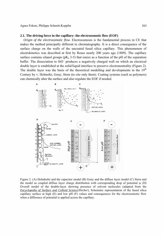
2.1. The driving force in the capillary: the electroosmotic flow (EOF) Origin of the electroosmotic flow. Electroosmosis is the fundamental process in CE that
makes the method principally different to chromatography. It is a direct consequence of the surface charge on the walls of the uncoated fused silica capillary. This phenomenon of electrokinetics was described at first by Reuss nearly 200 years ago (1809). The capillary surface contains silanol groups (pKa 3-5) that ionize as a function of the pH of the separation buffer. The dissociation to SiO– produces a negatively charged wall on which an electrical double layer is established at the solid/liquid interface to preserve electroneutrality (Figure 2). The double layer was the basis of the theoretical modelling and developments in the 19th Century by v. Helmoltz, Gouy, Stern (to cite only them). Coating systems (such as polymers) can chemically alter the surface and also regulate the EOF if needed.
Figure 2. (A) Helmholtz and the capacitor model (B) Gouy and the diffuse layer model (C) Stern and the model as coupled diffuse layer charge distribution with corresponding drop of potential (D) Overall model of the double-layer showing presence of solvent molecules (adapted from the Encyclopedia of Surface and Colloid Science/Decker), Schematic representation of the fused silica capillary surface at high (E) and low pH (F) values and consequences for the electroosmotic flow when a difference of potential is applied across the capillary.
Agnes Fekete, Philippe Schmitt-Kopplin 563

The potential drops to 0/e at a distance of x = –1, which is called the thickness of the diffuse double layer or also Debye length; –1 is dependent on the ionic strength I. An externally imposed tangential flow of the medium over the surface would lead to a distortion of the ions creating a “streaming potential”. This process is reversible and when a potential is applied, the counter-ions and their associated solvating water molecules migrate towards the cathode resulting in a flow of solution. This flow called as electroosmotic flow is an electrically driven pump towards the detector.
The electroosmotic flow (μeo) is proportional to the zeta potential ( ) and dependant on the chemistry, the viscosity ( ) and the dielectric constant ( ) of the separation buffer. The zeta potential measured at the plane of shear close to the liquid-solid interface (slipping plane in Figure3) is related to the inverse of the charge per unit surface area, the number of valence electrons, and the square root of the concentration of the electrolyte, an increase in the concentration of the electrolyte decreases the EOF. Strongly adsorbed ions on the inner wall of the capillary or a decrease in pH (lowering the surface charge of the capillary) of the separation electrolyte have the same effect.
Figure 3. Schematic representation of a charged particle and its different charged layers; identical rules as in the previous Fig.2
Capillary electrophoresis564
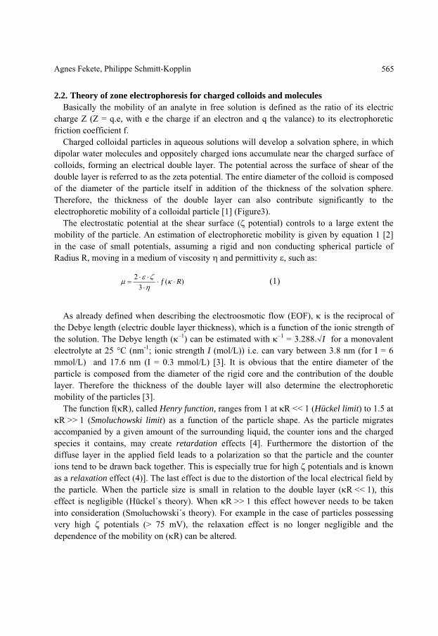
2.2. Theory of zone electrophoresis for charged colloids and molecules Basically the mobility of an analyte in free solution is defined as the ratio of its electric
charge Z (Z = q.e, with e the charge if an electron and q the valance) to its electrophoretic friction coefficient f.
Charged colloidal particles in aqueous solutions will develop a solvation sphere, in which dipolar water molecules and oppositely charged ions accumulate near the charged surface of colloids, forming an electrical double layer. The potential across the surface of shear of the double layer is referred to as the zeta potential. The entire diameter of the colloid is composed of the diameter of the particle itself in addition of the thickness of the solvation sphere. Therefore, the thickness of the double layer can also contribute significantly to the electrophoretic mobility of a colloidal particle [1] (Figure3).
The electrostatic potential at the shear surface ( potential) controls to a large extent the mobility of the particle. An estimation of electrophoretic mobility is given by equation 1 [2] in the case of small potentials, assuming a rigid and non conducting spherical particle of Radius R, moving in a medium of viscosity and permittivity , such as:
)(
32 Rf (1)
As already defined when describing the electroosmotic flow (EOF), is the reciprocal of
the Debye length (electric double layer thickness), which is a function of the ionic strength of the solution. The Debye length ( –1) can be estimated with –1 = 3.288. I for a monovalent electrolyte at 25 C (nm-1; ionic strength I (mol/L)) i.e. can vary between 3.8 nm (for I = 6 mmol/L) and 17.6 nm (I = 0.3 mmol/L) [3]. It is obvious that the entire diameter of the particle is composed from the diameter of the rigid core and the contribution of the double layer. Therefore the thickness of the double layer will also determine the electrophoretic mobility of the particles [3].
The function f( R), called Henry function, ranges from 1 at R << 1 (Hückel limit) to 1.5 at R >> 1 (Smoluchowski limit) as a function of the particle shape. As the particle migrates
accompanied by a given amount of the surrounding liquid, the counter ions and the charged species it contains, may create retardation effects [4]. Furthermore the distortion of the diffuse layer in the applied field leads to a polarization so that the particle and the counter ions tend to be drawn back together. This is especially true for high potentials and is known as a relaxation effect (4)]. The last effect is due to the distortion of the local electrical field by the particle. When the particle size is small in relation to the double layer ( R << 1), this effect is negligible (Hückel´s theory). When R >> 1 this effect however needs to be taken into consideration (Smoluchowski´s theory). For example in the case of particles possessing very high potentials (> 75 mV), the relaxation effect is no longer negligible and the dependence of the mobility on ( R) can be altered.
Agnes Fekete, Philippe Schmitt-Kopplin 565

2.3. The most important techniques in brief Many CE-techniques are routinely used in the analysis of biomolecules from small ions to
macromolecules [5] and have been applied for the analysis of food quality and food toxicants. The presented CE methods are all based on similar principles of open tubular separations (excepting capillary electrochromatography) with eletrokinetically driven buffer flow. Some variations in the quality of the separation buffer are used to tune and induce interactions between the buffer components and the analytes improving the selectivity in the separation. The frequently used CE methods in food analysis are presented herein in brief.
2.3.1. Capillary zone electrophoresis (CZE) By changing the quality of the separation buffer, optimized interactions with the sample
and with some buffer constituents allow an increased selectivity in the separation of charged or neutral analytes. CZE allows the separation of anions and cations as a direct function of their effective charge to size ratio [6].
The addition of solvents to the running buffer up to 100% in nonaqueous buffers (non-aqueous CE - NACE), can additionally allow specific selectivity and increased solubility for some analytes and is mostly used for pharmaceuticals or plant secondary metabolites [7] with low water solubility. The selectivity is governed by their effective charge and thus by the separation buffer pH and indirectly the electroosmotic flow (EOF).
Here the definitions of apparent and effective electrophoretic mobility need to be developed. With the polarity setup of the anode being at the injection inlet and the cathode at the outlet, neutral sample will move towards the detector with the velocity of the EOF; cations will move to the cathode first with a higher apparent velocity (apparently faster) and anions will move against the EOF with a reduced apparent velocity (apparently slower) as shown in
Figure4. The peaks are detected with increasing times in the electropherogram. The electrophoretic mobility is defined as being the field strength reduced velocity of the ions in the capillary. With an applied electric field E across a capillary of total length Lt the field strength is E/Lt. After a time t following the injection, the analytes (anion, neutral, cation) will cross the detector situated at a length Ld from the inlet one after the other; the observed velocity (v) of the analyte is thus equal to Ld/t. The electrophoretic mobility calculated from the observed velocity is called apparent electrophoretic mobility ( ap). The effective electrophoretic mobility ( ef) takes account of the velocity of the buffer towards the detector (EOF, ( eo)) and is thus the EOF-normalized electrophoretic mobility of the ions ( ef = ap –
eo); ef is equal to zero for neutral analytes, negative for anions and positive for cations. The effective electrophoretic mobility is an absolute parameter independent of the applied field or the column length and only dependant of the charge and size of the analyte. A conversion of the electropherograms from the time-scale into the effective mobility scale ( -scale) allows better reproducibility of separation patterns and of quantification parameters [8].
Capillary electrophoresis566

Figure 4. Illustration of separation by CZE
2.3.2. Micellar electrokinetic chromatography (MEKC) Micellar electrokinetic chromatography (micellar capillary electrophoresis) is a technique
introduced already 1984 by Terabe [9] – commercial CE systems were available only from 1988; the addition of charged surfactants (i.e. SDS or CTAB) to the separation buffer at concentration above their critical micellar concentration allow the separation of neutral or charged analytes (see Figure 5) as a function of their affinity to partition into micelles (hydrophobicity / Kow dependant). A partial filling technique of MEKC needs to be used when coupled to mass spectrometry avoiding ion suppression due to the presence of surfactants in the electrospray ionization and a lower contamination of the mass spectrometer.
Figure 5. Separation theory based on MEKC
Agnes Fekete, Philippe Schmitt-Kopplin 567

2.3.3. Chiral CZE and chiral MEKC The addition of a chiral ligand (for example different substituted cyclodextrins (CD),
antibiotics, sugar or peptide based ligands) to the CZE-separation buffer (Fig.6) allows the separation of ionic isomers (D/L, +/- enantiomers, stereomers). The separation of the isomers is based on different binding/partitioning constants between the chiral ligand and the D- or L-analyte. Non-racemic substances can also be separated based on their relative selective molecular binding occurring with the separation buffer constituents. The addition of a chiral ligand to the MEKC-buffer additionally permits the analysis of neutral isomers (enantiomers, stereoisomers). Similar results can be obtained when using charged cyclodextrines or other charged chiral ligands (i.e. chiral micelles).
Figure 6. Illustration of chiral separation based on CZE and MEKC
Capillary electrophoresis568

2.3.4. Capillary gel electrophoresis (CGE) Capillary gel electrophoresis allows the analysis of molecules based on their size. The
separation is done in physical gels (diluted polymer solutions such as methylcellulose, polyethylene glycols) which molecular size and concentration determine the selectivity by sieving effects as shown in Figure7. This technique is the basis of the gene sequencing techniques used in modern molecular biology.
Figure 7. Separation theory based on CGE
2.3.5. Capillary electrochromatography (CEC) Capillary electrochromatography is a relatively new coming capillary separation technique
that uses the same fused silica capillaries as in CE except that they are filled such as in nanobore liquid chromatography (nanoLC) with silica based materials or with polymeric sorbents [10-12] as illustrated in Figure8. Due to the pH-dependant surface charge of the silica or the polymer, the EOF can acts as a pump such as in CZE, moving the analytes with the buffer towards the detector; the separation occurs by interactions of the analytes with the immobilized active surface (hydrophobic, hydrogen and/or ionic bonds.) combined with their own electrophoretic mobility: CEC can be considered as a hybrid of CZE and nanoLC. The separation of selected lignin/humic substance (HS) degradation compounds by capillary
Agnes Fekete, Philippe Schmitt-Kopplin 569

electrochromatography with a methacrylate based monolithic column and a conventional column packed with 5 m octadecyl silica (ODS) particles was presented in [13] for the characterization of NOM. The effects of organic modifier concentration, pH of the mobile phase, ionic strength, applied voltage, and temperature on the separation of phenolic lignin monomers need to be investigated to determine the optimal separation conditions. The CEC of a soil fulvic acid with a monolithic column produced partly resolved broad bands; by means of NMR analysis a wide range of oxygen derived aromatic substitution patterns was found with prominent contributions from phenolic and carboxylic groups [13].
Figure 8. Illustration of capillaries used for CEC filled with silicagel and monolith.
3. Application of capillary electrophoresis in food analysis
The chemical and physical properties of the analytes, like dissociation coefficient (pKa) and octanol/water partition coefficient (logP) determine mainly the type of the CE separation technique. Substances with protonisation/deprotonisation ability can be separated applying zone electrophoresis. CZE is the most used technique in food analysis since several
Capillary electrophoresis570

components are highly polar and contain amino, carboxy and/or hydroxyl groups. Apart from food contaminants including antibiotics used in veterinary practice, pesticides and toxins, other components present in food can be separated and detected by CE techniques. The concentration of the contaminants is usually very low and the presence of the matrices may have a big influence on separation (shift and co-migration). Selective sample preparation techniques are often needed due to the similar chemical-physical properties of analytes and matrices. The found applications for analysis of matrices can be classified as:
- inorganic ions and low molecular weight organic acids, - biogenic amines used mostly for estimation of quality of sea products and wine, - amino acids useful for studies of adulteration and food quality - polyphenols and vitamins registered as the main components of functional food due to its
antioxidant activity - heterocyclic amines crucial for quality control due to their mutagenic effect, - and proteins paying big role in adulteration control. The differentiation between food contaminants and food constituents reflecting the food
quality sometimes is difficult and only based on the concentration of the analytes in the different type of food. For example the type and amount of biogenic amines determines the quality of the wines; biogenic amines are also used for distinguish fresh and spoiled fish products. The possible CE-methods applied for in food analysis are summarized briefly herein. Reviews dealing with the analysis of different substance class in food are shown in Table 1.
3.1. CZE methods for food contaminants and components Several capillary electrophoretic separations have been developed for the determination of
antibiotics, herbicides and toxins, however, only a few articles deals with their determination from food as matrix [14].
3.1.1. Veterinary drugs Most antibiotics including the group of (i) quinolones, (ii) tetracyclines, (iii) sulphonamides
and (iv) miscellaneous antibactericides are amphoteric compounds allowing their selective separation in wide pH range since they are cations at acidic condition and anions at high pH.
Quinolones are a group of widely used synthetic antibacterial agents in human and veterinary medicine and they may be found in contaminated meat products. Their CE analysis has not been used for real samples in routine practice; the methods were always applied to spiked samples mostly from chicken muscle. Oxolinic acid, flumequine, norfloxacin, difloxacin, ciprofloxacin, marbofloxacin and danofloxacin have been separated with phosphate based separation electrolyte at pH 8-9 in presence of methanol used as organic modifier [15-17]. A typical electropherogram of 8 quinolones is shown in Figure 9 [18].
Agnes Fekete, Philippe Schmitt-Kopplin 571

Table 1 General review on food analytic with use of CE
CE Analyte Goal, comment Matrix Ref
CE DNA; Gliadines, glutenins and proteins; Lactoglobulines, caseine, CMP, furosine; Inorganic ions and organic acids; amino acids; phenols, flavonoids; vitamins
-review on analytic in food authenticity
Cereals, fruits, milk, meat, wine, honey
[101
CE DNA; proteins and peptides, amino acids and amines; carbohydrates and gelling agents; inorganic and organic acids; phenolic compounds; vitamins; preservatives and colours; toxins and residues
-general review of published papers between 2002 and 2003
milk, cereal [102]
CE Proteins and amino acids; carbohydrates; vitamins; inorganic ions; additives; natural toxins; residues
-new detection systems -quality assessment;
[103]
CE milk protein; amino acids; carbohydrates; inorganic ions and organic acids; phenolic compounds; vitamins; food additives; biogenic amines; toxins and residues
-general review -development and difficulties
[104]
CE nucleic acid derivatives; carbohydrates; inorganic and organic anions; vitamins; phenolic acids; alditols and alcohols; amines; hop and beer bitter acids;
-analysis of beer components Beer [105]
CE cereal proteins; amino acids; vitamins; carbohydrates; fatty acids; biogenic amines; veterinary products; fungicides
-adapting CE to routine application -possibilities of automatisation
[106]
MEKC Enzymes, glucoamylases; amines -evaluation of mutagenicity [107] CE antibacterials, pesticides, toxins -organic contamination in food [14] CE, HPLC
oxytetracycline, tetracycline, chlortetracycline, doxycycline -analytic of tetracycline antibiotics -sample preparation methods
[21]
HPLC, CE
para-, diquat -analytic of quaternary amine herbicides
potato and serum [108]
GC, LC, CE
pesticides and metabolites -sample preparation methods [109]
GC, MEKC
polychlorinated biphenyls -sample preparation methods [110]
CE inorganic anions and cations -review on inorganic analysi Beverage, food [32] CE, LC heterocyclic amines - HAs in cooked meat meat, fish [46]
Capillary
electrophoresis572

CE Analyte Goal, comment Matrix Ref
CE, HPLC
catechins, caffeine, theanine, ascorbic acid; metal; ammonium, amines; organic acid
-review on analytic of tea Tea [84]
CE, HPLC
monosacharides, oligosaccharides ; alditols, alditol glycisides, polyols, amino sugars, deoxy sugars
-analytic of carbohydrates -sample preparation methods
[111]
CE catechins, isoflavones, antocyanins, resveratrol, vitamins -analytic of polyphenols in food
tea, soy, wine [48]
CE HPLC
caffeine, theanine, catechin, epicatechin, epigallocatechin, (epi)gallocatechin gallate,
-review on analytic of tea catechins
Tea [49]
CE, HPLC
cyanidin 3-glucoside, cyanidine 3-rutinoside, delphidin 3-glucoside,
-analytic of anthocyanins currant, candy, juice, jelly
[51]
CE cis and trans resveratrol -separation of isomers Wine [50] CE Lac, cochineal, cardenia, monascus, elderberry pigments -analysis of natural food
pigments [112]
CE ascorbic acid, nicacin, thiamine -water-soluble vitamin analytic meat, milk [113] CE, GC, HPLC
sugars; carboxylic acids; amino acids -separation techniques in food analytic
juices, citrus, apple, beer
[114]
CZE, MEKC
preservatives; antioxidants; sweeteners; colourings; nitrite, nitrate
-review on analytic of food additives until 2001
Beverage, vegetables [52]
CE, CEC Peptide; amino acids; organic acids; pesticides; sugar -review on chiral separation juices, wine, yogurt [87] CE-MS peptides and proteins; amino acids; carbohydrates;
polyphenols; toxins; pollutants - food analytic applying CE-MS
[115]
CE Proteins and peptides of animal origin -analytic of on proteins milk, egg, fish, meat [116] CE Proteins meat, milk cereal [61] CE DNA -combination with molecular
methods dairy products, meat [95]
Agnes
Fekete,PhilippeSchm
itt-Kopplin
573

However, these substances are water soluble, NACE have been also developed [19] to increase the selectivity of the separation of seven quinolones from pig kidney extract. Ammonium acetate buffer solved in methanol and acetonitrile at ration of 1:1 was used as background electrolyte (BGE) in presence of hexadimethrine bromide acting as EOF reserve agent speeding up the analysis time.
Figure 9. Electropherogram of a mixture of the quinolones. Electrophoretic conditions were 50mM phosphate buffer at pH 8.4, separation voltage at 20 kV and detection wavelength 260 nm. Migration order: (1) danofloxacin, (2 and 3) ciprofloxacin and sarafloxacin, (4) marbofloxacin, (5) enrofloxacin, (6) difloxacin, (7) oxolinic acid and (8) flumequine ( Reproduced from [18] with permission from Elsevier© 2004 )
Tetracycline antibiotics produced by Streptomyces are broad-spectrum antibiotics and they are administered to e.g. catfish to control bacterial infection. A CZE method using phosphate buffer at highly acidic pH (pH 2) has shown that oxytetracyclin is determinable from catfish and it is stable during the cooking process [20]. The method used UV-Vis detection at low wavelength, however, tetracyclines have native fluorescence due to the condensed ring that can be strongly intensified by application of alkaline earth ion in the separation electrolyte [21]. Good resolution of sulphonamides from pork extract have been achieved applying a phosphate buffer near to neutral pH showing the amphoteric character of this substance class [22]. Sensitive determination of seven sulphonamides has been achieved with combination of continuous flow system and CE-MS and the applicability was shown through spiked milk
Capillary electrophoresis574

samples [22; 23]. Adsorption of the cationic analytes onto the capillary wall was observed causing non-reproducible migration time, thus use of cationic coating was negligible. Capillary coating is a generally used method to govern the EOF when anions are in the interest of the determination or to prevent the adsorption of the sample components onto the inner surface of the fused-silica tube
3.1.2. Pesticides Due to the diverse function of pesticide several methods applying CE have been
determined; the most paper deals with the development and validation of these substances by CE. Pesticides like dithiocarbamates, acidic pesticides and some fungicides are determinable by CZE due to the ionisable group in the molecule, but the others like the triazines, urea derived, organic phosphorus substances and some multiple pesticides need to be separated with MEKC. Acidic pesticides including o-phenylphenol, ioxynil, haloxyfop, acifluoren und picloram were determined from orange juice as sample matrix applying CE-MS with off-line solid phase microextraction (SPME) used as selective sample preparation technique; the method describes the development and the performance characteristics [24]. CZE-MS have been used for the separation of pyrifenox, pirimicarb, cyprodinil and pyrimethanil function as multiple pesticides from orange juices with ammonium acetate based separation electrolyte at acidic pH [25]. Dithiocarbamates (ferbam, ziram, zineb, metham and maneb) have been selectively determined applying borate based BGE with pH of 9 from wheat extract, but no targets have been found from the examined samples [26].
3.1.3. Biological origin toxins Toxins are third big substance class of food contaminants determinable by CE techniques.
Naturally occurring toxins such as glycoalkaloids and microbial contaminants like food-born pathogens or toxin-producing microorganism have been determined by CE coupled to selective detection techniques like MS and fluorescence. Glycoalkaloids were determined from freeze dried potato extracts applying NACE coupled to MS and MS/MS [7; 27] and an electropherogram of standard solution of -solanine, -chaconine is shown in Figure10.
Domoic acid and paralytic shellfish poisoning (PSP) toxins were separated separately from contaminated mussel extracts by CE-UV [28]. Increasing the method sensitivity and selectivity, solid phase extraction (SPE) were included into the analysis process. Domoic acid contains amino and carboxylic acid groups thus it can be separated at wide pH range by CZE. Robust and fast separation was achieved by application of borate buffer in presence of methanol as organic modifier. PSP toxins were also separated with isotachophoretic (ITP) separation buffer containing morpholine at pH 5 [28].
Bacterial contaminations can be studied different ways applying CE. Lypopolysaccharides present in the wall of Gram-negative bacteria protonating at alkaline medium and have native fluorescence allowing their CE-LIF determination [29]. CE-LIF have been also used for the
Agnes Fekete, Philippe Schmitt-Kopplin 575

determination of microorganisms from meat; however improvement of the method is required due to the board peaks of the targets and relatively long analysis time [30]. Improved separation was achieved for the determination of eight microorganism applying CZE electrolyte containing phosphate buffer in presence of calcium and myoinosytol hexakisphoshate that decrease the peak width due to the formation of protein-myoinosytol sandwich bonds [31]. Even though the method applicability was shown by spiked juice and corn flakes samples, no targets were found from the selected samples.
Figure 10: SIM traces by NACE-ESI-MS of a 1 mg/L standard mixture of glycoalkaloids and relative glycones. Electrophoretic conditions: buffer: ACN-MeOH (90:10 v/v) containing 50 mmol/L ammonium acetate and 1.2 mol/L acetic acid; uncoated fused-silica capillary, Ld = 80 cm, 50 μm ID; injection: pressure, 0.5 psi for 5 s; effective voltage: 25.5 kV; T = 20oC. ESI-needle voltage, Ves = +4.5 kV; spray current, sheath gas flow rate was set at (N2) 0 AU; temperature of the aluminium capillary, Tcap = 180 oC and capillary voltage, 32 V; coaxial sheath liquid, methanol: water (1:1) with 1% of acetic acid at a flow rate of 2.5 μL/min. MS detection was performed in the SIM mode and the acquired masses were: 398, 416, 852, 868, 1034 for solanidine, tomatidine, -chaconine, -solanine, and -tomatine, respectively (Reproduced from [7] with permission from Wiley© 2006).
Capillary electrophoresis576

Table 2 Selected CZE methods for food components Detector Analyte Goals, comments Matrix Ref
ESI-MS Procymidone, thiabendazole -applying sample stacking and SPE grape, tomato [117] ESI-MS/MS dinoseb, pirimicarb, procymidone,
pyriphenox, pyrimethanil, thiabenzadole -application of SPE Peaches, nectarines [25]
MS, UV pyrimethanil, pyriphenox, cyprodinil, cyromazine, pirimicarb
-application of SPME -use of chemometrics in optimization
orange juices, grape juice
[118]
DAD Sulfamethazine, sufamerazine, sulfadiazine, sulfadimethoxine, sulfamonomethoxine,
-determination of sulfonamides used in veterinary practice; application of SPE
Meat [22]
conductivity Sulfite -separation on PMMA chip Wine [119] DAD domoic acid as ASP toxin, GTX 1-5 as PSP
toxin -paralytic and amnesic shellfish toxin contaminated
mussel [28]
DAD Versinia enterociliticia, Leuconostoc mesenteroides, Salmonella enteritidis, E. coli, Listeria monocitogenes
-monitoring of bacterial contamination juice, milk, corn flakes, baby food
[31]
UV isocitric, citric, tartaric and malic acid - study of adulteration orange juice [41] UV Nitrite, nitrate meat, vegetables [120] Indirect UV Oxalic-, malic-, aspartic-, glutamic-, quinic
acid -fingerprint at different tea infusions tea infusion, [40]
Indirect-UV chloride, nitrate, sulfate, phosphate, tartarate, malate, succinate, citrate, acetate, lactate, ascorbate
-simultaneous determination of organic and inorganic acids
orange juice and wine,
[38]
DAD Histamine -study of the state of the deterioration tuna fish [121] UV histamine, tyramine -comparison of CE and HPLC; use of SPE fish, cheese, meat [122]
Agnes
Fekete,PhilippeSchm
itt-Kopplin
577

Detector Analyte Goals, comments Matrix Ref
Indirect UV methyl-, hist-, ethanol-, propyl-, isopropyl-, isoamyl-, tyr- and phenethylamine, purescine
-use of on-line SPE system Wine [123]
ESI-MS 20 amino acid -analytic of free amino acid infant food [45] UV arginine, alanine, serine, aspargine,
tryptophan, glutamic acid, phenylalanine, tyrosine, proline
-determination of free amino acids -fingerprints of different beers
orange juice, beer [43]
UV adenosine, guanosine, uridine -fingerprints for quality control C. Mycelia [124] ESI-MS intact gellan gum -analytic of polysacharides fruit flavor drinks [125] Indirect UV sucrose, sucralose, glucose, fructose -applying chemometrics and SPE juice, yoghurt [60] DAD apigenin, baicalein, naringenin, luteolin,
hesperetin, galangin, kaempferol, quercetin, myricetine
-use of SPE -comparison of the method
Wine [126]
DAD Caffeine,theo-, di- and enprophylline, theobromine
-analytic of alkylxanthines chocolate [127]
UV
Caffeine, adenine, theophylline, epigallocatechin-3- and epicatechin-3 gallate, epigallo- and epicatechin
-coupling of flow injection system (automation) green tea [53]
electrochem Trans-resveratrol -analytic of compounds with antioxidant activity
wine, chinese medicinal herb,
[128]
amperomet. Chlorogenic-, genistic-, ferulic- and vanillic acid
-separation on microchip red wine [129]
ESI-MS Cytochrome C, lysozyme from egg white, ribonuclease A
-applying physically adsorbed polymer coating turkey and chicken egg white, beef
[130]
LIF -lactalbumin, -lactalbumin, bovine serum albumin
-on-capillary derivatisation Cheese from whey [131]
UV -lactalbumin, -lactalbumin -adulteration of cows and goat milk products cows and goat milk and cheese
[132]
UV -casein1, -casein2, -casein1, -casein2 -casein concentration during the ripening time Cheese (from ovine [70]
Capillary
electrophoresis578

Detector Analyte Goals, comments Matrix Ref
milk) UV para- -casein, -lactoglobulin -analysis of whey proteins and their degradation milk, dairy
products [133]
UV -lactalbumins, -lactoglobulins -use of ITP based BGE binary and ternary cheese
[69]
FL DNA -lab-on-microchip, including PCR -species identification
fish species [134]
Agnes
Fekete,PhilippeSchm
itt-Kopplin
579

3.1.4. Inorganic ions and low molecular weight acids Several minor components like halides, fat-soluble vitamins and metals are essential for
human health but too high levels can lead to health diseases; in this case even endogenous compounds can be assigned to pollutants. Solutes like nitrate and nitrate may be present originally in the food extract, but can also originate from packing materials like paper as food contaminants. Thus the analysis methods applying CE for substances playing role in the food quality are summarized shortly (see Table 2).
One of the strong points of CE comparing with the other separation techniques is the simplicity and fastness of the determination of inorganic ions and low molecular weight acids and bases. Several methods have been developed for the determination of metals from different foods samples like milk, cola, vegetables and wheat flour [32]. Since they are cations in water, they can be separated by CZE applying buffer at acidic pH. However, their detection is more delicate since these molecules do not absorb light in the UV-Vis range therefore they can not be directly detected spectrophotometrically. Since spectrophotometric detection is commercially available in the CE instruments, methods using indirect-UV mode have been developed for the analysis of inorganic cations. It uses the addition of an absorbing co-ion into the separation electrolyte and negative peaks are detected at the migration zones of the charged and non-UV active solutes. Applying indirect-UV detection, the mobility of the UV-absorbing ion present in the separation electrolyte has to be similar as the targets in order to reduce fronting and tailing of the analyte peaks. The concentration of the UV-absorbing ion in the BGE influence the sensitivity of the method, however it may cause instability of the baseline at too high concentration. Indirect-UV detection imposes severe limitations on the choice of electrolytes, their concentrations and additives. Moreover, this technique is the least selective and therefore the most subject to interferences. For the determination of ammonia, alkali, alkali-earth and other metals like manganese, nickel, zinc, cupper and chrome from different food extracts imidazole or a commercially available solute called UVCat-1 have been used as UV-absorbing agent. As counter mostly - hydroxyisobutyric acid were used due to its complexion ability with the inorganic cations increasing the separation selectivity [32]. Crown ether, 18-crown-6, was also ingredients of BGE in order to distinguish NH4
+ and K+ from each other since they have identical size and charge/mass ratio. Direct UV detection applying non-selective detection wavelength (200 nm) have been also used for the determination of Ca2+ and Mg2+ as EDTA complex from extracts of different vegetables [33]. The complexes were separated in a polyimide coated capillary with buffer containing 20 mmol/L borate at pH 9.2 and 2 mmol/L EDTA. Methyl mercury, which is a possible contamination factor due to its highly toxic property, was also determined from fish samples applying borate based separation electrolyte [34]. They were also separated as UV-active complex with agent of dithiozone sulphonate under acidic pH (pH 4.5) [35].
Capillary electrophoresis580

Several CZE methods have been developed for the determination of inorganic anions and LMW acids from different matrixes including food and have been reviewed elsewhere [32; 36; 37]. Inorganic anions including sulphite and sulphate used as antimicrobial agent and antioxidants; nitrate and nitrites added into the meat products as preservatives; halides which presence at high concentration are toxic; phosphate and carbonate are in the interest of food analysis. Organic acids are usually used for study of food quality and adulteration.
For the determination of inorganic anions and LMW acids, the EOF is generally reserved to achieve a suitable separation time. The most common method to control and reserve the EOF for the analysis of anionic species is the addition of surfactant into the separation electrolyte. The use of long-chain ternary alkylammonium cationic surfactants such as cetyltrimethylammonium bromide (CTAB), tetradecyltrimethylammonium bromide (TTAB) and dodecyltrimethylammonium bromide (DTAB) has been reported in applications requiring suppression or reversal of EOF [38]. In addition, amines like diethylenetriamine or tettraethylenepentaamine and alkylammonium hydroxide such as octadecyl-trimethylammonium hydroxide and tetradecyl-trimethylammonium hydroxide have been also used to reserve the EOF.
Since the LMW acids are lack of chromophor groups, the most used detection technique based on indirect-UV. The most common UV-absorbing anions within the range of mobilities of analyte anions of interest for food analysis are chromate, 1,3,5- benzenetricarboxylic acid and phthalate. Chromate based electrolyte was used for the determination of sulphite from different vegetables and wines [39] and for the determination of phosphate from cola as beverage matrix. Phthalic acid with combination of commercially available EOF-suppressor (OFM-Anion BT) has been applied for the determination of nine inorganic anions (halides, nitrate, nitrite, sulphate, sulphite, phosphate and carbonate). EDTA have been also added into the sample solution and into the separation buffer for the determination of LMW acids to avoid the mobility differences of the analytes caused by the high content of alkaline-earth ions [40]. The method applying chromate as UV-absorbing ion, TTAB as EOF reserve agent and EDTA as complexing agent was used for the determination of fluoride and LMW acids like oxalic, citrate, malic and glutamic acids from different beverages like wine and orange juice [40]. Organic acids were determined with direct UV detection too with use of non-absorbing electrolytes since the maximum absorbance of the analytes was observed at 200 nm. For the determination of citric, isocitric, malic and tartaric acid phosphate based buffer was used as a BGE at pH 7.5 where peaks causing interference were separated from the targets [41].
3.1.5. Amines and amino acids Biogenic amines are organic bases of low-molecular mass comprising aliphatic, mono-, di-
and poly-amines. Other amines like cathecolamines registered as functional food components, indoyl- and heterocyclic amines present as contaminants in different food products. Low-molecular weight amines like histamine, putrescine, cadaverine, tyramine and ethylamine are
Agnes Fekete, Philippe Schmitt-Kopplin 581
forming during microbial process and their presence in alcoholic beverages (wine and beer) reflect unsanitary condition. They are analyzed in different foods like fish, cheese and sea food used as quality indicators for raw food materials. Since they are weak bases they can be separated with CE under acidic condition according to their charge and size. Their detection is mostly based on indirect-UV detection due to the absence of chromophores. The advantages of this approach are that primary amines through quaternary amines can rapidly be separated. LMW amines have been mostly determined from wine and fish applying formate based electrolyte (pH 3-4) containing cupper-sulphate function as UV-absorbing ion and 18-crown-6 to increase the selectivity of the separated targets [42]. The signal sensitivity can be improved two or three order of magnitude when the amines are derivatised before their capillary electrophoretic determination. Moreover, the interferences of other present and non-derivatised cations with similar mobilities might be eliminated. However, the separation of the derivatised products is more difficult than of the non-derivatised amines and application of MEKC is required.
Free amino acids have been determined by CZE from different matrix like beverages or soy sauce. Due to their zwitter ionic nature they can be separated in cationic mode using low pH carrier electrolytes [43] or in an anionic mode using separation buffer with pH above 10 [44]. The latter approach provides faster separations due to the high EOF but up to now the resolution of all 20 common amino acids without derivatisation has not been achieved. Using highly acidic carrier electrolytes, the selectivity differs from those obtained with high pH buffers and the 20 amino acids were successfully separated from each other applying phosphate based separation electrolyte (pH 2.4) with presence of octanesulphonic acid [43]. For the detection of the amino acids, spectrophotometry was applied at detection wavelength of 185 nm, since some amino acids do not possess a strong chromophore making their sensitive detection difficult. Thus other detection techniques like indirect UV detection [44], conductivity or mass spectrometry (MS) [45] have been used for the analysis of non-derivatised amines. CE coupled to MS gives a selective determination; it does not require baseline separation of all solutes, however, it is limited by the applicable electrolyte system which has to be volatile. Ammonia or short-chain alkyl amines can be used as high-pH carrier electrolyte and low-pH electrolytes contain mostly formic-, acetic- or trifluoroacetic acid can be regarded as suitable BGE for the purpose of separation the non-derivatised amino acids. For the determination of amino acids from infant food, triethylamine was used because of its lower volatility and an increased stability of the electrolyte concentration than applying ammonia as separation buffer [45].
Heterocyclic amines have been found in cooked food and due to their mutagenic activity their determination is an important task [46]. They contain strong chromophor thus can be detected with spectrophotometry after their CZE separation as cations present in the solution with pH of 2-3. A method was developed for the determination of 13 heterocyclic amines from salmon and meat extracts applying sodium phosphate buffer containing methanol and
Capillary electrophoresis582
sodium chloride [47]. Before the MS detection of the heterocyclic amines were also separated with buffer containing ammonium acetate at pH 3 in polyvinyl alcohol-coated capillary to prevent the adsorption of the target onto the capillary surface.
3.1.6. Phenolic compouns and vitamins Different derivatives of phenols present naturally mostly associated to flavour and colour of
the foods or as additive due to their antioxidant property. Phenolic compounds are divided into catechins, isoflavons and antocyanins [48] that are generally contains benzol ring allowing their direct spectrophotometric detection. They are mostly weak acids containing more than one hydroxyl group with pKa value between 8 and 10 thus they can be separated at highly alkaline condition (pH 8-9) applying CZE. The developed and validated methods have been summarized in several reviews [48-52]. However, some derivatives of catechin present in food products are neutral at basic pH, therefore MEKC is preferred for their determination. Mostly borate based buffer at concentration of 20-150 mmol/L have been used as separation electrolyte [48] since borate have complex ability to -hydroxy groups increasing the selectivity of the analytes. Concerning the analysis of polyphenol derivatives, tea, soy sauce and wine has been the most studied food samples; tea leaves may contain catechins up to 30% of dry mass and resveratrol is the most investigated derivative of polyphenols in wine and grape. Eleven catechins including epigallocatechin-3-gallate, epigallocatechin, epicatechin-3-gallate, catechin, epicatechin and quercetin were separated with borate buffer at pH 8.5 applying an on-line coupled flow injection system used for microwave treatment [53]. Isoflavons have been determined from soy sauce with separation of borate based electrolyte. Antocyanins, a group of flavonoids, are the largest group of water-soluble natural pigments and they were also separated at basic pH, however, the sensitivity of the method was not sufficient and the analytes found to be unstable [54]. Thus anthocyanins present in berries have been separated at highly acidic (pH 1.5-2.1) condition applying phosphate based buffer [55].
Vitamins such as ascorbic acid have also antioxidant activity and they have been determined applying CE. Methods for the determination of water-soluble vitamins like riboflavin and niacin have been also developed and applied for different food samples like beverages (juice, beer, wine) or extracts of fruits and vegetables. Their capillary electrophoretic separation can be achieved at alkaline condition due to their ability of deprotionation and mainly borate or phosphate buffer have been used as BGE. Riboflavin and its related flavins, flavin mononucleotide, and flavin adenine dinucleotide are natively fluorescent which enables their detection by fluorescence overcoming the issues of selectivity and sensitivity that is an obstacle to the determination of other vitamins at trace levels in complex food matrices. The flavins are separable in CZE using a 30 mmol/L phosphate buffer with pH 9.8 and were detected by laser induced fluorescence detector [56-58]. This basic CZE method has been demonstrated as effective for determination of riboflavin in a number of
Agnes Fekete, Philippe Schmitt-Kopplin 583

foodstuffs, including wines, vegetables, wheat flour, and tomatoes. However, other vitamins like ascorbic acid and niacin have been frequently determined by CE and reviewed elsewhere [59].
3.1.7. Carbohydrates The main problem encountered in carbohydrate determination by CE is the neutral nature
of these compounds and the absence of chromophores for the direct application of spectrophotometric detection. To overcome the problem of the absence of charge, they have been ionised by complexing with certain ions like borate or metals; ionization by increasing the pH of BGE to the strongly alkaline pH range or by chemical derivatisation. The presence of complexation agent in the BGE improving the UV-absorbance of the carbohydrates too, thus mostly borate based buffer have been applied for their determination. The pKa values of the sugars are in the range of 12-14 thus their deprotonation take place at pH above 12. Having a separation electrolyte at this pH range with buffer capacity the choice of the buffer system is limited; phosphate is the frequently used buffer. Improving the resolution of carbohydrates and reducing the migration time, the EOF was reserved by inverting the polarity of the applied potential or by covering the negatively charged silica capillary surface with cationic surfactants as described for the determination of the LMW acids. Derivatisation is an approach to overcome the issue of absence of UV-absorbance too. Several derivatisation agents have been used like 4-aminobenzoic acid ethyl ester, 6-aminoquilonine and phenylethylamine. However, indirect UV detection was used for the determination of the sugars from beverages like orange juice. Potassium sorbate, 2,6-pyridinedicarboxylic acid, p-nitrophenol and naphthol blue-back have been used as absorbing ion in the separation electrolyte [60].
3.1.8. Proteins Proteins extracted from milk, egg, cereal and meet can be separated by CZE based on the
differences of their charge densities with use of separation electrolyte at broad pH range [61]. The most used buffer systems based on phosphate in the pH range of 1.5-8, borate at pH 8-10 and citrate at acidic pH (pH 3). Sarcoplasmic proteins from fish [62; 63] and whey proteins from milk were separated with phosphate buffer at pH 7.4 [62; 64] where the proteins are negatively charged and at pH 1.5-2.5 as cations. Whey proteins were also separated with borate based buffer at highly basic condition (pH 8-9) to differentiate the genetic variants of
-lactoglobulines [65-67]; high ionic strength at high pH increase the separation efficiency and prevent the formation of aggregates. Simple buffer system without additives is seldom used for the separation of protein extracts. The biggest problem in the capillary electrophoretic separation of the proteins is the interaction between the proteins and silanol groups present on the surface of capillary wall leading to distorted peak shapes and poor separation efficiency. However, working with highly acidic separation condition, the
Capillary electrophoresis584

electrostatic interaction is minimized because of the neutral form of the silanols; at high pH with combination of high-ionic strength may decrease the adsorption by the repulsion effect between the negatively charged proteins and silanol groups. Preventing the protein-wall interactions, polymer modifiers have been added into the BGE that are mostly cellulose based. For example natural and artificial shark fins were differentiated applying separation electrolyte containing 0.01% methylcellulose [68]. Other derivatives of cellulose like methylhydroxyethylcellulose [69; 70] and hydroxypropylcellulose [71] at volume ratio between 0.05% and 0.5% have generally been used. Recently, other type of polymers like poly(ethyleneoxide) and linear polyacrylamide were successfully applied as dynamic coating agents [72]. Both cationic and anionic polymeric additives have been assayed for the capillary electrophoretic separation of aqueous extracts of bovine and chicken muscles with BGE at pH 3-7. The protein-wall interactions were effectively minimized with polydiallyldimethylammonium chloride as cationic polyelectrolyte, but better resolution was achieved in the presence of co-additive sodium acetone sulphate. Anionic polymer, dextrane sulphate, provided high efficiencies in the separation of chicken muscle proteins that not only prevented the adsorption of the proteins but increased the selectivity of the separation. Other possibility to prevent the adsorption of proteins is the application of modified capillaries e.g. capillary masked by methylasition.
Other main task in the capillary electrophoretic separation of the proteins is the use of solubilization agents in the separation buffer. Mostly urea at high concentration (4-8 M) is used which additionally help in resolving the proteins like caseins from milk products. However organic solvents and detergents worked as well as urea in solubilizing the hydrophobic proteins [73]. Use of acetonitrile (ACN) at volume ratio of 20 % avoided the potential problems of crystallization and protein modification that are sometimes associated with urea and provide a buffer with good UV transmission properties and low viscosity.
Separation electrolyte uses sodium-based buffers generates substantial current due to the high conductivity of the sodium ions that turn limit the applied voltage. To apply higher voltage causing decreased separation time, glycine or -alanine have been used in place of sodium that generated mostly half times less current. Wheat proteins and oat/rice prolamines have been successfully separated using these electrolytes containing 20% acetonitrile. Maize proteins have been also separated however the content of ACN had to be increased to maintain the solubility of the hydrophobic maize and sorghum proteins. Another approach to overcome the limitations of sodium based buffers is the use of isoelectric buffers. These unique compounds have an isoelectric point roughly equal to their pH in solution, thus they can buffer without the need for co-ion. Due to their amperometric nature, they produce low current even at high separation voltage favouring high resolution with short migration time. Iminodiacetic acid (IDA) and aspartic acid have been used to determine proteins from wheat, rice oat, maize and sorghum [74]. IDA based buffer containing urea, hydroxymethylethylcellulose and Tween 20 was used for separation of milk proteins to
Agnes Fekete, Philippe Schmitt-Kopplin 585

determine the animal origin of globulines [69]. IDA has a lower pH in solution than aspartic acid that may provide fewer problems with the wall interactions and Tween 20 and other non-ionic surfactants have a discriminating effect on the retention behaviour of the bovine globulins related to the different strength of the protein-surfactant association complex.
Food samples often modify the wall of the capillary mostly by the presence proteins. The proteins might lead to fouling of the uncoated silica capillary causing increased migration times every injection of the sample. When the proteins migrate slower than the analytes, the capillary surface can be deprotonated during the rinsing to wash them away. However the determination of proteins by CE is an important task in food analysis and mostly applied for adulteration studies.
3.2. MEKC methods for food contaminants and components MEKC is mostly applied capillary electrophoretic technique when one or more target
components are neutral. Between the food components pesticides, toxins, polyphenols, derivatised amines, aminoalcohols and proteins have been determined from different food extracts. Methods developed for their determination with use of MEKC are summarized in Table 3.
3.2.1. Pesticides and toxins Among the pesticides fungicides, triazines, urea derived and multiple pesticides have been
analysed by MEKC coupled with spectrophotometric detection mostly from soft beverages. Eight widely used fungicides namely carbendazim, imazalil, methylthiophanate, o-phenylphenol, procymidone, prochloraz, triamidefon and thiabendazole were separated with BGE containing borate buffer at pH 9 and 75 mmol/L sodium cholate as surfactant [75]. The applicability have been shown through spiked food extracts from grape, tomato, lettuce and orange as shown in Figure 11 [75]. Sample stacking have been developed increasing the sensitivity of the MEKC method and successfully applied for non-spiked apple juice [76]. The separation electrolyte contained 10 mmol/L phosphate as buffer, 30 mmol/L SDS and isopropanol / isobutylalcohol function as organic modifier enhancing the selectivity of the separation. The sample stacking was achieved by salting the injected sample. Pre-concentration strategies like sweeping, stacking and modified staking with combination of MEKC have been studied for nine multiple pesticides determined from spiked carrot samples because the most problematic task in the CE analysis of food contaminant is the sensitivity of a method [14; 77]. Atrazine, a triazine type pesticide, have been determined by MEKC from juices with combination of supported liquid membrane and solid-phase extraction used for sample preparation to increase the selectivity and sensitivity of the analytical process [78].
Capillary electrophoresis586

Figure 11: Electropherogram of spiked grape at 1 mg kg. Conditions: buffer: 4 mmol/L borate buffer, pH 9.2, 75 mmol/L sodium cholate; detection: 210 nm; separation voltage: 15 kV; capillary: 50 cm effective length, 75 μm I.D. Peak identification: (1) Methylthiophanate; (2) carbendazim; (3) thiabendazole; (4) procymidone; (5) O-phenylphenol; (6) imazalil; (7) triadimefon; (8) prochloraz. (Reproduced from [75] with permission from Elsevier© 2001).
Toxins have been determined mostly from spiked food samples or standard solutions of the targets with MEKC. A method was developed and applied for apple cider for the determination of patulin a type of mycotoxin [79]. After a single liquid-liquid extraction, the patulin were separated from the matrices applying a borate buffer with presence of SDS at pH 9. -toxins an highly toxic metabolites of fungi like Aspergillus were also separated with application of SDS and cholate micelles in the separation electrolyte at highly alkaline condition from extracts of feeds and corn [80; 81].
3.2.2. Amino acids, additives and proteins As mentioned above, amines and amino acids can be determined after their derivatisation
with e.g. dansyl chloride, fluorescein isothiocyanate or 3-(4-carboxybenzoyl)-2-quinoline-carboxaldehyde for improving the method sensitivity. However, the separation of the products is difficult task thus MEKC have to be used for achieving baseline separation of the targets. The derivatised amino acids are generally separated applying separation buffer with SDS or polyoxyethylene 23 lauryl ether. Seven fluorescein derivatives of amino acids were analysed from different orange juices applying borate based electrolyte containing 30 mmol/L SDS and
Agnes Fekete, Philippe Schmitt-Kopplin 587

20 mmol/L -CD at pH 9.4 allowing to distinguish the chiral form of the targets [82]. Method for the determination of dansyl derivatised amino acids were also developed applying similar separation electrolyte as describe above [83].
MEKC have been also frequently applied method for the determination of polyphenols from different food samples. Derivatives of catechins are neutral even at high pH and the selectivity of the method can improve by addition of SDS into the separation electrolyte as reviewed elsewhere [48; 49; 84]. Mostly, borate/phosphate buffer has been used as BGE; phosphate were used to assure the eligible current for the electrophoretic separation [85; 86].
Proteins have been also separated with MEKC, however, applying CZE result in a determination method with better performances. Major milk proteins were separated with BGE containing 3 mmol/L sodium borate buffer (pH 9.5) with SDS added at its critical micelle concentration (8.2 mmol/L). This buffer allowed rapid separation of the milk proteins with low separation time and the proteins were reported to be separated by their hydrophobicity [86].
3.3. Chiral CZE and chiral MEKC methods for food contaminants and components Use of CE has emerged as a good alternative for enantiomer separation since several chiral
selectors are available and their consumption for one separation is low. An enantiomer separation in food chemistry is useful for study of adulteration, monitoring of fermentation processes and analysis of chiral metabolites and prochiral constituents in food and beverages. Methods applying CE for chiral separation of food components are reviewed elsewhere and selected methods are shown in Table 3 [87].
3.3.1. Pesticides Chiral determination of pesticides is an important task since organism degrade or produce
chiral compounds by stereospecific enzymatic processes, thus the ratio of the chiral solutes that accumulate in food might be different as the used one. A comprehensive review of CE determination of pesticides have been published but no food as matrix was mentioned [88]. The developed methods for the chiral separation of pesticides were mostly done with BGE containing CD as chiral selector. A method was developed for the enantiomeric separation of vincozoline in wine. This fungicide was determined by MEKC applying BGE with content of SDS and -CD after extraction of the sample by SPE [89].
Capillary electrophoresis588

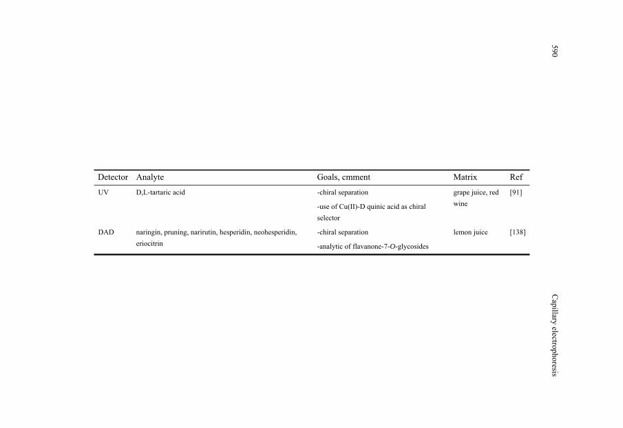
Table 3 Selected MEKC methods and chiral separation of food component
Detector Analyte Goals, cmment Matrix Ref
DAD acrinathrin, bitertanol, cyproconazole, fludioxonil, flutriafol, myclobutanil, pyriproxyfen, tebuconazole
-pesticide analytic
-comparison of SPE and SBSE
Lettuce, tomato, grape, strawberry
[135]
UV theanin, caffeine, ascorbic acid, (-)-epicatechin, (-)-epigallocatechin, (-)-epicatechin gallate, (-)-epigallocatechin gallate
-quality estimation of tea
- characterization of fresh tea leaves according to plucking dates
green tea, oolong tea, black tea
[86]
UV procyanidin B1, procyanidin B2, procyanidin B3 (+)-catechin, (-)-epicatechin, p- coumaric acid
-analytic of compounds with antibacterial activity
white and black beans
[85]
UV (+)-catechin, catechin gallate, (-)-epicatechin, epicatechin-3-gallate, epigallocatechin, epigallocatechin-3-gallate, theaflavin, theaflavin-3-monogallate, theaflavin-3`-monogallate, theaflavin-3,3`-gallate, caffeine, adenine, theophylline, quercetin, gallic acid, caffeic acid
-determination of tea polyphenols and tea ingredients
-comparison of HPLC and CE
green and black tea
[136]
Conduct. chloride, nitrite, nitrate, sulfate, fluoride, oxalate, malonate, pyruvate, tartrate,citrate malate, succinate, acetate, lactate, aspartate, glutamate, salycilate, gluconate, benzoate
-PMMA-microchip
-determination of organoleptic characteristics of wine
Wine [137]
ESI-MS D- and L- amino acid -chiral separation
-derivatisation of amino acids with dansyl chloride and fluorescein isothiocyanate
Orange juices
[115]
Agnes
Fekete,PhilippeSchm
itt-Kopplin
589
Detector Analyte Goals, cmment Matrix Ref
UV D,L-tartaric acid -chiral separation
-use of Cu(II)-D quinic acid as chiral selector
grape juice, red wine
[91]
DAD naringin, pruning, narirutin, hesperidin, neohesperidin, eriocitrin
-chiral separation
-analytic of flavanone-7-O-glycosides
lemon juice [138]
Capillary
electrophoresis590

3.3.2. Low molecular weight acids Chiral analysis of LMW organic acids is complicated due to its short chain that can hardly
makes the three-point interaction with the selector generally necessary for recognition. Target substances among the acids are mostly tartaric, malic and lactic acids applying the method mostly for food quality control. The chiral separation of these acids mostly based on ligand exchange process with chiral solutes. For the separation of D,L-tartaric acids a separation electrolyte containing cupper(II) ion and D-quinic acid at pH 5 and was applied for the determination of the targets in juices, jam and candies [90]. Direct spectrophotometric detection at selective wavelength could be used due to the light absorbing property of the complex. D,L-lactic acid have been separated from each other in yoghurt and beverages applying phosphate based separation electrolyte containing 2-hydroxypropyl- -CD as chiral selector in coated capillary to decrease the analysis time [91].
3.4. CGE methods for food contaminants and components Accurate quantitative determination of whey proteins was developed using cross-linked
polyacrylamide gel-filled capillaries under denaturing and non-denaturing conditions, that allowed the separation of the major whey proteins with high efficiency in less than 30 min [92]. CGE using gel-filled capillaries still presents some limitations, such as time-consuming capillary preparation, poor reproducibility and short capillary lifetime. Moreover, due to the high viscosity of the gel, the injection of the sample must be performed by electromigration, which is strongly influenced by the ion concentration of the sample. To avoid these limitations, CGE using replaceable gels of polyethylene glycol (PEG) and a cross-linked polyacrylamide coated capillary was employed for the separation of whey proteins, yielding good separation between -lactoglobulin and -lactalbumin. In order to decrease the absorbance of the polymer, the use of polyethyleneglycol and dextran as sieving mediums has been purposed. Ready-to-use polymer solutions have recently become commercially available and have been successfully employed [93]; e.g. commercial kit for SDS-CGE (Applied Biosystems) has been applied to the analysis of whey proteins achieving baseline separations of the globulins [94].
CGE combined with PCR have been used for separation of DNA fragments to detect e.g. generally modified microorganism, food borne pathogens or toxin-producing microorganism [95]. The higher sensitivity and the potential to distinguish similar strains of the same species make PCR-CGE suitable for fast studies.
Agnes Fekete, Philippe Schmitt-Kopplin 591

3.5. CEC methods for food contaminants and components In the past 10 years increase amount of papers deal with CEC, although its application for
determination purpose in food analytic is still rare [96]. Reversed phase CEC have been developed for the determination of 4 derivatives of phenylurea herbicides from vegetables and vegetable posessed food samples [97]. The method used capillary packed with non-end-capped C18 silica particles with average particle diameter of 5 μm at 8 cm long and a reduced amount of organic solvent in the mobile phase to increase the selectivity of the separation thus reduce the possibility of interference. Athough, matrix effect and high baseline noise was observed when spiked vegetable extracts were measured with the optimised process. Pressurized CEC with conventionally available capillary column packed with octadecylsilica particles at length of 20 cm were used for the separation of fluoroquinolones used as antimicrobial agent from extracts of spiked fish muscle [98]. To decrease the bubble formation that may generally occure during the CEC separation SDS was added into the mobile phase that reduce the surface tension at the solid-liquid interface. The mobile phase contained also triethylamine to mask the free silanol groups since at the separation pH they are deprotonised allowing ionic interaction with the analytes causing peak boardening [98]. Other solid phases have been also applied; methacrylate based monolith were used for the separation of acids from different foods like sirup or wine [99] and carotenoids were separated on silicagel with C30 functional group applying gradient CEC from algal extracts [100].
Capillary electrophoretic determination of the contaminants a in food became more popular because of the advantages of the technique comparing to the chromatographic techniques. However, the most of the papers describe methodological developments that is negligible to can use the method as a tool to solve specific problems. The poorer reliability and lower sensitivity could be one of the reason, although they loose their importance by the application of the “CE-way thinking”. Since the CE performances highly dependant on the physical-chemical properties of the solutes, small changes in the content of the BGE affect the separation eliminateing the possible interferences with the konwledge of the possible matrices determinable with the method optimised with standard solution of the target compounds. Thus methods applying for the determination of other food components such as LMW amines and phenols at trace level were also shortly discussed in this review. To increase the roboustness of the capillary electrophoretic separation, the separation electryole are aimed to dispose buffer capacity and phosphate have been generally used due to its low UV cut-off value and wide pH range with capacity. Changing the BGE from phosphate to borate, the separation could be highly affected if it composes complex with the analytes. However, not only the type of the electrolyte but other ingredients like organic modifier, surfactants or complax agents
Capillary electrophoresis592
4.Conclusion and future trends

have also big influence on the effective mobility of the solutes. Other possible way to increase the roubustness, is the application of mobility value not only for identification but also for quantification purpose if CZE is applied for the separation of food extracts. Increasing the method sensitivity, different concentration techniques such as sample stacking, electrokinetic injection or on-line SPE have been successfully applied for different analytes in different food matrix. Although UV-Vis detection is generally used, applying flourescence detection or coupling the CE to MS may impose more selective and sensitve method. With the help of the use of the concentration step, application of more roubust separation parameters and more selective detection systmes, the scale of the capillary electrophoretic separation could be decreased thus application of microchip in food analytic might be a promising thechnique allowing a low amount of required sample, highest determination speed and in-situ measurements.
References [1] M.J. Desai, D.W. Armstrong, Microbiol. Mol. Biol. Rev. 67 (2003) 38. [2] S.P. Radko, M. Stastna, A. Chrambach, Electrophoresis 21 (2000) 3583. [3] U. Schnabel, C.H. Fischer, E. Kenndler, J. Micro. Sep. 9 (1997) 529. [4] R.M. Fitch, Pollymer colloids: a comprehensive introduction, San Diego, CA, Academic Press,
1997. [5] P. Schmitt-Kopplin, Capillary electrophoresis; from small ions to macromolecules, Humana
Press, 2005. [6] P. Schmitt, T. Poiger, R. Simon, A.W. Garrison, D. Freitag, A. Kettrup, Anal. Chem. 69 (1997)
2559. [7] G. Bianco, P. Schmitt-Kopplin, G. De Benedetto, A. Kettrup, T.R. Cataldi, Electrophoresis 23
(2002) 2904.. [8] P. Schmitt-Kopplin, A.V. Garmash, A.V. Kudryavtsev, F. Menzinger, I.V. Perminova, N.
Hertkorn, D. Freitag, V.S. Petrosyan, A. Kettrup, Electrophoresis 22 (2001) 77. [9] S. Terabe, K. Otsuka, K. Ichikawa, A. Tsuchija, T. Ando, Anal. Chem. 56 (1984) 111. [10] G.C. Ping, W.B. Zhang, L.H. Zhang, P. Schmitt-Kopplin, Y.K. Zhang, A. Kettrup,
Chromatographia 57 (2003) 629. [11] G.C. Ping, Y.K. Zhang, W.B. Zhang, L. Zhang, L.H. Zhang, P. Schmitt-Kopplin, A. Kettrup,
Electrophoresis 25 (2004) 421. [12] G.C. Ping, L.H. Zhang, L. Zhang, W.B. Zhang, P. Schmitt-Kopplin, A. Kettrup, Y.K. Zhang, J.
Chromatogr. A 1035 (2004) 265. [13] G. Ping, P. Schmitt-Kopplin, N. Hertkorn, W.B. Zhang, Y.K. Zhang, A. Kettrup,
Electrophoresis 24 (2003) 958. [14] A. Juan-Garcia, G. Font, Y. Pico, J. Sep. Sci. 28 (2005) 793. [15] D. Barron, E. Jimenez-Lozano, S. Bailac, J. Barbosa, Anal. Chim. Acta 477 (2003) 21. [16] B. Saad, R. Mohamad, N. Mohamed, G.D. Lawrence, M.S. Jab, M.I. Saleh, Food Chem. 78
(2002) 383. [17] D. Barron, E. Jimenez-Lozano, J. Cano, J. Barbosa, J. Chromatogr. B 759 (2001) 73. [18] J.L. Beltran, E. Jimenez-Lozano, D. Barron, J. Barbosa, Anal. Chim. Acta 501 (2004) 137. [19] M. Hernandez, F. Borrull, M. Calull, Electrophoresis 23 (2002) 506.
Agnes Fekete, Philippe Schmitt-Kopplin 593

[20] T.S. Huang, W.X. Du, M.R. Marshall, C.I. Wei, J. Agric. Food Chem. 45 (1997) 2602. [21] H. Oka, Y. Ito, H. Matsumoto, J. Chromatogr. A 882 (2000) 109. [22] M.R.S. Fuh, S.Y. Chu, Anal. Chim. Acta 499 (2003) 215. [23] B. Santos, A. Lista, B.M. Simonet, A. Rios, M. Valcarcel, Electrophoresis 26 (2005) 1567. [24] R. Rodriguez, J. Manes, Y. Pico, Anal. Chem. 75 (2003) 452. [25] A. Juan-Garcia, G. Font, Y. Pico, Electrophoresis 26 (2005) 1550. [26] A.K. Malik, W. Faubel, Fresenius J. Anal. Chem. 367 (2000) 211. [27] G. Bianco, P. Schmitt-Kopplin, A. Crescenzi, S. Comes, A. Kettrup, T.R.I. Cataldi, Anal.
BioAnal. Chem. 375 (2003) 799. [28] N. Pineiro, J.M. Leao, A.G. Martinez, J.A.R. Vazquez, J. Chromatogr. A 847 (1999) 223.. [29] P. Venter, J.F.R. Lues, Int. J. Food Microbiol. 84 (2003) 245. [30] I.V. Kourkine, M. Ristic-Petrovic, E. Davis, C.G. Ruffolo, A. Kapsalis, A.E. Barron,
Electrophoresis 24 (2003) 655. [31] B. Palenzuela, B.M. Simonett, R.M. Garcia, A. Rios, M. Valcarcel, Anal. Chem. 76 (2004)
3012-3017. [32] J. Sadecka, J. Polonsky, J. Chromatogr. A 834 (1999) 401. [33] K. Fukushi, S. Takeda, S. Wakida, K. Higashi, K. Hiiro, J. Chromatogr. A 759 (1997) 211. [34] A.M. Carro-Diaz, R.A. Lorenzo-Ferreria, R. Cela-Torrijos, J. Chromatogr. A 730 (1996) 345. [35] P. Jones, S. Hardy, J. Chromatogr. A 765 (1997) 345-352. [36] V. Galli, A. Garcia, L. Saavedra, C. Barbas, Electrophoresis 24 (2003) 1951-1981. [37] D. Kaniansky, M. Masar, J. Marak, R. Bodor, J. Chromatogr. A 834 (1999) 133-178. [38] Y.S. Fung, K.M. Lau, Electrophoresis 24 (2003) 3224-3232. [39] V.C. Trenerry, Food Chem. 55 (1996) 299-303. [40] H. Horie, Y. Yamauchi, K. Kohata, J. Chromatogr. A 817 (1998) 139. [41] L. Saavedra, A. Garcia, C. Barbas, J. Chromatogr. A 881 (2000) 395. [42] M. Timm, B.M. Jorgensen, Food Chem. 76 (2002) 509. [43] C.W. Klampfl, W. Buchberger, M. Turner, G.S. Fritz, J. Chromatogr. A 804 (1998) 349. [44] T. Soga, G.A. Ross, J. Chromatogr. A 837 (1999) 231. [45] C.W. Klampfl, W. Ahrer, Electrophoresis 22 (2001) 1579. [46] P. Pais, M.G. Knize, J. Chromatogr. B-Anal. Technol. Biomedi. Life Sc. 747 (2000) 139. [47] J. Wu, M.K. Wong, H.K. Lee, B.L. Lee, C.Y. Shi, C.N. Ong, Food Add. Contam. 13 (1996)
851. [48] M. Herrero, E. Ibanez, A. Cifuentes, J. Sep. Sci. 28 (2005) 883. [49] J.J. Dalluge, B.C. Nelson, J. Chromatogr. A 881 (2000) 411. [50] X.L. Gu, Q.Y. Chub, M. O'Dwyer, M. Zeece, J. Chromatogr. A 881 (2000) 471. [51] C.T. da Costa, D. Horton, S.A. Margolis, J. Chromatogr. A 881 (2000) 403. [52] M.C. Boyce, Electrophoresis 22 (2001) 1447. [53] L. Arce, A. Rios, M. Valcarcel, J. Chromatogr. A 827 (1998) 113. [54] P. Bridle, C. GarciaViguera, Food Chem. 59 (1997) 299. [55] C.T. da Costa, B.C. Nelson, S.A. Margolis, D. Horton, J. Chromatogr. A 799 (1998) 321. [56] T.R.I. Cataldi, D. Nardiello, V. Carrara, R. Ciriello, G.E. De Benedetto, Food Chem. 82 (2003)
309. [57] T.R.I. Cataldi, D. Nardiello, G.E. De Benedetto, S.A. Bufo, J. Chromatogr. A 968 (2002) 229. [58] T.R.I. Cataldi, D. Nardiello, L. Scrano, A. Scopa, J. Agric. Food Chem. 50 (2002) 6643. [59] P.F. Cancalon, Lc Gc Europe 16 (2003) 148. [60] J. McCourt, J. Stroka, E. Anklam, Analytical and Bioanal. Chem. 382 (2005) 1269-1278.
Capillary electrophoresis594
[61] S.R. Bean, G.L. Lookhart, Electrophoresis 22 (2001) 4207-4215. [62] E.L. LeBlanc, S. Singh, R.J. LeBlanc, J. Food Sci. 59 (1994) 1267. [63] J.M. Gallardo, C.G. Sotelo, C. Pinerio, R.I. Perez-Martin, J. Agric. Food Chem. 43 (1995) 1238. [64] C. Stathakis, E.A. Arriaga, N.J. Dovichi, J. Chromatogr. A 817 (1998) 233. [65] N.M. Kinghorn, G.R. Paterson, D.E. Otter, J. Chromatogr. A 723 (1996) 371. [66] M. deFrutos, E. Molina, L. Amigo, Milchw-Milk Sci. Int. 51 (1996) 374. [67] J. Kim, M. Braunschweig, Z. Puhan, Milchw-Milk Sci. Int. 51 (1996) 435. [68] S.S. Chou, S.C. Su, H.W. Shiau, D.F. Hwang, P.C. Yu, S.C. Lee, J. Food Sci. 63 (1998) 782. [69] J.M. Herrero-Martinez, E.F. Simo-Alfonso, G. Ramis-Ramos, C. Gelfi, P.G. Righetti, J.
Chromatogr. A 878 (2000) 261. [70] A. Irigoyen, J.M. Izco, F.C. Ibanez, P. Torre, J. Chromatogr. A 881 (2000) 59. [71] S.R. Bean, M. Tilley, Cereal Chem. 80 (2003) 505. [72] S.R. Bean, J.A. Bietz, G.L. Lookhart, J. Chromatogr. A 814 (1998) 25. [73] S.R. Bean, G.L. Lookhart, J.A. Bietz, J. Agric. Food Chem. 48 (2000) 318. [74] P.G. Righetti, C. Gelfi, A. Bossi, E. Olivieri, L. Castelletti, B. Verzola, A.V. Stoyanov,
Electrophoresis 21 (2000) 4046. [75] R. Rodriguez, Y. Pico, G. Font, J. Manes, J. Chromatogr. A 924 (2001) 387. [76] M. Molina, M. Silva, Electrophoresis 21 (2000) 3625. [77] C.L. da Silva, E.C. de Lima, M.F.M. Tavares, J. Chromatogr. A 1014 (2003) 109. [78] M. Khrolenko, P. Dzygiel, P. Wieczorek, J. Chromatogr. A 975 (2002) 219. [79] R. Tsao, T. Zhou, J. Agric. Food Chem. 48 (2000) 5231. [80] R. Pena, M.C. Alcaraz, L. Arce, A. Rios, M. Valcarcel, J. Chromatogr. A 967 (2002) 303. [81] C.M. Maragos, J.I. Greer, J. Agric. Food Chem. 45 (1997) 4337. [82] C. Simo, C. Barbas, A. Cifuentes, J. Agric. Food Chem. 50 (2002) 5288. [83] C.F. Tsai, C.F. Li, H.M. Chang, J. Agric. Food Chem. 46 (1998) 979. [84] H. Horie, K. Kohata, J. Chromatogr. A 881 (2000) 425. [85] A. Cifuentes, B. Bartolome, C. Gomez-Cordoves, Electrophoresis 22 (2001) 1561. [86] H. Horie, K. Kohata, J. Chromatogr. A 802 (1998) 219. [87] C. Simo, C. Barbas, A. Cifuentes, Electrophoresis 24 (2003) 2431. [88] Z. El Rassi, Electrophoresis 18 (1997) 2465. [89] S. Kodama, A. Yamamoto, Y. Saitoh, A. Matsunaga, K. Okamura, R. Kizu, K. Hayakawa, J.
Agric. Food Chem. 50 (2002) 1312. [90] S. Kodama, A. Yamamoto, A. Matsunaga, K. Hayakawa, J. Chromatogr. A 932 (2001) 139. [91] S. Kodama, A. Yamamoto, A. Matsunaga, T. Soga, K. Minoura, J. Chromatogr. A 875 (2000)
371. [92] A. Cifuentes, M. de Frutos, J.C. Diez-Masa, Am. Lab. august (1994) 46. [93] D.B.L. Gomis, S. Junco, Y. Exposito, D. Gutierrez, Electrophoresis 24 (2003) 1391. [94] N.M. Kinghorn, C.S. Norris, G.R. Paterson, D.E. Otter, J. Chromatogr. A 700 (1995) 111. [95] V. Garcia-Canas, R. Gonzalez, A. Cifuentes, Trac-Trends Anal. Chem. 23 (2004) 637. [96] S. Eeltink, W.T. Kok, Electrophoresis 27 (2006) 84. [97] A. De Rossi, C. Desiderio, Chromatographia 61 (2005) 271. [98] H.X. Lu, X.P. Wu, Z.H. Xie, X.C. Lin, L.Q. Guo, C. Yan, G.N. Chen, J. Sep. Sci. 28 (2005)
2210. [99] H.Y. Huang, C.W. Chiu, I.Y. Huang, J.M. Yeh, Electrophoresis 25 (2004) 3237. [100] L.C. Sander, M. Pursch, B. Marker, S.A. Wise, Anal. Chem. 71 (1999) 3477. [101] F. Kvasnicka, J. Sep. Sci. 28 (2005) 813.
Agnes Fekete, Philippe Schmitt-Kopplin 595

[102] R.A. Frazier, A. Papadopoulou, Electrophoresis 24 (2003) 4095. [103] Y.Y. Dong, Trends in Food Sci, Technol.10 (1999) 87. [104] R.A. Frazier, Electrophoresis 22 (2001) 4197. [105] S. Cortacero-Ramirez, M.H.B. de Castro, A. Segura-Carretero, C. Cruces-Blanco, A.
Fernandez-Gutierrez, Trac-Trends Anal. Chem. 22 (2003) 440. [106] G. Castaneda, J. Rodriguez-Flores, A. Rios, J. Sep. Sci. 28 (2005) 915. [107] T. Watanabe, S. Terabe, J. Chromatogr. A 880 (2000) 295. [108] Y. Pico, G. Font, J.C. Molto, J. Manes, J. Chromatogr. A 885 (2000) 251. [109] F.E. Ahmed, Trac-Trends Anal. Chem. 20 (2001) 649. [110] F.E. Ahmed, Trac-Trends Anal. Chem. 22 (2003) 170. [111] C.M. Montero, M.C.R. Dodero, D.A.G. Sanchez, C.G. Barroso, Chromatographia 59 (2004) 15. [112] T. Watanabe, S. Terabe, J. Chromatogr. A 880 (2000) 311. [113] V.C. Trenerry, Electrophoresis 22 (2001) 1468. [114] I. Molnar-Perl, J. Chromatogr. A 891 (2000) 1. [115] C. Simo, A. Rizzi, C. Barbas, A. Cifuentes, Electrophoresis 26 (2005) 1432. [116] I. Recio, M. Ramos, R. Lopez-Fandino, Electrophoresis 22 (2001) 1489. [117] R. Rodriguez, Y. Pico, G. Font, J. Manes, J. Chromatogr. A 949 (2002) 359. [118] J. Hernandez-Borges, M.A. Rodriguez-Delgado, F.J. Garcia-Montelongo, A. Cifuentes,
Electrophoresis 25 (2004) 2065. [119] M. Masar, M. Dankova, E. Olvecka, A. Stachurova, D. Kaniansky, B. Stanislawski, J.
Chromatogr. A 1026 (2004) 31. [120] N. Oztekin, M.S. Nutku, F.B. Erim, Food Chem. 76 (2002) 103. [121] A.L. Cinquina, F. Longo, A. Cali, L. De Santis, R. Baccelliere, R. Cozzani, J. Chromatogr. A
1032 (2004) 79. [122] J. Lange, K. Thomas, C. Wittmann, J. Chromatogr. B-Anal. Technol. Biomed. Life Sci. 779
(2002) 229. [123] L. Arce, A. Rios, M. Valcarcel, J. Chromatogr. A 803 (1998) 249. [124] S.P. Li, Z.H. Song, T.T.X. Dong, Z.N. Ji, C.K. Lo, S.Q. Zhu, K.W.K. Tsim, Phytomedicine 11
(2004) 684. [125] D.H. Craston, P. Farnell, J.M. Francis, S. Gabriac, W. Matthews, M. Saeed, I.W. Sutherland,
Food Chem. 73 (2001) 103. [126] S.P. Wang, K.J. Huang, J. Chromatogr. A 1032 (2004) 273-279. [127] F. Regan, Y. Shakalisava, Anal. Chim. Acta 540 (2005) 103. [128] L.Y. Gao, Q.C. Chu, J.N. Ye, Food Chem. 78 (2002) 255. [129] M. Scampicchio, J. Wang, S. Mannino, M.P. Chatrathi, J. Chromatogr. A 1049 (2004) 189. [130] C. Simo, C. Elvira, N. Gonzalez, J.S. Roman, C. Barbas, A. Cifuentes, Electrophoresis 25
(2004) 2056. [131] M.T. Veledo, M. de Frutos, J.C. Diez-Masa, J. Sep. Sci. 28 (2005) 935. [132] G. Cartoni, F. Coccioli, R. Jasionowska, M. Masci, J. Chromatogr. A 846 (1999) 135. [133] B. Miralles, V. Rothbauer, M.A. Manso, L. Amigo, I. Krause, M. Ramos, J. Chromatogr. A 915
(2001) 225. [134] J.J. Dooley, H.D. Sage, M.A.L. Clarke, H.M. Brown, S.D. Garrett, J. Agric. Food Chem. 53
(2005) 3348. [135] A. Juan-Garcia, Y. Pico, G. Font, J. Chromatogr. A 1073 (2005) 229. [136] B.L. Lee, C.N. Ong, J. Chromatogr. A 881 (2000) 439.
Capillary electrophoresis596

[137] M. Masar, K. Poliakova, M. Dankova, D. Kaniansky, B. Stanislawski, J. Sep. Sci. 28 (2005) 905.
[138] N. Gel-Moreto, R. Streich, R. Galensa, J. Chromatogr. A 925 (2001) 279.
Agnes Fekete, Philippe Schmitt-Kopplin 597