effect of cr2o3 addition on their bioactivity and physico
TRANSCRIPT

Effect of Cr2O3 Addition on the Bioactivity and
Physico-Mechanical Properties of 45S5 Bioactive
Glass and Glass-Ceramic
Vikash Kumar Vyas
*, Arepalli Sampath Kumar, Himanshu Tripathi, S.P.Singh and
Ram Pyare Department of Ceramic Engineering,
Indian Institute of Technology (Banaras Hindu University)
Varanasi, India-221005.
Abstract-- The series of chromium oxide have been partially
substituted into the base bioactive glass and its effects on
bioactivity and mechanical behavior have been evaluated. On
addition of Cr2O3 in 45S5 bioactive glass, the nucleation and
crystallization temperatures were found to decrease which was
confirmed by Differential Thermal Analysis (DTA). The
controlled heat treatment was carried out to convert bio-glass
in to glass-ceramics. The developed crystalline phase was
identified by X-ray diffraction (XRD) technique. The
bioactivity of the prepared samples was evaluated after
soaking in simulated body fluid (SBF) for various time periods.
The formation of hydroxy apatite was identified by pH
measurement, FTIR Spectroscopy, X-ray diffraction and
Scanning Electron Microscopic (SEM) techniques. The
densities of prepared bio-glass and glass-ceramics densities
were found to increase. Flexural strength was also measured
and found to increase with an increase in the concentration of
chromium oxide in the base glass.
Key words: Bioactive glass, Transition Metal oxide (Cr2O3),
FTIR-Spectroscopy, X-ray diffraction, Bioglass-ceramic,
Mechanical properties and SEM.
I. INTRODUCTION
Generally, the purpose of biomaterials is to
substitute for damaged or diseased bone tissues.
Biomaterials with the property of bioactive materials were
considered to be the first generation materials applied
mainly at the host tissue so as to reduce the development of
any inflammation or wound. Bio-glass developed by Hench,
was to provide a controlled release of calcium and sodium
ions under physiological condition [1].These ions then
precipitated into amorphous calcium phosphate which later
crystallize
into hydroxyapatite to form new bone material(8-10).In
addition to provide the fundamental building block of
bone(HCA) the dissolution products of bioglasses were also
found to stimulate osteoblast acivity [2,3].More recently
bioactive glasses have been developed to provide a
controlled release mechanism for antimicrobial ions such as
Ag and Ga to combat infections[4,5].The key compositional
features that are responsible for the bioactivity of 45S5
bioactive glass are its low SiO2 ( network former)
content, high Na2O and CaO (glass network
modifiers)content, and high CaO/P2O5 ratio [6]. Although
45S5 bioactive glass is biocompatible and its shows high
bioactivity which is in fact clinically used for middle
ear prostheses and as end osseous ridge implants [7] but, it
has several limitations. A major disadvantage of 45S5
bioactive glass is connected to its fast degradation rate. In
addition, the mechanical properties of 45S5 bioactive glass
are not completely adequate for significant load - bearing
applications [8]. Lot of researches have been carried out for
preparation and characterization of bioglasses and glass-
ceramics, incorporated with some ions such as Ti, K, Zr, Li,
Fe, Zn, Mg and Sr because of their unique effect on
osteoblastic cell proliferation, differentiation and thus bone
mineralization [9-14].
The aim of substitution of chromium oxide in place of
silica in 45S5 system is to investigate the effect of
chromium ions on the, physicochemical properties,
bioactivity, mechanical properties and glass composition.
1479
International Journal of Engineering Research & Technology (IJERT)
Vol. 3 Issue 2, February - 2014
IJERT
IJERT
ISSN: 2278-0181
www.ijert.orgIJERTV3IS21112

Table 1: Composition of bioactive glasses (wt %)
SiO2 Na2O CaO P2O5 Cr2O3
45S5(Base) 45.00 24.50 24.50 6.00 0.00
Cr1 44.50 24.50 24.50 6.00 0.50
Cr2 44.00 24.50 24.50 6.00 1.00
Cr3 43.50 24.50 24.50 6.00 1.50
Cr4 43.00 24.50 24.50 6.00 2.00
Table 2:-Ion concentration (mM) of simulated body fluid and human blood plasma
Ion Na+ K+ Mg+ Ca2+ HCO3
- HPO4- SO4
2- Cl-
Simulated body fluid 142.0 5.0 1.5 2.5 4.2 1.0 0.5 147.8
Human blood plasma 140.0 5.0 1.5 2.5 27.0 1.0 0.5 103.0
Table.3 Heat treatment temperatures used for nucleation and crystal growth of bioglasses
Sample First nucleation
temperature (oC)
Soaking time (hrs) Crystallization temperature
(oC)
Soaking time (hrs)
Cr 0 609 6 755 3
Cr1 586 6 721 3
Cr2 581 6 712 3
Cr3 553 6 690 3
Cr4 547 6 676 3
Table 4:- Flexural strength and Density of bio-glasses and glass-ceramic
Density (g/cm3 ) Flexural strength (MPa)
Sample Bioglass
Glass-ceramic Bioglass
Glass-ceramic
45S5 2.6 2.81 44.48 104.27
Cr1 2.63
2.84
54.25
114.47
Cr2 2.64
2.87
61.42
120.21
Cr3 2.65
2.88
70.52
129.32
Cr4 2.67 2.89 77.58 135.95
1480
International Journal of Engineering Research & Technology (IJERT)
Vol. 3 Issue 2, February - 2014
IJERT
IJERT
ISSN: 2278-0181
www.ijert.orgIJERTV3IS21112

Table.5 Functional group of infrared wavenumbers in a bioactive glasses surface before and after SBF treatment
Wavenumber (cm-1) Functional Groups
400 - 500 Si-O-Si (bend)
500 - 560 P-O(Bend) (Crystalline)
560 - 600 P-O(Bend) (Amorphous)
720 - 840 Si-O-Si (Tetrahedral)
860 - 940 Si-O (Stretch)
1000 - 1100 Si-O-Si (Stretch)
1100 - 1200 P-O (Stretch)
1400 - 1530 C-O (Stretch)
1600 - 1900 C=O (Stretch)
3000 - 3700 O-H (Stretch)
Table.6The changes in infrared spectra of bioglasses samples peak were correlated with stages of formation of hydroxyl carbonate
apatite layer on the surface
Wave number
(cm−1) Vibration mode Surface reaction stage Surface reaction stage
980–850 Si–O–Si stretching mode of nonbridging oxygen atoms Stages 1 and 2
850–700
Si–O–Si symmetric stretch of nonbridging oxygen atoms
between tetrahedra Stage 3
620–520 P–O bending (amorphous) Stage 4
620–520 P–O bending (crystalline) Stage 5
520–560 P–O bending (crystalline) Stage 5
II. MATERIAL AND METHODS
A. Selection of composition
The bioactive glass composition was formulated from
Na2O–CaO–SiO2-P2O5 glass system. First the bioactive
glass 45S5 (Hench glass), having weight % composition 45
SiO2 - 24.5 Na2O - 24.5 CaO - 6 P2O5 was prepared. Then
the proposed bioglass containing chemical composition (45–
X) SiO2- 24.5 Na2O- 24.5CaO -6 P2O5 (Where X=0, 0.5, 1,
1.5 and 2 of Cr2O3) was further prepared. In this study the
CaO, Na2O and P2O5 percentage was kept constant and SiO2
was partially replaced. The compositions of bioactive
glasses are given in Table 1.
B. Preparation of the bioglasses.
The analytical reagent grade chemicals from Loba
Chemie, Mumbai were used for the experiment such as
quartz, sodium carbonate, calcium carbonate, and chromium
oxide and ammonium dihydrogen orthophosphate as a
source of SiO2, Na2O, CaO, Cr2O3 and P2O5 respectively.
All were introduced in the form of their respective
anhydrous state. The weighed batches were mixed
thoroughly for 30 minutes and melted in alumina crucibles
to get the desired bioglasses as given above in table 1. The
melting was carried out in an electric furnace at 1400±5oC
for 2 hours in air as furnace atmosphere and homogenized
melts were poured on preheated aluminum sheet. The
1481
International Journal of Engineering Research & Technology (IJERT)
Vol. 3 Issue 2, February - 2014
IJERT
IJERT
ISSN: 2278-0181
www.ijert.orgIJERTV3IS21112

prepared bioglass samples were directly transferred to a
regulated muffle furnace at 470oC for annealing. After 1
hour of annealing the muffle furnace was cooled to room
temperature with controlled rate of cooling at 10oC per min.
C. Preparation of SBF
Kokubo and his colleagues developed simulated body
fluid that has inorganic ion concentrations similar to those
of human body fluid in order to reproduce formation of
apatite on bioactive materials in vitro [15]. The SBF
solution was prepared by dissolving reagent-grade NaCl,
KCl, NaHCO3, MgCl2.6H2O, CaCl2 and KH2PO4 into
double distilled water and it was buffered at pH=7.4 with
TRIS (trishydroxymethyl aminomethane) and 1N HCl at 37
°C as compared to the human blood plasma (WBC). The ion
concentrations of SBF are given in the Table 2.
D. Characterization of samples Differential Thermal analysis measurements
(DTA)
Differential Thermal Analysis (SETARAM
Instrumentation, France) was carried out on powdered
bioglass samples in air up to 1000 ◦C using a powdered
alumina as a reference material and the heating rate was
10oC min
−1. The glass nucleation and crystallization
temperatures were obtained from the DTA results which
were used for proper heat treatment for converting glass to
their corresponding glass–ceramic derivatives with high
crystalline. The obtained temperatures are given in Table 3
for each specific composition.
Heat-treatment regime (conversion to glass–
ceramic)
The prepared bioglass samples were heat-treated in
two-step system, firstly nucleation temperature for the
formation of nuclei sites and after holding for the specific
time, it was then further heated to reach the second selected
crystal growth temperature after holding for the specific
time. The samples were left to cool inside the muffle
furnace to room temperature at a cooling rate of 10oC per
min. The temperatures were given in Table 3 for each
bioglass system.
Powder X-ray diffraction (XRD) measurements
In order to identity the crystalline phase present in
the heat-treated bioglass samples, the glass-ceramic samples
were ground to 75 microns and the fine powders were
subjected to X-ray diffraction analysis (XRD). A RIGAKU-
Miniflex II diffractometer adopted Cu-Kα radiation (λ =
1.5405A°) with a tube voltage of 40 kV and current of
35mA in a 2θ range between 20o and 80
o. The step size and
measuring speed was set to 0.02◦ and 1o per min
respectively, was used in the present investigation. The
JCPDS-International Centre for diffraction Data Cards were
used as a reference.
Structural analysis of bioglasses and glass-ceramic
by FTIR Reflection spectrometry
The structure of bioglasses and glass-ceramic were
measured at room in the frequency range of 2000–400 cm−1
using a Fourier transform infrared spectrometer, (VARIAN
scimitar 1000, USA). The fine bioglass and glass-ceramic
powder samples were mixed with KBr in the ratio 1:100 and
the mixtures were subjected to an evocable die at load of 10
tons/cm2 to produce clear homogeneous discs. The prepared
discs were immediately subjected to IR spectrometer to
measure the transmittance spectra in order to avoid moisture
attack.
In vitro bioactivity study of bioglass and bioglass-
ceramic
In order to investigate the formation of (calcium phosphate)
apatite layer on the surface of the samples after immersion
in SBF solution. The sample (1g) was immersed in 10ml of
SBF solution in a small plastic container at 37 oC with pH
7.40 in an incubator at static condition for the following
time period 1, 3, 7 and 15 days. After soaking, the samples
were filtered, rinsed with doubly distilled water, and dried
in an oven at 120 °C for 2 hours before analysis by FTIR
and SEM.
Density measurement by Archimedes principle
The densities of casted bioglasses and glass-
ceramic were measured by Archimedes principle with water
as the immersion fluid. The measurements were performed
at room temperature. Thin copper wire was used for
immersing the samples into water. The density was
determined from Eq 1.
Density =Ma
Ma−MiX 0.988 − −Eq 1
Where, Ma is the mass of glass in air and Mi is the mass of
immersed glass.
pH measurement
The pH of bioglass and glass-ceramic powders (1
g) was soaked in 10 ml of SBF solution at 37 oC for
different time period and the pH was measured using
Universal Bio microprocessor pH meter. The instrument
was calibrated each time with standard buffer solutions of
pH 4.00 and 7.00 at room temperature and pH values have
been recorded during different time periods at a fixed time
interval.
Surface morphology of bioglass sample by SEM
The SEM of bioglass powders (1 g) were pressed
(load of 10 MPa) into pellet form using an evocable die to
produce discs of 10 mm in dia. The pellets were immersed
1482
International Journal of Engineering Research & Technology (IJERT)
Vol. 3 Issue 2, February - 2014
IJERT
IJERT
ISSN: 2278-0181
www.ijert.orgIJERTV3IS21112

in SBF (10 ml) for 7 days at 37 oC. The surface morphology
of samples was analyzed before and after SBF treatment
using a scanning electron microscope (SEM - Inspect S50,
FEI). The samples were coated with gold (Au) by sputter
coating instrument before analyzing with SEM.
Flexural strength of bioglass and glass-ceramic.
The melts were casted in rectangular shape mould
and the resultant bioglass and glass-ceramic samples were
ground and polished for required dimension using grinding
machine then samples were subjected to three point bending
test. The test was performed at room temperature using
Instron Universal Testing Machine (AGS 10kND,
SHIMADZU) of cross-head speed of 0.5mm/min and full
scale load of 2500 kg.
III. RESULTS AND DISCUSSION
A. Differential Thermal Analysis (DTA) of Bioactive
Glasses
Table 3 shows the differential thermal analysis (DTA) of
bioglasses, the results shows that the nucleation temperature
reduced from 537 to 609 oC and crystallization temperature
reduced from 679 to 755oC. It can be seen from the Table-3
that the addition of chromium affected on both the glass
transition temperature (Tg) and crystallization temperature
(Tp), the peaks were decreased with increasing chromium
substitution. During heat treatment process the intermediate
oxides may switch their structural role into the glass;
however, apart from a slight shift of the thermo grams to
lower temperatures, there is no drastic change in the thermal
behavior.
B. X-Ray Diffraction (XRD) patterns of ceramic
derivatives of bioglasses
Figure.1shows the XRD patterns of bioglass sample
after controlled thermal treatment. The entire bioglass
samples show specific crystalline as Na2Ca2Si3O9, the
intensities of the sharp diffraction peaks match with the
standard PDF#: 22-1455. The same crystalline phase has
been reported in previous studies on sintered 45S5 bioactive
glass [16]. The minor phase found as Calcium phosphate
Ca (PO3)2) PDF#: 33-500584. It is well known that the 45S5
bioglass has the tendency to form the sodium-calcium-
silicate phase as the main phase silicate crystal which was
also confirmed by Hench [17].
1483
International Journal of Engineering Research & Technology (IJERT)
Vol. 3 Issue 2, February - 2014
IJERT
IJERT
ISSN: 2278-0181
www.ijert.orgIJERTV3IS21112

Figure1. XRD curves of bioactive glasses
1484
International Journal of Engineering Research & Technology (IJERT)
Vol. 3 Issue 2, February - 2014
IJERT
IJERT
ISSN: 2278-0181
www.ijert.orgIJERTV3IS21112
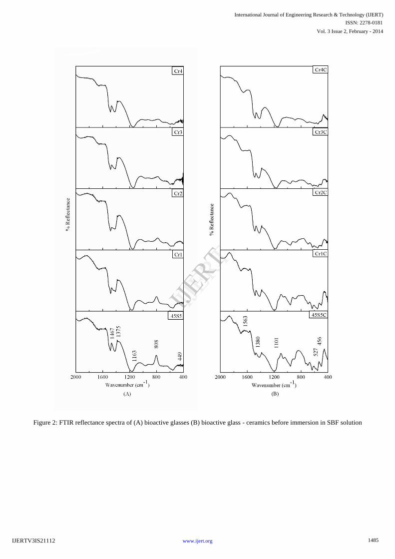
Figure 2: FTIR reflectance spectra of (A) bioactive glasses (B) bioactive glass - ceramics before immersion in SBF solution
1485
International Journal of Engineering Research & Technology (IJERT)
Vol. 3 Issue 2, February - 2014
IJERT
IJERT
ISSN: 2278-0181
www.ijert.orgIJERTV3IS21112

Figure 3: FTIR reflectance spectra of (A) 45S5 bioactive glasses (B) 45S5 bioactive glass - ceramics after soaking for different
day in SBF solution
1486
International Journal of Engineering Research & Technology (IJERT)
Vol. 3 Issue 2, February - 2014
IJERT
IJERT
ISSN: 2278-0181
www.ijert.orgIJERTV3IS21112

Figure 4: FTIR reflectance spectra of (A) Cr1doped bioactive glasses (B) Cr1doped bioactive glass - ceramics after soaking for
different day in SBF solution
1487
International Journal of Engineering Research & Technology (IJERT)
Vol. 3 Issue 2, February - 2014
IJERT
IJERT
ISSN: 2278-0181
www.ijert.orgIJERTV3IS21112

Figure 5: FTIR reflectance spectra of (A) Cr2 doped bioactive glasses (B) Cr2 doped bioactive glass - ceramics after soaking for
different day in SBF solution
1488
International Journal of Engineering Research & Technology (IJERT)
Vol. 3 Issue 2, February - 2014
IJERT
IJERT
ISSN: 2278-0181
www.ijert.orgIJERTV3IS21112
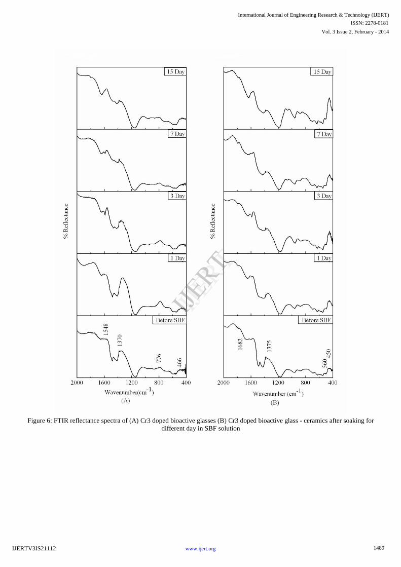
Figure 6: FTIR reflectance spectra of (A) Cr3 doped bioactive glasses (B) Cr3 doped bioactive glass - ceramics after soaking for
different day in SBF solution
1489
International Journal of Engineering Research & Technology (IJERT)
Vol. 3 Issue 2, February - 2014
IJERT
IJERT
ISSN: 2278-0181
www.ijert.orgIJERTV3IS21112

Figure 7: FTIR reflectance spectra of (A) Cr4 doped bioactive glasses (B) Cr4 doped bioactive glass - ceramics after soaking for
different day in SBF solution
1490
International Journal of Engineering Research & Technology (IJERT)
Vol. 3 Issue 2, February - 2014
IJERT
IJERT
ISSN: 2278-0181
www.ijert.orgIJERTV3IS21112

C. FTIR Reflection Spectra of bioglass and their
ceramic derivatives
Figure 2 shows the FTIR spectra of bioglass and their
ceramic derivative before SBF. In the bioglass peaks at
about 449, 808, 1163, 1375 and 1467 and glass-ceramic
peaks are 456, 527, 1101, 1389 and 1563 cm-1. The
reflection IR spectra peaks of bioglasses confirm the main
characteristic of silicate network. The resultant IR spectra at
449 cm-1
associated with a Si–O-Si symmetric bending
mode, the band at 808 cm-1
corresponds with Si–O–Si
symmetric stretch. The major band at about 1163 cm−1
can
be attributed to Si-O-Si stretching. The small band at 1467
cm-1
attributed to C–O vibrational mode. Table 5 depicts the
infrared frequencies and related functional structural groups
in the bioglasses [18].In glass-ceramic 45S5 and Cr doped
glass-ceramic has common peaks but 1101 cm-1 vanished in
Cr2, Cr3 and Cr4.The P-O bending (crystalline) peak at
550-620 cm-1 is the characteristics peaks of calcium
pyrophosphate which indicate the bioactivity of the glass
ceramic. The IR spectra peaks of Cr1, Cr2, Cr3 and Cr4
samples have the same behavior of Cr0 with small change in
the peaks intensities. The bioglasses substituted with
chromium are not showing noticeable changes in the IR
spectra bands.
D. pH behavior in SBF
The variation in pH values of simulated body
fluid (SBF) solution due to soaking of bioactive glasses and
glass - ceramics for various time periods is shown in Figure
8 & 9 respectively.
Figure.8 pH Bioglass Sample
It was observed that the addition of Cr2O3 in the base
bioactive glass (45S5) the pH values of SBF solution
increased during first 3 days of soaking due to faster
release of Ca and Na ions from the sample surface caused
an increase in the pH. Then it is found to decrease and
attained nearly a constant value in all cases. The crystallized
bioactive glasses also showed the same behavior of
decreases in pH. The reactions occurred are in favor of
formation of hydroxy apatite like layer on the surface of the
samples.
Figure.9 pH Bioglass-ceramic
E. In Vitro bioactivity of bioglasses and glass-
ceramic by Reflection spectroscopy
Figures.3–7 show the FTIR Reflection spectra
bands of the bioglasses and glass-ceramic before and after
immersion in SBF for different time periods 0, 1, 3, 7 and
15 days.
Hench [17] studied the stages of apatite formation
on the surface of the samples and spectral wavenumbers
were correlated with Table 6 that the changes in the IR
spectra bands after immersion in SBF for prolonged time
period was also as suggested by Kim et al. [18] and
Filgueira [20].
It may be seen from infrared spectra Figure 3-7 that
the chromium substitutions in base bioglasses are not
affected in apatite formation. Glass-ceramic before
immersion in SBF solution shows the similar peaks except
in glass-ceramic 1 has been extra peak at 1556 cm-1 one
peaks in before SBF 1101 cm-1 vanished. After immersion
in SBF solution from 1to 15 days all sample has common
peaks at around 527,813 and 1356 cm-1.The P-O stretching
peak at 1000-1220 cm-1 indicating the formation of
amorphous CaO.P2O5 rich layer. The P-O bending
(crystalline) peak at 550-620 cm-1 is the characteristics
peaks of calcium pyrophosphate which indicate the
bioactivity of the glass-ceramic.
E. Surface morphology of bioglass sample by SEM
The SEM micrographs Figure 12 & 13 shows the bioglass
samples (Cr0, Cr1, Cr2 and Cr3 ) before and after the
immersion in the simulated body fluid for 7 days at 37.0 oC.
The micrographs exhibits the polycrystalline fine texture
formed on surface layer which encourage the same
1491
International Journal of Engineering Research & Technology (IJERT)
Vol. 3 Issue 2, February - 2014
IJERT
IJERT
ISSN: 2278-0181
www.ijert.orgIJERTV3IS21112

crystallinity and also comparison with the surface layer
formed on the base bioglass samples [36].
F. Density of bioglasses and glass-ceramic
measured by Archimedes principle
Figure 10 shows the densities of bioglasses and
glass-ceramic were measured corresponding with chromium
substitution (45S5-2.60, Cr1-2.63, Cr2-2.64, Cr3-2.65 and
Cr4-2.67) and glass-ceramic (45S5C-2.81, Cr1C-2.84,
Cr2C-2.87, Cr3C-2.88 and Cr4C-2.89). It is observed that
the densities of the samples were increased with increasing
chromium content from 2.60 to 2.89 gm/cm3, it may be due
to partial replacement of SiO2 with Cr2O3, which is
attributed to the replacement of a light element (density of
SiO2 -2.63) with a heavier one (Cr2O35.22) in the
composition.
Figure.10 Density of bioglass & glass-ceramic
G. Flexural strength of bio-glasses and glass-
ceramic
Fig 11 shows the results of the flexural strength for
Cr0, Cr1, Cr2, Cr3 and Cr4 samples. The results
demonstrate an increasing tendency in flexural strength as
the percentage of chromium increases (44.48, 54.25, 61.42,
70.52 and 77.58) and glass-ceramic (104.27, 114.47,
120.20, 129.32 and 135.95 respectively).
Figure.11 Flexural Strength of bioglass & glass-ceramic
Figure 12. Scanning Electron Microscope (SEM) of bioactive glasses after immersion in SBF for 7 days
1492
International Journal of Engineering Research & Technology (IJERT)
Vol. 3 Issue 2, February - 2014
IJERT
IJERT
ISSN: 2278-0181
www.ijert.orgIJERTV3IS21112

Figure.13. Scanning Electron Microscope (SEM) of bioactive glass-ceramics after immersion in SBF for 7 days
IV. CONCLUSIONS In the present investigation, a comparative study was
made on physical and bioactive properties of Cr2O3
substituted 45S5 bioactive glasses and glass - ceramics. The
Following conclusions are obtained from this investigation.
The chromium substituted base bioglass showed decrease in
both nucleation and crystallization temperatures. It is
concluded that an increase in chromium oxide content in
this series of glasses found to increase in bioactivity, this is
also supported by pH and SEM analysis. Crystallized
bioactive glasses decreases their bioactivity but increased
their density and flexural strength compared to bioglass.
The prepared bioglasses and glass-ceramics FTIR results
showed the silicate network structure. The crystallized
bioglasses showing main phase as Na2Ca2Si3O9 (Sodium
calcium silicate)
REFERENCES
[1] L. L. Hench,J. Mater. Sci.: Mater. Med., 2006, 17, 967.
[2] I. D. Xynos, A. J. Edgar, L. D. K. Buttery, L. L. Hench and J. M. Polak,Biochem. Biophys. Res. Commun., 2000, 276, 461.
[3] I. D. Xynos, A. J. Edgar, L. D. K. Buttery, L. L. Hench and J. M.
Polak,J. Biomed. Mater. Res., 2001, 55, 151. [4] R. M. Moss, D. M. Pickup, I. Ahmed, J. C. Knowles, M. E. Smith
and R. J. Newport,Adv. Funct. Mater., 2008, 18, 634.
[5] S. P. Valappil, D. Ready, E. A. Abou Neel, D. M. Pickup, W. Chrzanowski, L. A. O'Dell, R. J. Newport, M. E. Smith, M. Wilson
and J. C. Knowles,Adv. Funct. Mater., 2008, 18,732.
[6] ElBatal HA, Khalil EMA, Hamdy YM (2009) Ceram Int 35:1195. [7] Hench LL, Wilson J (eds) (1993) An introduction to bioceramics
world scientific. Singapore.
[8] Serra J, Gonzalez P, Liste S, Chiussi S, Leon B, Perez-Amor M,Ylanen HO, Hupa M (2002) J Mater Sci Mater Med 13:1221.
[9] Fatma H. ElBatal, Amany ElKheshen, Materials Chemistry and
Physics 110 (2008) 352–362. [10] Oki A, Parveen B, Hossain S, Adeniji S, Donahue H, J Biomed Mater
Res A. 2004; 69: 216–21.
[11] Saboori A, Sheikhi M, Moztarzadeh F, Rabiee M, Hesaraki S, Tahriri M, Adv Apll Ceram. 2009;108:155–61.
[12] Balamurugan A, Rebelo AH, Lemos AF, Rocha JH, Ventura JM,
Ferreira JM, Dent Mater. 2008;24:1374–80. [13] Balamurugan A, Balossier G, Kannan S, Michel J, Rebelo AH,
Ferreira JM, Acta Biomater. 2007;3: 255–62.
[14] M. D. O’Donnell, P. L. Candarlioglu, C. A. Miller, E. Gentleman and M. M. Stevens, J. Mater. Chem., 2010, 20, 8934–8941.
[15] Kokubo T, Takadama H, Biomaterials 2006; 27:2907–15.
[16] Oana Bretcanu a,1, Xanthippi Chatzistavrou a, Konstantinos Paraskevopoulos, Journal of the European London, 1989, p. 59.
[17] L.L. Hench, Journal of American Ceramic Society 81 (1998) 1705–
1728. [18] J. P. Nayak, S. Kumar, and J. Bera, Journal of Non-Crystalline Solids,
356, 1447–1451 (2010).
[19] V.R. Mastelaro, E.D. Zanotto, N. Lequeux, R. Cortes, J. Non-Cryst. Solids 262 (2000) 191–199.
[20] Rehman I, Karsh M, Hench LL, Bonfield W, J Biomed Mater Res.
2000 May;50(2):97-100.
Corresponding author : Vikash Kumar Vyas
Indian Institute of Technology(BHU), Varanasi
1493
International Journal of Engineering Research & Technology (IJERT)
Vol. 3 Issue 2, February - 2014
IJERT
IJERT
ISSN: 2278-0181
www.ijert.orgIJERTV3IS21112