dvt: a life‐threatening condition - rn.com · deep vein thrombus (dvt) and pulmonary embolism...
TRANSCRIPT

DVT: A Life‐Threatening Condition
Contact Hours: 2.0 First Published: August 1, 2012
Course Expires: December 31, 2019
Copyright© 2015 by RN.com All Rights Reserved
Reproduction and distribution of these materials is prohibited without the
express written authorization of RN.com

Acknowledgements
RN.com acknowledges the valuable contributions of…
Suzan Miller‐Hoover, DNP, RN, CCNS, CCRN
Disclaimer
RN.com strives to keep its content fair and unbiased. The author, planning committee, and reviewers have no conflicts of interest in relation to this course. Conflict of Interest is defined as circumstances a conflict of interest that an individual may have, which could possibly affect Education content about products or services of a commercial interest with which he/she has a financial relationship.
There is no commercial support being used for this course. Participants are advised that the accredited status of RN.com does not imply endorsement by the provider or ANCC of any commercial products mentioned in this course.
There is no "off label" usage of drugs or products discussed in this course. You may find that both generic and trade names are used in courses produced by RN.com. The use of trade names does not indicate any preference of one trade named agent or company over another. Trade names are provided to enhance recognition of agents described in the course.
Note: All dosages given are for adults unless otherwise stated. The information on medications contained in this course is not meant to be prescriptive or all‐encompassing. You are encouraged to consult with physicians and pharmacists about all medication issues for your patients.

Purpose
The purpose of this course is to update nurses on current guidelines for prophylaxis, treatment, and complications arising from venous thromboembolism (VTE) formation. Learning Objectives
After successful completion of this continuing education course the learner will:
1. Recognize the signs, symptoms and risk factors for venous thromboembolism. 2. List criteria anticoagulant selection for an individual patient. 3. Identify differences between unfractionated heparin and low molecular weight heparin. 4. Discuss complications of venous thromboembolism. 5. Review prophylactic measures used to prevent complications. 6. Review post‐thrombotic syndrome and discuss pulmonary embolism (PE). 7. Identify content for patient teaching.

Introduction
Venous thromboembolism (VTE) is the formation of a venous blood clot; usually in a large vein in the lower extremities, but VTE may form in the large veins of the chest especially when a central venous catheter is in place. Deep vein thrombus (DVT) and pulmonary embolism (PE) are manifestations of VTE.
By the time a patient has reached the age of puberty, the clotting cascade has matured and the risk for VTE formation is high. VTE formation was once thought to be caused by restricted motility; however, recent data shows that a fully mobile patient may still be at risk if other factors are present. This module will discuss the mobility issue as well as the additional risk factors that contribute to the development of a VTE. Venous thromboembolism has been added to the Center for Medicare Services“never event” list. (CMS, 2014)
NOTE: Third party payers (insurance companies) do not reimburse costs accrued after a “never event” occurs. Therefore it is essential that the patient is assessed thoroughly for the presence of a “never event” on admission. Complete documentation in the medical record will help ensure that the facility receives the reimbursement due for care given. Did you know? Hospitals are responsible for a VTE even when it appears after discharge? VTE are reportable if they occur up to 30 days post discharge.
Statistics
Venous thromboembolism affects 300,000 to 600,000 Americans annually. The rate of VTE varies depending on the age, gender and race of the patient. In patients equal to or older than 80 years, the rate is approximately 1 per 100; however in the younger population, the rate is approximately 1 per 1000. Black men have a higher incidence than other ethnicities and gender, but during reproductive years, women have a higher rate of VTE (Beckman, Hooper, Critchley, & Ortel, 2010; Agency for Healthcare Research and Quality [AHRQ], 2012). According to the Agency for Healthcare Research and Quality (AHRQ), 2012, VTE constitute the largest cause of preventable hospital deaths. 100,000 deaths are directly caused by VTE and VTE contribute to another 100,000 deaths. The hospital length of stay is increased by 2‐5 days and the cost of each VTE is estimated to be $7,500 (AHRQ, 2012). Test Yourself: The Centers for Medicare Services has indicated that VTE should be an event that never occurs during a hospitalization. As a result of this designation: a. Third party payers can decide if they are going to pay for costs incurred for VTE treatment b. Third party payers will not pay for costs incurred for VTE treatment
c. The designation of a never event has no effect on third party payment Answer: B Rationale: Third party payers (insurance companies) do not reimburse costs accrued after a “never event” occurs. Therefore it is essential that the patient is assessed thoroughly for the presence of a “never event” on admission.
Evidence‐Based Guidelines for VTE Prophylaxis
The American College of Chest Physicians published the “Diagnosis of DVT: Antithrombotic Therapy and Prevention of Thrombosis, 9th edition, 2012; evidence‐based clinical practice guidelines for VTE prophylaxis. These guidelines detail which conditions increase the risk of VTE and which prophylactic approaches should be utilized for VTE prevention (Bates et at., 2012). This document was validated via external and internal peer review. This document does not cover:
• People younger than 18 years of age • Outpatients • Emergency Department (ED) patients • Patients admitted with a diagnosis of VTE
The following sections are the major recommendations of these nationally recognized guidelines. To review the entire document, refer to the AHRQ National Guideline Clearing
House at http://www.guideline.gov/content.aspx?id=24106&search=dvt+risk#tiptop
Diagnosis of DVT: Antithrombotic Therapy and Prevention of Thrombosis, 9th edition, 2012; Evidence‐based clinical practice guidelines for VTE prophylaxis.
Assessment: Upon admission all patients should be assessed for VTE risk. Additionally as the patient’s condition changes; surgery, admission to the critical care unit, or transfer to an acute care unit; reassessment should occur. When the patient is determined to be at risk for VTE prevention strategies should be implemented, patient education conducted, and medical record documentation completed. Recognizing Risk Factors for VTE Formation
• Active cancer or cancer treatment • Age over 60 years • Critical care admission • Dehydration • Known thrombophilia • Obesity (BMI over 30 kg/m2) • One or more significant medical comorbidities (heart disease, metabolic, endocrine or
respiratory pathologies; acute infectious diseases; inflammatory conditions) • History of VTE • Use of hormone replacement therapy • Use of estrogen‐containing contraceptive therapy • Varicose veins with associated phlebitis

• Fracture of pelvis/hip/lower extremity • Indwelling central venous catheter • Immobility • Major surgery
(AHRQ, 2012) Assessing the Risks of Venous Thromboembolism Medical patients are at an increased risk for VTE if they:
• Have or expected to have significantly reduced mobility for 3 days or more • Have or expected to have reduced mobility plus one of the above risk factors • Virchow’s Triad: vein injury, venous stasis, and hypercoagulability
Surgical patients are at an increased risk for VTE if they have a:
• Surgical procedure that lasts greater than 90 minutes (anesthesia and surgical time) o Minor surgery for patients aged 40 to 60 o Major surgery in patients under 40
• Pelvic or lower extremity surgical procedure that lasts greater than 60 minutes (anesthesia and surgical time)
• Acute surgical admission with an inflammatory or intra‐abdominal condition • Expected significantly reduced mobility • One or more of the above risk factors
(Bates, et al., 2012; National Institute for Health and Clinical Excellence [NICE], 2010)
Prevention Strategies VTE prevention strategies should be implemented as soon as the patient has been assessed for risk and contraindications.
The best prevention strategy is to stay mobile The evidence‐based clinical guidelines for VTE prevention states:
• Dehydration should be prevented unless clinically indicated • Encourage mobility as soon as possible • Aspirin or other antiplatelet agents are NOT adequate prophylaxis • Consider temporary inferior vena cava filters for:
o Patients at very high risk (previous VTE event or active malignancy) o Mechanical and pharmacologic prophylaxis is contraindicated
Mobility: The first step in prevention of VTE is to assess the patient’s mobility and encourage early ambulation. Inpatients may benefit from the use of a mobility scoring system. One scoring system, Braden Scale for Predicting Pressure Sore Risk, contains a mobility score. This score or similar scores, objectify the ability of the patient to move about; which allows the healthcare worker determine whether VTE prophylaxis is appropriate. To view the Braden Scale please click on this link

http://www.bradenscale.com/products.htm The second step is to apply mechanical VTE prophylaxis, compression stockings and/or sequential compression devices. These mechanical devices help avoid venous stasis in immobile or partially immobile patients. The third step is the use of pharmacotherapeutic agents, heparin, low molecular weight heparin, or warfarin.
NOTE: VTE development is not limited to healthcare facilities; VTE may develop during long periods of sedentary activity such as long flights, train rides or car trips. Advise patients, friends, and family to avoid prolonged sitting (longer than six hours) during travels. Encourage exercise; walking in place, walking the aisles periodically, and to arrange trips with shorter segments of confinement. One additional hint to encourage mobility; drink lots of water as this will facilitate more trips to the bathroom and therefore more mobility.
Mechanical Devices: Contraindications: Anti‐embolism devices are contraindicated in patients with
• Stroke • Suspected or proven peripheral arterial disease • Peripheral arterial bypass grafting • Arteriosclerosis • Severe peripheral neuropathy • Massive leg edema • Pulmonary edema • Cardiac failure • Local skin/soft tissue diseases such as recent skin graft or dermatitis • Extreme deformity of the leg • Gangrenous limb • Doppler pressure index <0.8 • Cellulitis • Known allergy to material of manufacture • Unusual leg size or shape • Venous wounds/ulcers
(Bates et al., 2012, NICE, 2010)
Use of Mechanical Devices
Compression stockings, sequential compression devices, and foot pumps help the calf muscle to increase venous return, improve circulation and tissue oxygenation, and reduce swelling thus reducing the risk of VTE (Sachdeva, Dalton, Amaragiri, &Lees, 2014). All three devices can be used independently or in conjunction with each other, depending on the patient’s condition. Nursing staff may place compression stockings, sequential compression devices, and foot pumps without a physician order. However, in many facilities, an order is required to bill for

the devices. Check with your facility to see if an order is required.
Note: Compression stockings may not be well tolerated and can be difficult to put on patients. Thigh‐high devices, while distributing the mechanical effect over a larger proportion of the leg, are less tolerated, due to the heat generated from wearing them. Use of knee‐high sequential compression devices may encourage compliance and may be easier to place on patients. Recent research indicates that knee‐high sequential compression devices are as efficacious as thigh‐high sequential compression devices (Sajid, Desai, Morris, & Hamilton, 2012). Additionally, knee high sequential compression devices are less expensive than thigh high. To facilitate the use of sequential compression devices in the operating room, knee high devices are more conducive to accessing the patient. Daily Use:
• Use devices that provide graduated compression and produce a calf pressure of 14‐15 mmHg
• Measure the leg for correct sizing • Remove devices for daily hygiene • Remove devices 2‐3 times a day for skin inspection
Discontinue device: • When patient is mobile • If there are markings, blisters, or discoloration of the skin • Pain or discomfort occur
Note: Mechanical devices should remain on the patient as long as possible each day, including when up in a chair, as the efficacy of their use increases with time in use. When the devices are removed for an extended period of time or when a vascular line is removed; a venous ultrasound should be considered to verify the presence or absence of a thrombus.
Use of Pharmacologic Prophylaxis Contraindications:
• Active bleeding • Acquired bleeding disorders (such as acute liver failure) • Concurrent use of anticoagulants known to increase the risk of bleeding (such as warfarin
with international normalized ratio [INR] higher than 2) • Lumbar puncture/epidural/spinal anesthesia expected within the next 12 hours • Lumbar puncture/epidural/spinal anesthesia within the previous four hours • Acute stroke • Major trauma • Recent neurosurgery • Thrombocytopenia (platelets less than 75 x 109/l) • Uncontrolled hypertension (230/120 mmHg or higher) • Untreated inherited bleeding disorders (such as hemophilia and von Willebrand's disease) • Pregnancy and impending delivery date • Unwillingness or inability to take oral anticoagulation

Indications: • Hemorrhagic stroke has been excluded
Risk of bleeding assessed to be low • Major restriction of mobility • Previous history of VTE • Dehydration • Comorbidities (see risk for VTE list earlier in module)
(NICE, 2010)
Anticoagulant Use
Anticoagulants used to prevent venous thrombi include unfractionated heparin (UFH), low molecular weight heparin (LMWH), and warfarin. Heparin inhibits a variety of factors in the coagulation cascade and ultimately prevents fibrin deposition. Warfarin reduces the formation of blood clots primarily by inhibiting the vitamin K dependent coagulation factors.
Unfractionated heparin and low molecular weight heparin may be utilized for VTE prevention. However, due to the monitoring required by unfractionated heparin, low molecular weight heparin has become the drug of choice; especially when warfarin is not applicable (Bates et al., 2012). In the case of a severe anticoagulant overdose resulting severe hemorrhage; fresh frozen plasma or clotting factor transfusions may be necessary.
Note: Anticoagulants prevent thrombi formation and/or extension of formed thrombi; however, they will not breakdown established clots.
Did you know?
Heparin‐induced thrombocytopenia (HIT) is a potentially devastating immune mediated adverse drug reaction to all types of heparin. This drug reaction is caused by antibodies that activate platelets in the presence of heparin resulting in thromboembolic complications involving both the arterial and venous systems. Treatment options focus on inhibiting thrombin formation or direct thrombin inhibition. Warfarin should not be used until the platelet count has recovered (Ahmed, Majeed, & Powell, 2008).
Low Molecular Weight Heparin (LMWH) The dosage for LMWH is based on weight, age, and renal function. LMWHs come in ampules, syringes, and vials. Two of the most common types of LMWH are:
• Enoxaparin sodium • Dalteparin sodium
The action and benefits of LMWH are more predictable than UFH or warfarin. Thus, close monitoring of aPTT and PT/INR is NOT necessary. However, LMWH should be used in caution with patient with renal insufficiency or failure as LMWH is excreted by the kidneys.

LMWH is the better option for DVT prophylaxis; as it is given daily, has less bleeding tendencies, and carries a lower risk for the development of Heparin Induced Thrombocytopenia (HIT). Patients may be discharged home on LMWH if appropriate.
In the case of an overdose of LMWH, protamine sulfate is the antidote. The following table provides guidance on LMWH dosing. The lower end of the range should be considered for patients with mild (creatinine clearance 50‐80 mL/min) to moderate (creatinine clearance 30‐50 mL/min) renal impairment (RxList a, 2015).
Indication Dosage Regimen
Prophylaxis in abdominal surgery 30 ‐ 40 mg administered sub q once daily
Prophylaxis in hip or knee replacement surgery
30 ‐ 40 mg administered sub q once daily
Prophylaxis in medical patients during acute illness
30 ‐ 40 mg administered sub q once daily
Inpatient treatment of acute deep vein thrombosis with or without pulmonary embolism, when administered in conjunction with warfarin sodium
1 mg/kg administered sub q once or twice daily OR 1.5 mg/kg once a day
Outpatient treatment of acute deep vein thrombosis without pulmonary embolism, when administered in conjunction with warfarin sodium
1 mg/kg administered sub q once or twice daily
Adapted from rxlist.com/Lovenox‐drug/indications‐dosage.htm Oral Anticoagulation: Warfarin The dosage for warfarin is individualized and adjusted according to protime level (PT) and international normalized ratio (INR), a standardized value. Adjust the warfarin dose to maintain a target INR of 2.5 (INR range, 2.0‐3.0) for all treatment durations. (Rxlistc, 2015)
Administer warfarin at the same time daily. Do not administer the oral form with food.
If your patient has bleeding, fever, or rash, hold the dose and check with the prescriber.
In the case of an overdose of warfarin, vitamin K given orally or IV is the antidote.

Warfarin is affected by many foods, especially those foods high in vitamin K. Patient education should include information regarding foods that interfere with warfarin.
Unfractionated Heparin (UFH): UFH is most commonly available in the form of heparin sodium, and can be administered intravenously or subcutaneously. UFH dosing is based on and titrated to the activated partial thromboplastin time (aPTT) results and individualized to weight, age, and health. Adjust the dosage to maintain the activated partial thromboplastin time (aPTT) of 1.5 to 2 times normal or the whole blood clotting time of 2.5 to 3 times the control value (Rxlistb, 2015). Prior to administering heparin draw a baseline platelet count. In the case of an overdose of unfractionated heparin, protamine sulfate is the antidote. (Arnold, et al., 2010)
For more detailed information on Heparin, please see RN.com's course entitled: Patient Safety: Honing In On Heparin.
Did you Know: UHF may be the drug of choice for patients with several renal failure as LMWH is excreted by the kidney. Test your knowledge: True or False: HIT is an adverse medication event that manifests as severe hemorrhage. Answer: False Rationale: Heparin‐induced thrombocytopenia (HIT) is a potentially devastating immune mediated adverse drug reaction to all types of heparin. This drug reaction is caused by antibodies that activate platelets in the presence of heparin resulting in thromboembolic complications involving both the arterial and venous systems. Bridging from Heparin to Warfarin Warfarin will be started when the patient is deemed to stable enough to be discharged. Within a few days of discharge; heparin titration will begin and the warfarin dose will be calculated and started. The heparin is discontinued once the patient shows a stable international ratio (INR) value between 2 and 3.5 for two consecutive days. This bridging may take five to seven days. Clinical Presentation

Venous thromboembolism is often asymptomatic, and thus often challenging to recognize. The following signs and symptoms may be present:
• Redness, warmth and swelling in the affected extremity • Unilateral pitting edema with collateral venous
engorgement, localized warmth and erythema • Dilated superficial veins in the affected extremity • Pain, tenderness, or a dull ache in the affected extremity

Confirmation of a Suspected VTE Diagnosis Deep Vein Thrombosis
A definitive diagnosis of VTE can be established by the use of one or more of the following diagnostic tools:
Diagnostic Imaging Venous ultrasonography Venous ultrasound is the method of choice for diagnosing VTE as it is non‐invasive and accurate. This test can be completed at the bedside.
Venography An iodine‐based contrast medium is injected into a dorsal vein in the foot; detailed X‐rays are taken; detecting venous filling defects. Venography is a useful diagnostic tool particularly when there are poor views on ultrasound and high clinical suspicion for VTE. This diagnostic tool is not a risk‐free strategy and it has many disadvantages. This test cannot be completed at the bedside. Additionally, invasive techniques can be painful and increase the likelihood of localized infection and there can be problems obtaining venous access in the foot. Adverse reactions to the contrast can include urticaria, bronchospasm, and deterioration in renal function.
Confirmation of a Suspected VTE Diagnosis (Cont) Pulmonary Embolism
Chest X‐ray A chest X‐ray is obtained in patients presenting with a suspected pulmonary embolism (PE) to assist in determining the appropriate scanning technique to use. For example, a ventilation/perfusion scan cannot exclude PE in the presence of an abnormal chest X‐ray. Note: A normal chest X‐ray will not exclude PE.
Electrocardiogram (ECG) An ECG is of limited value as the ECG can be normal in the presence of PE and should be used in conjunction with the appropriate diagnostic scans..
Ventilation/Perfusion Scan (V/Q scan) A pulmonary V/Q scan is a combination of two tests that use inhaled and injected radioisotopes to measure the air and blood flow to the lungs. The tests can be used together or separately. The perfusion scan is sometimes used alone for the exclusion or detection of PE in a patient with a normal chest X‐ray and low clinical suspicion of PE. If there is a high clinical suspicion of PE and a negative or an equivocal V/Q scan, a computed tomography pulmonary angiogram is

required to confirm diagnosis.
Computed Tomography Pulmonary Angiogram (CTPA) CTPA is the gold standard test for diagnosing PE and has a sensitivity of greater than 95%. This diagnostic test is more accurate, enables an alternative diagnosis to be made if PE is excluded, and is diagnostic even with an abnormal chest X‐ray or underlying lung disease. Signs and Symptoms: Pulmonary Embolism Pulmonary emboli are a life‐threatening complication caused by obstruction of the pulmonary arteries, which increases the workload of the heart. The heart may be unable to maintain sufficient circulation to support blood pressure, resulting in hypovolemic shock.
Estimates suggest that 60,000‐100,000 Americans die of DVT/PE (Centers for Disease Control, 2011). Small emboli may silently infarct a portion of the lung; a huge embolus at the bifurcation of the main pulmonary artery (called a saddle embolus) will create obvious symptoms.

The arrow s pul
Image c
shows filling monary emb
courtesy of AH
defect due to olism.
RQ, 2012.
Early signs & symptoms of a PE may include: • Dyspnea • Tachypnea • Tachycardia • Chest pain • Anxiety • Cough • Hemoptysis • Fever • Arrhythmias • Hypertension • Syncope • Diaphoresis

Pharmacological Management of VTE’s VTE’s can be pharmacologically treated with anticoagulants and thrombolytics: anticoagulants help prevent further formation of clots and thrombolytics breakdown the existing clot and aid in absorption of the clot. Safe anticoagulation therapy requires striking a balance between thrombosis and bleeding to ensure that the patient receives safe and beneficial therapy. There are several considerations in selecting anticoagulant therapy:
Always familiarize yourself with your facility's protocol for anticoagulation and thrombolytic therapy.
Contraindications to thrombolytic therapy:
• Any previous history of hemorrhagic stroke • History of stroke, dementia, or central nervous system damage within one year • Head trauma or brain surgery within 6 months • Known intracranial neoplasm • Suspected aortic dissection • Internal bleeding within six weeks • Active bleeding or known bleeding disorder • Major surgery, trauma, or bleeding within three weeks • Traumatic cardiopulmonary resuscitation within three weeks
Thrombolytics:
Thrombolytics act by breaking up and dissolving blood clots and are administered intravenously or through a catheter directed at the thrombus.
Thrombolytics can be used for the lysis of acute, extensive thrombi of deep veins such as those involving the popliteal and more proximal vessels. It can also be used for the lysis of pulmonary emboli. However, it is not indicated for arterial emboli originating from the left side of the heart, due to the risk of cerebral embolism (RxLista, 2015).
Thrombolytics should be instituted as soon as possible after onset of the thrombotic event, preferably within seven days. Side effects include:
• Nausea and dizziness • Hypotension • Mild fever • Headaches • Flushing, rapid or abnormal heartbeat with or without chest pain
Nursing Alert! Monitor for excessive bleeding every 15 minutes for the first hour of thrombolytic therapy, and avoid intramuscular injections, IV starts, or surgical
procedures immediately following the implementation of thrombolytics. Other Management Strategies for Diagnosed VTE’s
VTE patients who do not respond to anticoagulants or where anticoagulant therapy is contraindicated may need invasive management strategies.
Venous Thrombectomy The clot is suctioned out via a catheter or surgically removed.
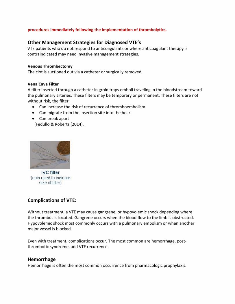
Vena Cava Filter A filter inserted through a catheter in groin traps emboli traveling in the bloodstream toward the pulmonary arteries. These filters may be temporary or permanent. These filters are not without risk, the filter: • Can increase the risk of recurrence of thromboembolism • Can migrate from the insertion site into the heart • Can break apart (Fedullo & Roberts (2014).
Complications of VTE:
Without treatment, a VTE may cause gangrene, or hypovolemic shock depending where the thrombus is located. Gangrene occurs when the blood flow to the limb is obstructed. Hypovolemic shock most commonly occurs with a pulmonary embolism or when another major vessel is blocked.
Even with treatment, complications occur. The most common are hemorrhage, post‐thrombotic syndrome, and VTE recurrence. Hemorrhage Hemorrhage is often the most common occurrence from pharmacologic prophylaxis.

Signs and Symptoms of Hemorrhage
This is what to look for: Unusual bruising Joint pain or edema Bleeding gums Epistaxis Hematuria Heavy menstrual flow Constipation Black, tarry stools Back pain Vertigo Weakness Headache
Complications of VTE: Heparin Induced Thrombocytopenia
Heparin‐induced thrombocytopenia (HIT) is caused when patients develop an antibody to heparin and develop abnormal clotting.
HIT is characterized by:
• A reduced platelet count of 50% less than the pre‐heparin baseline • Hypercoagulability (abnormal clotting) • Venous and arterial thrombosis
Note: HIT can occur any time after heparin is started within a range of 30 minutes to a month; classic onset occurs within four to 14 days. Argatroban, a direct thrombin inhibitor, is anticoagulant therapy for patients with HIT.
For more information on HIT, please refer to RN.com's course: Heparin Induced Thrombocytopenia.
Complications of VTE: Post Thrombotic Syndrome
Post Thrombotic Syndrome is tissue injury that may occur 2 years following a VTE. This syndrome presents with pain, swelling, paresthesia (abnormal sensation), venous ulceration, edema, and skin changes in the affected extremity. This is due to impaired venous circulation related to VTE residual damage in the vein and valves. The incidence of residual damage is decreased when thrombolytics are given and when graduated compression stockings are used (Lowe, 2010).

Patient Education
In‐Patient: Compliance is key to the prevention of VTE. It is essential that the patient and family understand the risks and complications of VTE to ensure compliance with VTE management. Patients and families should receive verbal and written information regarding:
• Risks and possible complications • Importance of VTE prophylaxis • Correct use of VTE prophylaxis • Types of VTE prophylaxis and the inherent risks associated with the treatment • Signs and symptoms of thrombosis formation
This education should be documented in the electronic medical record. Discharge: Discharge education should include
• Signs and symptoms of venous thrombosis • Recommended duration of VTE prophylaxis • Importance of the correct and continuing use of VTE prophylaxis • When to notify their physician • Medication use and side effects
Ideally, more than one caretaker should be educated. Allocate plenty of time to implement your teaching. Divide patient teaching into several short sessions, allowing time for the patient to perform a return demonstration when applicable. Document teaching provided and to whom in the medical record. If the discharge assessment indicates that the patient and/or family are not able to comply with this regime, discuss the possibility of obtaining in‐home medical assistance.
Warfarin: Inform patients to take warfarin at the same time every day without food. If a dose is missed and the omission is noticed quickly, advise the person to take missed dose as soon as possible. However, when an entire day has passed, they should NOT to double the next dose.
Warfarin interacts with numerous medications, foods, and herbs; provision of a written list to avoid or minimize should be given to the patient at discharge.
Key points to emphasize are:
• Keep routine laboratory appointments • Avoid excessive use of over‐the‐counter salicylates such as aspirin;
acetaminophen (Tylenol®); and non‐steroidal anti‐inflammatory drugs (NSAID). • Be alert to increased bleeding after taking antibiotics. o Antibiotics deplete vitamin K, thereby increasing warfarin’s anticoagulant effect •
Encourage moderation and consistency in eating foods high in vitamin K, rather than strict avoidance of these foods.
o Limit foods high in vitamin K including broccoli, lettuce, asparagus, peas, brussel sprouts, spinach, cabbage and cauliflower

o Limit intake of substances that interfere with warfarin, such as vitamin C, green tea, cranberry juice, garlic, ginger, ginseng, and goldenseal • Consume alcohol in moderation; large amounts increase warfarin’s anticoagulant effect (Lippincott, Williams & Wilkens, 2012)
• Ensure that the provider and pharmacist are fully informed of all other medications and supplements the patient uses
• Stop warfarin three days prior to any invasive dental or surgical procedure with physician guidance
• Encourage exercise with caution to avoid injuries that might cause bleeding
Safety precautions to minimize risk of bleeding: • Encourage use of an electric razor and soft toothbrush • Install good lighting • Remove area rugs to minimize risk of falls • Install handrails where necessary • Avoid wax or polish on floors
Emphasize the importance of reporting hemorrhage. Look for and report nosebleeds, unusual bruising, black or tarry stools, dark or blood‐tinged urine, weakness and/or back pain, or an unusually heavy menstrual period.
Teach patients to identify the signs and symptoms of thrombosis and to report any chest pain, extremity pain, redness, or swelling; discoloration of fingers or toes or tingling or numbness in arms or legs; and/or slurred speech to their provider
Case Study:
Ann Tate, a 24‐year‐old travel journalist, is admitted with a persistent dull ache in her upper right leg. On examination you observe that the leg is edematous, reddened, and warmer than the other leg. Ann’s symptoms started on the long return flight back from an overseas assignment. She reports that soaking in a hot tub and taking aspirin made no difference. You learn that Ann started taking oral contraceptives three months ago. She has not had any pregnancies, surgeries, or traumas. She recalls a cousin had multiple miscarriages related to a genetic blood disorder. A diagnosis is made after an ultrasound shows VTE in the leg proximal to the site of the ache.
Blood tests show fibrin degradation products indicating the presence of clots. Ann is started on 1mg/kg subcutaneous enoxaparin (Lovenox®). Oral warfarin (Coumadin®) is started the same day.
What will your patient education include? You will need to teach Ann how to give her own injections, signs and symptoms of hemorrhage, foods and drug interactions, and initiate a discussion regarding the risk of continuing with oral contraception.

Case Study:
Bob Jackson, a 52‐year‐old male, breaks his femur during a ski accident. The open fracture is surgically set, casted, and you meet Bob three weeks later in the ED. Bob presents with intolerable pain in his injured leg. He states the pain is quite different than what he previously experienced. His toes are blue and edematous. You assist in removing the cast.
Bob Jackson tells you he was given some form of heparin in the hospital and now takes warfarin (Coumadin®) to prevent clotting.
Despite his medication, Bob’s INR is only 1.3, indicating a need for a dosage increase. When questioned about his last value, Bob admits to erratic compliance with having his blood drawn.
What diagnosis do you anticipate? A VTE, most likely a deep vein thrombosis from immobilization due to the fracture and cast.
What is the anticipated management of this patient? Bob will most likely receive thrombolytic therapy to dissolve the clot, followed by a continuous infusion of unfractionated heparin. You will need to monitor the aPTT, for signs and symptoms of bleeding or possible PE. Prior to discharge, the UFH infusion will be changed to oral warfarin or a LMWH injection.

Case Study: Charlene Doe is an 80‐year‐old ICU patient who had an ischemic stroke 2 days ago. She is receiving anti‐platelet therapy and oxygen. A central venous catheter is in place. You are alarmed when she exhibits sudden cardiac arrhythmia and diaphoresis.
Her respirations and pulse increase and her BP falls. She appears highly anxious, clutching her chest, and gasping for breath.
What do you expect is happening? Charlene is most likely experiencing a PE related to an undiagnosed VTE in a lower extremity or from her central venous catheter.
What is your priority nursing intervention and rationale? Recognizing that Charlene’s condition may be caused by a large PE (small PE may be asymptomatic), notify the provider immediately, prepare to reverse impending shock and prepare for thrombolytic therapy. What type of treatment would you anticipate for this patient? Charlene will most likely receive thrombolytic therapy followed by a continuous infusion of unfractionated heparin. You will need to monitor the aPTT, for signs and symptoms of bleeding.
Conclusion
Prevention is key. Assessment for VTE risk is essential to determining the type of VTE prophylaxis that will be indicated for the particular patient. Early ambulation, use of mechanical prophylaxis and concurrent use of anticoagulants can significantly reduce the risk of VTE in an immobile patient or in a patient who is somewhat mobile and has comorbidities. Sequential compression devices, compression stockings, and foot pumps are an efficacious and safe treatment to prevent venous thromboembolism. Nurses should advocate for this type of prophylaxis. In the event that these devices are contraindicated, pharmacologic prophylaxis should be used. Pharmacologic prophylaxis is used in conjunction with mechanical devices or alone. Anticoagulation is a high risk treatment, healthcare workers play a key role in administering and monitoring these drugs to avoid untoward patient outcomes. Anticoagulants prevent thrombus formation but do not breakdown the clot. When VTEs occur, the provision of patient care becomes increasingly complex and costly. The use of thrombolytic medication increases the risk of untoward effects. The nurse plays an essential role in preventing poor outcomes in these patients. The nurse’s role, in conjunction with the physician and pharmacist, are crucial to venous thrombus prevention, especially for those inpatients at high risk. Knowledge about etiology, prevention, and treatment of VTE is essential to achieve a successful outcome.

References
Agency for Healthcare Quality & Research [AHRQ], (2012). Trauma centers vary in screening for deep vein thrombosis. Patient Safety & Quality, 381,
Arnold, J. , Dart, M., Barker, D., Maxwell, R., Burkhalter, H., Mejia, V., Smith, P.& Longley, J. (2010). Unfractionated heparin three times a day versus Enoxaparin in the prevention of deep vein thrombosis in trauma patients. The American Surgeon, 76, 563‐570.
Bates, S., M., Jaeschke, R., Stevens, S., M., Goodacre, S., Wells, P., S., Stevenson, M., D., … & Guyatt, G.,H. (2012). Diagnosis of DVT: Antithrombotic therapy and prevention of thrombosis, (9th ed): American College of Chest Physicians evidence‐based clinical practice guidelines. Chest, 141(2 Suppl), e351S‐418S. Retrieved from the AHRQ National Guideline clearing house at http://www.guideline.gov/content.aspx?id=24106&search=dvt+risk#tiptop
Centers for Disease Control and Prevention. (2011). Deep vein thrombosis (dvt) / Pulmonary embolism (PE) — blood clot forming in a vein. Retrieved from http://www.cdc.gov/ncbddd/dvt/facts.html
Centers for Medicare Services, (ND). Venous thromboembolism. Retrieved from http://partnershipforpatients.cms.gov/p4p_resources/tsp‐venusthromboembolism/toolvenousthromboembolismvte.html
Fedullo, P. & Roberts, A. (2014). Placement of vena cava filters and their complications. Retrieved from http://www.uptodate.com/contents/placement‐of‐vena‐cava‐filters‐and‐their‐complications.
Lippincott, Williams & Wilkins. (2012). Nursing 2012 Drug Handbook. Philadelphia, Baltimore & New York: Wolters Kluwer Company.
Lowe, G.D. (2010). Management of deep vein thrombosis to reduce the incidence of post‐ thrombotic syndrome. Phlebology 2010, 25 Suppl 1, 9–13.
National Collaborating Centre for Acute and Chronic Conditions. (2010). Venous thromboembolism: reducing the risk. Reducing the risk of venous thromboembolism (deep vein thrombosis and pulmonary embolism) in patients admitted to hospital. Retrieved from the AHRQ National Guideline clearing house at http://www.guideline.gov/content.aspx?id=24106&search=dvt+risk#tiptop

RxList a. (2015). Lovenox. Retrieved from http://www.rxlist.com/lovenox‐drug/indications‐dosage.htm RxList b. (2015). Unfractionated Heparin. Retrieved from http://www.rxlist.com/heparin‐drug/indications‐dosage.htm RxList c. (2015). Warfarin. Retrieved http://www.rxlist.com/coumadin‐drug.htm
Sajid, M.S., Desai, M., Morris, R. W., & Hamilton, G. (2012). Knee length versus thigh length graduated compression stockings for prevention of deep vein thrombosis in postoperative surgical patients. Cochrane Peripheral Vascular Diseases Group. DOI: 10.1002/14651858.CD007162.pub2 Sachdeva, A., Dalton, M., Amaragiri, S. A., & Lees, T. (2014). Graduated compression stockings for prevention of deep vein thrombosis. Cochrane Peripheral Vascular Diseases Group. DOI: 10.1002/14651858.CD001484.pub3 Please Read
This publication is intended solely for the use of healthcare professionals taking this course, for credit, from RN.com. It is designed to assist healthcare professionals, including nurses, in addressing many issues associated with healthcare. The guidance provided in this publication is general in nature, and is not designed to address any specific situation. This publication in no way absolves facilities of their responsibility for the appropriate orientation of healthcare professionals.
Hospitals or other organizations using this publication as a part of their own orientation processes should review the contents of this publication to ensure accuracy and compliance before using this publication. Hospitals and facilities that use this publication agree to defend and indemnify, and shall hold RN.com, including its parent(s), subsidiaries, affiliates, officers/directors, and employees from liability resulting from the use of this publication. The contents of this publication may not be reproduced without written permission from RN.com.