durable pharmacological responses from the peptide...
TRANSCRIPT

Durable Pharmacological Responses from the Peptide ShK-186, a Specific Kv1.3 Channel Inhibitor That Suppresses T CellMediators of Autoimmune Disease□S
Eric J. Tarcha, Victor Chi, Ernesto J. Munoz-Elías, David Bailey, Luz M. Londono,Sanjeev K. Upadhyay, Kayla Norton, Amy Banks, Indra Tjong, Hai Nguyen, Xueyou Hu,Greg W. Ruppert, Scott E. Boley, Richard Slauter, James Sams, Brian Knapp,Dustin Kentala, Zachary Hansen, Michael W. Pennington, Christine Beeton,K. George Chandy, and Shawn P. IadonatoKineta Inc., Seattle, Washington (E.J.T., E.J.M.-E., L.M.L., K.N., A.B., S.P.I.); Departments of Physiology and Biophysics andSurgery, University of California, Irvine, California (V.C., S.K.U., I.T., H.N., K.G.C.); Department of Molecular Physiology andBiophysics, Baylor College of Medicine, Houston, Texas (X.H., C.B.); MPI Research, Mattawan, Michigan (D.B., G.W.R., S.E.B.,R.S., J.S., B.K., D.K., Z.H.); and Peptides International, Louisville, Kentucky (M.W.P.)
Received January 18, 2012; accepted May 23, 2012
ABSTRACTThe Kv1.3 channel is a recognized target for pharmaceuticaldevelopment to treat autoimmune diseases and organ rejec-tion. ShK-186, a specific peptide inhibitor of Kv1.3, has shownpromise in animal models of multiple sclerosis and rheumatoidarthritis. Here, we describe the pharmacokinetic-pharmacody-namic relationship for ShK-186 in rats and monkeys. The phar-macokinetic profile of ShK-186 was evaluated with a validatedhigh-performance liquid chromatography-tandem mass spec-trometry method to measure the peptide’s concentration inplasma. These results were compared with single-photon emis-sion computed tomography/computed tomography data col-lected with an 111In-1,4,7,10-tetraazacyclododecane-1,4,7,10-tetraacetic acid-conjugate of ShK-186 to assess whole-bloodpharmacokinetic parameters as well as the peptide’s absorp-tion, distribution, and excretion. Analysis of these data support
a model wherein ShK-186 is absorbed slowly from the injectionsite, resulting in blood concentrations above the Kv1.3 channel-blocking IC50 value for up to 7 days in monkeys. Pharmacody-namic studies on human peripheral blood mononuclear cellsshowed that brief exposure to ShK-186 resulted in sustainedsuppression of cytokine responses and may contribute to pro-longed drug effects. In delayed-type hypersensitivity, chronicrelapsing-remitting experimental autoimmune encephalomyeli-tis, and pristane-induced arthritis rat models, a single dose ofShK-186 every 2 to 5 days was as effective as daily adminis-tration. ShK-186’s slow distribution from the injection site andits long residence time on the Kv1.3 channel contribute to theprolonged therapeutic effect of ShK-186 in animal models ofautoimmune disease.
IntroductionThe Kv1.3 channel has been an active target of pharma-
ceutical development for more than 15 years (Chi et al.,2012). The interest in this channel derives from its importantfunction in activated effector-memory T (TEM) cells, whichare major mediators of autoimmune disease (Wulff et al.,2003; Beeton et al., 2006). Engagement of the T cell receptorby antigen-presenting cells results in an influx of calciuminto the cytoplasm, initially from the endoplasmic reticulum
This work was supported by the National Institutes of Health NationalInstitute of Allergy and Infectious Diseases Extramural Activities [GrantR43-AI085691] (to S.I.); the National Institutes of Health National Institute ofNeurological Disorders and Stroke [Grant R01-NS48252] (to K.G.C); and theRegents of the University of California (Irvine) [UC Discovery Grant UCOPBi009R-156245] (to K.G.C.).
Article, publication date, and citation information can be found athttp://jpet.aspetjournals.org.
http://dx.doi.org/10.1124/jpet.112.191890.□S The online version of this article (available at http://jpet.aspetjournals.org)
contains supplemental material.
ABBREVIATIONS: TEM, effector-memory T; ACN, acetonitrile; ANOVA, analysis of variance; AUC, area under the concentration-time curve; CFA,complete Freund’s adjuvant; CS, clinical score; CT, computed tomography; SPECT, single-photon emission CT; DA, dark agouti; DTH,delayed-type hypersensitivity; EAE, experimental autoimmune encephalomyelitis; CR-EAE, chronic-relapsing EAE; HPLC, high-pressure liquidchromatography; Gd, gadolinium; IACUC, institutional animal care and use committee; ID, injected dose; IDRI, Infectious Disease ResearchInstitute; IL, interleukin; In, indium; MS, mass spectrometry; MS/MS, tandem MS; OVA, ovalbumin; PBMC, peripheral blood mononuclear cell; PD,pharmacodynamics; PIA, pristane-induced arthritis; PK, pharmacokinetics; SD, Sprague-Dawley; TFA, trifluoroacetic acid.
1521-0103/12/3423-642–653$25.00THE JOURNAL OF PHARMACOLOGY AND EXPERIMENTAL THERAPEUTICS Vol. 342, No. 3Copyright © 2012 by The American Society for Pharmacology and Experimental Therapeutics 191890/3785526JPET 342:642–653, 2012
642
http://jpet.aspetjournals.org/content/suppl/2012/05/25/jpet.112.191890.DC1Supplemental material to this article can be found at:
at ASPE
T Journals on Septem
ber 16, 2018jpet.aspetjournals.org
Dow
nloaded from

but subsequently from the extracellular space via the Ca2�
release-activated Ca2� channel (Cahalan and Chandy, 2009).The opening of voltage-gated Kv1.3 and calcium-activatedKCa3.1 potassium channels in the T cell membrane and theresulting efflux of potassium ions promotes calcium entryand sustains intracellular calcium at concentrations neces-sary for T cell activation (Cahalan and Chandy, 2009). Rest-ing T cells express a mixture of both K� channels. However,upon activation, naive and central-memory T cells increaseexpression of the KCa3.1 channel, whereas TEM cells up-regulate Kv1.3 channel expression (Wulff et al., 2003). In thelatter cell population, the degree of Kv1.3 expression is ameasure of cell activation, and the channel is required for themaintenance of the TEM cell phenotype (Hu et al., 2007,2012).
We and others have shown previously that Kv1.3HIGH
CCR7�-activated TEM cells are present at sites of inflamma-tion in autoimmune disease, and auto-reactive T cells fromsubjects with multiple sclerosis, type 1 diabetes, and rheu-matoid arthritis express the Kv1.3HIGH phenotype of acti-vated TEM cells (Rus et al., 2005; Beeton et al., 2006). Inaddition, specific Kv1.3 inhibitors have been found to beeffective in numerous animal models of inflammation includ-ing chronic relapse-remitting experimental autoimmune en-cephalomyelitis (CR-EAE) and adoptive EAE (Beeton et al.,2005, 2006), pristane-induced arthritis (PIA) (Beeton et al.,2006), the delayed-type hypersensitivity (DTH) reaction (Kooet al., 1997; Beeton et al., 2005; Matheu et al., 2008), allergiccontact dermatitis (Azam et al., 2007), allogeneic kidneytransplant (Grgic et al., 2009), spontaneous autoimmune di-abetes (Beeton et al., 2006), vascular neointima hyperplasia(Cheong et al., 2011), antiglomerular basement membraneglomerulonephritis (Hyodo et al., 2010), and psoriasis (Gilharet al., 2011). For these reasons, numerous groups have fo-cused on developing specific and potent inhibitors of theKv1.3 channel for the treatment of inflammation and auto-immune disease (Cahalan and Chandy 2009, Rangaraju etal., 2009). Much of this effort has focused on developingsmall-molecule inhibitors of Kv1.3, but identifying com-pounds that are adequately specific has proved challenging,and to date, no drug specifically targeting Kv1.3 has enteredclinical trials.
Our group is developing ShK-186, a 37-amino acid selec-tive peptide inhibitor of Kv1.3, as a therapeutic for autoim-mune diseases. To date, the Food and Drug Administrationhas approved more than 55 peptide drugs in approximately33 mechanistic classes, making peptides one of the mostactive areas of biologics drug research. However, the peptidefield has suffered from a perception that frequent dosing isrequired for sustained pharmacodynamic (PD) activity andpatients have a poor acceptance of parenteral therapies.Therefore, careful attention to dose frequency and dose pre-sentation are important to the development of a commer-cially viable peptide drug. Here, we report on the optimiza-tion of ShK-186 dose and dose frequency in animal models ofautoimmune disease as part of our recently completed non-clinical program for the peptide.
Previous studies from our group have characterized thelevel of Kv1.3 channel-blocking activity in the serum of Lewisrats after single subcutaneous injections of three relatedanalogs, ShK(L5), ShK-186, and ShK-192 (Beeton et al.,2005; Matheu et al., 2009; Pennington et al., 2009). Peak
serum drug activity occurred 30 min after injection and re-turned to baseline by 7 h postdose, but �200 pM concentra-tion of functionally active peptide was detected in the blood24 and 48 h after injection, and approximately 100 pM wasdetectable at 72 h (Beeton et al., 2005; Matheu et al., 2009;Pennington et al., 2009). However, previous animal studiesthat investigated ShK peptides were performed by using aonce-daily or more than once-daily dosing frequency (Beetonet al., 2001, 2005, 2006; Matheu et al., 2008; Pennington etal., 2009). Here, we demonstrate that ShK-186 has a long-lasting therapeutic effect in three different rat models ofautoimmune disease because of its slow release from the siteof subcutaneous injection and its tight binding to and slowrelease from the Kv1.3 channel on T cells.
Materials and MethodsAnimals. DA and Lewis rats (8–10 weeks old) were purchased
from Harlan (Indianapolis, IN) and housed under pathogen-freeconditions with food and water ad libitum. DTH trials were approvedby the Baylor College of Medicine and Infectious Disease ResearchInstitute (IDRI; Seattle, WA) institutional animal use and care com-mittees (IACUCs). EAE trials were approved by the IACUCs of theUniversity of California at Irvine and the IDRI. PIA trials wereapproved by the Baylor College of Medicine IACUC.
Sprague-Dawley (Crl:CDSD) rats (6–9 weeks old) were purchasedfrom Charles River Laboratories, Inc. (Wilmington, MA) and housedin a temperature (64°C-79°C)- and humidity (30–70%)-controlledfacility. Food and water were ad libitum. PK studies in SD rats wereapproved by either the MPI Research or IDRI IACUCs.
Non-naive cynomolgus (Macaca fascicularis) and squirrel mon-keys (Saimiri boliviensis) were between 2 and 5 years of age andtransferred from the MPI Research stock colony to the study site(Mattawan, MI). All cynomolgus monkeys were of Chinese origin.The squirrel monkey was of Bolivian origin and obtained originallyfrom the University of Texas MD Anderson Cancer Center (Houston,TX). Animals were housed individually in stainless-steel cages in anenvironmentally controlled room. The monkeys were provided en-vironmental enrichment; fluorescent lighting was provided 12h/day. Temperature was maintained between 64 and 84°C; humid-ity was 30 to 70%. Animals were provided Certified Primate Diet(PMI Nutrition International, Inc., St. Louis, MO) twice daily.Primatreats and other enrichment foods were provided on a reg-ular basis. Water was available ad libitum. Primate studies wereapproved by the MPI Research IACUC.
Kv1.3 Peptide Inhibitors. ShK-186 and ShK-198 (derivatives ofShK; accession no. P29187) were manufactured by using an fluore-nylmethyloxycarbonyl-tertiary butyl solid-phase strategy on anamide resin. All of the coupling steps were mediated with 6-Cl-N-hydroxybenzotriazole in the presence of diisopropyl carbodiimide.Fluorenylmethyloxycarbonyl removal was facilitated with 20% pip-eridine in dimethylformamide containing 0.1 M N-hydroxybenzotria-zole to buffer the piperidine and minimize potential racemization atthe six Cys residues. After assembly, the peptide was cleaved fromthe resin and simultaneously deprotected by using a TFA cleavagecocktail (reagent K) containing aromatic cationic scavengers for 2 hat room temperature. The crude peptide was filtered from the spentresin and subsequently isolated by precipitation into ice-cold diethylether. The crude peptide was dissolved in 50% acetic acid and sub-sequently diluted into 3 liters of H2O. The pH of this peptide solutionwas adjusted to 8.0 with NH4OH and allowed to slowly stir over-night. Disulfide bond formation was mediated by air oxidation orthrough the addition of a glutathione exchange system. ShK and itsderivatives spontaneously fold to a major thermodynamically fa-vored isomer, which is the biologically active form of the peptides.The folded peptide was loaded onto a preparative RP-HPLC column
Durable Pharmacological Responses from ShK-186 643
at ASPE
T Journals on Septem
ber 16, 2018jpet.aspetjournals.org
Dow
nloaded from

and purified by using a gradient of ACN versus H2O containing0.05% TFA. The fractions containing the desired peptide purity werepooled together and lyophilized to produce an acetate salt. Drug wasformulated at 0.5 to 25 mg/ml in 10 mM sodium phosphate, 0.8%NaCl, and 0.05% polysorbate 20, pH 6.0.
Serum and Plasma Stability Studies. Shk-186 peptide wassolubilized at a concentration of 10 mg/ml in formulation buffer.Twenty five microliters of the stock peptide solution was spiked into475 �l of a 1:1 dilution of serum, plasma, or whole blood with RPMIto a final concentration of 0.5 mg/ml and incubated at 37°C for theindicated period. Fifty microliters of the sample was then mixed with50 �l of 4% trichloroacetic acid, and the sample was vortexed for 30 sand placed at 4°C for 15 min. The trichloroacetic acid mixture wascentrifuged at 12,000 rpm for 5 min to pellet the precipitated pro-teins, and the supernatant was analyzed on an Agilent Technologies(Santa Clara, CA) 1100 HPLC system fitted with a Grace (Deerfield,IL) Vydac C18, 5.0-�m, 300-Å, 4.6 � 250-mm column. Mobile phaseA was composed of 0.1% TFA in 95% water/5% ACN, and mobilephase B was composed of 0.1% TFA in 5% water/95% ACN. Thesystem flow rate was 1 ml/min.
HPLC-MS/MS Method for Measuring ShK-186 and Metabo-lites in Plasma. Whole-blood samples were collected into K2EDTA-containing tubes and processed by centrifugation to plasma. Plasmawas supplemented 20:1 (v/v) with the HALT Phosphatase InhibitorCocktail (Thermo Fisher Scientific, Waltham, MA) and stored frozenat �70°C until analysis. Fifty microliters of an internal standard [2�g/ml ShK (parent peptide; accession number P29187)] in 90:10H2O/ACN (v/v) (Bachem, Bubendorf, Switzerland) were added to 100�l of plasma. The combined sample was diluted with 300 �l of H2Oand purified by using a Waters (Milford, MA) Sep-Pak tC18, 25-mg,96-well SPE plate. Samples were eluted in 500 �l of TFA/H2O/ACN(1:70:30, v/v) and evaporated under N2. The residue was reconsti-tuted in 200 �l of TFA/H2O/ACN (0.02:90:10, v/v/v) and analyzed byHPLC-MS/MS. HPLC was performed on an Agilent 1200 series in-strument fitted with an ACE 5 C18-PFP 50 � 2.1-mm, 5-�m column(Advanced Chromatography Technologies, Aberdeen, UK). Mobilephase A was formic acid/H2O (2:1000, v/v), and mobile phase B wasformic acid/H2O/ACN (2:500:500, v/v/v). The flow rate was 300 �l/min. Mass spectrometry was performed on a SCIEX API 5000 in-strument (AB Sciex, Foster City, CA) using turbo ion spray in thepositive mode. Mass detection was performed by using multiplereaction monitoring (ShK-186, m/z 741.53 841.0; ShK-198, m/z 7283 840.6; internal standard, m/z 676.83 769.0). The dwell times forthe internal standard, ShK-186, and ShK-198 were each 500 ms.
SPECT/CT Scanning of Radiolabeled ShK-221. ShK-221 (100�g) was radiolabeled with 2 mCi 111indium chloride (GE Healthcare,Chalfont St. Giles, Buckinghamshire, UK) in a 300-�l reaction con-taining 50 mM sodium acetate, pH 5.0 for 30 min at 95°C. Thereaction was quenched by the addition of EDTA to a final concentra-tion of 50 mM, and the radiolabeling efficiency was assessed byreverse-phase HPLC [Luna 5� C18(2) 100A 250 � 4.6-mm column;Phenomenex, Torrance, CA] on an Agilent 1100 system using anIN/US Systems Gamma RAM model 4 radio-HPLC detector (LabLogicSystems, Brandon, FL). The labeling efficiency varied from 89 to 98% bythis method. SPECT/CT scanning (NanoSPECT/CT Preclinical Imager;Mediso, Budapest, Hungary) was carried out on anesthetized animalsin four 15-min scans during the first hour and one scan each at 4, 8, 24,48, 72, 120, and 160 h postdose. The individual projection frame time foreach helical SPECT was set such that the duration of each scan wouldlast for approximately 15 to 45 min (varying by time point to account forisotope decay) and allow for significant collection of statistics withineach frame. The characteristic peaks detected from the spectra for 111Inwere 245 and 171 keV (primary and secondary, respectively). Theresulting projection data were reconstructed after each scan by using aniterative model that takes advantage of the pinhole geometry to achievea resolution of approximately 2 mm.
Approximately 10 �l of blood samples were collected after eachscan, and the amount of radioactivity in the sample was measured by
using a Wallac Wizard 1470 scintillation counter (PerkinElmer Lifeand Analytical Sciences, Waltham, MA). Drug concentrations werecomputed by taking into account the specific activity of the admin-istered dose, the half-life of 111In (67.3 h), and the counting efficiencyof the instrument.
Suppression of Cytokine Responses in Peripheral BloodMononuclear Cells. PBMCs were isolated from human whole bloodby using CPT Vacutainers (BD Biosciences, San Jose, CA) and dis-persed into RPMI media. One hundred microliters of media contain-ing 2 � 105 PBMCs were added to each well of a 96-well dish andtreated with the addition of 50 �l of ShK-186 in media at varyingconcentrations for 1 h at 37°C, 5% CO2. Cells were washed twice withRPMI, resuspended in 200 �l of fresh media supplemented with 40�M thapsigargin, and stimulated for 48 h at 37°C, 5% CO2. Cytokineproduction was measured in the overlying media by using a LuminexAssay (Millipore Corporation, Billerica, MA) specific for IL-2.
DTH Model in Rat. Active DTH was induced and monitored asdescribed previously (Beeton and Chandy, 2007). In brief, Lewis ratswere immunized in the flanks with ovalbumin (OVA) (200 �g/rat)emulsified in complete Freund’s adjuvant (CFA) (Sigma, St. Louis,MO). Seven to nine days later, animals were challenged under iso-flurane anesthesia in the pinna of one ear with 20 �g of OVAdissolved in saline and in the other ear with saline. Animals receivedone or two subcutaneous injections of either ShK-186 or vehicle atthe time of challenge or on the 4 days before challenge. Thickness ofboth ears was measured 24 h after challenge with a spring-loadedmicrometer (Mitutoyo America Corporation, Aurora, IL).
CR-EAE Model in Rat. The CR-EAE model in DA rats (Lorent-zen et al., 1995) was used with minor modifications. In brief, animalswere immunized by subcutaneous injection at the base of the tailwith 0.2 ml of a 1:1 emulsion of homogenized SD rat spinal cord(Bioreclamation LLC, Westbury, NY) in CFA supplemented to 4mg/ml Mycobacterium tuberculosis H37Ra (Sigma) under isofluraneanesthesia. Each rat received �80 mg of spinal cord and 400 �g ofH37Ra. Either ShK-186 or placebo was administered subcutane-ously on alternate flanks as appropriate. Animals were observeddaily by measuring their body weight and assessing clinical signs ofdisease. Animals were included in the study and randomized intoexperimental groups sequentially as they reached a clinical score of1. Scores were assigned as follows: 0, no illness; 0.5, no tail coil; 1, notail coil and flaccid tail (tail dropped straight down five consecutivetimes); 2, mild paraparesis, wobbling; 3, moderate to severe parapa-resis, falling on its side, unable to stand on hind legs; 3.5, one-limbparalysis; 4, two-limb paralysis; 5, two-limb paralysis with inconti-nence; 6, death. A cumulative clinical score (CS) was calculated foreach rat by adding the daily scores from the day of disease onset(CS � �1) until the end of treatment and averaged to obtain a meancumulative clinical score. Disease prevalence was calculated as thenumber of animals with CS �0.5 divided by the number of livinganimals per day and expressed as a percentage. During the chronicphase, an animal was considered to have a relapse if its clinical scorewas more than 1.
Pristane-Induced Arthritis in Rats. Female DA rats received150 �l of pristane (2,6,10,14-tetramethylpentadecane; CosmoBio,Carlsbad, CA) by subcutaneous injection in two sites at the base ofthe tail under isoflurane anesthesia. ShK-186 or vehicle was admin-istered subcutaneously in the scruff of the neck daily or every otherday for the duration of the trial. All four limbs were monitored forarthritis as described previously (Rintisch et al., 2009). In brief, ascore of one point was given for each swollen and red toe, and eachmidfoot, digit, or knuckle, and five points for each swollen ankle orwrist (maximum score per limb, 15; maximum score per animal, 60).All of these studies were done under an IACUC-approved protocol atthe Baylor College of Medicine.
Statistical and Computational Analysis. Statistical analysiswas carried out by using one-way ANOVA (EAE model and cytokineexpression studies), the two-tailed, Mann-Whitney U test (DTHmodel), or the paired t test (pharmacokinetic studies). Goodness of
644 Tarcha et al.
at ASPE
T Journals on Septem
ber 16, 2018jpet.aspetjournals.org
Dow
nloaded from

model fit was determined by using the R2 statistic. Pharmacokineticcalculations were as follows: Cmax and Tmax were as observed in thedataset. AUC was computed by using a linear trapezoidal method.The terminal elimination half-life was computed from the slope ofthe regression with the best adjusted R2 value. AUCt-� was calcu-lated by dividing the last observed drug concentration by the termi-nal elimination slope.
ResultsPharmacokinetic Properties of ShK-186 in Repre-
sentative Species. As a precursor to PK studies of ShK-186in nonclinical species, we evaluated the stability of the pep-tide in serum, plasma, and whole blood from humans, SDrats, and cynomolgus monkeys (M. fascicularis). Spiking-instudies showed the formation of a single metabolite in allsamples analyzed from all three species. The metabolite wascharacterized by mass spectrometry and shown to be thedephosphorylated form of ShK-186 (referred to as ShK-198;Supplemental Fig. 1, A-C). ShK-198 is identical to the previ-ously described analog ShK(L4) with the exception of con-taining a C-terminal amide in place of a carboxyl (Beeton etal., 2005). ShK(L4) blocks Kv1.3 with an IC50 of 48 pM(Beeton et al., 2005), which is similar to the potency of ShK-198 (Fig. 1, A and C; IC50 � 41.4 7.25 pM; n � 5). Conver-sion of ShK-186 to ShK-198 occurred most readily in serumand in plasma samples treated with citrate or heparin as theanticoagulant. ShK-186 is most stable in plasma containingK2EDTA or specific phosphatase inhibitors (sodium fluoride,sodium orthovanadate, sodium pyrophosphate, and -glycer-ophosphate), suggesting that endogenous phosphatases areresponsible for its conversion.
We developed and validated an HPLC-MS/MS method tomeasure concentrations of ShK-186 and ShK-198 in K2EDTAplasma of rats and cynomolgus monkeys. The method had alower limit of quantitation of 2 ng/ml (�450 pM) for eachanalyte and showed equivalent recovery of ShK-186 fromboth spiked plasma and spiked buffer QC samples. Using thismethod, we measured the in vivo pharmacokinetic propertiesof ShK-186 after a single subcutaneous administration (theintended clinical route of administration) to rats and mon-keys. Both rats and monkeys have TEM cells that expresslarge numbers of the Kv1.3 channel after activation and aretherefore relevant nonclinical species for testing ShK-186(Chi et al., 2012).
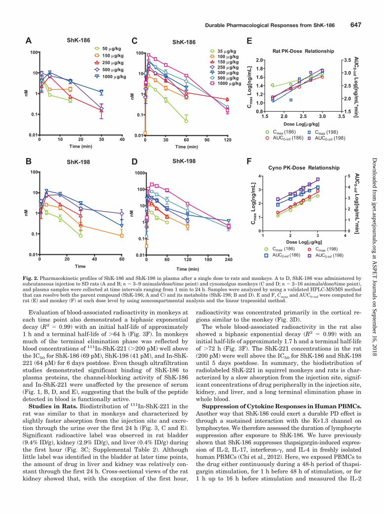
The PK profile of ShK-186 is characterized by a shortresidence time in the central compartment of rat and monkeyspecies. In rats, after a single administration, ShK-186reaches a maximum plasma concentration between 1 and 5min, whereas its metabolite reaches its Cmax between 1 and15 min (Fig. 2, A and B; Tables 1 and 2). Although the Cmax
and AUC for both the parent and metabolite increase withdose in rats, the relationship between Cmax and dose waslargely nonlinear (R2 � 0.8), whereas the relationship be-tween AUC and dose was approximately linear through the500 �g/kg dose (R2 � 0.97; Fig. 2E). The half-life in ratranged from 4.4 to 9.2 min for ShK-186 and from 6.1 to 16.4min for ShK-198 (Tables 1 and 2). Clearance values for ShK-186 and ShK-198 in rats were not significantly different(paired t test; t4 � 2.2; p � 0.1) and ranged from 441.4 to1453.3 ml/kg � min.
The PK of ShK-186 (Fig. 2C) and ShK-198 (Fig. 2D) after asingle subcutaneous administration to monkeys demon-
strated a linear relationship between dose and both AUC andCmax (slope � 1.02–1.27; R2 � 0.97; Fig. 2F) through theentire range of doses from 35 to 1000 �g/kg. The monkeyhalf-life ranged from 8.8 to 23.8 min for ShK-186 and from16.7 to 73.5 min for ShK-198. Clearance rates in the monkeywere generally lower for ShK-198 than ShK-186 and rangedfrom 18.7 to 141.3 ml/kg � min (Tables 1 and 2).
These data indicate that ShK-186 and its metabolite ShK-198 reach a maximum concentration in plasma 1 to 15 minafter a single subcutaneous injection of drug in both species,and both are rapidly cleared from the central compartment.The limit of quantitation of the HPLC-MS method is approx-imately 10-fold higher than the IC50 of ShK-186 and ShK-198on the Kv1.3 channel and thus lacks the sensitivity to esti-mate the terminal elimination phase at low, but potentiallytherapeutic, concentrations.
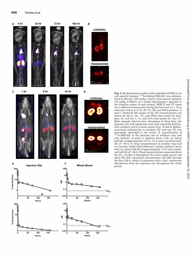
Absorption, Distribution, Metabolism, and Excre-tion Studies with a Radiolabeled Analog of ShK-186.We developed a radiolabeled analog of ShK-186 to measurethe biodistribution of the peptide and evaluate its total con-centration in whole blood. ShK-186 contains a single iodinat-able tyrosine at position 23. However, iodine incorporationinto the ring, which is predicted to interact within the poreregion of the Kv1.3 channel (Pennington et al., 1996), resultsin disruption of the channel binding properties of the peptide.We therefore modified the amino terminus of ShK-198 with asix-carbon linker attached via a peptide bond to one of thecarboxylic acids of a 1,4,7,10-tetraazacyclododecane-1,4,7,10-tetraacetic acid chelate (Supplemental Fig. 2A). The 1,4,7,10-tetraazacyclododecane-1,4,7,10-tetraacetic acid conjugate,designated ShK-221, was readily coordinated with indium orgadolinium (Supplemental Fig. 2, B and C). In patch-clampexperiments, the gadolinium- and indium-labeled ShK-221peptides blocked Kv1.3 with IC50 values similar to ShK-186and ShK-198: Gd-ShK-221, 58.23 1.38 pM (n � 5); In-ShK-221, 63.8 2.25 pM; (n � 3); ShK-186, 68.99 4.01 pM (n � 5);and ShK-198, 41.4 7.25 pM (n � 5) (Fig. 1C). We preparedand administered 111In-labeled ShK-221 by subcutaneous in-jection to SD rats (1.0 mCi; 100 �g/kg) and squirrel monkeys(0.83 mCi; 35 �g/kg). The radiolabeling efficiency ranged from89 to 98% over the series of experiments as determined byHPLC. Biodistribution of radiolabeled ShK-221 was evaluatedby SPECT imaging continuously for the first hour postdose, andthen at 4, 8, 24, 48, 72, 120, and 160 h (Supplemental Videos1–12). The background level in the detection system was ap-proximately 0.1�Ci/ml (�5 ng/ml of ShK-221 at the initial timepoint and 26 ng/ml at the last time point). Blood samples werecollected after each scan, and total radioactivity in whole bloodwas measured by gamma counting. Computed tomography wasperformed at each time point to enable colocalization of theradiolabel with key anatomical structures.
Studies in Squirrel Monkeys. Biodistribution of 111In-ShK-221 in squirrel monkeys was characterized principallyby slow absorption from the injection site over the entire160-h period (Fig. 3, A and E). The quantity of 111In-ShK-221present at the injection site followed a biphasic exponentialdecay (R2 � 0.95) with an initial half-life of approximately 1to 1.5 h and a terminal half-life of �48 h (Fig. 3E). During thefirst hour, significant radioactivity could be observed in thekidney, increasing in intensity through 1 h [�1% injecteddose (ID)/g; Supplemental Table 1] and slowly declining toapproximately baseline by 48 h. Radioactivity in the monkey
Durable Pharmacological Responses from ShK-186 645
at ASPE
T Journals on Septem
ber 16, 2018jpet.aspetjournals.org
Dow
nloaded from

kidney was observed primarily in the cortical and medullaryregions during all time points and was comparatively absentin the renal pelvis except for the first hour (Fig. 3B). Signif-icant bladder associated radioactivity (Tmax � 0.75–1 h;0.34% ID/g) was observed only during the first 4 h, after
which relatively little radiolabel was detected in bladder. Noother organ showed significant levels of radioactivity exceptfor liver, which peaked at 0.75 to 1 h after dose administra-tion (0.166% ID/g). Muscle, heart, and brain all had �0.1%ID/g at all time points measured (Supplemental Table 1).
50 msec
2 nA
Control1 pM
10 pM
50 pM
100 pM 1000 pM
50 msec
2 nA
Control1 pM
10 pM
50 pM
100 pM 1000 pM
25 pM
B
D E
A
C
Fig. 1. Biophysical properties of ShK-186, ShK-186 dephosphorylated form (ShK-198), and ShK-186 analogs. A, representative whole-cell Kv1.3currents in the absence and presence of dephosphorylated ShK-186 (ShK-198). B, representative whole-cell Kv1.3 currents in the absence and presenceof In-ShK-221 (a ShK-186 analog). In-ShK-221 was in 100% serum. C, comparison of dose-response curves for ShK-186, ShK-198, Gd-ShK-221, andIn-ShK-221 on Kv1.3. The IC50 values were: ShK-186, 68.99 4.01 pM (n � 5); ShK-198, 41.4 7.25 pM (n � 5); Gd-ShK-221, 58.23 1.38 pM (n �5), and In-ShK-221, 63.8 2.25 pM (n � 3). D, dose-response curves of ShK-186 on Kv1.3 in the presence of varying concentrations of serum. E,dose-response curves of In-ShK-221 in the presence or absence of serum. Electrophysical recordings were carried out in the whole-cell configurationof the patch-clamp technique as described previously (Beeton et al., 2005; Wulff et al., 2000). The external solution was sodium Ringer’s, and thepipette solution was potassium fluoride (300 mOsm). Kv1.3 currents were elucidated by 200-ms depolarizing pulses from a holding potential of �80to 40 mV. All analogs were each tested at several concentrations. The reduction in peak current at 40 mV for each concentration was used to generatea dose-response curve by using Origin software (OriginLab Corp., Northhampton, MA).
TABLE 1ShK-186 pharmacokinetic parameters in Sprague Dawley rats andcynomolgus monkeys
Species Dose n Tmax t1⁄2 Cmax AUC0-inf_obs Clearance
�g/kg min ng/ml ng/ml � min ml/kg � min
Monkey 35 10 5 8.8 12.8 247.6 141.3100 14 5 14.5 33.6 823.1 121.8150 16 5 23.8 69.8 1935.9 78.5250 2 5 11.3 106.6 2220.9 112.2300 5 5 14.0 173.8 3496.4 86.0500 2 5 15.8 192.5 4734.0 105.6
1000 2 5 13.0 358.0 9552.7 104.6Rat 50 6 1 4.4 26.2 115.9 441.4
150 6 1 9.2 27.0 249.5 640.9250 3 5 4.7 29.7 282.9 886.3500 9 1 5.2 42.7 587.5 847.2
1000 3 5 7.9 48.0 690.6 1453.3
TABLE 2ShK-198 pharmacokinetic parameters in Sprague Dawley rats andcynomolgus monkeys
Species Dose n Tmax t1⁄2 Cmax AUC0-inf_obs Clearance
�g/kg min ng/ml ng/ml � min ml/kg � min
Monkey 35 10 5 22.7 23.0 771.9 46.0100 14 5 24.5 55.6 2298.4 43.6150 16 5 16.7 94.2 2555.9 58.7250 2 15 22.8 117.0 5943.2 42.1300 5 5 73.5 168.0 8588.5 35.1500 2 15 30.8 321.0 17,750.6 28.2
1000 2 15 25.4 852.5 53,389.9 18.7Rat 50 6 1 6.1 11.8 102.5 486.6
150 6 5 10.4 11.6 243.9 617.9250 3 5 7.5 36.4 480.5 522.1500 9 15 16.4 33.9 1039.6 488.5
1000 3 5 8.7 53.9 1219.2 819.9
646 Tarcha et al.
at ASPE
T Journals on Septem
ber 16, 2018jpet.aspetjournals.org
Dow
nloaded from
Evaluation of blood-associated radioactivity in monkeys ateach time point also demonstrated a biphasic exponentialdecay (R2 � 0.99) with an initial half-life of approximately1 h and a terminal half-life of �64 h (Fig. 3F). In monkeysmuch of the terminal elimination phase was reflected byblood concentrations of 111In-ShK-221 (�200 pM) well abovethe IC50 for ShK-186 (69 pM), ShK-198 (41 pM), and In-ShK-221 (64 pM) for 6 days postdose. Even though ultrafiltrationstudies demonstrated significant binding of ShK-186 toplasma proteins, the channel-blocking activity of ShK-186and In-ShK-221 were unaffected by the presence of serum(Fig. 1, B, D, and E), suggesting that the bulk of the peptidedetected in blood is functionally active.
Studies in Rats. Biodistribution of 111In-ShK-221 in therat was similar to that in monkeys and characterized byslightly faster absorption from the injection site and excre-tion through the urine over the first 24 h (Fig. 3, C and E).Significant radioactive label was observed in rat bladder(9.4% ID/g), kidney (2.9% ID/g), and liver (0.4% ID/g) duringthe first hour (Fig. 3C; Supplemental Table 2). Althoughlittle label was identified in the bladder at later time points,the amount of drug in liver and kidney was relatively con-stant through the first 24 h. Cross-sectional views of the ratkidney showed that, with the exception of the first hour,
radioactivity was concentrated primarily in the cortical re-gions similar to the monkey (Fig. 3D).
The whole blood-associated radioactivity in the rat alsoshowed a biphasic exponential decay (R2 � 0.99) with aninitial half-life of approximately 1.7 h and a terminal half-lifeof �72 h (Fig. 3F). The ShK-221 concentrations in the rat(200 pM) were well above the IC50 for ShK-186 and ShK-198until 5 days postdose. In summary, the biodistribution ofradiolabeled ShK-221 in squirrel monkeys and rats is char-acterized by a slow absorption from the injection site, signif-icant concentrations of drug peripherally in the injection site,kidney, and liver, and a long terminal elimination phase inwhole blood.
Suppression of Cytokine Responses in Human PBMCs.Another way that ShK-186 could exert a durable PD effect isthrough a sustained interaction with the Kv1.3 channel onlymphocytes. We therefore assessed the duration of lymphocytesuppression after exposure to ShK-186. We have previouslyshown that ShK-186 suppresses thapsigargin-induced expres-sion of IL-2, IL-17, interferon-�, and IL-4 in freshly isolatedhuman PBMCs (Chi et al., 2012). Here, we exposed PBMCs tothe drug either continuously during a 48-h period of thapsi-gargin stimulation, for 1 h before 48 h of stimulation, or for1 h up to 16 h before stimulation and measured the IL-2
ShK-186
0 30 60 90 1200.01
0.1
1
10
100 35 µg/kg100 µg/kg150 µg/kg250 µg/kg300 µg/kg500 µg/kg1000 µg/kg
ShK-198
0 60 120 180 2400.01
0.1
1
10
100
1000
Time (min)
nM
A
B
ShK-186
0 10 20 30 400.01
0.1
1
10
10050 µg/kg150 µg/kg250 µg/kg500 µg/kg1000 µg/kg
Time (min) Time (min)
nM
nM
ShK-198
0 20 40 600.01
0.1
1
10
100
Time
nM
C
D
Rat PK-Dose Relationship
1.5 2.0 2.5 3.0 3.50.81.0
1.21.41.61.82.0
1.5
2.0
2.5
3.0
3.5
Cmax (186)AUC0-inf (186)
Cmax (198)AUC0-inf (198)
E
FCyno PK-Dose Relationship
1 2 3 40
1
2
3
4
0
1
2
3
4
5
Cmax (186)AUC0-inf (186)
Cmax (198)AUC0-inf (198)
Dose Log[µg/kg]
Dose Log[µg/kg]
Cm
ax L
og[n
g/m
L]C
max
Log
[ng/
mL]
AUC0-inf Log[ng/m
L*min]
AUC0-inf Log[ng/m
L*min]
Fig. 2. Pharmacokinetic profiles of ShK-186 and ShK-198 in plasma after a single dose to rats and monkeys. A to D, ShK-186 was administered bysubcutaneous injection to SD rats (A and B; n � 3–9 animals/dose/time point) and cynomolgus monkeys (C and D; n � 2–16 animals/dose/time point),and plasma samples were collected at time intervals ranging from 1 min to 24 h. Samples were analyzed by using a validated HPLC-MS/MS methodthat can resolve both the parent compound (ShK-186; A and C) and its metabolite (ShK-198; B and D). E and F, Cmax and AUC0-inf were computed forrat (E) and monkey (F) at each dose level by using noncompartmental analysis and the linear trapezoidal method.
Durable Pharmacological Responses from ShK-186 647
at ASPE
T Journals on Septem
ber 16, 2018jpet.aspetjournals.org
Dow
nloaded from
4 Hr 24 Hr 72 Hr 160 Hr
CORONAL
TRANSVERSE
A B
1 Hr 8 Hr 24 HrC
CORONAL
TRANSVERSE
D
E F Whole Blood
IC50
0 50 100 150
1
10
100
Hours
% In
ject
ed D
ose
Injection Site
0 50 100 150
1
10
100
Hours
% In
ject
ed D
ose
0 50 100 1500.01
0.1
1
10
100
Hours
nM
0 50 100 1500.01
0.1
1
10
100
Hours
nM
IC50
Fig. 3. Biodistribution studies with radiolabeled ShK in ratand squirrel monkey. 111In-labeled ShK-221 was adminis-tered to SD rats (100 �g/kg; 1.0mCi) and squirrel monkeys(35 �g/kg; 0.84mCi) as a single subcutaneous injection tothe scapular region of each animal. SPECT and CT scanswere collected continuously during the first hour (4 � 15-mintervals) and at 4, 8, 24, 48, 72, 120, and 160 h postdose. Aand C, flattened 2D images of the 3D reconstructions areshown for the 4-, 24-, 72-, and 160-h time points for mon-keys (A) and the 1-, 8-, and 24-h time points for rats (C).Both animals showed slow absorption of drug from theinjection site and significant early and sustained distribu-tion to kidney and to a lesser extent liver. B and D, kidney-associated radioactivity in monkeys (B) and rats (D) wasprincipally identified in the cortex. E, quantification of111In-ShK-221 at the injection site in monkeys (top) andrats (bottom) revealed a biphasic decay with an initialhalf-life of approximately 0.5 to 1.5 h and a terminal half-life of �48 h. F, drug concentrations in monkey (top) andrat (bottom) whole blood followed a similar biphasic decaywith an initial half-life of approximately 1.5 h and a termi-nal half-life of �64 h. Blood concentrations remained abovethe IC50 for Kv1.3 throughout the entire study period andabove the 80% saturation concentration (233 pM) throughthe first 120 h, which is consistent with a slow, continuousdistribution from the injection site throughout the studyperiod.
648 Tarcha et al.
at ASPE
T Journals on Septem
ber 16, 2018jpet.aspetjournals.org
Dow
nloaded from

responses by enzyme-linked immunosorbent assay (Fig. 4).In cases of transient exposure, the cells were thoroughlywashed with media before thapsigargin treatment. Therewas no statistically significant difference (one-way ANOVA;F3,12 � 0.40; p � 0.76)between continuous drug treatment ver-sus transient drug exposure up to 16 h before thapsigarginstimulation. These data are consistent with a model whereShK-186 rapidly associates with, but slowly dissociates from,the Kv1.3 channel of lymphocytes.
Suppression of the DTH Response In Vivo: Dose Fre-quency and Dose-Response Studies. Our absorption, dis-
tribution, metabolism, and excretion studies suggested that alow, but therapeutically, relevant drug concentration per-sists in the blood for several days after a single administra-tion of ShK-186. We therefore evaluated the relationshipbetween dose frequency and therapeutic efficacy in animalmodels of effector-memory T cell-mediated disease. The sim-plest model for assessing drug effects is the DTH reaction, animmune response largely mediated by skin-homing effector-memory CD4� T cells (Soler et al., 2003; Matheu et al., 2008).We immunized groups of five to eight Lewis rats with OVA inCFA followed 7 to 9 days later by elicitation of the DTHresponse in the ears of immunized animals. Ear swelling wasmeasured 24 h postelicitation. The durability of drug effectwas assessed by treating animals with ShK-186 at varyingtimes before or coincident with the elicitation phase. The dayof ear challenge is referred to as day 0 for reference.
Animals that were treated with two doses of 10 or 100�g/kg ShK-186 on days 0 and 1 (the standard regimen) werecompared with single 100 �g/kg doses administered on days0, �1, �2, �3, and �4. Two doses administered on days 0 and1 and single doses administered on days �1 to �4 yieldedstatistically significant reductions in ear swelling relative toplacebo-treated animals (two-tailed Mann-Whitney test;Fig. 5A). Single doses administered before day �4 did notresult in a significant reduction in ear swelling relative to thecontrol group (data not shown). We conclude that a singlesubcutaneous injection of ShK-186 is sufficient to suppressthe DTH response in rat for 5 days, consistent with the longterminal elimination half-life of radiolabeled ShK-221.
To establish a relationship between dose level and the timeof dosing, we evaluated the suppressive effect of a singleascending dose (0.1–100 �g/kg) of ShK-186 administered 2days before ear challenge. A dose-dependent reduction in earswelling was observed over the three-log dose range, withdoses �1 �g/kg achieving statistical significance relative tovehicle-treated animals (Fig. 5B).
Treatment Strategies in the CR-EAE and PIA Mod-els. CR-EAE is widely used as a model of multiple sclerosis.This model has been extensively characterized in guinea pigs(Raine et al., 1977; Keith et al., 1979) and DA rats (Feurer et
ShK + Thapsigargin (48 h)
Thapsigargin (48 h)ShK (1h)
Thapsigargin (48 h)ShK (1h) Media (16 h)
IL-2 Secretion
0 1 100 100000
50
100
150
pM ShK-186
pg/m
L
AB
CFig. 4. Durable effect of drug treatment on human PBMCs. Time oftreatment studies were conducted in human PBMCs in which the effect ofdrug treatment timing and duration on thapsigargin-stimulated IL-2responses was measured. Continuous drug treatment during the 48-hstimulation period (A; black bars) was compared with drug pretreatmentfor 1 h followed by a brief washout and stimulation for 48 h (B; open bars)or drug pretreatment (1 h) followed by washout, media incubation (16 h),and thapsigargin stimulation (48 h) (C; hatched bars). Drug treatment fora 1-h period up to 16 h before stimulation was statistically indistinguish-able from continuous drug treatment. None of the treatment times anddurations were significantly different (one-way ANOVA; F3,12 � 0.40; p �0.76).
A B
0 0.1 1 10 1000.0
0.2
0.4
0.6
0.8
1.0
1.2
***
**
µg/kg ShK-186
POC
0.0
0.2
0.4
0.6
0.8
1.0
1.2
* **
****
***
POC
Dose: PL 10 100 100 100 100 100 100Day: 0/1 0/1 0/1 0 -1 -2 -3 -4
Fig. 5. Time of administration and dose-dependent suppression of the DTH response. Lewis rats were immunized with ovalbumin in CFA andchallenged 7 to 9 days later by injection of ovalbumin to the ear pinna. The day of challenge is represented as day 0. The DTH reaction was measured24 h postchallenge (day 1). A, ShK-186 administered at 10 or 100 �g/kg in two doses on days 0 and 1 or as a single 100 �g/kg dose on days �1, �2,�3, and �4 resulted in a significant reduction in ear swelling relative to placebo (PL)-treated animals when measured on day 1 postchallenge(two-tailed Mann-Whitney). These data indicate that a single ShK-186 dose can suppress the DTH response for up to 5 days. B, using the DTH model,a dose-response study was conducted with a single dose of ShK-186 administered on day �2. Doses between 0.1 and 100 �g/kg showed a cleardose-response relationship, with all but 0.1 �g/kg achieving statistical significance relative to placebo. �, p � 0.05; ��, p � 0.01.
Durable Pharmacological Responses from ShK-186 649
at ASPE
T Journals on Septem
ber 16, 2018jpet.aspetjournals.org
Dow
nloaded from

al., 1985; Lorentzen et al., 1995), and unlike the acute EAEmodels includes relapses and some degree of demyelination(Lassman and Wisniewski, 1979; Lorentzen et al., 1995;Matheu et al., 2008). Moreover, because of its protractednature, it is an adequate model for testing dosing schedules.The initial wave of disease is largely adjuvant mediated andcomposed primarily of central memory T cells, whereas sub-sequent waves of disease are principally TEM cell-mediatedand therefore Kv1.3-dependent (Matheu et al., 2008). Wehave previously shown ShK-186 to be effective in the CR-EAE model by using daily dosing at 100 �g/kg (Matheu et al.,2008). In the present study, we explored the effect of lessfrequent dose administration on drug efficacy in this model.
Two study designs were explored. In the first, a lead-inperiod with daily drug administration was initiated at thetime of disease induction and continued throughout the firstwave of disease. Seven days after disease onset, animals wererandomized to receive 100 �g/kg ShK-186 every 2 or 3 daysthrough the remainder of the study. Daily drug treatmentcaused a statistically significant reduction in mean clinicalscore compared with placebo-treated animals during the firstwave of disease (Fig. 6A). After lead-in, drug administrationevery 2 or 3 days resulted in continued significantly lowermean daily clinical score in treated relative to control ani-mals. The average cumulative clinical score was 161.1 162.3, 72.5 98.1, and 71.3 103.3 for the placebo, every-2-days, and every-3-days dosing groups, respectively. Ani-mals were scored twice daily.
In a second series of experiments, we evaluated the effectof reduced dose frequency on EAE without the drug lead-inperiod. In this study, groups of DA rats were randomized toreceive placebo, daily ShK-186, or ShK-186 every 3 daysbeginning at a clinical score �1. Both drug-treated groupsexhibited a significantly lower mean daily clinical score thanplacebo-treated animals (Fig. 6B). However, there was nostatistically significant difference between daily and every-third-day dose administration. Mean cumulative clinicalscores were 49.7 26.7, 36.9 20.6, and 30 19.3 for theplacebo, daily, and every-third-day dosing groups, respec-tively. The percentage of rats with one or more relapses was70% (9/13), 21% (3/14), and 21% (3/14), and the total numberof relapses observed during the chronic phase were 15, 6, and
-10 -5 0 5 10 15 20 25 30 35
0.5
1.0
1.5
2.0
2.5
3.0
3.5
Placebo Every 2 days (100µg/kg) Every 3 days (100µg/kg)day post onset
Clin
ical
Sco
re
A Daily 100 µg/kgE2D 100 µg/kg
E3D 100 µg/kg
0 5 10 15 20 25 300.0
0.5
1.0
1.5
2.0
2.5
3.0
3.5
Daily Placebo (P6N) Daily (100 µg/kg) Every 3 Days (100 µg/kg)
day post onset
Clin
ical
Sco
re
BDaily or E3D (100µg/kg)
0 10 20 300
20
40
60
80
day post onset
Dis
ease
Pre
vale
nce
(%)
0 5 10 15 20 25 30 35 400.0
0.5
1.0
1.5
2.0
2.5
3.0
3.5
4.0
4.5
Placebo Daily (100 µg/kg)
Days post onset
Clin
ical
Sco
re
C Daily 100 µg/kg
Fig. 6. Alternate dosing schedules are effective in the chronic EAE model.A, an initial study evaluated lead-in dosing followed by less than dailydrug administration. Male DA rats were immunized with spinal cordhomogenate in CFA and administered ShK-186 (100 �g/kg/day; n � 25) orvehicle (n � 12) during the prodromal period and for 7 days after theonset of EAE, after which drug-treated animals were randomized toreceive a maintenance dose of 100 �g/kg ShK-186 every 2 days (n � 12)
or 3 days (n � 13). ShK-186 significantly reduced (p � 0.001) the severityof the initial episode of disease. Maintenance therapy administered at 48-or 72-h intervals significantly (p � 0.001; one-way repeated-measuresANOVA) reduced the severity of CR-EAE. B, a second study measuredthe effect of infrequent drug administration beginning at disease onset.Female DA rats (n � 13/group) were immunized with spinal cord homog-enate in CFA to elicit a chronic, relapsing EAE. After the onset ofsymptoms (clinical score �1), animals were randomized to receive dailyplacebo, daily ShK-186, or ShK-186 every 2 or 3 days. Drug treatmentresulted in significantly lower clinical scores and a lower frequency ofdisease incidence (inset) during the chronic phase in the groups treateddaily and groups treated every third day compared with placebo (repeat-ed-measures ANOVA; F2,60 � 39.4; p � 0.0001). Mean clinical scores werenot lower relative to placebo in the group treated on alternate days in thisstudy (data not shown). C, a final study measured the durability ofresponse of a period of drug therapy. Female DA rats (n � 23) wereimmunized and allowed to proceed to CR-EAE. Ten days after the onsetof symptoms, animals were randomized to receive daily placebo (n � 10)or 100 �g/kg ShK-186 (n � 13) for 14 days. Therapy was discontinued onday 24, and the time required for treated animals to return to baselinedisease was evaluated. The previously treated group had mean clinicalscores significantly lower than placebo controls on days 24, 25, 26, and 28(Supplemental Table 2), which is consistent with a 5-day window fordisease recurrence.
650 Tarcha et al.
at ASPE
T Journals on Septem
ber 16, 2018jpet.aspetjournals.org
Dow
nloaded from
5 for the placebo, ShK186 daily, and every-third day-treatedgroups, respectively. Animals were scored once daily.
Finally, we wanted to evaluate the durability of diseasesuppression in the CR-EAE model after cessation of drugadministration. Disease was elicited in groups of DA rats,and the animals were allowed to develop a relapsing disease.On day 10 after disease onset (clinical score �1), animalswere randomized to receive daily placebo or 100 �g/kg ShK-186 for 14 days. Drug treatment was discontinued on day 24,and the animals were monitored for an additional 15 days.There was no difference between the placebo- and drug-treated groups before drug treatment (Fig. 6C). During the14 days of daily drug administration, there was a statisticallysignificant reduction in mean clinical score in drug-treatedversus control animals. Once treatment was withdrawn, themean clinical score in the drug-treated group slowly returnedto the level of placebo over approximately 5 days (Supple-mental Table 3). However, there was no rebound effect ob-served in the previously treated animals.
As a final test of the effectiveness of less than daily dosing,we evaluated ShK-186 administration every other day in thePIA model of rheumatoid arthritis (Beeton et al., 2006). ShK-186 administered once daily (100 �g/kg) has been previouslyreported to reduce the number of joints affected and theseverity of joint swelling in the PIA model in DA rats (Beetonet al., 2006). Here, we demonstrate that ShK-186 adminis-tered on alternate days (100 �g/kg) was as effective in ame-liorating disease (Fig. 7) as our previously reported effectwith once-daily administration (Beeton et al., 2006).
The data from these four studies are consistent with amodel of durable drug effects after a single subcutaneousdose of ShK-186 and observations made using the DTHmodel.
DiscussionHere, we present five complementary types of data that
relate the PK properties of ShK-186 to its therapeutic effi-cacy in rat models of autoimmune diseases. Through serum/plasma stability studies, we identify the sole metabolite ofShK-186 in rat, monkey, and human samples as the dephos-phorylated peptide ShK-198. ShK-198 blocks Kv1.3 (IC50 40pM) with approximately the same potency as ShK-186 (69
pM). Second, we demonstrate that the Cmax of both ShK-186and ShK-198 is rapidly reached after subcutaneous injectionto rats and monkeys. Both parent and metabolite have ashort apparent half-life in the central compartment with�90% of the Cmax eliminated by 2 h in both species at alltested doses. The half-life is shorter and the apparent clear-ance greater for ShK-186 than ShK-198, probably reflectingthe independent action of endogenous phosphatases on theconversion of ShK-186 to its metabolite. Third, SPECT im-aging of rats and monkeys administered an 111In-labeledShK-186 analog (ShK-221; IC50 65 pM) by subcutaneousinjection revealed that the peptide is released slowly from theinjection site and has an extended terminal half-life, withwhole-blood levels above the IC50 value for Kv1.3 block for 5days in rats and 7 days in monkeys. The Kv1.3 channel-blocking affinity of ShK-186 and ShK-221 is not affected bythe presence of serum, indicating that plasma protein bind-ing, if any, does not affect the functional activity of thesepeptides. Fourth, in vitro proliferation assays demonstratethat brief 1-h exposure of PBMCs to ShK-186 is sufficient tosuppress interleukin-2 production 64 h later. This suggeststhat the peptide, once bound to the Kv1.3 channel, dissoci-ates very slowly. Finally, in three different rat models of TEM
cell-mediated inflammatory diseases (DTH, CR-EAE, andPIA) ShK-186 is as effective in ameliorating disease whenadministered every 2 to 5 days as when it is administeredonce daily. The durable PD effect of ShK-186 is contrary toconventional wisdom that frequent administration of peptidetherapeutics is required to sustain PD activity.
Our findings highlight a potential advantage of developingpeptides from venoms as drug candidates. SPECT imagingstudies with 111In-labeled ShK-221 revealed a biphasic (fastthen slow) release from the subcutaneous injection site over7 days in rats and monkeys. This type of rate-limiting ab-sorption resulting in prolonged plasma exposure has beendescribed for whole animal venoms (Barral-Netto et al., 1990;Audebert et al., 1994). The long absorption phase has beenused to explain the frequent relapse in envenomation pa-tients long after exposure and/or treatment with antivenom(Dart et al., 2001; Seifert and Boyer, 2001; Gutierrez et al.,2003), and venoms are often described as having a depoteffect. The parent peptide of ShK-186 was originally isolatedfrom the venom of Stichodactyla helianthus (Castaneda etal., 1995), and ShK-186 may share some of the in vivo char-acteristics of the complex peptide mixtures found in animalvenom. Venom and toxin PK parameters are also character-ized by large apparent volumes of distribution and slow elim-ination from deep and shallow peripheral compartments (Is-mail et al., 1996).The apparent volume of distribution forShK-186 is large in both rats (2743–16,458 ml/kg) and mon-keys (1729–2664 ml/kg), consistent with that reported forother animal venoms. The blood concentration of 111In-ShK-221 mimicked the absorption of the peptide from the injectionsite and was characterized by a rapid initial phase and a verylong terminal phase. The terminal half-life computed by us-ing 111In-ShK-221 was �64 h in monkeys with sustainedblood levels for 7 days above the IC50 values for ShK-186 (69pM) and ShK-198 (40 pM). The presence of serum does notaffect channel-blocking affinity of these peptides, indicatingthat plasma protein binding, if any, does not affect functionalactivity.
0 5 10 15 200
10
20
30
ControlShK-186 EOD
Days after onset of clinical signs
Clin
ical
sco
re
Fig. 7. Pristane-induced arthritis treated with alternate day administra-tion of ShK-186. Arthritis was induced in DA rats by injection of 75 �l ofpristane at the base of the tail. Disease was allowed to evolve to a clinicalscore of 1 when animals were randomized to receive placebo or 100 �g/kgShK-186 every other day (EOD). Clinical score is computed as accordingto Gillett et al. (2010). Overall disease severity in the study was high.Animals receiving ShK-186 every other day had a statistically significantreduction in mean score (placebo, 20.4 5.9; drug, 14.6 4.1; paired ttest; p � 0.0001).
Durable Pharmacological Responses from ShK-186 651
at ASPE
T Journals on Septem
ber 16, 2018jpet.aspetjournals.org
Dow
nloaded from

Glomerular filtration is the principal elimination pathwayfor the peptide shortly after subcutaneous injection. Signifi-cant amounts of radioactivity were observed in the bladder ofboth rats (�17% injected dose) and monkeys (�1% injecteddose) at the earliest time points after administration of 111In-ShK-221. The large amount of drug excreted by the rat in thefirst hour is most likely a reflection of the increased metab-olism of the rat compared with the monkey. After 1 h in ratsand approximately 4 h in monkey, little radioactivity wasobserved in bladder or renal pelvis, whereas significantamounts of radioactivity could still be observed in the kidneycortex. Cortical concentration has been reported for numer-ous radiolabeled versions of peptide drugs including oc-treotide, bombesin, exendin, and gastrin (Gotthardt et al.,2007). The mechanism of cortical retention has been mostthoroughly described for octreotide. Tubular reabsorption ofthe cationic octapeptide is mediated by megalin, a scavengerreceptor expressed in the proximal kidney tubule (de Jong etal., 2005). Mice with a kidney-specific disruption of the re-ceptor lack the cortical retention of radiolabeled octreotideseen in wild-type mice. Renal uptake of octreotide is partiallymediated by charge and can be disrupted by coinfusion of thepositively charged amino acids L-lysine and L-arginine (Bodeiet al., 2003). ShK-186 carries a net �6 charge at physiologicalpH, so its cortical retention may be mediated by a similarmechanism.
Another contributing factor to the long PD effect of ShK-186along with an extended terminal half-life could be its slowdissociation from the Kv1.3 channel. Studies with PBMCs showthat there is essentially no difference between continuous ShK-186 treatment during thapsigargin stimulation and treatmentup to 16 h before stimulation. Although we did not formallymeasure an off-rate of the drug on the Kv1.3 channel by usingtraditional methods, we did show that ShK-186’s PD effect canpersist for at least 72 h after its brief exposure to PBMCs. Thereceptor dissociation half-life of some animal toxins has beenreported to be as long as 1 week (Berg and Hall, 1975, Changand Huang, 1975). Future receptor binding studies using theradiolabeled analog will allow determination of the off-rate ofShK-186 from Kv1.3.
Absorption, distribution, metabolism, and excretion stud-ies with 111In-labeled ShK-221 suggest that a single dose ofpeptide can provide therapeutically meaningful blood con-centrations for up to 5 days in rats and 7 days in monkeys.These data are consistent with data from the rat DTH modelwhere a single administered dose of ShK-186 provided astatistically significant reduction in ear swelling for up to 5days. Likewise, dose administration every 2 to 3 days in theCR-EAE and PIA models were as effective in amelioratingdisease as daily ShK-186 administration. Because ShK-186’stherapeutic effects are long-lasting, we anticipate adminis-tering the peptide to humans once weekly or less frequently.From a therapeutic and commercial perspective, ShK-186will compete favorably with pills taken daily or multipletimes a day or injectables.
Acknowledgments
We thank Srikant Rangaraju for testing the Kv1.3 channel-blocking activity of ShK-186 in the presence or absence of serum.
Authorship Contributions
Participated in research design: Tarcha, Chi, Munoz-Elías, Bailey,Ruppert, Boley, Beeton, Slauter, Knapp, Pennington, Chandy, andIadonato.
Conducted experiments: Tarcha, Chi, Munoz-Elías, Bailey, Upad-hyay, Norton, Banks, Beeton, Tjong, Nguyen, Hu, Kentala, Hansen,and Iadonato.
Contributed new reagents or analytic tools: Tarcha, Bailey, andPennington.
Performed data analysis: Tarcha, Chi, Munoz-Elías, Bailey, Upa-dhyay, Nguyen, Knapp, Beeton, Chandy, and Iadonato.
Wrote or contributed to the writing of the manuscript: Tarcha, Chi,Munoz-Elías, Chandy, and Iadonato.
ReferencesAudebert F, Urtizberea M, Sabouraud A, Scherrmann JM, and Bon C (1994) Phar-
macokinetics of Vipera aspis venom after experimental envenomation in rabbits.J Pharmacol Exp Ther 268:1512–1517.
Azam P, Sankaranarayanan A, Homerick D, Griffey S, and Wulff H (2007) Targetingeffector memory T cells with the small molecule Kv1.3 blocker PAP-1 suppressesallergic contact dermatitis. J Invest Dermatol 127:1419–1429.
Barral-Netto M, Schriefer A, Vinhas V, and Almeida AR (1990) Enzyme-linkedimmunosorbent assay for the detection of Bothrops jararaca venom. Toxicon 28:1053–1061.
Beeton C and Chandy KG (2007) Induction and monitoring of active delayed typehypersensitivity (DTH) in rats. J Vis Exp 6:237.
Beeton C, Pennington MW, Wulff H, Singh S, Nugent D, Crossley G, Khaytin I,Calabresi PA, Chen CY, Gutman GA, et al. (2005) Targeting effector memory Tcells with a selective peptide inhibitor of Kv1.3 channels for therapy of autoim-mune diseases. Mol Pharmacol 67:1369–1381.
Beeton C, Wulff H, Barbaria J, Clot-Faybesse O, Pennington M, Bernard D, CahalanMD, Chandy KG, and Beraud E (2001) Selective blockade of T lymphocyte K�
channels ameliorates experimental autoimmune encephalomyelitis, a model formultiple sclerosis. Proc Natl Acad Sci U S A 98:13942–13947.
Beeton C, Wulff H, Standifer NE, Azam P, Mullen KM, Pennington MW, Kolski-Andreaco A, Wei E, Grino A, Counts DR, et al. (2006) Kv1.3 channels are atherapeutic target for T cell-mediated autoimmune diseases. Proc Natl Acad SciU S A 103:17414–17419.
Berg DK and Hall ZW (1975) Loss of -bungarotoxin from junctional and extrajunc-tional acetylcholine receptors in rat diaphragm muscle in vivo and in organculture. J Physiol 252:771–789.
Bodei L, Cremonesi M, Zoboli S, Grana C, Bartolomei M, Rocca P, Caracciolo M,Macke HR, Chinol M, and Paganelli G (2003) Receptor-mediated radionuclidetherapy with 90Y-DOTATOC in association with amino acid infusion: a phase 1study. Eur J Nucl Med Mol Imaging 30:207–216.
Cahalan MD and Chandy KG (2009) The functional network of ion channels in Tlymphocytes. Immunol Rev 231:59–87.
Castaneda O, Sotolongo V, Amor AM, Stocklin R, Anderson AJ, Harvey AL, Eng-strom A, Wernstedt C, and Karlsson E (1995) Characterization of a potassiumchannel toxin from the Caribbean Sea anemone Stichodactyla helianthus. Toxicon33:603–613.
Chang CC and Huang MC (1975) Turnover of junctional and extrajunctional acetyl-choline receptors of the rat diaphragm. Nature 253:643–644.
Cheong A, Li J, Sukumar P, Kumar B, Zeng F, Riches K, Munsch C, Wood IC, PorterKE, and Beech DJ (2011) Potent suppression of vascular smooth muscle cellmigration and human neointimal hyperplasia by KV1.3 blockers. Cardiovasc Res89:282–289.
Chi V, Pennington MW, Norton RS, Tarcha EJ, Londono LM, Sims-Fahey B, Upa-dhyay SK, Lakey JT, Iadonato S, Wulff H, et al. (2012) Development of a seaanemone toxin as an immunomodulator for therapy of autoimmune diseases.Toxicon 59:529–546.
Dart RC, Seifert SA, Boyer LV, Clark RF, Hall E, McKinney P, McNally J, KitchensCS, Curry SC, Bogdan GM, et al. (2001) A randomized multicenter trial of crotali-nae polyvalent immune Fab (ovine) antivenom for the treatment for crotalinesnakebite in the United States. Arch Intern Med 161:2030–2036.
de Jong M, Barone R, Krenning E, Bernard B, Melis M, Visser T, Gekle M, WillnowTE, Walrand S, Jamar F, et al. (2005) Megalin is essential for renal proximaltubule reabsorption of 111In-DTPA-octreotide. J Nucl Med 46:1696–1700.
Feurer C, Prentice DE, and Cammisuli S (1985) Chronic relapsing experimentalallergic encephalomyelitis in the Lewis rat. J Neuroimmunol 10:159–166.
Gilhar A, Bergman R, Assay B, Ullmann Y, and Etzioni A (2011) The beneficial effectof blocking Kv1.3 in the psoriasiform SCID mouse model. J Invest Dermatol131:118–124.
Gillett A, Marta M, Jin T, Tuncel J, Leclerc P, Nohra R, Lange S, Holmdahl R, OlssonT, Harris RA, et al. (2010) TNF production in macrophages is genetically deter-mined and regulates inflammatory disease in rats. J Immunol 185:442–450.
Gotthardt M, van Eerd-Vismale J, Oyen WJ, de Jong M, Zhang H, Rolleman E,Maecke HR, Behe M, and Boerman O (2007) Indication for different mechanismsof kidney uptake of radiolabeled peptides. J Nucl Med 48:596–601.
Grgic I, Wulff H, Eichler I, Flothmann C, Kohler R, and Hoyer J (2009) Blockade ofT-lymphocyte KCa3.1 and Kv1.3 channels as novel immunosuppression strategyto prevent kidney allograft rejection. Transplant Proc 41:2601–2606.
Gutierrez JM, Leon G, and Lomonte B (2003) Pharmacokinetic-pharmacodynamicrelationships of immunoglobulin therapy for envenoming. Clin Pharmacokinet42:721–741.
652 Tarcha et al.
at ASPE
T Journals on Septem
ber 16, 2018jpet.aspetjournals.org
Dow
nloaded from
Hu L, Gocke AR, Knapp E, Rosenzweig JM, Grishkan IV, Baxi EG, Zhang H,Margolick JB, Whartenby KA, and Calabresi PA (2012) Functional blockade of thevoltage-gated potassium channel Kv1.3 mediates reversion of T effector to centralmemory lymphocytes through SMAD3/p21cip1 signaling. J Biol Chem 287:1261–1268.
Hu L, Pennington M, Jiang Q, Whartenby KA, and Calabresi PA (2007) Character-ization of the functional properties of the voltage-gated potassium channel Kv1.3in human CD4� T lymphocytes. J Immunol 179:4563–4570.
Hyodo T, Oda T, Kikuchi Y, Higashi K, Kushiyama T, Yamamoto K, Yamada M,Suzuki S, Hokari R, Kinoshita M, et al. (2010) Voltage-gated potassium channelKv1.3 blocker as a potential treatment for rat anti-glomerular basement mem-brane glomerulonephritis. Am J Physiol Renal Physiol 299:F1258–F1269.
Ismail M, Aly MH, Abd-Elsalam MA, and Morad AM (1996) A three-compartmentopen pharmacokinetic model can explain variable toxicities of cobra venom andtheir alpha toxins. Toxicon 34:1011–1026.
Keith AB, Arnon R, Teitelbaum D, Caspary EA, and Wisniewski HM (1979) Theeffect of Cop 1, a synthetic polypeptide, on chronic relapsing experimental allergicencephalomyelitis in guinea pigs. J Neurol Sci 42:267–274.
Koo GC, Blake JT, Talento A, Nguyen M, Lin S, Sirotina A, Shah K, Mulvany K,Hora D Jr, Cunningham P, et al. (1997) Blockade of the voltage-gated potassiumchannel Kv1.3 inhibits immune responses in vivo. J Immunol 158:5120–5128.
Lassmann H and Wisniewski HM (1979) Chronic relapsing experimental allergicencephalomyelitis: effect of age at the time of sensitization on clinical course andpathology. Acta Neuropathol 47:111–116.
Lorentzen JC, Issazadeh S, Storch M, Mustafa MI, Lassman H, Linington C, Klares-kog L, and Olsson T (1995) Protracted, relapsing and demyelinating experimentalautoimmune encephalomyelitis in DA rats immunized with syngeneic spinal cordand incomplete Freund’s adjuvant. J Neuroimmunol 63:193–205.
Matheu MP, Beeton C, Garcia A, Chi V, Rangaraju S, Safrina O, Monaghan K,Uemura MI, Li D, Pal S, et al. (2008) Imaging of effector memory T cells during adelayed-type hypersensitivity reaction and suppression by Kv1.3 channel block.Immunity 29:602–614.
Pennington MW, Beeton C, Galea CA, Smith BJ, Chi V, Monaghan KP, Garcia A,Rangaraju S, Giuffrida A, Plank D, et al. (2009) Engineering a stable and selective
peptide blocker of the Kv1.3 channel in T lymphocytes. Mol Pharmacol 75:762–773.
Pennington MW, Mahnir VM, Khaytin I, Zaydenberg I, Byrnes ME, and Kem WR(1996) An essential binding surface for ShK toxin interaction with rat brainpotassium channels. Biochemistry 35:16407–16411.
Raine CS, Snyder DH, Stone SH, and Bornstein MB (1977) Suppression of acute andchronic experimental allergic encephalomyelitis in Strain 13 guinea pigs. A clinicaland pathological study. J Neurol Sci 31:355–367.
Rangaraju S, Chi V, Pennington MW, and Chandy KG (2009) Kv1.3 potassiumchannels as a therapeutic target in multiple sclerosis. Expert Opin Ther Targets13:909–924.
Rintisch C, Forster M, and Holmdahl R (2009) Detection of arthritis-susceptibilityloci, including Ncf1, and variable effects of the major histocompatibility complexregion depending on genetic background in rats. Arthritis Rheum 60:419–427.
Rus H, Pardo CA, Hu L, Darrah E, Cudrici C, Niculescu T, Niculescu F, Mullen KM,Allie R, Guo L, et al. (2005) The voltage-gated potassium channel Kv1.3 is highlyexpressed on inflammatory brain infiltrates in multiple sclerosis brain. Proc NatlAcad Sci U S A 102:11094–11099.
Seifert SA and Boyer LV (2001) Recurrence phenomena after immunoglobulin ther-apy for snake envenomations: Part 1. Pharmacokinetics and pharmacodynamics ofimmunoglobulin antivenoms and related antibodies. Ann Emerg Med 37:189–195.
Soler D, Humphreys TL, Spinola SM, and Campbell JJ (2003) CCR4 versus CCR10in human cutaneous TH lymphocyte trafficking. Blood 101:1677–1682.
Wulff H, Calabresi PA, Allie R, Yun S, Pennington M, Beeton C, and Chandy KG(2003) The voltage-gated Kv1.3 K� channel in effector memory T cells as newtarget for MS. J Clin Invest 111:1703–1713.
Wulff H, Miller MJ, Hansel W, Grissmer S, Cahalan MD, and Chandy KG (2000)Design of a potent and selective inhibitor of the intermediate-conductance Ca2�-activated K� channel, IKCa1: a potential immunosuppressant. Proc Natl Acad SciU S A 97:8151–8156.
Address correspondence to: Eric J. Tarcha, Kineta Inc., 219 Terry Ave N.,Suite 300, Seattle, WA 98109-5208. E-mail: [email protected]
Durable Pharmacological Responses from ShK-186 653
at ASPE
T Journals on Septem
ber 16, 2018jpet.aspetjournals.org
Dow
nloaded from