drug metabolism: significance and...
TRANSCRIPT
P1: PIC/PIC P2: ABCc01 JWBK262/Xie September 18, 2008 18:59 Printer Name: Yet to Come
1DRUG METABOLISM: SIGNIFICANCEAND CHALLENGES
Chandra Prakash and Alfin D. N. VazDepartment of Pharmacokinetics, Dynamics and Metabolism, Pfizer Global Research andDevelopment, Pfizer Inc., Groton, CT, USA
1.1 INTRODUCTION
Searching for new drugs is a very time-consuming and expensive endeavor, takingapproximately 10–12 years and on the order of $900 million to bring a new drug tomarket [1, 2]. It has been estimated that for every 5000 new chemical entities (NCEs)evaluated in a discovery program, only 1 is approved for market [3]. Even after a drugis marketed, there is the possibility of some undesired side effects, which were not seenin earlier clinical trials. In these cases, the drug is either withdrawn from the marketor acquires a warning label (black box). Therefore, efforts are being made to reduceattrition of drug candidates during the various stages of their development to bringsafer compounds to market. The major reasons for the failure of the NCEs are lackof in vivo efficacy, serious undesired side effects, and unfavorable drug metabolismand pharmacokinetics (DMPK). Therefore, in addition to potency and selectivity,drug candidates are selected on the basis of DMPK properties, such as desiredclearance, oral bioavailability, low potential of drug–drug interactions, and acceptablemetabolism/toxicology profiles in preclinical species [4, 5]. In support of this need,and as a consequence of increased knowledge within the drug metabolism discipline,new approaches have been developed that include extensive in vitro methods usinghuman and animal hepatic cellular and subcellular systems, recombinant human drug-metabolizing enzymes, transgenic animals and cell lines stably expressing humantransporters, increased automation for higher throughput screens, sensitive analytical
Nuclear Receptors in Drug Metabolism Edited by Wen XieCopyright C© 2009 John Wiley & Sons, Inc.
1
COPYRIG
HTED M
ATERIAL
P1: PIC/PIC P2: ABCc01 JWBK262/Xie September 18, 2008 18:59 Printer Name: Yet to Come
2 DRUG METABOLISM: SIGNIFICANCE AND CHALLENGES
Efficacy27%
Clinicalsafety13%
PK10%
Commercial20%
Others10%
Preclinicalsafety20%
Efficacy30%
Clinicalsafety10%
PK bioavailability40%
Com
mercial
Others6%
Preclinicalsafety10%
6%
(a) (b)
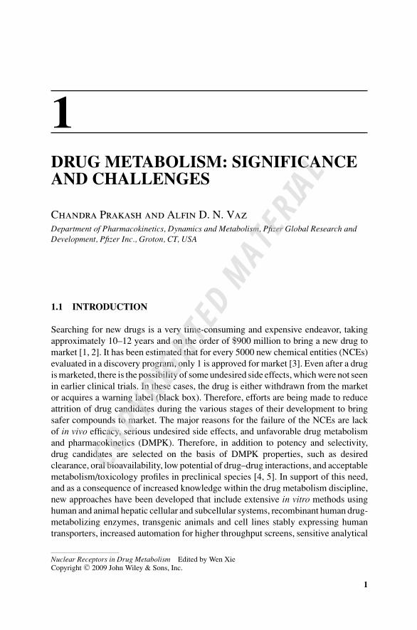
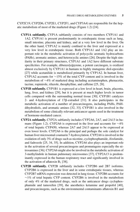
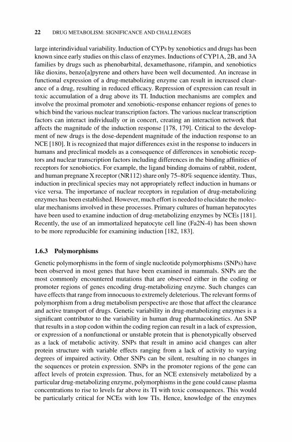
FIGURE 1.1 Reasons for attrition of NCEs in drug development: (a) 1990 and (b) 2000.(Modified from Ref. 6)
technologies, and in silico computational models to assess drug metabolism aspectsof the NCEs. Recent data suggest that these approaches have reduced the attrition dueto DMPK issues from 40% of total attrition in 1990 to 10% in 2000 (Figure 1.1) [6].
The predictive power of in vitro studies using animal and human hepatocellular andsubcellular fractions and/or recombinant enzymes [7–9], in silico models [10–12],and in vivo studies using a range of experimental animal models [13, 14] has advancedconsiderably due to an ever increasing understanding of the relationships between invitro and in vivo drug metabolism and disposition. These in vitro studies include
1. absorption/transport studies in Caco-2 cells or cell lines over expressing varioustransporters;
2. metabolic stability and metabolite formation in liver microsomes, S-9,hepatocytes or recombinant cytochrome P450 enzymes;
3. cytochrome P450 inhibition and induction;
4. plasma protein binding;
5. reactive metabolite (glutathione adduct formation).
The in vivo studies include
1. pharmacokinetic studies in laboratory animals via various routes of adminis-tration (oral, intravenous, subcutaneous, etc.);
2. tissue distribution (e.g., brain penetration);
3. metabolite identification and clearance pathways in animals and humans usingradiotracers;
4. PK/PD relationship;
5. specific studies using genetically engineered mouse models.
P1: PIC/PIC P2: ABCc01 JWBK262/Xie September 18, 2008 18:59 Printer Name: Yet to Come
PHASE I DRUG-METABOLIZING ENZYMES 3
Among ADME (absorption, distribution, metabolism, and excretion) properties,metabolism of an NCE by the host system can be one of the most important de-terminants of its pharmacokinetic disposition. It is the biochemical process by whichcompounds are converted to more hydrophilic (water-soluble) entities, which notonly enhance their elimination from the body but also lead to compounds that aregenerally pharmacologically inactive and relatively nontoxic. However, metabolictransformation of an NCE at times can lead to the formation of metabolites withpharmacological activity [15] and/or toxicity [16–18]. Additionally, metabolism canbe the main cause of poor bioavailability and drug–drug interactions via inhibi-tion or induction of drug-metabolizing enzymes [19, 20]. Therefore, determinationof metabolic rate and biotransformation pathways of an NCE, in animals and hu-mans, and evaluation of pharmacological and toxicological consequences of itsmetabolites are very critical to pharmaceutical development [21]. At the lead op-timization stage, information on the metabolic fate of the NCEs can direct medic-inal chemists to synthesize metabolically more stable analogs by blocking sites ofmetabolism, and potentially creating NCEs with superior pharmacology and safetyprofiles. Knowledge of the major human metabolites of an NCE early in its de-velopment is useful to enable the judicious selection of animal species used forsafety evaluation, to ensure that the selected animal species are exposed to all majormetabolites formed in humans [22, 23]. Subsequently, major circulatory metabo-lites in humans can be synthesized for the evaluation of their pharmacologicalactivity.
The metabolism of drugs has traditionally been classified into two reaction classes,phase I and phase II. Phase I reactions include hydroxylation, dealkylation, deam-ination, N- or S-oxidation, reduction, and hydrolysis. These reactions introduce orunmask a functional group (e.g., –OH, –COOH, –NH2, or –SH) within a molecule toenhance its hydrophilicity. Phase II or conjugation biotransformations include glu-curonidation, sulfation, methylation, acetylation, and amino acid (glycine, glutamicacid, and taurine) and glutathione (GSH) conjugation. The cofactors of these reac-tions react with functional groups that are either present on the NCE or are introducedduring phase I biotransformation. Most phase II biotransformation reactions resultin a large increase in drug hydrophilicity, thus greatly promoting the excretion offoreign chemicals via urine and/or bile, and, with few exceptions, are generally phar-macologically inactive. While phase I and phase II reactions are thought of as actingsequentially in the biotransformation of drugs, NCEs, and xenobiotics, these reac-tions occur independently, and often phase II enzymes become the primary metabolicroute.
1.2 PHASE I DRUG-METABOLIZING ENZYMES
The phase I reactions are mediated primarily by liver enzymes such as cytochromeP450 (CYP450), FAD-containing mono-oxygenase (FMO), monoamine oxidase(MAO), molybdenum hydroxylase (aldehyde oxidase/xanthine oxidase; AO/XO),aldo-ketoreductase (AKR), epoxide hydrolase (EH), and esterase.
P1: PIC/PIC P2: ABCc01 JWBK262/Xie September 18, 2008 18:59 Printer Name: Yet to Come
4 DRUG METABOLISM: SIGNIFICANCE AND CHALLENGES
1.2.1 Cytochrome P450
CYP450 is a superfamily of hemoproteins, responsible for the oxidative metabolismas well as metabolic activation of the vast majority of xenobiotics (drugs, dietary com-ponents, and pollutants) and endogenous substrates (e.g., steroids, cholesterol, andbile acids). The CYP450 system possesses three known types of activities. CYP450enzymes, acting as mono-oxygenases, activate molecular oxygen with electrons fromNADPH via NADPH-CYP450 reductase, and insert one atom of molecular oxygeninto the substrate while reducing the other atom of oxygen to water (equation 1.1).As a result, the xenobiotics can undergo hydroxylations, epoxidations, N-, S-, orO-dealkylations, deaminations, N- or S-oxidations, and oxidative dehalogenations:
RH + O2 + NADPH + H+ → ROH + H2O + NAD(P)+ (1.1)
The second activity commonly referred to as the oxidase activity of CYP450 involveselectron transfer from reduced CYP450 to molecular oxygen with the formation ofsuperoxide anion radical and H2O2 (equations 1.2a and 1.2b):
NADPH + O2 → O·−2 + NAD(P)+ (1.2a)
2NADPH + 2H+ + O2 → H2O2 + NAD(P)+ (1.2b)
The third activity of P450 system, known as reductase activity, involves direct electrontransfer to reducible substrates such as quinones and proceeds readily under anaerobicconditions.
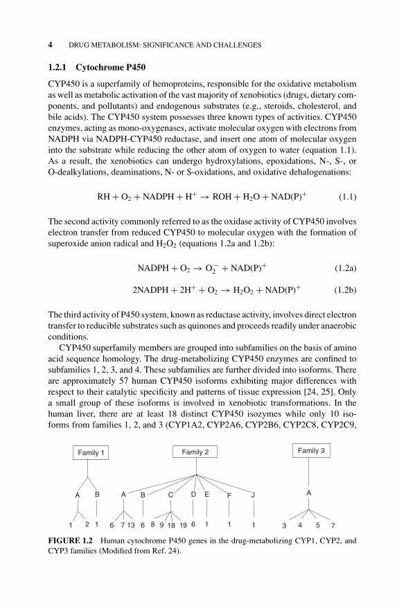
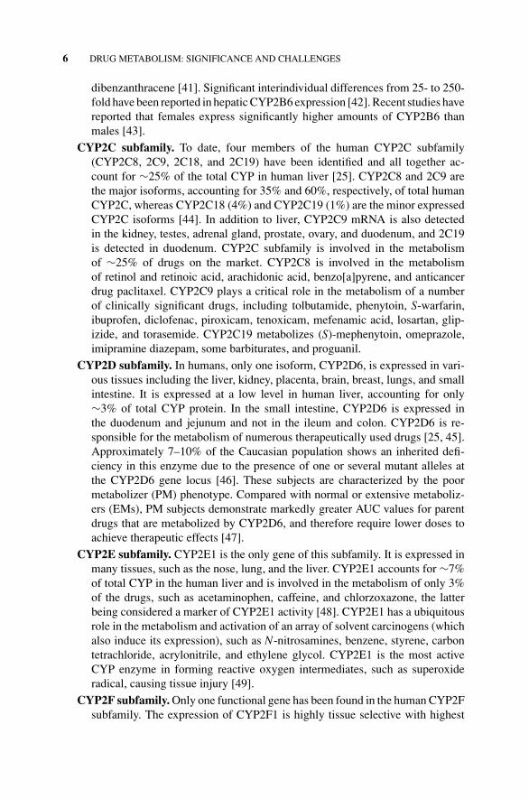
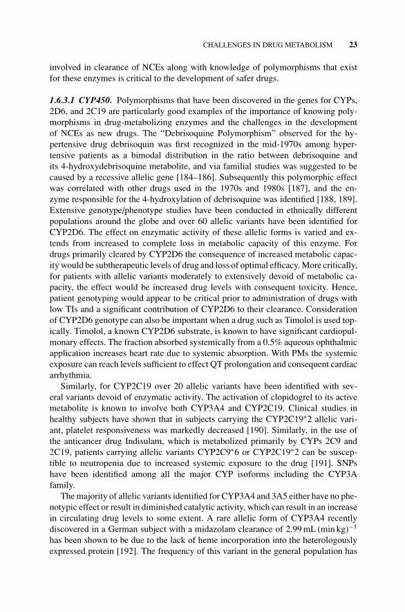
CYP450 superfamily members are grouped into subfamilies on the basis of aminoacid sequence homology. The drug-metabolizing CYP450 enzymes are confined tosubfamilies 1, 2, 3, and 4. These subfamilies are further divided into isoforms. Thereare approximately 57 human CYP450 isoforms exhibiting major differences withrespect to their catalytic specificity and patterns of tissue expression [24, 25]. Onlya small group of these isoforms is involved in xenobiotic transformations. In thehuman liver, there are at least 18 distinct CYP450 isozymes while only 10 iso-forms from families 1, 2, and 3 (CYP1A2, CYP2A6, CYP2B6, CYP2C8, CYP2C9,
Family 3
A B A
3 4 5 7
Family 2Family 1
1 2 6
A
1 7
B
6
C
9 18
D
13 8 19 6
E
1
F
1
J
1
FIGURE 1.2 Human cytochrome P450 genes in the drug-metabolizing CYP1, CYP2, andCYP3 families (Modified from Ref. 24).
P1: PIC/PIC P2: ABCc01 JWBK262/Xie September 18, 2008 18:59 Printer Name: Yet to Come
PHASE I DRUG-METABOLIZING ENZYMES 5
CYP2C19, CYP2D6, CYP2E1, CYP2F1, and CYP3A4) are responsible for the hep-atic metabolism of most of the marketed drugs (Figure 1.2) [24].
CYP1A subfamily. CYP1A subfamily consists of two members CYP1A1 and1A2. CYP1A1 is present predominantly in extrahepatic tissue such as lung,small intestine, placenta and kidney, and at a very low level in the liver. Onthe other hand, CYP1A2 is mainly confined to the liver and expressed at avery low level in extrahepatic tissue. Both CYP1A1 and 1A2 play an im-portant role in the metabolic activation of polycyclic aromatic hydrocarbons(PAHs), aromatic amines, and heterocyclic amines [26]. Despite the high sim-ilarity in their primary structures, CYP1A1 and 1A2 have different substratespecificities. For example, dibenzo[a]pyrene, a potent carcinogen, is oxidizedalmost exclusively by CYP1A1 in humans to highly mutagenic diol-epoxides[27] while acetanilide is metabolized primarily by CYP1A2. In human liver,CYP1A2 accounts for ∼15% of the total CYP content and is involved in themetabolism of ∼4% of marketed drug including acetaminophen, phenacetin,tacrine, ropinirole, riluzole, theophylline, and caffeine [25, 28].
CYP1B subfamily. CYP1B1 is expressed at a low level in heart, brain, placenta,lung, liver, and kidney [29], but it is present at much higher levels in tumorcells compared with the surrounding normal tissue [30]. CYP1B1 catalyzes2- and 4-hydroxylation of 17�-estradiol [31]. It also participates in themetabolic activation of a number of procarcinogens, including PAHs, PAH-dihydrodiols, and aromatic amines [32, 33]. CYP1B1 is also involved in themetabolism of some clinically relevant anticancer agents used in the treatmentof hormone-mediated cancer.
CYP2A subfamily. CYP2A subfamily includes CYP2A6, 2A7, and 2A13 in hu-mans (Figure 1.2). CYP2A6 is expressed in the liver and accounts for ∼4%of total hepatic CYP450, whereas 2A7 and 2A13 appear to be expressed ateven lower levels. CYP2A6 is the principal and perhaps the sole catalyst forhuman liver microsomal coumarin 7-hydroxylation. CYP2A6 is involved in theoxidation of only 3% of drugs such as nicotine, cyclophosphamide, ifosfamide,and fadrozole [25, 34, 35]. In addition, CYP2A6 also plays an important rolein the activation of several procarcinogens and promutagens especially the ni-trosamines [36]. CYP2A6 might also be involved in the metabolic activation of3-methylindole in human pulmonary microsomes [37]. CYP2A13 is predom-inantly expressed in the human respiratory tract and significantly involved inthe activation of aflatoxin B1 [38].
CYP2B subfamily. CYP2B subfamily includes CYP2B6 and 2B7 isoforms.CYP2B6 is expressed in the liver and in some extrahepatic tissues, whereasCYP2B7 mRNA expression was detected in lung tissue. CYP2B6 accounts for∼1% of total hepatic CYP content. CYP2B6 is involved in the metabolismof only 4% of the marketed drugs, such as the anticancer drugs cyclophos-phamide and tamoxifen [39], the anesthetics ketamine and propofol [40],and procarcinogens, such as the environmental contaminants aflatoxin B1 and
P1: PIC/PIC P2: ABCc01 JWBK262/Xie September 18, 2008 18:59 Printer Name: Yet to Come
6 DRUG METABOLISM: SIGNIFICANCE AND CHALLENGES
dibenzanthracene [41]. Significant interindividual differences from 25- to 250-fold have been reported in hepatic CYP2B6 expression [42]. Recent studies havereported that females express significantly higher amounts of CYP2B6 thanmales [43].
CYP2C subfamily. To date, four members of the human CYP2C subfamily(CYP2C8, 2C9, 2C18, and 2C19) have been identified and all together ac-count for ∼25% of the total CYP in human liver [25]. CYP2C8 and 2C9 arethe major isoforms, accounting for 35% and 60%, respectively, of total humanCYP2C, whereas CYP2C18 (4%) and CYP2C19 (1%) are the minor expressedCYP2C isoforms [44]. In addition to liver, CYP2C9 mRNA is also detectedin the kidney, testes, adrenal gland, prostate, ovary, and duodenum, and 2C19is detected in duodenum. CYP2C subfamily is involved in the metabolismof ∼25% of drugs on the market. CYP2C8 is involved in the metabolismof retinol and retinoic acid, arachidonic acid, benzo[a]pyrene, and anticancerdrug paclitaxel. CYP2C9 plays a critical role in the metabolism of a numberof clinically significant drugs, including tolbutamide, phenytoin, S-warfarin,ibuprofen, diclofenac, piroxicam, tenoxicam, mefenamic acid, losartan, glip-izide, and torasemide. CYP2C19 metabolizes (S)-mephenytoin, omeprazole,imipramine diazepam, some barbiturates, and proguanil.
CYP2D subfamily. In humans, only one isoform, CYP2D6, is expressed in vari-ous tissues including the liver, kidney, placenta, brain, breast, lungs, and smallintestine. It is expressed at a low level in human liver, accounting for only∼3% of total CYP protein. In the small intestine, CYP2D6 is expressed inthe duodenum and jejunum and not in the ileum and colon. CYP2D6 is re-sponsible for the metabolism of numerous therapeutically used drugs [25, 45].Approximately 7–10% of the Caucasian population shows an inherited defi-ciency in this enzyme due to the presence of one or several mutant alleles atthe CYP2D6 gene locus [46]. These subjects are characterized by the poormetabolizer (PM) phenotype. Compared with normal or extensive metaboliz-ers (EMs), PM subjects demonstrate markedly greater AUC values for parentdrugs that are metabolized by CYP2D6, and therefore require lower doses toachieve therapeutic effects [47].
CYP2E subfamily. CYP2E1 is the only gene of this subfamily. It is expressed inmany tissues, such as the nose, lung, and the liver. CYP2E1 accounts for ∼7%of total CYP in the human liver and is involved in the metabolism of only 3%of the drugs, such as acetaminophen, caffeine, and chlorzoxazone, the latterbeing considered a marker of CYP2E1 activity [48]. CYP2E1 has a ubiquitousrole in the metabolism and activation of an array of solvent carcinogens (whichalso induce its expression), such as N-nitrosamines, benzene, styrene, carbontetrachloride, acrylonitrile, and ethylene glycol. CYP2E1 is the most activeCYP enzyme in forming reactive oxygen intermediates, such as superoxideradical, causing tissue injury [49].
CYP2F subfamily. Only one functional gene has been found in the human CYP2Fsubfamily. The expression of CYP2F1 is highly tissue selective with highest
P1: PIC/PIC P2: ABCc01 JWBK262/Xie September 18, 2008 18:59 Printer Name: Yet to Come
PHASE I DRUG-METABOLIZING ENZYMES 7
expression observed in the lung and little or no hepatic expression [50].Substrates for CYP2F1 include ethoxycoumarin, propoxycoumarin, and pen-toxyresorufin, but not ethoxyresorufin. Recombinant CYP2F1 is capable ofactivating two prototypical pneumotoxicant, 3-methylindole and naphthalene.CYP2F1 metabolizes naphthalene to its highly toxic intermediate, naphthalene-1,2-epoxide, and 3-methyl indole to its dehydrogenated pneumotoxic metabo-lite 3-methyleneindolenine [51, 52].
CYP2J subfamily. CYP2J2 is the only gene of this subfamily. It is known to beexpressed in many extrahepatic tissues and may play a role in the oxidativebioactivation of arachidonic acid to form epoxyeicosatrienoic acids, whichmodulate bronchial smooth muscle tone and airway transepithelial ion transport[53]. CYP2J2 is also active toward other compounds such as linoleic acid andtestosterone. Recently, it has been reported that CYP2J2 is involved in theintestinal first-pass metabolism of an antihistamine drug, astemizole [54].
CYP2S subfamily. In humans, CYP2S1 appears to be the sole member of anew subfamily, CYP2S. The CYP2S1 gene is located at the proximal end onchromosome 19q13.2 CYP2 gene cluster [55]. Of interest, CYP2S1 is closelyrelated to CYP2F1 (47–49% identity) and is induced by dioxin in a human lungepithelial cell line, suggesting the possibility that CYP2S1 may participate inthe metabolism of toxic and carcinogenic compounds [39]. Recent studies usingheterologously expressed CYP2S1 in yeast have shown that CYP2S1 is ableto metabolize naphthalene [56]. Therefore, it is speculated that CYP2S1 mightalso play a role in naphthalene-induced lung toxicity [57].
CYP3A subfamily. The CYP3A subfamily of CYP450 in humans is composedof several enzymes and accounts for ∼28% of total hepatic P450 content.The human CYP3A family is clinically very important because it has beenshown to catalyze the metabolism of an amazingly large number of struc-turally diverse xenobiotics and endobiotics. It is estimated that CYP3A formsparticipate in the metabolism of 35–50% of all marketed drugs [25, 58]. Thehuman CYP3A subfamily includes CYP3A4, CYP3A5, CYP3A7 [59], andCYP3A43 [60]. CYP3A4 is the major human liver CYP3A enzyme, whereasCYP3A5 is present in only ∼20% of human liver. CYP3A4 and CYP3A5 arealso expressed in the stomach, lungs, small intestine, and renal tissue. Mostof the CYP3A4 substrates are also metabolized by CYP3A5 [61]. CYP3A7and CYP3A43 isozymes seem to play only a minor role in the metabolismof drugs. In fact, CYP3A7 is only present in fetal liver, whereas CYP3A43,which is expressed in liver, appears to be very restricted, both in terms of itsactivity and expression [62]. The highest concentration of transcript expressionof CYP3A43 is in the prostate, whereas hepatic mRNA concentration is only0.2–5% that of CYP3A4 [62]. Some examples of drugs metabolized by CYP3Aare terfenadine, the benzodiazepines midazolam and triazolam, quinidine, li-docaine, carbamazepine, nifedipine, tacrolimus, dapsone, erythromycin, anddextromethorphan [63]. In addition to drugs, CYP3A is involved in the oxi-dation of a variety of endogenous substrates, such as steroids, bile acids, andretinoic acid [64].
P1: PIC/PIC P2: ABCc01 JWBK262/Xie September 18, 2008 18:59 Printer Name: Yet to Come
8 DRUG METABOLISM: SIGNIFICANCE AND CHALLENGES
1.2.2 Flavin Mono-Oxygenase (FMO)
FMOs are NADPH-dependent and oxygen-dependent microsomal flavoenzymes,which oxygenate a number of drugs and xenobiotics that contain a “soft-nucleophile”heteroatom such as nitrogen, sulfur, and phosphorus. Unlike CYPs, which generallyuse sequential one-electron transfer, the FMOs are two-electron oxygenating en-zymes for N-oxidation. The microsomal FMO enzyme family is comprised of fiveisozymes (designated FMO1 to FMO5), whose expression is tissue specific. FMO1 ispredominantly expressed in human kidney and FMO2 in lung and kidney [65]. FMO3is the prominent isozyme in adult human liver, FMO4 is more broadly distributedin liver, kidney, small intestine and lung, and FMO5 is expressed in human liver,lung, small intestine, and kidney [66]. Of all the FMOs isozymes, FMO3 has a widesubstrate specificity, including the physiologically and plant-derived tertiary amine,trimethylamine, tyramine, and nicotine; commonly used drugs including cimeti-dine, ranitidine, clozapine [65, 66], methimazole, itopride, ketoconazole, tamoxifen,and sulindac sulfide; and agrichemicals, such as organophosphates and carbamates[65, 66].
1.2.3 Monoamine Oxidase
MAOs are mitochondrial flavoproteins containing one covalently bound FAD cofac-tor. Two isozymes, termed as MAO-A and MAO-B, are known for the MAO en-zyme family. They catalyze the oxidative deamination of structurally diverse aminesincluding neurotransmitters dopamine, norepinephrine, serotonin, tyramine, and 2-phenylethylamine, and some drugs and xenobiotics that contain cyclic and acyclicalkylamine functional groups [67, 68]. The MAO reaction cycle involves two halfreactions, as shown in equations 1.3a and 1.3c:
RCH2NH2 + FAD → RCH = NH2 + FADH2 (1.3a)
RCH = NH2 + H2O → RCH = O + NH3 (1.3b)
FADH2 + O2 + 2H+ → FAD + H2O2 (1.3c)
A two-electron oxidation results in the imine and reduced protein-bound FAD (equa-tion 1.3a). The imine is then nonenzymatically hydrolyzed to the carbonyl compound(equation 1.3b). In the second half reaction, the reduced FAD (FADH2) is reoxidizedby molecular oxygen producing hydrogen peroxide (equation 1.3c).
MAOs are expressed in most mammalian tissues that complicate pharmacokineticpredictions when MAOs are involved in metabolism. Inhibitors of MAOs are used inpsychiatry for the treatment of depressive disorders and in neurology for the treatmentof Parkinson’s disease. MAO-A and MAO-B play a critical role in the bioactivationof 1-methyl-4-phenyl-1,2,3,6-tetrahydropyridine (MPTP) to a toxic metabolite thatinduces Parkinson-like effects [68].
P1: PIC/PIC P2: ABCc01 JWBK262/Xie September 18, 2008 18:59 Printer Name: Yet to Come
PHASE I DRUG-METABOLIZING ENZYMES 9
1.2.4 Molybdenum Hydroxylases
Molybdenum hydroxylases (i.e., AO and XO) are flavoproteins that contain in ad-dition to a FAD, a pterine cofactor coordinated to a molybdenum atom, and an ironsulfur center for their catalytic activity. They catalyze the two-electron oxidation ofsubstrates with transfer to molecular oxygen to produce H2O2, and insert an atomof oxygen from water into a wide range of N-heterocycles and aldehydes via two-electron redox reaction as shown in equation 1.4:
RH + H2O → ROH + 2e− + 2H+ (1.4)
AO and XO are cytosolic enzymes and are closely related. However, they differin their substrate/inhibitor specificities. AO is involved in the metabolism of severalclinically significant drugs such as famciclovir, zaleplon, zonisamide, and ziprasidone[69–72]. XO has a narrower substrate specificity than AO and is mainly active towardpurines and pyrimidines. XO plays a role in the oxidation of several chemotherapeuticagents and has been implicated in the bioactivation of mitomycin B [73].
1.2.5 Epoxide Hydrolase
EH converts potentially reactive epoxides to trans-dihydrodiols. Originally, it wasthought to be localized solely in the endoplasmic reticulum; subsequent studiesdemonstrated there are distinct microsomal (EH1) and cytosolic (EH2) forms of theenzyme. The EH1 gene is polymorphic and the expressed enzyme has two metabolicfunctions, detoxification and bioactivation, depending on the particular substrate. EH1hydrolyzes epoxides, derived from aromatics and alkenes by CYP450 enzymes, to thecorresponding dihydrodiols through the trans addition of water. Dihydrodiols fromPAHs can be substrates for further transformation by CYP450 enzymes to dihydrodiolepoxides such as (+)-anti–benzo[a]pyrene (B[a]P)-7,8-diol-9,10 epoxide, the mostmutagenic and carcinogenic metabolite of B[a]P [74].
1.2.6 Esterase/Amidase
These enzymes are widely distributed in various tissue types and belong to the neu-tral, acidic, or metalloproteinase classes. While their roles have been recognizedwhen drug structures contain ester or amide functions, very little characterizationof the specific enzymes involved have been reported. Commonly, the ester functionis introduced into drug structures as a means of masking carboxylate functions toincrease absorption by increasing lipophilicity as with the antiviral drugs such as val-ganciclovir and oseltamivir that are converted in vivo to ganciclovir and oseltamivircarboxylate by intestinal and hepatic esterases [75, 76]. Amide functions are com-monly used in linking drug substructures. These bonds are generally not susceptibleto amidase activity since typically they are not natural amino acid derived. However,when such functions do present themselves in the NCEs, the potential for hydrolysisas a mechanism for clearance should be considered and stability in whole blood andhepatic subcellular matrices should be determined.
P1: PIC/PIC P2: ABCc01 JWBK262/Xie September 18, 2008 18:59 Printer Name: Yet to Come
10 DRUG METABOLISM: SIGNIFICANCE AND CHALLENGES
1.3 PHASE II CONJUGATIVE ENZYMES
Phase II reactions are catalyzed by conjugative enzymes, such as UDP-glucuronosyltransferase (UGT), sulfotransferase (SULT), glutathione S-transferase(GST), N-acetyltransferase (NAT) and methyltransferase (N-methyl-, thiomethyl-,and thiopurinemethyl-). Glutathione conjugates are further metabolized to cysteineand N-acetyl cysteine adducts. Most phase II reactions result in a compound’s con-comitant increase in hydrophilicity and decrease in volume of distribution (VDss),which together greatly facilitate its excretion from the body.
1.3.1 Uridine Diphosphate Glucuronosyltransferase (UGT)
UGT family of enzymes catalyzes the transfer of glucuronic acid from UDP glu-curonic acid to available substrates to form the water-soluble glucuronide conju-gates, suitable for excretion. Thus, glucuronidation is a major detoxification pathwayof endo- and xenobiotics in man and other animals. Beside human liver, the gut andkidneys are two important sites of glucuronidation. The UGT family of enzymes issubdivided into two subfamilies, UGT1 (1A1, 1A3, 1A4, 1A5 1A6, 1A7, 1A8, 1A9,and 1A10) and UGT2 (2A1, 2B4, 2B7, 2B10, 2B11, 2B15, 2B17, and 2B28), on thebasis of sequence homology [77]. All classes of drugs containing a wide range ofacceptor groups including phenols, alcohols, aliphatic and aromatic amines, thiols,acidic carbon atoms, and carboxylic acids are substrates for UGTs and this pathwayhas been estimated to account for ∼35% of all drugs metabolized by phase II drug-metabolizing enzymes [78]. Human UGTs can also form N-linked glucuronides ofseveral tertiary amine drugs [79]. Some acyl glucuronides are reactive intermediatesthat bind covalently to macromolecules causing potential toxicity. Several drugs thatcontain a carboxylic acid such as diclofenac, ketoprofen, suprofen, and tolmetin areglucuronidated to form a reactive acyl glucuronide that could be associated with someof the observed toxicity of these drugs [80].
1.3.2 Sulfotransferase
SULT family of enzymes catalyzes the transfer of sulfite (SO3−) from 3′-
phosphoadenosine 5′-phosphosulfate (PAPS) to a hydroxyl or an amino group onan acceptor molecule to form the water-soluble sulfonate or sulfamate conjugatesand thereby aid in their excretion via the kidneys or bile. SULTs are capable ofsulfonating hydroxyl group of a wide range of substrates including phenols, primaryand secondary alcohols, N-hydroxy arylamines, and N-hydroxy heterocyclic amines.Amino groups of arylamines such as 2-naphthylamine are also sulfonated by SULTs.SULTs have a wide tissue distribution and play an important role in the detoxifi-cation, metabolism, and bioactivation of numerous xenobiotics, many dietary andenvironmental mutagens, drugs, neurotransmitters, and hormones [81]. In humans,three SULT families, SULT1, SULT2, and SULT4, have been identified that contain atleast 13 distinct members. SULT1 and SULT2 families are the largest and are respon-sible for sulfonating the greatest number of endogenous and xenobiotic compounds.
P1: PIC/PIC P2: ABCc01 JWBK262/Xie September 18, 2008 18:59 Printer Name: Yet to Come
PHASE II CONJUGATIVE ENZYMES 11
Studies using recombinant enzymes demonstrated that many promutagens are acti-vated with high selectivity by an individual SULT form.
1.3.3 Glutathione S-Transferase
The GST family of enzymes catalyzes the nucleophilic attack of the tripeptide(� -glu-cys-gly) (GSH) on a wide variety of soft electrophiles such as epoxides andquinones, formed during phase I oxidation of xenobiotics, generally resulting in theirelimination and detoxification. There are two GST superfamilies: (1) the membrane-bound GST isozymes and leukotriene C4 synthetase and (2) the cytosolic-solubleenzymes, each of which displays different intracellular distribution and distinct cat-alytic as well as noncatalytic binding properties. Thirteen different human GSTsubunits, GSTA1 through GSTA4, GSTM1 through GSTM5, GSTP1, GSTT1 andGSTT2, and GSTZ1, have been identified belonging to seven distinct classes: alpha(�), mu (�), omega (�), pi (�), sigma (�), theta (�), and zeta () [82]. GSTs appear tobe ubiquitously distributed in human tissues. Some examples of clinically significantdrugs that form glutathione conjugates include acetaminophen, sulfonamides, irinote-can, carbamazepine, rotonavir, clozapine, procainmide, hydralazine, cyclosporine A,diclofenac, estrogens, and tamoxifen [83].
1.3.4 N-Acetyltransferase
NAT family of enzymes catalyzes the acetyl-CoA-dependent acetylation of arylamines and arylhydrazines and N-hydroxyarylamine including many drugs and car-cinogens. In most cases, this reaction is generally considered to result in the detoxifi-cation of potentially toxic exogenous compounds. However, NATs are also involvedin bioactivation reactions via O-acetylation of N-hydroxyarylamines to unstable ace-toxy esters that decompose to highly reactive mutagens that form adducts with cellularmacromolecules. Humans express two distinct isozymes, designated NAT1 and NAT2[84]. Clinically relevant substrates of NAT include isoniazid, procainamide, aminog-lutethimide, sulphamethoxazole, 5-aminosalicylic acid, hydralazine, phenelzine, anddapsone. Also, NATs play an important role in the metabolism of industrial and envi-ronmental carcinogens, including 2-naphthylamine, benzidine, 2-aminofluorene, and4-aminobiphenyl, as well as of potentially carcinogenic heterocyclic amines foundin well-cooked red meat and cigarette smoke [85].
1.3.5 Methyl Transferase (MT)
MT family of enzymes catalyzes the O-, S-, or N-methylation of drugs, hormones, andneurotransmitters, and utilizes S-adenosyl-l-methionine (SAM) as a methyl donor.Catechol-O-methyltransferase (COMT) is the most extensively investigated drug-metabolizing MT. It plays an important role in the biotransformation of both endoge-nous and exogenous catechols. COMT has a rather broad substrate specificity forstructures that contain catechol moieties and is often involved in the methylation of
P1: PIC/PIC P2: ABCc01 JWBK262/Xie September 18, 2008 18:59 Printer Name: Yet to Come
12 DRUG METABOLISM: SIGNIFICANCE AND CHALLENGES
drugs that have been metabolized by the CYP system to generate catechol functions[86, 87].
S-Methylation is also an important pathway in the biotransformation ofmany sulfur-containing drugs. At least two separate enzymes, thiol methyltrans-ferase (TMT) and thiopurine methyltransferase (TPMT) are known to catalyzeS-methylation in humans [87]. TMT, a membrane-bound enzyme, catalyzes theS-methylation of captopril, d-penicillamine, and other aliphatic sulfhydryl com-pounds such as 2-mercaptoethanol. On the other hand TPMT, a cytosolic enzyme,catalyzes the S-methylation of aromatic and heterocyclic sulfhydryl compounds in-cluding 6-mercaptopurine and other thiopurines. Recently, S-methyltransferase hasbeen shown to play a critical role in the metabolism of the antipsychotic drug, ziprasi-done, in humans [72, 88]. Both TPMT and TMT have been shown to be geneticallypolymorphic in humans.
Histamine-N-methyltransferase is a hepatic enzyme, although its inhibition byvarious drugs has been reported [87, 89], its ability to methylate drugs has not beenestablished. Several protein arginine methyltransferases (PRMTs) are known andtheir physiological role in producing mono- and dimethylated arginine is known[90]. However, their role in N-methylating drug molecules has yet to be identified.Thus, except for COMT and S-methyltransferases the other MTs including DNAmethylating enzymes are not known to play a role in drug metabolism.
1.4 DRUG EFFLUX TRANSPORTERS
Carrier-mediated transport of drugs and their metabolites has recently been recog-nized as an important issue in pharmaceutical research. There is a wealth of infor-mation that suggests that transporters are responsible both for the uptake and effluxof drugs and other xenobiotics, and may be key determinants of the disposition of adrug [91, 92]. Transporter proteins are divided into two categories: (1) the adenosinetriphosphate (ATP) binding cassette (ABC) transporter superfamily and (2) the solutecarrier (SLC) family of proteins.
1.4.1 ABCB1 (P-Glycoprotein, MDR1)
The first member of ABC efflux transporter family to be discovered wasP-glycoprotein (P-gp; gene ABCB1 or MDR1). The importance of P-gp or MDR1 wasfirst recognized in the multidrug resistance of tumor cells in response to chemotherapytreatment [93]. However, constitutive expression of P-gp in many normal tissues suchas liver, kidney, intestine, blood–brain barrier, and lymphocytes demonstrates its rolein modulation of the absorption, distribution, metabolism, excretion, and toxicologybehaviors of some drugs and NCEs in development [94]. Experiments with mdr1aknock-out mice revealed that P-gp not only limits the CNS entry of drugs, but it alsoreduces the oral absorption of drugs by extruding them from enterocytes back intothe intestinal lumen [95]. Therefore, the inhibition and induction of P-gp transportercan lead to significant drug–drug interaction and, most importantly, to drug treatment
P1: PIC/PIC P2: ABCc01 JWBK262/Xie September 18, 2008 18:59 Printer Name: Yet to Come
DRUG EFFLUX TRANSPORTERS 13
resistance. Talinolol, fexofenadine, and digoxin are the most commonly used probesubstrates for assessing the modulation of P-gp.
1.4.2 ABCC1 (Multidrug Resistance Related Protein1, MRP1)
ABCC1 or MRP1 is a 190-kDa transport protein that was originally cloned froma doxorubicin-selected lung cancer cell line. MRP1 has now been found to be ex-pressed in a range of solid and hematological tumors, and has been demonstrated totransport a wide array of structurally diverse anticancer drugs such as oxorubicin,vincristine, and methotrexate [96]. In addition, MRP1 also transports many glu-tathione, glucuronide, and sulfate conjugated organic anions, such as leukotriene C4,17�-estradiol-glucuronide, and estrone 3-sulfate, respectively. MRP1 is also foundin normal tissues throughout the human body and plays a significant role in tissuedefense from toxic agents. Thus the expression levels and activity of MRP1 areimportant considerations in drug development and chemical toxicity.
1.4.3 ABCC2 (Multidrug Resistance Related Protein2, MRP2)
ABCC2 or MRP2 is located on the bile canalicular membrane and is involved inthe biliary excretion of glucuronide, glutathione, and sulfate conjugates of lipophiliccompounds (i.e., drugs) as well as endogenous compounds such as hormone andbilirubin conjugates. In addition, MRP2 can also transport uncharged compounds incotransport with glutathione and thus can modulate the disposition of many drugs.MRP2 is predominantly expressed at the hepatocyte canalicular membrane [97]. Itis also expressed in other tissues such as the kidney, as well as the intestinal entrocytes[97, 98] and apical membrane of brain capillary endothelial cells [99]. In humans,a genetic deficiency of MRP2 results in a disease known as Dubin–Johnson syn-drome [100]. In addition, altered MRP2 function can change the ADME propertiesof many clinically important drugs including cancer chemotherapeutics (irinotecan,methotrexate, and vinblastine), antibiotics (ampicillin and rifampin), antihyperlipi-demics, and angiotensin-converting enzyme inhibitors.
1.4.4 ABCG2 (Breast Cancer Resistance Protein, BCRP)
ABCG2 or BCRP, a 655 amino acid peptide with an ability to extrude a wide varietyof chemical compounds from the cells, was first cloned from mitoxantrone andanthracycline-resistant breast and colon cancer cell lines [101, 102]. BCRP is a half-transporter efflux pump with an intracellular N and C terminus and an intracellularATP binding site, followed by six putative transmembrane segments. It is expressedin liver, small intestine, placenta, kidney, mammary gland, and endothelial cells atthe blood–brain barrier, and is believed to be involved in protecting tissues fromxenobiotic accumulation and resulting toxicity [103]. It is capable of transportingsulfate and glucuronide conjugated organic anions, at least in vitro. It affects intestinalabsorption/bioavailability, organ distribution, hepatic/renal elimination, and plasmaclearance of substrate drugs such as topotechan [104]. Downregulation of BCRP was

P1: PIC/PIC P2: ABCc01 JWBK262/Xie September 18, 2008 18:59 Printer Name: Yet to Come
14 DRUG METABOLISM: SIGNIFICANCE AND CHALLENGES
suggested to contribute to cellular adaptation to folate deficiency and homeostasis[105].
1.5 DRUG UPTAKE TRANSPORTERS
The drug uptake (SLC) family of transporter is the largest superfamily of transporters.This family includes 31 transporters from organic anion transporter polypeptides(OATPs), organic anion transporters (OATs), organic cation transporters (OCTs), pep-tide cotransporters (PEPTs), and sodium–bile acid cotransporter classes. Only OATP,OAT, OCT, and PEPT are primarily involved with the transport of drugs/xenobiotics.
1.5.1 Organic Anion Transporter Polypeptides
The OATP transport proteins function as organic solute exchangers with broad sub-strate specificity that includes organic anions, cations, and neutral compounds [106].The OATP transporters are expressed in organs such as the intestine, liver, andblood–brain barrier and considered to be the key determinant in the cellular uptake ofmany endogenous and exogenous chemicals, including drugs in clinical use. To date,eight different OATPs (OATP1A2, OATP1B1, OATP2B1, OATP3A1, OATP4A1,OATP1C1, OATP1B3, and OATP 2A1) have been cloned from human tissues [107].
1.5.2 Organic Cation Transporter
OCTs are responsible for reabsorption and excretion of a wide variety of organiccations, such as quinidine, cimetidine, procainamide, vecuronium, and cardiac gly-cosides as well as endogenous substances including, dopamine, epinenephrine, nore-pinephrine, creatinine, and choline. The OCT transporters are expressed in humanorgans such as the intestine, liver, proximal tubules of kidney, brain, arota, skeletalmuscle, prostate, adrenal gland, salivary gland, and fetal lung. To date, three differentOCTs (OCT1, OCT2, and OCT3) have been identified in humans [108, 109].
1.5.3 Organic Anion Transporter
OAT family plays a critical role in the renal excretion and detoxification of a wide va-riety of compounds including drugs, toxins, hormones, and neurotransmitter metabo-lites. OATs are primarily expressed in the kidney. In addition, OAT expression hasbeen detected in liver, placenta, brain, and choroid plexus. At least five differentOATs (OAT1, OAT2, OAT3, OAT4, and Urat1) have been detected in human tissues[109].
1.5.4 Peptide Transporter
The PEPTs are hydrogen ion-dependent transporters that transport small peptidesand proteinlike compounds such as cephalosporins, penicillin, enalapril, and cap-topril. Humans express two distinct PEPTs designated PEPT1 and PEPT2. PEPT1
P1: PIC/PIC P2: ABCc01 JWBK262/Xie September 18, 2008 18:59 Printer Name: Yet to Come
CHALLENGES IN DRUG METABOLISM 15
is involved with the oral absorption of drugs from small intestine while PEPT2contributes to the proximal tubular reabsorption of drugs [108].
1.6 CHALLENGES IN DRUG METABOLISM
1.6.1 Prediction of Metabolism and Pharmacokinetics in Humans
1.6.1.1 In silico Computational Tools. In recent years, several in silico computa-tional methods have become available for prediction of metabolism and pharmacoki-netics of the NCEs and are increasingly becoming a part of the drug discovery processto select candidates with desirable ADME properties [110–112]. De Graaf et al. [110]have described how in silico computational approaches can be used to understand,rationalize, and predict the activity and substrate selectivity of CYPs. Mechanism-based quantum chemical calculations on substrates and the enzyme, pharmacophoremodeling of ligands, and protein homology modeling in combination with automateddocking and molecular dynamics simulations have been used to rationalize and pre-dict ligand binding and formation of metabolites. Several of these models, especiallyfor CYP2D6 [113] and CYP2C9 [114], have been shown to have a reasonably goodpredictive value concerning qualitative metabolism and substrate inhibitor selectivity.Programs such as Meteor and Metasite use database-based evaluations or de novocomputational methods based on bond energies and active site docking to predictmetabolic transformations and hot spots. While these methods assist in predictingmetabolic outcomes, they are still limited and neither software is capable of predictingrates of metabolism at the sites that are predicted. These tools cannot be used to guidenew chemical synthesis that incorporates metabolic concerns without experimentalverification.
1.6.1.2 In Vitro–In Vivo Extrapolation. The ability to predict human pharmacoki-netic from in vitro and in vivo models with a reasonable degree of accuracy (2.0-foldvariation) is an ongoing challenge with many different approaches yielding varyingdegrees of success. Interspecies allometric scaling of pharmacokinetic parametersis the earliest of the approaches and is still a tool used in predicting human phar-macokinetic behavior of drugs. The method is based on empirically observed re-lationships between physiological processes and body weights of mammals [115].Several enhancements have been introduced to the original method such as maximumlife-span potential, brain weight, body surface area, rule of exponents, and proteinbinding [115–117]. The allometric methods assume that clearance mechanisms aresimilar across species and do not consider interspecies specificity differences in drug-metabolizing enzymes and metabolic pathways. A recent analysis of 103 compoundsfor allometric scaling incorporating the enhanced methods where data were availablefor rat, dog, and monkey showed that the success rate for human projection frompreclinical models was suboptimal (18–53%) with or without the enhanced methods[118]. A major limitation of the allometric methods is the lack of incorporation ofmetabolic differences in preclinical species and humans.
P1: PIC/PIC P2: ABCc01 JWBK262/Xie September 18, 2008 18:59 Printer Name: Yet to Come
16 DRUG METABOLISM: SIGNIFICANCE AND CHALLENGES
In vitro methods using liver slices, hepatocytes, and liver subcellular fractions,such as microsomes, have also been developed, and are used to varying extents inclearance prediction. Methods based on half-life of NCEs in metabolically active livermicrosomes are used to project hepatic extraction from which clearance predictionsare calculated [119]. The limitation of this approach is that prediction is limited tothose NCEs for which hepatic metabolism by oxidative enzymes of the endoplasmicreticulum dominate the overall clearance mechanism. When drug-metabolizing en-zymes other than CYPs or FMOs dominate the metabolic clearance, this approachfalls short. Hepatocytes contain the entire cellular metabolic machinery and wouldbe considered the better choice for hepatic clearance prediction. However, unlike hu-man liver microsomes, which can be prepared from frozen liver tissue and effectivelystored at −80◦C, fresh human hepatocytes are not readily accessible, and the activitiesof some drug-metabolizing enzymes have been shown to decline with age of hepato-cyte cultures, thus limiting their use. Cryopreserved human hepatocytes are used ina limited capacity to predict hepatic clearance and in evaluating overall metabolismof NCEs. When allometric scaling was combined with in vitro metabolism-basedscaling for 10 highly metabolized drugs, a significant improvement was noted for thepredicted human clearance compared to either method alone [120]. A recent retro-spective analysis of pharmacokinetic data for a large set of diverse drugs suggeststhat human pharmacokinetic parameters can be predicted from rat pharmacokineticdata within a threefold variation by a fixed exponent approach [121]. This methodallows for early binning of NCEs into low-, medium-, or high-clearance categoriesat a very early stage in drug discovery.
In the past two decades, success in predicting human pharmacokinetics frompreclinical and in vitro techniques has dropped the attrition rate for NCEs due tofailure to achieve the pharmacokinetic thresholds necessary for drug action in hu-mans from ∼40% to less than 10% [6]. Thus, while success has been achieved inthis area, clearly much progress is still necessary in preclinical models to achievegreater success in eliminating attrition due to pharmacokinetics. A more direct ap-proach using drug microdosing in humans to predict pharmacokinetic behavior earlyin the drug development process is under investigation. In this approach, NCEs areadministered at doses significantly below therapeutic levels (�100-fold lower thanprojected therapeutic dose or 100 µg) in order to get an early readout on their phar-macokinetic behavior [122, 123]. A microdosing study with five marketed drugs(warfarin ZK253 (schering), midazolam, erythromycin, and diazepam), known to beproblematic in extrapolation to humans from either in vitro or animal models, hasshown good concordance in the pharmacokinetic parameters obtained under micro-and therapeutic-dosing conditions for all except erythromycin [124]. Because of theextrmely low dose, this method does not require the same extent of animal safetytesting as is required by a traditional investigative new drug (IND) application to theFood and Drug Administration [124–126]. Microdosing has become possible becauseof newer analytical tools such as liquid chromatography coupled high sensitivity softionization mass spectrometry and more recently at significantly higher sensitivitywith accelerator mass spectrometry [127].
P1: PIC/PIC P2: ABCc01 JWBK262/Xie September 18, 2008 18:59 Printer Name: Yet to Come
CHALLENGES IN DRUG METABOLISM 17
1.6.1.3 Species Specificity and Limitation of Animal Models. The primary goalsin the use of preclinical studies in animals, typically rodent (rat and mouse), dog,monkey, and rabbit, are to extrapolate pharmacokinetic parameters and establishclearance mechanisms applicable to human drug administration, determine toxicitylimiting dosages, and evaluate long-term carcinogenic and teratogenic potential ofthe NCEs. Drug-metabolizing enzymes in mammals have common ancestral roots.However, the substrate and reaction rate specificities of NCEs are known to varyconsiderably among species [128, 129]. These variations in activity and specificityare particularly important for pharmacokinetic parameters when drug clearance ismetabolically driven, and also when preclinical species are considered for the toxi-cological model. For toxicological studies, identification of major metabolites fromin vitro and in vivo studies in rodent, dog, and monkey compared to those from humanin vitro reactions with hepatocytes and microsomes are particularly useful to identifythe preclinical species with optimal coverage of exposure to potential circulatingmetabolites in humans. Interspecies specificity of some drug-metabolizing enzymescan differ significantly. For example, when AO is involved in metabolic clearance,species selection for pharmacokinetics and toxicology testing is critical. AO activityis high in monkey and human, moderate in rodent, and not detected in dog liver.Complicating the situation is that significant species differences exist in both speci-ficity and activity of this enzyme. Thus, in addition to identifying metabolites, thereaction phenotype for the major metabolites of an NCE at early stages in the drugdevelopment process can be critical in selection of the species for pharmacokineticand toxicological testing [130].
1.6.2 Drug–Drug Interactions
Drug–drug interactions (DDIs) have received considerable attention in the pharma-ceutical industry because in recent years several prominent drugs have been with-drawn from the market in the US and Europe due to serious adverse events as a resultof significant DDIs [131]. DDIs are caused when the clearance of one drug is influ-enced by a coadministered drug [131]. Therefore, the consequences of these DDIscan range from loss of therapeutic efficacy to the introduction of potential lethal toxiceffects. Although DDIs can occur during absorption, distribution, and eliminationphases following initial drug administration, metabolism seems to be the predom-inant mechanism for such interactions. Since most marketed drugs are eliminatedfrom the body at least in part by metabolism, inhibition and/or induction of DMEsby one drug could have a significant effect on the disposition of another drug. TheNCEs can either be perpetrators or victims of such interactions, and early evaluationof NCEs for their potential to function in either capacity is critical to the developmentof new drug candidates and involves the identification of drug-metabolizing enzymesinhibited by NCEs (in particular CYPs) as well as the metabolic pathways by whichthey are cleared [130]. High specificity substrates for in vitro/in vivo use in druginteraction assays are known for six major isoforms of CYP enzymes involved indrug metabolism.
P1: PIC/PIC P2: ABCc01 JWBK262/Xie September 18, 2008 18:59 Printer Name: Yet to Come
18 DRUG METABOLISM: SIGNIFICANCE AND CHALLENGES
1.6.2.1 Biotransformation Reaction Phenotyping. As described earlier, a majorityof drugs are cleared by metabolic transformation, generally first by oxidation tomore polar metabolites, by hepatic enzymes. Knowledge of the specific enzyme(s)especially CYPs, involved in the metabolism of and inhibition by the NCE, particu-larly of the major pathways, is critical for evaluation of the potential liabilities thatan NCE may have with respect to DDI [130]. Metabolic flux of an NCE throughseveral CYP-dependent pathways where each pathway contributes less than 30% ofthe overall metabolic clearance is always preferable to flux through a unique CYPisoform since inhibition of any one of the pathways by a coadministered drug wouldnot significantly increase the systemic exposure of the NCE, and consequently poseno significant risk. Terbinafine, an oral antifungal agent, is an example of such adrug where multiple pathways for its metabolism predict lack of significant clinicalDDI for its clearance [132], which has been clinically confirmed [133]. However,terbinafine is an inhibitor of CYP2D6 and has been shown to increase the AUC ofdesipramine when coadministered to CYP2D6 EMs, suggesting that caution mustbe used with coadministration of terbinafine to patients on medications with narrowtherapeutic index (TI), and its metabolism is mediated primarily by CYP2D6. Thus,terbinafine can be considered as an example of a drug that is only a perpetrator fora drug interaction that involves coadministered drugs that are metabolized primarilyby CYP2D6.
If the metabolic flux of an NCE is greater than 60% through a unique CYP isoform,especially by a polymorphic enzyme (CYP2D6), the potential for DDI is extremeand careful consideration has to be given to the forward development strategy forsuch an NCE [134]. The case of the antihistaminic drug terfenadine stands out asan excellent example of this process. Terfenadine is primarily metabolized to itspharmacologically active form by CYP3A4. Inhibition of CYP3A4 by a variety ofdrugs, best exemplified by ketoconazole, results in dramatic increases in the AUC ofterfenadine to toxic levels [135] that can prolong the QT interval and produce poten-tially fatal ventricular arrhythmias. This example shows how ketoconazole functionsas the perpetrator of the DDI while terfenadine, because of its major flux throughCYP3A4, becomes victim. When the TI for the NCE is narrow, knowledge of theCYP isoform(s) involved is critical and close attention is needed to the potential forDDI by coadministered drugs.
Cyclosporine, paclitaxel, tacrolimus, and warfarin are examples of drugs withnarrow TIs where even dietary habits can play significant roles in toxicity. Forexample, in the late 1980s clinical observations noted a dramatic effect of grapefruitjuice consumption on the levels of dihydropyridine calcium ion channel blockers.Flavanoids present in grapefruit juice were speculated on the basis of studies thatshowed Naringenin, the aglycone component of the flavanoid naringin, which isabundant in grapefruit juice, as inhibiting the in vitro metabolism of dihydropyridinesby human liver microsomes [136]. Clinical studies on renal transplant patients showedthat grapefruit juice significantly enhanced the systemic exposure of cyclosporine,while having no significant effects on prednisone or prednisolone [137]. Furtherinvestigations have established that the effect is on drugs that are primarily cleared byCYP3A4 and show low bioavailability due to presystemic metabolism by CYP3A4
P1: PIC/PIC P2: ABCc01 JWBK262/Xie September 18, 2008 18:59 Printer Name: Yet to Come
CHALLENGES IN DRUG METABOLISM 19
in the entrocytes. The increase in systemic exposure arises from the inactivation ofCYP3A4 in the entrocytes.
Epoxyfuranocoumarins formed from grapefruit juice components like bergamottinand related natural products have been identified as responsible for self-inactivationof CYP3A4 [138]. The inhibitory effect of grapefruit juice on enteric metabolism byCYP3A4 is temporal and its duration is limited to the duration of consumption[139]. Dietary effects from the consumption of other foods and natural remedieson the metabolism of some drugs have also been reported [131–143]. Examples ofsuch processes with other CYP450 enzymes abound in the literature with over 500published articles since 1990 that examine in vitro, in vitro–in vivo correlations, andin vivo drug interactions via CYP450 enzymes demonstrating the importance of suchstudies in drug development. Thus, early phenotyping of metabolic pathways for anNCE and the inhibitory effect of the NCE on CYP isoforms are important aspects ofthe development process for NCEs as drug candidates that provide an understandingof potential clinical DDI issues that may arise for which early clinical evaluationscan be conducted. Reaction phenotyping is done by several methods including theuse of specific inhibitors for some CYP isoforms, recombinant CYP isoforms andrelative activity factors, inhibitory antibodies, and interindividual correlations [130,144–148].
As with CYP isoforms, UGTs are also more commonly encountered in themetabolism of drugs and xenobiotics. There are several drugs and xenobiotics whereglucuronidation contributes to the overall metabolic clearance mechanism and insome instances is the predominant metabolic pathway. Examples include zidovudine(AZT) [149], mycophenolate [150], retigabine [151], mitiglinide [152], fenamates[153], and gemcabene [154]. In contrast to extensive DDIs caused by binding ofdrugs to CYP isoforms, DDIs with the UGT family enzymes are relatively few, andare confined to compounds where UGTs are either the primary clearance mecha-nism or contribute substantially to it. The cancer drug irinotecan is metabolized toits active form SN-38 (7-ethyl-10-hydroxy-camptothecin), which is cleared by glu-curonidation by UGT1A1. Ketoconazole increases the circulating levels of SN-38due to potent inhibition of its glucuronidation by UGT1A1 [155]. Since irinotecanhas a small TI, patients should be genotyped for UGT1A1 prior to commencement oftherapy. Another example of a UGT-dependent drug interaction is that of statins andthe lipid-lowering drug, gemfibrozil [156]. A significant component of simvastatin’sclearance is via an unusual lactonization mechanism involving an intermediate acylglucuronide catalyzed by UGTs [157]. Gemfibrozil, which is also metabolized to itsglucuronide by UGTs, inhibits the glucuronidation of simvastatin with the resultantpharmacokinetic effect of an increase in simvastatin’s AUC and decrease in biliarylevels of simvastatin lactone and glucuronide [158]. UGT1A1 has a critical physi-ological function in the elimination of bilirubin as its glucuronide. Surprisingly, noreports on drug interactions with UGT1A1 such as that of ketoconazole have beenreported to show significant increases in the blood levels of bilirubin. A possibleexplanation for this may be that the in vivo concentrations of substrates or inhibitorscleared primarily by the particular UGT under consideration may often be below theircorresponding Km and Ki values, resulting in partial site occupancy and consequently
P1: PIC/PIC P2: ABCc01 JWBK262/Xie September 18, 2008 18:59 Printer Name: Yet to Come
20 DRUG METABOLISM: SIGNIFICANCE AND CHALLENGES
insignificant in vivo interactions. Hence, when considering the potential for drug in-teractions with the UGTs, dose and concentrations in the blood and in the liver areimportant parameters to consider. Reaction phenotyping of UGTs is less developedthan that for CYPs. Few high specificity low Km or Ki substrates or inhibitors havebeen identified, except for UGT1A1 where bilirubin is a highly specific substrate.However, it is only useful for in vitro inhibition assays. Phenotyping of UGT reactionsis generally conducted in vitro using recombinant forms of the enzymes.
Drug interactions with MAOs are particularly relevant when patients are on therapywith MAO inhibitors and the MAOs are significant contributors to the clearance ofa drug. As with UGTs, the Km or Ki for MAO substrates tend to be generallyhigh and drug interactions at therapeutic levels are less pronounced than with CYPenzymes. MAO-A and MOA-B can be selectively inactivated by Chlorgyline andDeprenyl, respectively [68]. Thus, distinguishing between these enzymes can bereadily achieved using either liver microsomal suspension where MAO-A and Bare present as contaminants from fractured mitochondrial membrane particles orrecombinant enzymes [68].
AO has been shown to be responsible for the primary metabolism of several drugsthat contain an aromatic nitrogen heterocycle [72, 88, 159, 160]. In general, when drugstructures contain aromatic nitrogen heterocycles, a role for AO should be consideredin preclinical and in vitro tests. The contribution of this enzyme to drug clearancecan be defined by examining metabolism of an NCE by cytosolic preparations andinhibition by either menadione or raloxifene [161, 162]. DDIs with AO have not yetbeen reported in the literature.
Several in vitro models including cell-based, cell-free, and yeast systems as wellas in vivo models such as genetic knock-out, gene-deficient, and chemical knock-outanimals have recently been developed in order to understand the interaction betweendrugs and transports [163]. However, unlike the CYP-mediated drug interactions, theevidence for the transport-mediated drug interactions is often less conclusive due toa broad overlap of substrate specificities between transporters and drug-metabolizingenzymes. Some evidences for the interaction of digoxin, a good P-gp substrate, whencoadministered with valspodar or verapamil (potent and selective P-gp inhibitors)have been reported [164].
1.6.2.2 Metabolism-Based Inactivation/Bioactivation. The metabolic activation ofan NCE to a reactive intermediate is an important consideration in the developmentof NCEs as new drug candidates. Metabolic activation can be effected by phase 1 orphase 2 enzymes. Depending on the mechanism and the substrate, oxidative reactionscan yield reactive intermediates such as epoxides, quinone methides, quinone imines,and quinones, which are alkylating agents, which react with cellular constituentssuch as DNA and proteins. A comprehensive listing of known bioactivation reactionsand mechanisms has been presented [83, 165]. These modifications are thought to beresponsible for DNA damage that can result in tumor initiation and promotion, andidiosyncratic reactions that are observed in patients post marketing of a new drug. TheAmes test and micronucleus assay are routinely used to determine the carcinogenicpotential of an NCE early in the development process. In addition, metabolism-based
P1: PIC/PIC P2: ABCc01 JWBK262/Xie September 18, 2008 18:59 Printer Name: Yet to Come
CHALLENGES IN DRUG METABOLISM 21
inactivation (MBI) assays have been established to detect either an enzymatic activityloss at a single concentration or an IC50 shift when an NCE is preincubated witheither pooled human liver microsomes or recombinant CYP enzymes, and humanhepatocytes. However, the relationships between protein covalent binding by reactiveintermediates and toxicity are not well understood.
An in vitro test commonly used to establish reactive intermediates is the trapping ofa reactive intermediate by glutathione [166–170]. While this test shows the potentialfor a reactive intermediate to be generated from an NCE by the oxidative enzymesin hepatic microsomes, it does not imply that toxicity will necessarily result fromsuch an intermediate. Acetaminophen (Tylenol) and ethynylestradiol are examples ofdrugs that, when used in the proper manner, have proven to be extremely safe, andyet are positive for reactive intermediates when tested with the glutathione reactiveintermediate screen. Accordingly, a positive reaction for an NCE in this assay shouldby itself not constitute a kill shot, rather it should serve as a flag for enhancedvigilance in toxicological testing, and such information should be used cautiouslywhen selecting between NCEs for progression of compounds for drug development[171].
As described earlier, conjugation of xenobiotics to glucuronide or sulfate is gen-erally associated with detoxification. However, phase II enzymes can also catalyzethe bioactivation of drugs. For example, acyl glucuronides formed by UGTs fromcarboxylic acid functions present in drugs, NCEs or their metabolites can be reac-tive intermediates that acylate macromolecules by transferring the aglycone moiety.The modified macromolecules may be recognized by the immune system and elicitan immunological response either to the aglycone or the modified macromolecule.Consequences of such events can be hypersensitization to the drug or induction ofauto immunity [80, 172–175]. Acyl glucuronides undergo intramolecular acyl groupmigration, which is used as a measure of reactivity. A recent NMR study provides arapid means of assessing the migration half-life and the competing hydrolytic process.A general observation is that increased substitution at the �-carbon of the carboxylicacid increases the migration half-life and decreases toxicity [176]. Clearly, a betterunderstanding of the relationships between reactive intermediates, macromolecularadducts, and toxicity is necessary.
NATs are also involved in bioactivation reactions via O-acetylation ofN-hydroxylamines formed from CYP-mediated N-hydroxylation of arylamines.These bioactivation reactions form unstable acetoxy esters that decompose to highlyreactive species, which bind to cellular DNA [83]. The O-sulfonation of compoundscatalyzed by SULTs can also result in the formation reactive intermediates. Recently,it has been shown that �-hydroxytamoxifen (derived from CYP-mediated hydroxy-lation of tamoxifen) is bioactivated by SULTs [177].
1.6.2.3 Induction and Repression of Drug-Metabolizing Enzymes. Among the var-ious classes of drug-metabolizing enzymes, members of the CYP family have been themost extensively studied for susceptibility to regulation by xenobiotic and endobioticmediators. FMOs and MAOs have not been shown to be susceptible to induction orrepression by xenobiotics, and UGTs have not been sufficiently investigated despite
P1: PIC/PIC P2: ABCc01 JWBK262/Xie September 18, 2008 18:59 Printer Name: Yet to Come
22 DRUG METABOLISM: SIGNIFICANCE AND CHALLENGES
large interindividual variability. Induction of CYPs by xenobiotics and drugs has beenknown since early studies on this class of enzymes. Inductions of CYP1A, 2B, and 3Afamilies by drugs such as phenobarbital, dexamethasone, rifampin, and xenobioticslike dioxins, benzo[a]pyrene and others have been well documented. An increase infunctional expression of a drug-metabolizing enzyme can result in increased clear-ance of a drug, resulting in reduced efficacy. Repression of expression can result intoxic accumulation of a drug above its TI. Induction mechanisms are complex andinvolve the proximal promoter and xenobiotic-response enhancer regions of genes towhich bind the various nuclear transcription factors. The various nuclear transcriptionfactors can interact individually or in concert, creating an interaction network thataffects the magnitude of the induction response [178, 179]. Critical to the develop-ment of new drugs is the dose-dependent magnitude of the induction response to anNCE [180]. It is recognized that major differences exist in the response to inducers inhumans and preclinical models as a consequence of differences in xenobiotic recep-tors and nuclear transcription factors including differences in the binding affinities ofreceptors for xenobiotics. For example, the ligand binding domains of rabbit, rodent,and human pregnane X receptor (NR112) share only 75–80% sequence identity. Thus,induction in preclinical species may not appropriately reflect induction in humans orvice versa. The importance of nuclear receptors in regulation of drug-metabolizingenzymes has been established. However, much effort is needed to elucidate the molec-ular mechanisms involved in these processes. Primary cultures of human hepatocyteshave been used to examine induction of drug-metabolizing enzymes by NCEs [181].Recently, the use of an immortalized hepatocyte cell line (Fa2N-4) has been shownto be more reproducible for examining induction [182, 183].
1.6.3 Polymorphisms
Genetic polymorphisms in the form of single nucleotide polymorphisms (SNPs) havebeen observed in most genes that have been examined in mammals. SNPs are themost commonly encountered mutations that are observed either in the coding orpromoter regions of genes encoding drug-metabolizing enzyme. Such changes canhave effects that range from innocuous to extremely deleterious. The relevant forms ofpolymorphism from a drug metabolism perspective are those that affect the clearanceand active transport of drugs. Genetic variability in drug-metabolizing enzymes is asignificant contributor to the variability in human drug pharmacokinetics. An SNPthat results in a stop codon within the coding region can result in a lack of expression,or expression of a nonfunctional or unstable protein that is phenotypically observedas a lack of metabolic activity. SNPs that result in amino acid changes can alterprotein structure with variable effects ranging from a lack of activity to varyingdegrees of impaired activity. Other SNPs can be silent, resulting in no changes inthe sequences or protein expression. SNPs in the promoter regions of the gene canaffect levels of protein expression. Thus, for an NCE extensively metabolized by aparticular drug-metabolizing enzyme, polymorphisms in the gene could cause plasmaconcentrations to rise to levels far above its TI with toxic consequences. This wouldbe particularly critical for NCEs with low TIs. Hence, knowledge of the enzymes
P1: PIC/PIC P2: ABCc01 JWBK262/Xie September 18, 2008 18:59 Printer Name: Yet to Come
CHALLENGES IN DRUG METABOLISM 23
involved in clearance of NCEs along with knowledge of polymorphisms that existfor these enzymes is critical to the development of safer drugs.
1.6.3.1 CYP450. Polymorphisms that have been discovered in the genes for CYPs,2D6, and 2C19 are particularly good examples of the importance of knowing poly-morphisms in drug-metabolizing enzymes and the challenges in the developmentof NCEs as new drugs. The “Debrisoquine Polymorphism” observed for the hy-pertensive drug debrisoquin was first recognized in the mid-1970s among hyper-tensive patients as a bimodal distribution in the ratio between debrisoquine andits 4-hydroxydebrisoquine metabolite, and via familial studies was suggested to becaused by a recessive allelic gene [184–186]. Subsequently this polymorphic effectwas correlated with other drugs used in the 1970s and 1980s [187], and the en-zyme responsible for the 4-hydroxylation of debrisoquine was identified [188, 189].Extensive genotype/phenotype studies have been conducted in ethnically differentpopulations around the globe and over 60 allelic variants have been identified forCYP2D6. The effect on enzymatic activity of these allelic forms is varied and ex-tends from increased to complete loss in metabolic capacity of this enzyme. Fordrugs primarily cleared by CYP2D6 the consequence of increased metabolic capac-ity would be subtherapeutic levels of drug and loss of optimal efficacy. More critically,for patients with allelic variants moderately to extensively devoid of metabolic ca-pacity, the effect would be increased drug levels with consequent toxicity. Hence,patient genotyping would appear to be critical prior to administration of drugs withlow TIs and a significant contribution of CYP2D6 to their clearance. Considerationof CYP2D6 genotype can also be important when a drug such as Timolol is used top-ically. Timolol, a known CYP2D6 substrate, is known to have significant cardiopul-monary effects. The fraction absorbed systemically from a 0.5% aqueous ophthalmicapplication increases heart rate due to systemic absorption. With PMs the systemicexposure can reach levels sufficient to effect QT prolongation and consequent cardiacarrhythmia.
Similarly, for CYP2C19 over 20 allelic variants have been identified with sev-eral variants devoid of enzymatic activity. The activation of clopidogrel to its activemetabolite is known to involve both CYP3A4 and CYP2C19. Clinical studies inhealthy subjects have shown that in subjects carrying the CYP2C19∗2 allelic vari-ant, platelet responsiveness was markedly decreased [190]. Similarly, in the use ofthe anticancer drug Indisulam, which is metabolized primarily by CYPs 2C9 and2C19, patients carrying allelic variants CYP2C9∗6 or CYP2C19∗2 can be suscep-tible to neutropenia due to increased systemic exposure to the drug [191]. SNPshave been identified among all the major CYP isoforms including the CYP3Afamily.
The majority of allelic variants identified for CYP3A4 and 3A5 either have no phe-notypic effect or result in diminished catalytic activity, which can result in an increasein circulating drug levels to some extent. A rare allelic form of CYP3A4 recentlydiscovered in a German subject with a midazolam clearance of 2.99 mL (min kg)−1
has been shown to be due to the lack of heme incorporation into the heterologouslyexpressed protein [192]. The frequency of this variant in the general population has
P1: PIC/PIC P2: ABCc01 JWBK262/Xie September 18, 2008 18:59 Printer Name: Yet to Come
24 DRUG METABOLISM: SIGNIFICANCE AND CHALLENGES
not been established, and its effect on drug metabolism by such allelic carriers re-mains to be established. Tacrolimus, cyclosporine, and some other CYP3A cleareddrugs that have low TIs are particularly sensitive to allelic variants with diminishedmetabolic capacity, and care in dose administration is critical. Clinical studies in re-nal transplant patients have shown that the CYP3A5 genotype correlated with dose,and the mean dose required to achieve efficacious concentrations was lowest for theCYP3A5∗1\∗1 allele. Tacrolimus is nephrotoxic and hence genotyping of subjects iscritical for effective dose administration [193, 194].
Allelic variants for other CYP isoforms involved in the metabolism of drugs havebeen documented and in some cases effects of allelic variants have been examined.A compendium of allelic variants for human CYP isoforms in families 1, 2, 3, and4 is available on the Web at the home page of the Human Cytochrome P450 (CYP)Allele Nomenclature Committee (http://www.cypalleles.ki.se/cypalleles.html).
1.6.3.2 UGTs and SULTs. Polymorphisms have been identified in all the UGTisoforms that have been examined. The mutations are observed in the coding andpromoter regions of the genes. A compendium of allelic variants in families 1 and2 is available on the Web at http://galien.pha.ulaval.ca/alleles/alleles.html. Sixty-twoallelic variants have been reported for the UGT1A1 gene. Bilirubin is cleared as itsglucuronide only by UGT1A1. Several syndromes are associated with mutations inthe UGT1A1 gene. The type I Crigler–Najar (CN) syndrome is lethal and is due toany of 36 allelic forms of UGT1A1 that result in complete loss of UGT1A1 activity.Systemic accumulation of bilirubin results in severe neurological toxicity. Currently,only liver transplant is the approach used to treat such patients [195]. Gene and celltherapies hold promise for future treatment of this disease [196, 197]. Sixteen allelicvariants of UGT1A1 result in the less lethal, type II CN syndrome. Gilbert syndromeis a form of inherited mild hyperbilirubinemia that results from mutations in theTATA box upstream of the UGT1A1 gene resulting in altered gene expression [198].The magnitude of severity in type II CN syndrome is determined by the intrinsicactivity of allelic forms toward bilirubin and treatments vary from phenobarbital andphototherapy to dietary control [197].
The active metabolite of Irinotecan, SN-38, and the cancer drug Etoposide aredrugs that are cleared primarily by UGT1A1 and have narrow TIs. To maintain drugconcentrations within the therapeutic window, it would appear critical to genotypepatients to be treated with such drugs. Therefore, from a drug development perspec-tive, in vitro knowledge of the involvement of UGT1A1 in primary clearance of, orinhibition by, an NCE or its metabolite and their potential to limit bilirubin clearancewould appear useful to conduct at an early drug discovery stage. Such knowledge isuseful either to conduct early drug interaction studies to limit late stage attrition oras indicators for patient genotyping prior to commencement of therapy.
Polymorphisms have also been identified among several SULT members, andsome secondary effects on metabolism have been documented. However, since thisenzyme system has not yet been identified in any critical drug metabolism function,the effects of such mutations have been far less defined [199].
P1: PIC/PIC P2: ABCc01 JWBK262/Xie September 18, 2008 18:59 Printer Name: Yet to Come
CHALLENGES IN DRUG METABOLISM 25
1.6.3.3 Other Drug-Metabolizing Enzymes. MOA-A and MOA-B are critical formaintenance of neurotransmitter homeostasis. Polymorphisms in these genes havebeen encountered and reports generally address polymorphisms in the promoterregions of these enzymes that result in alterations in enzyme levels [68]. No reportsexist in the literature where polymorphisms encountered in the coding region of thegenes have dramatically altered the catalytic activities for these enzymes.
There are five human flavin monooxygenase (FMO) gene families. Polymorphismsare known for these enzymes. However, due to a lack of significant primary roles indrug clearance or demonstrated DDIs, polymorphisms among these enzymes have notgained much attention [200]. FMO3 has received the highest attention because of itsphysiological role in clearance of trimethylamine. Polymorphic forms of FMO3 resultin variable loss of trimethamine metabolic capacity from moderate to severe. Thisresults in varying degrees of trimethylaminuria (fish odor syndrome) and consequentpsychological effects [200].
AO/XO belongs to the family of molybdenum cofactor dependent enzymes asdescribed earlier. XO has received the most attention because of its role in themetabolism of purines. Type I xanthinuria is caused by mutations in the XO genethat result in a lack of protein expression or inactive protein [201, 202]. Type IIxanthinurias are caused by potentially inactive XO polymorphism and due to lack ofthe molybdenum cofactor caused by mutations in the molybdenum cofactor sulfurasegene [203, 204]. Literature on polymorphic forms of AO is limited. However, withincreasing involvement of AO in drug clearance, attention needs to be focused onpolymorphic forms of this enzyme. A recent report has identified Donryu rats withsignificant variability in clearance of the IND RS-8359. The variability was shownto be associated with mutations in the AO gene [205].
While no critical physiological function for the two forms of NAT1 and NAT2have been identified, these enzymes have been associated with drug and xenobioticmetabolism, and with polycyclic aromatic amine-induced cancer. Humans have beenclassified as high, medium, and PMs with respect to these genes. Various reports havetried to link acetylator status with disease states. However, while trends have beennoted, no definitive linkages have been established [206]. SNPs in the genes encodingfor NAT1 and NAT2 are responsible for the high degree of variability observed inN-acetylator status. The antituberculosis drug isoniazid is cleared by NATs, andconsequently the NAT genotype does influence the dose requirement for treatment oftuberculosis. Thus, from a drug metabolism perspective, it may be important to knowif NATs play a role in clearance of NCEs. However, these enzymes have not becomefrontline screens in the drug discovery and development paradigm.
1.6.3.4 Transporters. If the NCEs are substrates for transporters, systemic or targettissue availability of the NCE may become limiting, and consequently influence thepharmacokinetics and pharmacodynamics of the NCE. Efflux transporters such asP-gp present in intestinal epithelia can have negative effects on the bioavailability ofNCEs, particularly those that have poor membrane permeability. Whereas for NCEsthat have good membrane permeability, efflux transporters do not play as criticalrole in intestinal absorption since a dose-dependent saturation of the efflux pump
P1: PIC/PIC P2: ABCc01 JWBK262/Xie September 18, 2008 18:59 Printer Name: Yet to Come
26 DRUG METABOLISM: SIGNIFICANCE AND CHALLENGES
is possible. While systemic exposure may be achieved by overcoming the intestinalefflux barrier, target tissues that express such transporters may function as efficientbarriers to tissue penetration from systemic circulation. For example, P-gp presentin the blood/brain barrier and in tumor cells is an effective hindrance to brain andtumor cell access of P-gp substrates [207, 208]. Uptake and efflux transporters inthe liver and kidney can have similar influences on systemic exposure of NCEs[209]. Inhibition of transporter function can, depending on the endogenous role ofa transporter, result in toxicity. For example, inhibition of bile salt transporters canresult in cholestasis and hepatic failure [210]. Conversely, inhibition of P-gp effluxtransporters can enhance efficacy of antineoplastic agents [211]. While extensiveknowledge has been gained in transporters, techniques for rapid evaluation of theirroles in pharmacokinetics is still lacking and major strides are needed to effectivelyincorporate transporters in the drug discovery paradigm.
SNPs in many classes of transporter genes are known, particularly among thoseinvolved in bile salt homeostasis [212]. Polymorphisms in P-gp have also beenidentified and are thought in part to contribute to variability in P-gp function. However,insufficient studies have thus far been conducted to establish distinct roles for SNPs[213].
1.7 SUMMARY
Since the early 1980s a majority of the human enzymes involved in drug metabolismsuch as the CYPs, UGTs, MAOs, FMOs, and some of the lesser encountered drug-metabolizing enzymes have been isolated or heterologously expressed and charac-terized, and more recently some crystal structures have been solved, providing abetter understanding of the structural basis for their broad specificity. Knowledge oftransporters such as P-gp, MRPs, OATPs, and OCTs has expanded with constructionof cell systems incorporating specific transporters for use in characterizing their rolein drug efflux or uptake. Progress in molecular genetics of drug-metabolizing andrelated enzyme systems has enabled some understanding of the molecular basis forinherited traits such as PMs and EMs. Several CYP isoform-specific inhibitors andsubstrates are known that allow for the elucidation of some primary metabolic path-ways and an evaluation of the potential for DDIs. The enzyme kinetic parameters(Km, Ki, Vm) for the isoforms involved in clearance or inhibition can be accuratelydetermined and used to assess the potential for drug interactions and issues that maybe associated with metabolism by, or inhibition of, polymorphic forms that can alterprojected pharmacokinetic parameters. With this information preclinical and clinicalstudies can be designed to address and evaluate such concerns. Expansion of ourknowledge of the reaction mechanisms of CYP enzymes, their substrate specificities,and crystal structures allows for in silico predictive models to be constructed. Thiscan minimize exhaustive experimental testing by selectively examining compoundsin a series. Advances in structure elucidation by soft ionization mass spectroscopyallow for rapid characterization of metabolites and identification of metabolic hotspots. When done in an iterative manner, this can aid in designing compounds withbetter pharmacokinetic properties.
P1: PIC/PIC P2: ABCc01 JWBK262/Xie September 18, 2008 18:59 Printer Name: Yet to Come
REFERENCES 27
Advances in our understanding of mechanisms of induction through nuclear re-ceptors are expanding and allow for early screening to identify compounds with thepotential to induce CYP isoforms using in vitro ligand binding assays for nuclear re-ceptors rather than preclinical animal models where outcomes often do not translateto humans. This knowledge allows for appropriate nonclinical or clinical testing to beconducted at an early stage to mitigate late stage attrition due to enzyme induction.
Progress in efflux and uptake transporters allows for assessment of the role of themore commonly encountered transporters in achieving targeted drug concentrationsat the site of action. This is particularly important in CNS and cancer programswhere P-gp plays a critical role in the blood–brain barrier and tumor cell surface bypreventing drug entry. Thus if CNS or cancer drugs are P-gp substrates, dependingon the intrinsic efflux parameter, TI, and safety window, such drugs may not achievetheir targeted concentrations for efficacy. For drugs not targeted to organs or sites withhigh P-gp content, the presence of P-gp in the intestinal wall, although limiting drugentry, is not as critical since saturation of the pathway can be achieved by increasingthe oral dose to overcome the efflux. Biliary efflux pumps can also play a significantrole in limiting systemic exposure. As tools to assess the roles of such transportersbecome available, the ability to more accurately predict behavior in human subjectsbecomes a reality.
Knowledge of non-CYP metabolic pathways has also expanded. Tools are avail-able to assess if enzymes, such as MAOs, UGTs, aldehyde or xanthine oxidases,contribute to the clearance. Lacking are means of assessing the magnitude of thecontribution made by these enzymes to the overall clearance. As medicinal chemistsbecome more effective in limiting CYP-mediated metabolism of NCEs, non-CYPmetabolic pathways will become increasingly involved in drug clearance and con-sequently better tools are necessary to evaluate the role such enzymes may play inmetabolism of NCEs early in the drug development process.
Most drug testing is conducted in adult humans between the ages of 18 and 60years. Pediatric use of drugs has generally been limited to allometric scaling fromadult pharmacokinetic studies. Developmental changes in the hepatic expression ofsome drug-metabolizing enzymes have been noted; however, far greater understand-ing and statistical knowledge is needed for projection of pediatric pharmacokinet-ics and simulation thereof by predictive tools such as SimCYP can be effectivelyemployed.
Despite major strides in understanding of drug metabolic processes, knowledgein several areas is still lacking, and predictive tools, while significantly improved,need further enhancement. In silico tools are improving with better ability to predictmetabolic hot spots (Meteor and Metasite). As technologies evolve, strategies usedin drug metabolism studies in early drug discovery will also evolve to fit changingparadigms.
REFERENCES
1. Grabowski, H., Vernon, J., and DiMasi, J. (2002) Returns on research and developmentfor 1990 drug introductions. Pharmacoeconomics 20, 11–29.

P1: PIC/PIC P2: ABCc01 JWBK262/Xie September 18, 2008 18:59 Printer Name: Yet to Come
28 DRUG METABOLISM: SIGNIFICANCE AND CHALLENGES
2. Caldwell, J. (1996) The role of drug metabolism studies for efficient drug discoveryand development: opportunities to enhance time- and cost-efficiency. J. Pharm. Sci. 2,117–119.
3. DiMasi, J. A., Hansen, R. W., and Grabowski, H. G. (2003) The price of innovation: newestimates of drug development costs. J. Health Econ. 22, 151–185.
4. Alavijeh, M. S. and Palmer, A. M. (2004) The pivotal role of drug metabolism andpharmacokinetics in the discovery and development of new medicines. Invest. Drugs 7,755–763.
5. Segall, M. D., Beresford, A. P., Gola, J. M. R., Hawksley, D., and Tarbit, M. H.(2006) Focus on success: using a probabilistic approach to achieve an optimal bal-ance of compound properties in drug discovery. Expert Opin. Drug Metab. Toxicol. 2 (2),325–337.
6. Kola, I. and Landis, J. (2004) Can the pharmaceutical industry reduce attrition rats? Nat.Rev. Drug Discov. 3, 711–715.
7. Baranczewski, P., Stanczak, A., Kautianinen, A., Sandin, P., and Edlund, P.-O. (2006)Introduction to early in vitro identification of metabolites of new chemical entities indrug discovery and development. Pharmacol. Rep. 58, 341–352.
8. Bachmann, K. A. and Ghosh, R. (2001) The use of in vitro methods to predict in vivopharmacokinetics and drug interactions. Curr. Drug Metab. 3, 299–314.
9. Hewitt, N. J., Gomez Lechon, M. J., Houston, B. J., Hallifax, D., Brown, H. S.,Maurel, P., Kenna, J. G., Gustavsson, L., Lohmann, C., Skonberg, C., et al. (2007)Primary hepatocytes: current understanding of the regulation of metabolic enzymes andtransporter proteins, and pharmaceutical practice for the use of hepatocytes in metabolism,enzyme induction, transporter, clearance, and hepatotoxicity studies. Drug Metab. Rev.39, 159–234.
10. Beresford, A. P., Segall, M. D., and Tarbit, M. H. (2004) In silico prediction of ADMEproperties: are we making progress? Curr. Opin. Drug Discov. Dev. 7, 36–42.
11. Noller, J., Schmitt, J., Hartmann, T., and Loffler, T. (2005) Just married-in vitro assaysand in silico ADME prediction. Screening 1, 32–33.
12. Bajorath, J. (2002) Integration of virtual and high-throughput screening. Nat. Rev. DrugDiscov. 1, 882–894.
13. Mahmood, I. (1998) Interspecies scaling predicting volumes, mean residence time andelimination half-life. Some considerations. J. Pharm. Pharmacol. 50, 493–499.
14. Jang, J. R., Harris, R. Z., and Lau, D. T. (2001) Pharmacokinetics and its role in smallmolecule drug research. Med. Res. Rev. 21, 382–396.
15. Fura, A., Shu, Y., Zhu, M., Hanson, R. L., Roongta, V., and Humphreys, W. G. (2004)Discovering drugs through biological transformation: role of pharmacologically activemetabolites in drug discovery. J. Med. Chem. 47, 4339–4351.
16. Baillie, T. A. (2003) Drug Metabolizing Enzymes: Cytochrome P450 and Other Enzymesin Drug Discovery and Development, Marcel Dekker, New York, pp. 147–154.
17. Evans, D. C., Watt, A. P., Nicoll-Griffith, D. A., and Baillie, T. (2004) Drug-proteinadducts: an industry perspective on minimizing the potential for drug bioactivation indrug discovery and development. Chem. Res. Toxicol. 17, 3–6.
18. Kalgutkar, A. S., Gardner, I., Obach, R. S., Shaffer, C. L., Callegari, E., Henne,K. R., Mutlib, A. E., Dalvie, D. K., Lee, J. S., Nakai, Y., et al. (2005) A comprehensivelisting of bioactivation pathways of organic functional groups. Curr. Drug Metab. 6,161–225.
P1: PIC/PIC P2: ABCc01 JWBK262/Xie September 18, 2008 18:59 Printer Name: Yet to Come
REFERENCES 29
19. Clark, S. E. and Jones, B. C. (2002) Human cytochrome P450 and their role in metabolism-based drug–drug interactions. In: Drug and Pharmaceutical Sciences: Drug–Drug Inter-actions, Rodrigues, A. D., ed., Marcel Dekker, New York, pp. 55–88.
20. Obach, R. S. (2003) Drug–drug interactions: an important negative attribute in drugs.Drug Discov. Today 39, 301–338.
21. Watt, A. P., Mortishire-Smith, R. J., Gerhard, U., and Thomas, T. (2003) Metaboliteidentification in drug discovery. Drug Discov. Dev. 6, 57–65.
22. Baillie, T. A., Cayen, M. N., Fouda, H. G., Gerson, R. J., Green, J. D., Grossman, S. J.,Klunk, L. J., LeBlanc, B., Perkins, D. G., and Shipley, L. A. (2002) Drug metabolites insafety testing. Toxicol. Appl. Pharmacol. 182, 188–196.
23. Smith, D. A. and Obach, R. S. (2005) Seeing through the MIST: abundance versuspercentage. Commentary on metabolites in safety testing. Drug Metab. Dispos. 33,1409–1417.
24. Guengerich, F. P. (2003) Cytochrome P450, drugs, and diseases. Mol. Interv. 3 (4),194–204.
25. Rendic, S. (2002) Summary of information on human CYP enzymes: human P450metabolism data. Drug Metab. Rev. 34, 83–448.
26. Shou, M., Korzekwa, K. R., Crespi, C. L., Gonzalez, F. J., and Gelboin, H. V. (1994)The role of 12 cDNA-expressed human, rodent, and rabbit cytochrome P450 in themetabolism of benzo[a]pyrene and benzo[a]pyrene trans-7,8-dihydrodiol. Mol. Carcinog.10 (3), 159–68.
27. Shou, M., Krausz, K. W., Gonzalez, F. J., and Gelboin, H. V. (1996) Metabolic activationof the potent carcinogen dibenzo[a,l]pyrene by human recombinant cytochrome P450,lung and liver microsomes. Carcinogenesis 17, 2429–2433.
28. Baldwin, S. J., Bloomer, J. C., Smith, G. J., Ayrton, A. D., Clarke, S. E., and Chenery,R. J. (1995) Ketoconazole and sulphaphenazole as the respective selective inhibitors ofP4503A and 2C9. Xenobiotica 25, 261–270.
29. Sutter, T. R., Tang, Y. M., Hayes, C. L., Wo, Y., Jabs, E. W., Li, X., Yin, H., Cody,C. W., and Greenlee, W. F. (1994) Complete cDNA sequence of a human dioxin-induciblemRNA identifies a new gene subfamily of cytochrome P450 that maps to chromosome2. J. Biol. Chem. 269, 13092–13099.
30. Murray, G. I., Taylor, M. C., Mcfadyen, M. C., McKay, J. A., Greenlee, W. F., Burke,M. D., and Melvin, W. T. (1997) Tumor-specific expression of cytochrome P450 CYP1B1.Cancer Res. 57, 3026–3031.
31. Belous, A. R., Hachey, D. L., Dawling, S., Roodi, N., and Parl, F. F. (2007) Cytochrome P450 1B1-mediated estrogen metabolism results in estrogen-deoxyribonucleoside adductformation. Cancer Res. 67 (2), 812–817.
32. Hukkanen, J., Pelkonen, O., Hakkola, J., and Raunio, H. (2002) Expression and regulationof xenobiotic-metabolizing cytochrome P450 (CYP) enzymes in human lung. Crit. Rev.Toxicol. 32 (5), 391–411.
33. Nebert, D. W., Dalton, T. P., Okey, A. B., and Gonzalez, F. J. (2004) Role of arylhydrocarbon receptor-mediated induction of the CYP1 enzymes in environmental toxicityand cancer. J. Biol. Chem. 279 (23), 23847–23850.
34. Guengerich, F. P. (1997) Comparisons of catalytic selectivity of cytochrome P450 sub-family enzymes from different species. Chem. Biol. Interact. 106 (3), 161–82.
35. Honkakoski, P. and Negish, M. (1997) The Structure, function, and regulation of cy-tochrome P450 2A enzymes. Drug Metab. Rev. 29, 977–996.
P1: PIC/PIC P2: ABCc01 JWBK262/Xie September 18, 2008 18:59 Printer Name: Yet to Come
30 DRUG METABOLISM: SIGNIFICANCE AND CHALLENGES
36. Smith, G. B. J., Bend, J. R., Bedard, J. J., Reid, K. R., Petsikas, D., and Massey,T. E. (2003) Biotransformation of 4-(methylnitrosamino)-1-(3-pyridyl)-1-butanone(NNK) in peripheral human lung microsomes. Drug Metab. Dispos. 31 (9), 1134–1141.
37. Ruangyuttikarn, W., Appleton, M. L., and Yost, G. S. (1991) Metabolism of 3-methylindole in human tissues. Drug Metab. Dispos. 19 (5), 977–84.
38. He, X. Y., Tang, L., Wang, S. L., Cai, Q. S., Wang, J. S., and Hong, J. Y. (2006)Efficient activation of aflatoxin B1 by cytochrome P450 2A13, an enzyme predominantlyexpressed in human respiratory tract. Int. J. Cancer 118, 2665–2671.
39. Rivera, S. P., Saarikoski, S. T., and Hankinson, O. (2002) Identification of a novel dioxin-inducible cytochrome P450. Mol. Pharmacol. 61 (2), 255–259.
40. Court, M. H., Duan, S. X., Hesse, L. M., Venkatakrishnan, K., and Greenblatt, D. J.(2001) Cytochrome P450 2B6 is responsible for interindividual variability of propofolhydroxylation by human liver microsomes. Anesthesiology 94, 110–119.
41. Lang, T. A., Klein, K. A., Fischer, J. A., Nussler, A. K. B., Neuhaus, P. B., Hofmann,U. A., Eichelbaum, M. A., Schwab, M. A., and Zanger, U. M. A. (2001) Extensive geneticpolymorphism in the human CYP2B6 gene with impact on expression and function inhuman liver. Pharmacogenetics 11, 399–415.
42. Code, E. L., Crespi, C. L., Penman, B. W., Gonzalez, F. J., Chang, T. K., andWaxman, D. J. (1997) Human cytochrome P4502B6: interindividual hepatic expres-sion, substrate specificity, and role in procarcinogen activation. Drug Metab. Dispos. 25,985–993.
43. Lamba, V., Lamba, J., Yasuda, K., Strom, S., Davila, J., Hancock, M. L., Fackenthal,J. D., Rogan, P. K., Ring, B., Steven, A., et al. (2003) Hepatic CYP2B6 expression: genderand ethnic differences and relationship to CYP2B6 genotype and CAR (constitutiveandrostane receptor) expression. J. Pharmacol. Exp. Ther. 307, 906–922.
44. Romkes, M., Faletto, M. B., Blaisdell, J. A., Raucy, J. L., and Goldstein, J. A. (1991)Cloning and expression of complementary DNAs for multiple members of the humancytochrome P450IIC subfamily. Biochemistry 30, 3247–3255.
45. Zuber, R., Anzenbacherova, E., and Anzenbacher, P. (2002) Cytochrome P450 and ex-perimental models of drug metabolism. J. Cell. Mol. Med. 6, 189–198.
46. Hanioka, N., Kimura, S., Meyer, U. A., and Gonzalez, F. J. (1990) The human CYP2D6locus associates with a common genetic defect in drug oxidation: a G1934 to a basechange in intron 3 of a mutant CYP2D6 allele results in an aberrant 3′ splice recognitionsite. Am. J. Hum. Genet. 47, 994–1001.
47. Mulder, H., Herder, A., Wilmink, F. W., Tamminga, W. J., Belitser, S. V., and Egberts,A. C. G. (2006) The impact of cytochrome P450-2D6 genotype on the use and interpreta-tion of therapeutic drug monitoring in long-stay patients treated with antidepressant andantipsychotic drugs in daily psychiatric practice. Pharmacoepidemiol. Drug Saf. 15 (2),107–114.
48. Lofgren, S., Hagbjork, A., Ekman, S., Fransson-Steen, R., and Terelius, Y. (2004)Metabolism of human cytochrome P450 marker substrates in mouse: a strain and gendercomparison. Xenobiotica 34, 811–834.
49. Ronis, M. J. J., Lindros, K. O., Ingelman-Sundberg, M., Ioannides, C., and Parke,D. V., eds. (1996) Cytochromes P450: Metabolic and Toxicological Aspects. 9, TheCYP2E Subfamily, CRC Press, Boca Raton, FL, pp. 211–239.
P1: PIC/PIC P2: ABCc01 JWBK262/Xie September 18, 2008 18:59 Printer Name: Yet to Come
REFERENCES 31
50. Carr, B. A., Wan, J., Hines, R. N., and Yost, G. S. (2003) Characterization of the humanlung CYP2F1 gene and identification of a novel lung-specific binding motif. J. Biol.Chem. 278 (18), 15473–15483.
51. Lanza, D. L., Code, E., Crespi, C. L., Gonzalez, F. J., and Yost, G. S. (1999) Specific de-hydrogenation of 3-methylindole and epoxidation of naphthalene by recombinant humanCYP2F1 expressed in lymphoblastoid cells. Drug Metab. Dispos. 27, 798–803.
52. Thornton-Manning, J., Appleton, M. L., Gonzalez, F. J., and Yost, G. S. (1996)Metabolism of 3-methylindole by vaccinia-expressed P450 enzymes: correlation of 3-methyleneindolenine formation and protein-binding. J. Pharmacol. Exp. Ther. 276 (1),21–29.
53. Zeldin, D. C., Foley, J., Ma, J., Boyle, J. E., Pascual, J. M., Moomaw, C. R., Tomer,K. B., Steenbergen, C., and Wu, S. (1996) CYP2J subfamily P450s in the lung: expres-sion, localization, and potential functional significance. Mol. Pharmacol. 50 (5), 1111–1117.
54. Matsumoto, S., Hirama, T., Matsubara, T., Nagata, K., and Yamazoe, Y. (2002) Involve-ment of CYP2J2 on the intestinal first-pass metabolism of antihistamine drug, astemizole.Drug Metab. Dispos. 30 (11), 1240–1245.
55. Hoffman, S. M., Nelson, D. R., and Keeney, D. S. (2001) Organization, structure andevolution of the CYP2S gene cluster on human chromosome 19. Pharmacogenetics 11,687–698.
56. Karlgren, M., Miura, S., and Ingelman-Sundberg, M. (2005) Novel extrahepatic cy-tochrome P450s. Toxicol. Appl. Pharmacol. 207 (2, Suppl.), S57–S61.
57. Buckpitt, A., Boland, B., Isbell, M., Morin, D., Shultz, M., Baldwin, R., Chan, K.,Karlson, A., Lin, C., Taff, A., et al. (2002) Naphthalene-induced respiratory tract toxicity:metabolic mechanisms of toxicity. Drug Metab. Rev. 34 (4), 791–820.
58. Wrighton, S. A., VandenBranden, M., and Ring, B. J. (1996) The human drug metabo-lizing cytochrome P450. J. Pharmacokinet. Biopharmacol. 24, 461–473.
59. Li, A. P., Kaminsky, D. L., and Rasmussen, A. (1995) Substrate of human hepaticcytochrome P450 3A4. Toxicol. 104, 1–8.
60. Domanski, D. L., Finta, C., Halpert, J. R., and Zaphiropoulos, P. G. (2001) cDNAcloning and initial characterization of CYP3A43, a novel human cytochrome P450. Mol.Pharmacol. 59, 386–392.
61. Williams, J. A., Ring, B. J., Cantrell, V. E., Jones, D. R., Eckstein, J., Ruterbories,K., Hamman, M. A., Hall, S. D., and Wrighton, S. A. (2002) Comparative metaboliccapabilities of CYP3A4, CYP3A5, and CYP3A7. Drug Metab. Dispos. 30, 883–891.
62. Gellner, K., Eiselt, R., Hustert, E., Arnold, H., Koch, I., Haberl, M., Deglmann, C. J.,Burk, O., Buntefuss, D., Escher, S., et al. (2001) Genomic organization of the humanCYP3A locus: identification of a new inducible CYP3A gene. Pharmacogenetics 11,111–121.
63. Nebert, D. W. and Russell, D. W. (2002) Clinical importance of the cytochromes P450.Lancet 360, 1155–1162.
64. Marill, J., Cresteil, T., Lanotte, M., and Chabot, G. G. (2000) Identification of human cy-tochrome P450s involved in the formation of all-trans-retinoic acid principal metabolites.Mol. Pharmacol. 58, 1341–1348.
65. Cashman, J. R. (2000) Human flavin-containing monooxygenase: substrate specificityand role in drug metabolism. Curr. Drug Metab. 1, 1037–1045.

P1: PIC/PIC P2: ABCc01 JWBK262/Xie September 18, 2008 18:59 Printer Name: Yet to Come
32 DRUG METABOLISM: SIGNIFICANCE AND CHALLENGES
66. Zhang, J. and Cashman, J. R. (2006) Quantitative analysis of FMO gene mRNA levels inhuman tissues. Drug Metab. Dispos. 34 (1), 19–26.
67. Shih, J. C., Chen, K., and Ridd, M. J. (1999) Monoamine oxidase from genes to behavior.Annu. Rev. Neurosci. 22, 197–217.
68. Kalgutkar, A. S., Dalvie, D. K., Castagnoli, N., Jr., and Taylor, T. J. (2001) Interactionsof nitrogen-containing xenobiotics with monoamine oxidase (MAO) isozymes A and B:SAR studies on MAO substrates and inhibitors. Chem. Res. Toxicol. 14 (9), 1139–1162.
69. Rashidi, M. R., Smith, J. A., Clarke, S. E., and Beedham, C. (1997) In vitro oxidation offamciclovir and 6-deoxypenciclovir by aldehyde oxidase from human, guinea pig, rabbit,and rat liver. Drug Metab. Dispos. 25, 805–813.
70. Lake, B. G., Ball, S., Kao, J., Renwick, A. B., Price, R. J., and Scatina, J. A. (2002)Metabolism of zaleplon by human liver: evidence for involvement of aldehyde oxidase.Xenobiotica 32, 835–847.
71. Sugihara, K., Kitamura, S., and Tatsumi, K. (1996) Involvement of mammalian livercytosols and aldehyde oxidase in reductive metabolism of zonisamide. Drug Metab.Dispos. 24, 199–202.
72. Kamel, A., Prakash, C., and Obach, S. (2002) Determination of the enzyme involvedin the reductive metabolism of ziprasidone to dihydroziprasidone and the formation ofS-methyl dihydroziprasidone using human in vitro systems and electrospray ionizationtandem mass spectrometry. Proceedings of the 50th ASMS Conference, Orlando, USA.
73. Pristos, C. A. and Gustafson, D. L. (1994) Xanthine dehydrogenase and its role in cancerchemotherapy. Oncol. Res. 6, 477.
74. Benhamou, S., Reinikanen, M., Bouchardy, C., Dayer, P., and Hirvonen, A. (1998)Association between lung cancer and microsomal epoxide hydrolase genotypes. CancerRes. 58, 5291–5293.
75. Razonable, R. R. and Paya, C. V. (2004) Valganciclovir for the prevention and treatmentof cytomegalovirus disease in immunocompromised hosts. Expert Rev. Anti–infect. Ther.2 (1), 27–42.
76. McClellan, K. and Perry, C. M. (2001) Oseltamivir: a review of its use in influenza.Drugs 61 (2), 261–283.
77. Tukey, R. H. and Strassburg, C. P. (2000) Human UDP-glucuronosyltransferases:metabolism, expression, and disease. Annu. Rev. Pharmacol. Toxicol. 40, 581–616.
78. Evans, R. W. and Relling, M. V. (1999) Pharmacogenomics: translating functional ge-nomics into rational therapeutics. Science 286, 487–491.
79. Hawes, E. (1998) N+-glucuronidation, a common pathway in human metabolism of drugswith tertiary amine group. Drug Metab. Dispos. 26 (9), 830–837.
80. Benet, L. Z. and Spahn, H. (1988) Acyl migration and covalent binding of drugglucuronides—potential toxicity mediators. Colloq. INSERM 173, 261–269.
81. Kauffman, F. C. (2004) Sulfonation in pharmacology and toxicology. Drug Metab. Rev.36, 823–843.
82. Hayes, J. D. and Pulford, D. J. (1995) The glutathione S-transferase supergene family:regulation of GST and the contribution of the isoenzymes to cancer chemoprotection anddrug resistance. Crit. Rev. Biochem. Mol. Biol. 30, 445–600.
83. Zhou, S., Chan, E., Duan, W., Huang, M., and Chen, Y.-Z. (2005) Drug bioactivation:covalent binding to target proteins and toxicity relevance. Drug Metab. Rev. 37 (1),41–213.
P1: PIC/PIC P2: ABCc01 JWBK262/Xie September 18, 2008 18:59 Printer Name: Yet to Come
REFERENCES 33
84. Grant, D. M. (1993) Molecular genetics of N-acetyltransferases. Pharmacogenetics 3,45–50.
85. Hanna, P. E. (1996) Metabolic activation and detoxification of arylamines. Curr. Med.Chem. 3, 195–210.
86. Tenhunen, J., Salminen, M., Lundstrom, K., Kiviluoto, T., Savolainen, R., and Ulmanen,I. (1994) Genomic organization of the human catechol-O-methyltransferase gene and itsexpression from two distinct promoters. Eur. J. Biochem. 223, 1049–1059.
87. Weinshilboum, R. M., Otterness, D. M., and Szumlanski, C. L. (1999) Methylationpharmacogenetics: catechol O-methyltransferase, thiopurine methyltransferase, and his-tamine N-methyltransferase. Annu. Rev. Pharmacol. Toxicol. 39, 19–52.
88. Prakash, C., Kamel, A., Gummerus, J., and Wilner, K. (1997) Metabolism and excretionof the antipsychotic drug, ziprasidone, in humans. Drug Metab. Dispos. 25, 863–872.
89. Pacifici, G. M., Donatelli, P., and Giuliani, L. (1992) Histamine N-methyl transferase:inhibition by drugs. Br. J. Clin. Pharmacol. 34 (4), 322–327.
90. Pahlich, S., Zakaryan, R. P., and Gehring, H. (2006) Protein arginine methylation: cel-lular functions and methods of analysis. Biochim. Biophys. Acta, Protein. Proteomics1764 (12), 1890–1903.
91. Beringer, P. M. and Salughter, R. L. (2005) Transporters and their impact on drugdisposition. Ann. Pharmacother. 39, 1097–1108.
92. van Montfoort, J. E., Hagenbuch, B., Groothuis, H., Koepsell, H., Meier, P. J., and Meijer,D. K. (2003) Drug uptake systems in liver and kidney. Curr. Drug Metab. 4, 185–211.
93. Chen, C. J., Chin, J. E., Ueda, K., Clark, D. P., Pastan, I., Gottesman, M. M., and Robinson,I. B. (1986) Internal duplication and homology with bacterial transport proteins in themdr1 (P-glycoprotein) gene from multidrug-resistant human cells. Cell 47, 381–389.
94. Ambudkar, S. V., Kimchi-sarfaty, C., Sauna, Z. E., and Gottesman, M. M. (2003)P-glycoprotein: from genomics to mechanism. Oncogene 22, 7468–7485.
95. Marzolini, C., Paus, E., Buclin, T., and Kim, R. B. (2004) Polymorphisms in humanMDR1 (P-glycoprotein): recent advances and clinical relevance. Clin. Pharmacol. Ther.75, 13–33.
96. Deeley, R. G. and Cole, S. P. C. (2006) Substrate recognition and transport by multidrugresistance protein 1 (ABCC1). FEBS Lett. 580, 1103–1111.
97. Sandusky, G. E., Mintze, K. S., Pratt, S. E., and Dantzig, A. H. (2002) Expression of mul-tidrug resistance-associated protein 2 (MRP2) in normal human tissues and carcinomasusing tissue microarrays. Histopathology 41, 65–74.
98. Hirohashi, T., Suzuki, H., Chu, X. Y., Tamai, I., Tsuji, A., and Sugiyama, Y. (2000)Function and expression of multidrug resistance-associated protein family in humancolon adenocarcinoma cells (Caco-2). J. Pharmacol. Exp. Ther. 292, 265–270.
99. Loscher, W. and Potschka, H. (2005) Drug resistance in brain diseases and the role ofdrug efflux transporters. Nat. Rev. Neurosci. 6, 591–602.
100. Kartenbeck, J., Leuschner, U., Mayer, R., and Keepler, D. (1996) Absence of the canalic-ular isoform of the MRP gene-encoded conjugate export pump from the hepatocytes inDubin–Johnson Syndrome. Hepatology 23, 106–066.
101. Doyle, L. A., Yang, W., Abruzzo, L. V., Krogmann, T., Gao, Y., Rishi, A. K., and Ross,D. D. (1998) A multidrug resistance transporter from human MCF-7 breast cancer cells.Proc. Natl Acad. Sci. USA 95, 15665–15670.

P1: PIC/PIC P2: ABCc01 JWBK262/Xie September 18, 2008 18:59 Printer Name: Yet to Come
34 DRUG METABOLISM: SIGNIFICANCE AND CHALLENGES
102. Miyake, K., Mickley, L., Litman, T., Zhan, Z., Robey, R., Cristensen, B., Brangi, M.,Greenberger, L., Dean, M., Fojo, T., et al. (1999) Molecular cloning of CDNAs whichare highly over expressed in mitoxantrone-resistant cells: demonstration of homology toABC transport genes. Cancer Res. 59, 8–13.
103. Maliepaard, M., Scheffer, G. L., Faneyte, I. F., van Gastelen, M. A., Pijnenborg,A. C., Schinkel, A. H., van De Vijver, M. J., Scheper, R. J., and Schellens, J. H. (2001)Subcellular localization and distribution of the breast cancer resistance protein transporterin normal human tissues. Cancer Res. 61, 3458–3464.
104. Jonker, J. W., Smit, J. W. M., Brinkhuis, R. F., Maliepaard, M., Beijnen, J. H.,Schellens, J. H., and Schinkel, A. H. (2000) Role of breast cancer resistance pro-tein in the bioavailability and fetal penetration of topotecan. J. Natl. Cancer Inst. 92,1651–1656.
105. Ifergan, A., Shafran, G., Jansen, J., Hooijberg, H., Scheffer, G. L., and Assaraf, Y. G.(2004) Folate deprivation results in the loss of breast cancer resistance protein(BCRP/ABCG2) expression. A role for BCRP in cellular folate homeostasis. J. Biol.Chem. 279, 25527–25534.
106. Suzuki, H. and Sugiyama, Y. (2000) Transport of drugs across the hepatic sinu-soidal membrane: sinusoidal drug influx and efflux in the liver. Semin. Liver Dis. 20,251–263.
107. Hagenbuch, B. and Meier, P. J. (2003) The superfamily of organic anion transportingpolypeptides. Biochim. Biophys. Acta 1, 1–18.
108. Lee, W. and Kim, R. B. (2004) Transporter and renal drug elimination. Annu. Rev.Pharmacol. Toxicol. 44, 137–166.
109. Sweet, D. H. and Pritchard, J. B. (1999) The molecular biology of renal organic anionand organic cation transporters. Cell. Biochem. Biophys. 31, 89–118.
110. De Graff, C., Vermeulen, N. P. E., and Feenstra, K. A. (2005) Cytochrome P450 in silico:an integrative modeling approach. J. Med. Chem. 48 (8), 2725–2755.
111. Ekins, S., Nikolsky, Y., Nikolskaya, T., and GeneGo, S. J. (2005) Techniques: applicationof systems biology to absorption, distribution, metabolism, excretion and toxicity. TrendsPharmacol. Sci. 26 (4), 202–209.
112. Afzelius, L., Arnby, C. H., Broo, A., Carlsson, L., Isaksson, C., Ulrik Jurva, U.,Kjellander, B., Kolmodin, K., Nilsson, K., Raubacher, F., et al. (2007) State of the art toolsfor computational site of metabolism predictions: comparative analysis, mechanisticallyinsights, and future applications. Drug Metab. Rev. 39, 61–86.
113. De Groot, M. J., Auckland, M. J., Horne, V. A., Alex, A. A., and Jones, B. C. (1999)A novel approach to predicting P450-mediated drug metabolism. CYP2D6 catalyzedN-dealkylation reactions and qualitative metabolite predictions using a combined proteinand pharmacophore model for CYP2D6. J. Med. Chem. 42, 4062–4070.
114. Afzelius, L., Zamora, I., Masimirembwa, C. M., Karle, A., Anderson, T. B., Mecucci,S., Baroni, M., and Cruciani, G. (2004) Conformer- and alignment independent modelfor predicting structurally diverse competitive CYP2C9 inhibitors. J. Med. Chem. 47,907–914.
115. Boxenbaum, H. (1984) Interspecies pharmacokinetic scaling and the evolutionary-comparative paradigm. Drug Metab. Rev. 15, 1071–1121.
116. Mahmood, I. and Balian, J. D. (1996) Interspecies scaling: prediction clearance of drugsin humans: three different approaches. Xenobiotica 26, 887–895.
P1: PIC/PIC P2: ABCc01 JWBK262/Xie September 18, 2008 18:59 Printer Name: Yet to Come
REFERENCES 35
117. Feng, M. R., Lou, X., Brown, R. R., and Hutchaleelaha, A. (2000) Allometric pharma-cokinetic scaling: towards the prediction of human oral pharmacokinetics. Pharm. Res.17, 410–418.
118. Nagilla, R. and Ward, K. W. (2004) A comprehensive analysis of the role of correctionfactors in the allometric predictivity of clearance from rat, dog, and monkey to humans.J. Pharm. Sci. 93 (10), 2522–2534.
119. Obach, R. S., Baxter, J. G., Liston, T. E., Silber, B. M., Jones, B. C., Macintyre, F., Rance,D. J., and Wastall, P. (1997) The prediction of human pharmacokinetic parameters frompreclinical and in vitro metabolism data. J. Pharmacol. Exp. Ther. 283 (1), 46–58.
120. Lave, T., Dupin, S., Schmitt, C., Chou, R. C., Jaeck, D., and Coassolo, P. F. (1997)Integration of in vitro data into allometric scaling to predict hepatic metabolic clearancein man: application to 10 extensively metabolized drugs. J. Pharm. Sci. 86 (5), 584–590.
121. Caldwell, G. W., Masucci, J. A., Yan, Z., and Hageman, W. (2004) Allometric scaling ofpharmacokinetic parameters in drug discovery: can human CL, Vss and t1/2 be predictedfrom in-vivo rat data? Eur. J. Drug Metab. Pharmacokinet. 29 (2), 133–143.
122. McLean, M. A., Tam, C. J., Baratta, M. T., Holliman, C. L., Ings, R. M., and Galluppi,G. R. (2007) Accelerating drug development: methodology to support first-in-man phar-macokinetic studies by the use of drug candidate microdosing. Drug Dev. Res. 68 (1),14–22.
123. Balani, S. K., Nagaraja, N. V., Qian, M. G., Costa, A. O., Daniels, J. S., Yang, H.,Shimoga, P. R., Wu, J. T., Gan, L., Lee, F. W., et al. (2006) Evaluation of microdosingto assess pharmacokinetic linearity in rats using liquid chromatography-tandem massspectrometry. Drug Metab. Dispos. 34 (3), 384–388.
124. Lappin, G., Kuhnz, W., Jochemsen, R., Kneer, J., Chaudhary, A., Oosterhuis, B.,Drijfhout, W., Rowland, M., and Garner, R. C. (2006) Use of microdosing to predictpharmacokinetics at the therapeutic dose: experience with 5 drugs. Clin. Pharmacol.Ther. 80 (3), 203–215.
125. Sarapa, N. (2007) Exploratory IND: a new regulatory strategy for early clinical drugdevelopment in the United States. Ernst Schering Res. Found. Workshop 59, 151–163,116b.
126. Bertino, J. S., Jr., Greenberg, H. E., and Reed, M. D. (2007) American College of ClinicalPharmacology position statement on the use of microdosing in the drug developmentprocess. J. Clin. Pharmacol. 47 (4), 418–422.
127. Prakash, C., Shaffer, C., Tremaine, L., Liberman, R., Skipper, P., Flarakos, J., andTannenbaum, S. (2007) Application of liquid chromatography-accelerator mass spec-trometry (LC-AMS) to evaluate the metabolic profiles of a drug candidate in humanurine and plasma. Drug Metab. Lett., 1 (3), 226–231.
128. Souhaili, E. A., Batt, A. M., and Siest, G. (1986) Comparison of cytochrome P-450 contentand activities in liver microsomes of seven animal species, including man. Xenobiotica16 (4), 351–358.
129. Labedzki, A., Buters, J., Jabrane, W., and Fuhr, U. (2002) Differences in caffeine andparaxanthine metabolism between human and murine CYP1A2. Biochem. Pharmacol.63 (12), 2159–2167.
130. Bjornsson, T. D., Callaghan, J. T., Einolf, H. J., Fischer, V., Gan, L., Grimm, S., Kao, J.,King, S. P., Miwa, G., Ni, L., et al. (2003) The conduct of in vitro and in vivo drug–druginteraction studies: a PhRMA perspective. J. Clin. Pharmacol. 43, 443–469.
P1: PIC/PIC P2: ABCc01 JWBK262/Xie September 18, 2008 18:59 Printer Name: Yet to Come
36 DRUG METABOLISM: SIGNIFICANCE AND CHALLENGES
131. Wienkers, L. C. and Heath, H. G. (2005) Risk assessment for drug–drug interactionscaused by metabolism-based inhibition of CYP3A4 using automated in vitro assaysystems and its application in early discovery process. Drug Metab. Dispos. 35 (7),1232–1238.
132. Vickers, A. E. M., Sinclair, J. R., Zollinger, M., Heitz, F., Glanzel, U., Johanson, L., andFischer, V. (1999) Multiple cytochrome P-450s involved in the metabolism of terbinafinesuggest a limited potential for drug–drug interactions. Drug Metab. Dispos. 27 (9),1029–1038.
133. Elewski, B. and Tavakkol, A. (2005) Safety and tolerability of oral antifungal agentsin the treatment of fungal nail disease: a proven reality. Ther. Clin. Risk Manag. 1 (4),299–306.
134. Gibbs, J. P., Hyland, R., and Youdim, K. (2006) Minimizing polymorphic metabolismin drug discovery: evaluation of the utility of in vitro methods for predictingpharmacokinetic consequences associated with CYP2D6 metabolism. Drug Metab. Dis-pos. 34, 1516–1522.
135. Honig, P. K., Wortham, D. C., Zamani, K., Conner, D. P., Mullin, J. C., and Cantilena, L. R.(1993) Terfenadine–ketoconazole interaction. Pharmacokinetic and electrocardiographicconsequences. J. Am. Med. Assoc. 269 (12), 1513–1518.
136. Guengerich, F. P. and Kim, D. H. (1990) In vitro inhibition of dihydropyridine oxidationand aflatoxin B1 activation in human liver microsomes by naringenin and other flavonoids.Carcinogenesis 11 (12), 2275–2279.
137. Hollander, A. A., van Rooij, J., Lentjes, G. W., Arbouw, F., van Bree, J. B., Schoemaker,R. C., van Es, L. A., Van Der Woude, F. J., and Cohen, A. F. (1995) The effect ofgrapefruit juice on cyclosporine and prednisone metabolism in transplant patients. Clin.Pharmacol. Ther. 57 (3), 318–324.
138. Bailey, D. G., Malcolm, J., Arnold, O., and Spence, J. D. (1998) Grapefruit juice–druginteractions. Br. J. Clin. Pharmacol. 46 (2), 101–110.
139. Greenblatt, D. J., von Moltke, L. L., Harmatz, J. S., Chen, G., Weemhoff, J. L., Jen,C., Kelley, C. J., LeDuc, B. W., and Zinny, M. A. (2003) Time course of recovery ofcytochrome P450 3A function after single doses of grapefruit juice. Clin. Pharmacol.Ther. 74 (2), 121–129.
140. Hidaka, M., Okumura, M., Fujita, K., Ogikubo, T., Yamasaki, K., Iwakiri, T., Setoguchi,N., and Arimori, K. (2005) Effects of pomegranate juice on human cytochrome P4503A (CYP3A) and carbamazepine pharmacokinetics in rats. Drug Metab. Dispos. 33 (5),644–648.
141. Egashira, K., Ohtani, H., Itoh, S., Koyabu, N., Tsujimoto, M., Murakami, H., and Sawada,Y. (2004) Inhibitory effects of pomelo on the metabolism of tacrolimus and the activitiesof CYP3A4 and P-glycoprotein. Drug Metab. Dispos. 32 (8), 828–833.
142. Harris, R. Z., Jang, G. R., and Tsunoda, S. (2003) Dietary effects on drug metabolismand transport. Clin. Pharmacokinet. 42 (13), 1071–1088.
143. Guo, L., Taniguchi, M., Xiao, Y., Baba, K., Ohta, T., and Yamazoe, Y. (2000) Inhibitoryeffect of natural furanocoumarins on human microsomal cytochrome P450 3A activity.Japan. J. Pharmacol. 82 (2), 122–129.
144. Venkatakrishnan, K., von Moltke, L. L., and Greenblatt, D. J. (1998) Relative quantitiesof catalytically active CYP 2C9 and 2C19 in human liver microsomes: application of therelative activity factor approach. J. Pharm. Sci. 87 (7), 845–853.
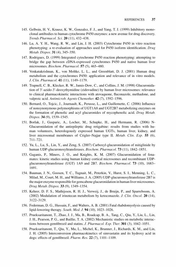
P1: PIC/PIC P2: ABCc01 JWBK262/Xie September 18, 2008 18:59 Printer Name: Yet to Come
REFERENCES 37
145. Gelboin, H. V., Krausz, K. W., Gonzalez, F. J., and Yang, T. J. (1999) Inhibitory mono-clonal antibodies to human cytochrome P450 enzymes: a new avenue for drug discovery.Trends Pharmacol. Sci. 20 (11), 432–438.
146. Lu, A. Y. H., Wang, R. W., and Lin, J. H. (2003) Cytochrome P450 in vitro reactionphenotyping: a re-evaluation of approaches used for P450 isoform identification. DrugMetab. Dispos. 31 (4), 345–350.
147. Rodrigues, D. (1999) Integrated cytochrome P450 reaction phenotyping: attempting tobridge the gap between cDNA-expressed cytochromes P450 and native human livermicrosomes. Biochem. Pharmacol. 57 (5), 465–480.
148. Venkatakrishnan, K., von Moltke, L. L., and Greenblatt, D. J. (2001) Human drugmetabolism and the cytochromes P450: application and relevance of in vitro models.J. Clin. Pharmacol. 41 (11), 1149–1179.
149. Trapnell, C. B., Klecker, R. W., Jamis-Dow, C., and Collins, J. M. (1998) Glucuronida-tion of 3′-azido-3′-deoxythymidine (zidovudine) by human liver microsomes: relevanceto clinical pharmacokinetic interactions with atovaquone, fluconazole, methadone, andvalproic acid. Antimicrob. Agents Chemother. 42 (7), 1592–1596.
150. Bernard, O., Tojcic, J., Journault, K., Perusse, L., and Guillemette, C. (2006) Influenceof nonsynonymous polymorphisms of UGT1A8 and UGT2B7 metabolizing enzymes onthe formation of phenolic and acyl glucuronides of mycophenolic acid. Drug Metab.Dispos. 34 (9), 1539–1545.
151. Borlak, J., Gasparic, A., Locher, M., Schupke, H., and Hermann, R. (2006) N-Glucuronidation of the antiepileptic drug retigabine: results from studies with hu-man volunteers, heterologously expressed human UGTs, human liver, kidney, andliver microsomal membranes of Crigler–Najjar type II. Metab. Clin. Exp. 55 (6),711–721.
152. Yu, L., Lu, S., Lin, Y., and Zeng, S. (2007) Carboxyl-glucuronidation of mitiglinide byhuman UDP-glucuronosyltransferases. Biochem. Pharmacol. 73 (11), 1842–1851.
153. Gaganis, P., Miners, J. O., and Knights, K. M. (2007) Glucuronidation of fena-mates: kinetic studies using human kidney cortical microsomes and recombinant UDP-glucuronosyltransferase (UGT) 1A9 and 2B7. Biochem. Pharmacol. 73 (10), 1683–1691.
154. Bauman, J. N., Goosen, T. C., Tugnait, M., Peterkin, V., Hurst, S. I., Menning, L. C.,Milad, M., Court, M. H., and Williams, J. A. (2005) UDP-glucuronosyltransferase 2B7 isthe major enzyme responsible for gemcabene glucuronidation in human liver microsomes.Drug Metab. Dispos. 33 (9), 1349–1354.
155. Kehrer, D. F. S., Mathijssen, R. H. J., Verweij, J., de Bruijn, P., and Sparreboom, A.(2002) Modulation of irinotecan metabolism by ketoconazole. J. Clin. Oncol. 20 (14),3122–3129.
156. Federman, D. G., Hussain, F., and Walters, A. B. (2001) Fatal rhabdomyolysis caused bylipid-lowering therapy. South. Med. J. 94 (10), 1023–1026.
157. Prueksaritanont, T., Zhao, J. J., Ma, B., Roadcap, B. A., Tang, C., Qiu, Y., Liu, L., Lin,J. H., Pearson, P. G., and Baillie, T. A. (2002) Mechanistic studies on metabolic interac-tions between gemfibrozil and statins. J. Pharmacol. Exp. Ther. 301 (3), 1042–1051.
158. Prueksaritanont, T., Qiu, Y., Mu, L., Michel, K., Brunner, J., Richards, K. M., and Lin,J. H. (2005) Interconversion pharmacokinetics of simvastatin and its hydroxy acid indogs: effects of gemfibrozil. Pharm. Res. 22 (7), 1101–1109.
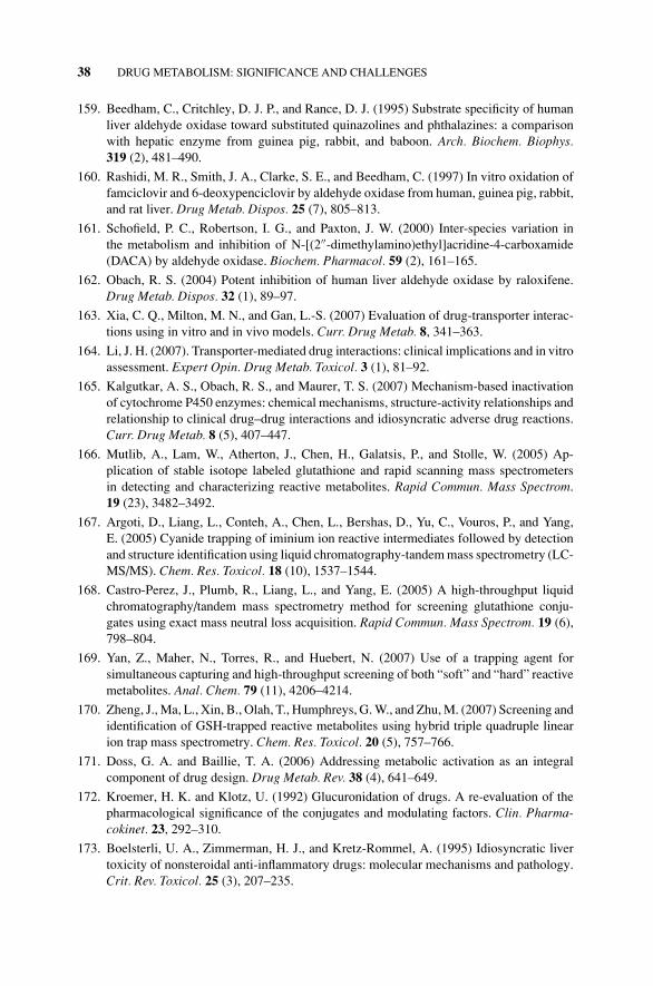
P1: PIC/PIC P2: ABCc01 JWBK262/Xie September 18, 2008 18:59 Printer Name: Yet to Come
38 DRUG METABOLISM: SIGNIFICANCE AND CHALLENGES
159. Beedham, C., Critchley, D. J. P., and Rance, D. J. (1995) Substrate specificity of humanliver aldehyde oxidase toward substituted quinazolines and phthalazines: a comparisonwith hepatic enzyme from guinea pig, rabbit, and baboon. Arch. Biochem. Biophys.319 (2), 481–490.
160. Rashidi, M. R., Smith, J. A., Clarke, S. E., and Beedham, C. (1997) In vitro oxidation offamciclovir and 6-deoxypenciclovir by aldehyde oxidase from human, guinea pig, rabbit,and rat liver. Drug Metab. Dispos. 25 (7), 805–813.
161. Schofield, P. C., Robertson, I. G., and Paxton, J. W. (2000) Inter-species variation inthe metabolism and inhibition of N-[(2′′-dimethylamino)ethyl]acridine-4-carboxamide(DACA) by aldehyde oxidase. Biochem. Pharmacol. 59 (2), 161–165.
162. Obach, R. S. (2004) Potent inhibition of human liver aldehyde oxidase by raloxifene.Drug Metab. Dispos. 32 (1), 89–97.
163. Xia, C. Q., Milton, M. N., and Gan, L.-S. (2007) Evaluation of drug-transporter interac-tions using in vitro and in vivo models. Curr. Drug Metab. 8, 341–363.
164. Li, J. H. (2007). Transporter-mediated drug interactions: clinical implications and in vitroassessment. Expert Opin. Drug Metab. Toxicol. 3 (1), 81–92.
165. Kalgutkar, A. S., Obach, R. S., and Maurer, T. S. (2007) Mechanism-based inactivationof cytochrome P450 enzymes: chemical mechanisms, structure-activity relationships andrelationship to clinical drug–drug interactions and idiosyncratic adverse drug reactions.Curr. Drug Metab. 8 (5), 407–447.
166. Mutlib, A., Lam, W., Atherton, J., Chen, H., Galatsis, P., and Stolle, W. (2005) Ap-plication of stable isotope labeled glutathione and rapid scanning mass spectrometersin detecting and characterizing reactive metabolites. Rapid Commun. Mass Spectrom.19 (23), 3482–3492.
167. Argoti, D., Liang, L., Conteh, A., Chen, L., Bershas, D., Yu, C., Vouros, P., and Yang,E. (2005) Cyanide trapping of iminium ion reactive intermediates followed by detectionand structure identification using liquid chromatography-tandem mass spectrometry (LC-MS/MS). Chem. Res. Toxicol. 18 (10), 1537–1544.
168. Castro-Perez, J., Plumb, R., Liang, L., and Yang, E. (2005) A high-throughput liquidchromatography/tandem mass spectrometry method for screening glutathione conju-gates using exact mass neutral loss acquisition. Rapid Commun. Mass Spectrom. 19 (6),798–804.
169. Yan, Z., Maher, N., Torres, R., and Huebert, N. (2007) Use of a trapping agent forsimultaneous capturing and high-throughput screening of both “soft” and “hard” reactivemetabolites. Anal. Chem. 79 (11), 4206–4214.
170. Zheng, J., Ma, L., Xin, B., Olah, T., Humphreys, G. W., and Zhu, M. (2007) Screening andidentification of GSH-trapped reactive metabolites using hybrid triple quadruple linearion trap mass spectrometry. Chem. Res. Toxicol. 20 (5), 757–766.
171. Doss, G. A. and Baillie, T. A. (2006) Addressing metabolic activation as an integralcomponent of drug design. Drug Metab. Rev. 38 (4), 641–649.
172. Kroemer, H. K. and Klotz, U. (1992) Glucuronidation of drugs. A re-evaluation of thepharmacological significance of the conjugates and modulating factors. Clin. Pharma-cokinet. 23, 292–310.
173. Boelsterli, U. A., Zimmerman, H. J., and Kretz-Rommel, A. (1995) Idiosyncratic livertoxicity of nonsteroidal anti-inflammatory drugs: molecular mechanisms and pathology.Crit. Rev. Toxicol. 25 (3), 207–235.
P1: PIC/PIC P2: ABCc01 JWBK262/Xie September 18, 2008 18:59 Printer Name: Yet to Come
REFERENCES 39
174. Bailey, M. J. and Dickinson, R. G. (2003) Acyl glucuronide reactivity in perspective:biological consequences. Chem. Biol. Interact. 145, 117–137.
175. Boelsterli, U. A. (2003) Diclofenac-induced liver injury: a paradigm of idiosyncraticdrug toxicity. Toxicol. Appl. Pharmacol. 192, 307–322.
176. Walker, G. S., Atherton, J., Bauman, J., Kohl, C., Lam, W., Reily, M., Lou, Z., and Mutlib,A. (2007) Determination of degradation pathways and kinetics of acyl glucuronides byNMR spectroscopy. Chem. Res. Toxicol. 20, 876–886.
177. Chen, G., Yin, S., Maiti, S., and Shao, X. (2002) 4-Hydroxytamoxifen sulfationmetabolism. J. Biochem. Mol. Toxicol. 16, 279–285.
178. Plant, N. (2007) The human cytochrome P450 subfamily: transcriptional regulation,inter-individual variation and interaction networks. Biochim. Biophys. Acta 1770 (3),478–488.
179. Dickins, M. (2004) Induction of cytochromes P450. Curr. Top. Med. Chem. 4 (16),1745–1766.
180. Smith, D. A., Dickins, M., Fahmi, O. A., Iwasaki, K., Lee, C., Obach, R. S., Padbury,G., De Morais, S. M., Ripp, S. L., Stevens, J., et al. (2007) The time to move cytochromeP450 induction into mainstream pharmacology is long overdue. Drug Metab. Dispos.35 (4), 697–698.
181. Hewitt, N. J., Lechon, M. J. G., Houston, J. B., Hallifax, D., Brown, H. S., Maurel,P., Kenna, J. G., Gustavsson, L., Lohmann, C., Skonberg, C., et al. (2007) Primaryhepatocytes: current understanding of the regulation of metabolic enzymes and transporterproteins, and pharmaceutical practice for the use of hepatocytes in metabolism, enzymeinduction, transporter, clearance, and hepatotoxicity studies. Drug Metab. Rev. 39 (1),159–234.
182. Mills, J. B., Rose, K. A., Sadagopan, N., Sahi, J., and De Morais, S. M. (2004) Inductionof drug metabolism enzymes and MDR1 using a novel human hepatocyte cell line. J.Pharmacol. Exp. Ther. 309, 303–309.
183. Youdim, K. A., Tyman, C. A., Jones, B. C., and Hyland, R. (2007) Induction of cy-tochrome P450: assessment in an immortalized human hepatocyte cell line (Fa2N4)using a novel higher throughput cocktail assay. Drug Metab. Dispos. 35, 275–282.
184. Mahgoub, A., Idle, J. R., Dring, L. G., Lancaster, R., and Smith, R. L. (1977) Polymorphichydroxylation of debrisoquine in man. Lancet 2 (8038), 584–586.
185. Tucker, G. T., Silas, J. H., Iyun, A. O., Lennard, M. S., and Smith, A. J. (1977) Polymor-phic hydroxylation of debrisoquine. Lancet 2 (8040), 718.
186. Evans, D. A., Mahgoub, A., Sloan, T. P., Idle, J. R., and Smith, R. L. (1980) A familyand population study of the genetic polymorphism of debrisoquine oxidation in a whiteBritish population. J. Med. Genet. 17 (2), 102–105.
187. Eichelbaum, M. (1984) Polymorphic drug oxidation in humans. Fed. Proc. 43 (8),2298–2302.
188. Gut, J., Gasser, R., Dayer, P., Kronbach, T., Catin, T., and Meyer, U. A. (1984)Debrisoquine-type polymorphism of drug oxidation: purification from human liver ofa cytochrome P450 isozyme with high activity for bufuralol hydroxylation. FEBS Lett.173 (2), 287–290.
189. Inaba, T., Nakano, M., Otton, S. V., Mahon, W. A., and Kalow, W. (1984) A hu-man cytochrome P-450 characterized by inhibition studies as the sparteine-debrisoquinemonooxygenase. Can. J. Physiol. Pharmacol. 62 (7), 860–862.
P1: PIC/PIC P2: ABCc01 JWBK262/Xie September 18, 2008 18:59 Printer Name: Yet to Come
40 DRUG METABOLISM: SIGNIFICANCE AND CHALLENGES
190. Hulot, J.-S., Bura, A., Villard, E., Azizi, M., Remones, V., Goyenvalle, C., Aiach, M.,Lechat, P., and Gaussem, P. (2006) Cytochrome P450 2C19 loss-of-function polymor-phism is a major determinant of clopidogrel responsiveness in healthy subjects. Blood108 (7), 2244–2247.
191. Zandvliet, A. S., Huitema, A. D. R., Copalu, W., Yamada, Y., Tamura, T., Beijnen, J. H.,and Schellens, J. H. M. (2007) CYP2C9 and CYP2C19 polymorphic forms are relatedto increased indisulam exposure and higher risk of severe hematologic toxicity. Clin.Cancer Res. 13 (10), 2970–2976.
192. Westlind-Johnsson, A., Hermann, R., Huennemeye, A., Hauns, B., Lahu, G., Nassr,N., Zech, K., Ingelman-Sundberg, M., and von Richter, O. (2008) Identification andcharacterization of CYP3A4∗20, a novel rare CYP3A4 allele without functional activity.Clin. Pharmacol. Ther. 79 (4), 339–349.
193. Thervet, E., Anglicheau, D., King, B., Schlageter, M.-H., Cassinat, B., Beaune, P.,Legendre, C., and Daly, A. K. (2003) Impact of cytochrome P450 3A5 genetic polymor-phism on tacrolimus doses and concentration-to-dose ratio in renal transplant recipients.Transplantation 76 (8), 1233–1235.
194. MacPhee, I. A. M., Fredericks, S., and Holt, D. W. (2005) Does pharmacogenetics havethe potential to allow the individualization of immunosuppressive drug dosing in organtransplantation? Expert Opin. Pharmacother. 6 (15), 2593–2605.
195. Rela, M., Muiesan, P., Vilca-Melendez, H., Dhawan, A., Baker, A., Mieli-Vergani, G., andHeaton, N. D. (1999) Auxiliary partial orthotopic liver transplantation for Crigler–Najjarsyndrome type I. Ann. Surg. 229 (4), 565–569.
196. Nguyen, T. H., Birraux, J., Wildhaber, B., Myara, A., Trivin, F., Le Coultre, C., Trono,D., and Chardot, C. (2006) Ex vivo lentivirus transduction and immediate transplantationof uncultured hepatocytes for treating hyperbilirubinemic Gunn rat. Transplantation82 (6), 794–803.
197. Li, Q., Murphree, S. S., Willer, S. S., Bolli, R., and French, B. A. (1998) Gene therapywith bilirubin-UDP-glucuronosyltransferase in the Gunn rat model of Crigler–Najjarsyndrome type 1. Hum. Gene Ther. 9 (4), 497–505.
198. Hirschfield, G. M. and Alexander, G. J. (2006) Gilbert’s syndrome: an overview forclinical biochemists. Ann. Clin. Biochem. 43 (5), 340–343.
199. Nowell, S. and Falany, C. N. (2006) Pharmacogenetics of human cytosolic sulfotrans-ferases. Oncogene 25 (11), 1673–1678.
200. Hisamuddin, I. M. and Yang, V. W. (2007) Genetic polymorphisms of human flavin-containing monooxygenase 3: implications for drug metabolism and clinical perspectives.Pharmacogenomics 8 (6), 635–643.
201. Ichida, K., Amaya, Y., Kamatani, N., Nishino, T., Hosoya, T., and Sakai, O. (1997)Identification of two mutations in human xanthine dehydrogenase gene responsible forclassical type I xanthinuria. J. Clin. Invest. 99 (10), 2391–2397.
202. Yamamoto, T., Moriwaki, Y., Shibutani, Y., Matsui, K., Ueo, T., Takahashi, S., Tsutsumi,Z., and Hada, T. (2001) Human xanthine dehydrogenase cDNA sequence and protein inan atypical case of type I xanthinuria in comparison with normal subjects. Clin. Chim.Acta 304 (1–2), 153–158.
203. Yamamoto, T., Moriwaki, Y., Takahashi, S., Tsutsumi, Z., Tuneyoshi, K., Matsui, K.,Cheng, J., and Hada, T. (2003) Identification of a new point mutation in the humanmolybdenum cofactor sulfurase gene that is responsible for xanthinuria type II. Metab.Clin. Exp. 52 (11), 1501–1504.
P1: PIC/PIC P2: ABCc01 JWBK262/Xie September 18, 2008 18:59 Printer Name: Yet to Come
REFERENCES 41
204. Peretz, H., Naamati, M. S., Levartovsky, D., Lagziel, A., Shani, E., Horn, I., Shalev,H., and Landau, D. (2007) Identification and characterization of the first mutation(Arg776Cys) in the C-terminal domain of the human molybdenum cofactor sulfurase(HMCS) associated with type II classical xanthinuria. Mol. Genet. Metab. 91 (1), 23–29.
205. Itoh, K., Masubuchi, A., Sasaki, T., Adachi, M., Watanabe, N., Nagata, K., Yamazoe, Y.,Hiratsuka, M., Mizugaki, M., and Tanaka, Y. (2007) Genetic polymorphism of aldehydeoxidase in donryu rats. Drug Metab. Dispos. 35 (5), 734–739.
206. Brockton, N., Little, J., Sharp, L., and Cotton, S. C. (2000) N-acetyltransferase polymor-phisms and colorectal cancer: a HuGE review. Am. J. Epidemiol. 151 (9), 846–861.
207. Benedetti, M. S., Whomsley, R., Espie, P., and Baltes, E. (2004) The role of the effluxtransporter P-glycoprotein (P-gp) on the disposition of antiepileptic drugs: implicationsfor drug interactions. In: Focus on Epilepsy Research, Benjamin Shawn, M., (ed.), NovaScience, New York, pp. 199–220.
208. Robey, R. W., Polgar, O., Deeken, J., To, K. W., and Bates, S. E. (2007) ABCG2:determining its relevance in clinical drug resistance. Cancer Metastasis Rev. 26 (1),39–57.
209. Geier, A., Wagner, M., Dietrich, C. G., and Trauner, M. (2007) Principles of hepaticorganic anion transporter regulation during cholestasis, inflammation and liver regener-ation. Biochim. Biophys. Acta Mol. Cell Res. 1773 (3), 283–308.
210. Sakurai, A., Kurata, A., Onishi, Y., Hirano, H., and Ishikawa, T. (2007) Prediction ofdrug-induced intrahepatic cholestasis: in vitro screening and QSAR analysis of drugsinhibiting the human bile salt export pump. Expert Opin. Drug Saf. 6 (1), 71–86.
211. Mahadevan, D. and Shirahatti, N. (2005) Strategies for targeting the multidrug resistance-1 (MDR1)/P-gp transporter in human malignancies. Curr. Cancer Drug Targets 5 (6),445–455.
212. Jansen, P. L. M. and Muller, M. (2001) Genetic transport defects as causes of cholestasis.Falk Symposium. (Hepatology) Vol. 117, pp. 27–33.
213. Leschziner, G. D., Andrew, T., Pirmohamed, M., and Johnson, M. R. (2007) ABCB1genotype and P-gp expression, function and therapeutic drug response: a critical reviewand recommendations for future research. Pharmacogenomics J. 7 (3), 154–179.

P1: PIC/PIC P2: ABCc01 JWBK262/Xie September 18, 2008 18:59 Printer Name: Yet to Come