downloaded from by guest …atvb.ahajournals.org/content/atvbaha/7/5/441.full.pdfwere cut an d...
TRANSCRIPT


442 ARTERIOSCLEROSIS VOL 7, No 5, SEPTEMBER/OCTOBER 1987
Table 1. Diet Composition
Diet ingredients (g/100)
Monkey mealfCoconut oll§Fish oil ||CholesterolAusman-Hayes vitamin mixGelatinOrange juiceH
Group I
71.2525.000.002.0000.251.50
15.00
Group II
71.2512.5012.502.0540.251.50
15.00
Group III*
71.250.00
25.002.1080.251.50
15.00
Group lilt
71.256.25
18.752.0810.251.50
15.00
'Diet fed for 1.5 months initially. tDiet fed for the remainder of the study. ^Purina Monkey Chow, specially formulatedwithout added fat. §Durkee Foods, Chicago, Illinois. ||Menhaden, Zapata Haynie, Reedville, Virginia. ^Excluded fromtotal dry weight.
Table 2.ments
Fatty Acid Content of Dietary Fat Supple-
Fatty acids
6:08:0
10:012:014:016:016:116:2-418:018:118:218:318:420:1-320:420:522:1-422:522:6Others
TotalTotal n-6Total n-3
Group I
0.121.751.50
12.004.752.25——
0.751.500.38————————
—
25.000.380
Group II
0.060.880.756.003.423.021.450.760.711.940.410.230.430.250.292.000.080.491.350.48
25.000.704.50
Group III
0.030.440.383.002.753.412.181.140.692.160.430.340.650.380.433.010.120.742.030.69
25.000.866.77
Values are given in grams per 100 g dry weight.The Group I diet contained 25% coconut oil; the Group II diet
contained 12.5% coconut oil and 12.5% fish oil; the Group III dietcontained 6.25% coconut oil and 18.75% fish oil.
12-month experimental period by previously describedmethods.28
Laboratory TechniquesThe animals were autopsled after 12 months following
exsanguination with thiamylal sodium anesthesia. Aorta,carotid, and femoral arteries were cleaned of adventitiaand were opened, and the extent and severity of athero-sclerosis was evaluated independently by two observersusing a point counting technique.27 Standard samples fromaorta, carotid, and femoral arteries less than 3 mm in thick-ness were fixed at 4° C for 24 hours in 4% paraformalde-hyde (wt/vol) containing 7.5% sucrose (wt/vol), followed bya 24-hour washing at 4° C in 1 % gum acacia (wt/vol) con-taining 30% sucrose (wt/vol). Serial frozen sections (10
were cut and stained for lipid with oil red O28 and formacrophages by histochemical localization of acid lipaseactivity.29'M The atherosclerotic lesions and their compo-nents were quantitated by computer-assisted morphome-tric analysis. The system used has been previously de-scribed by Glagov et al.31 and other investigators.32'M
Standard samples of the thoracic and abdominal aortaswere analyzed biochemically for composition and enzymeactivities. The aorta was split longitudinally, and adjacentsamples from the right side of the thoracic aorta beginningat the second intercostal were sampled for enzyme assays(2 cm) and chemical composition (1 cm). The abdominalaorta was sampled 3 cm distal to the renals for chemicalcomposition (1 cm) and enzyme assay (2 cm). Aortic lipidswere extracted by the method of Bligh and Dyer34 withcoprostanol as an internal standard. The quantitation oftotal, free, and esterrfied cholesterol was performed ac-cording to the method of Ishikawa et al.35 as modified byBates and Wissler.36 Briefly, the relative volume of thecholesterol peak to the nearby coprostanol peak was usedto demonstrate the free cholesterol content of the sampleusing gas liquid chromatography. After the free cholesterolcontent of a sample aliquot was determined, the entiresample was saponified. Chromatographic determinationon this saponified sample provided quantitation of the totalcholesterol in the original sample. The cholesteryl estercontent was determined by subtraction of the free choles-terol content from the post-saponified determination.
Samples for enzyme assay were homogenized in dis-tilled, deionized water at 0° C in an all-glass homogenizer(1/10, wt/vol). The homogenate was then centrifuged at5000 g for 10 minutes, and the supernatant was collectedfor enzyme activity studies.
Cholesteryl ester hydrolytic activity was determined us-ing a radioactively labelled vesicle substrate system modi-fied from the method of Brecher et al.37 Vesicles wereprepared with egg-yolk lecithin containing unlabelled cho-lesteryl oleate and cholesteryl-1 -14C oleate in a 66:1 phos-pholipid/substrate molar ratio. These mixtures were driedand lyophilized, mixed with 0.01 M Tris-HCI buffer (pH 7.4),containing 0.1 M NaCI and 0.02% sodium azide, and soni-cated. The vesicle mixture (100 /xl) was then added to anequal volume of 0.15 M acetate buffer (pH 4.5) (100 /xl),and 50 ̂ il of tissue homogenate was assayed. The finalsubstrate concentration in the incubation mixture was 1 mgof egg-yolk lecithin and 14 /xg of cholesteryl oleate. Thereaction was terminated after a 4-hour incubation by add-
by guest on June 30, 2018http://atvb.ahajournals.org/
Dow
nloaded from

FISH OIL INHIBITS ATHEROSCLEROSIS Davis et al 443
1400
1200
1000
~ BOO—e 600
400
200
0mos.
I TOTAL CHOL
I UJL CHOL
0 B0 1 3GROUP
6 12I
0 1 3GROUP
6 12II
0 1 3 6 12GROUP I I I
Figure 1. Serum total cholesterol and low density lipoprotein(LDL) cholesterol levels of the three groups of monkeys fed highfat, high cholesterol diets for 12 months. The mean total cholester-ol levels ± SEM are represented by the total bars and the meanLDL cholesterol levels by the hatched portion.
ing 6 ml of benzene/chloroform/ethanol (1.0:0.5:1.2).Free oleate was extracted by the method of Pittman et al.38
In brief, the mixture was made alkaline with 1.2 ml of 0.3 MNaOH. After phase separation, 0.5 ml of the upper phasewas counted by liquid scintillation spectrometry. Countingefficiencies were determined from sample channel ratio(SCR) values, and quenching curves were derived fromquenching standards (Amersham). Appropriate blanksand controls were included in each assay.
Acid lipase substrate micelles were prepared using 1.0mM alpha-naphthyl palmitate and 10 mM Triton X-100 in0.1 M sodium acetate buffer (pH 4.2) containing 0.1% fattyacid-poor bovine serum albumin.29 Fifty /xl of homogenizedtissue supernatant was assayed in 1 ml of substrate at25° C with continuous agitation for 1 to 4 hours. The reac-tion was stopped by placing the samples in boiling waterfor 2 minutes. One ml of 1.0 M sodium acetate buffer (pH4.2) containing 10% Tween 20 (wt/vol) and 0.5 mg fastgarnet GBC salt was added to each sample. Diazocou-pling of liberated alpha-naphthol was performed at 25° Cfor 16 hours. Absorbance of samples, heat-inactivatedblanks, and alpha-naphthol standards were then mea-sured at 535 nm.
Acyl-CoA cholesterol acyl transferase (ACAT) activitywas determined by a modified method of Brecher andChobanian.39 The incubation medium consisted of 40 pM(1-14C) palmiryl-CoA, 2 mg/ml fatty acid-free bovine serumalbumin (BSA) and 2 mM dithiothreitol in 0.1 M potassiumphosphate buffer (pH 7.4). The aortic supernatants (20 n\)were assayed for 60 minutes at 37° C and the reaction wasstopped by the addition of methanol. The lipids were ex-tracted by the method of Bligh and Dyer.34 The solventswere evaporated under N2; the lipids were resuspended inchloroform and spotted on silica gel IB2 (Baker-flex) thin-layer chromatography plates. The plates were developedin hexane/ethyl ether/acetic acid, 83:16:1; the spots werevisualized in l2 vapors; the lipid classes were cut out andcounted by liquid scintillation spectrometry.
125
100
mos. o 1 3 6 12GROUP I
0 1 3 6 12GROUP II
0 1 3 6 12GROUP III
Figure 2. Serum high density lipoprotein (HDL) cholesterol lev-els of the three groups of monkeys over the 12-month dietaryperiod. Values are means ± SEM at selected time points.
Statistical AnalysisStatistical differences between groups were analyzed
using the Wilcoxon test and significance was assumed forp values less than 0.05.
Results
Serum LipidsThe average total cholesterol for all three groups at the
start of the experiment was 172 mg/dl. The animals inGroup I which were fed 25% coconut oil and 2% cholester-ol rapidly increased their average serum cholesterol levelsto 683 mg/dl after 1 month and maintained a running aver-age of 875 mg/dl over the twelve months (Figure 1). Theanimals in Group II which were fed coconut oil/fish oil (1:1)increased their average serum cholesterol levels to 571mg/dl at 1 month which fell to a 12-month running averageof 463 mg/dl which was significantly lower than Group I(p< 0.05). Group III with a 1:3 coconut oil/fish oil diet hadsimilar changes in serum cholesterol as seen in Group II(Figure 1) with a 12-month average of 405 mg/dl.
The HDL cholesterol levels In all three groups were re-duced by the high-fat diets (Figure 2). Group I demonstrat-ed the least reduction from a baseline level of 83 mg/dl toan average of 49 mg/dl over the 12 months. Fish oil in thediets of Groups II and III caused a more significant reduc-tion in HDL cholesterol to average levels of 29 mg/di and20 mg/dl, respectively, over the 12 months.
Serum triglyceride levels were never increased overnormal and were below 40 mg/dl, and there were no differ-ences among the three groups.
Atherosclerotic Lesion DistributionThe degree of aortic, carotid, and femoral artery athero-
sclerosis involvement was evaluated and was expressedas the percentage of total surface area covered by lesions(Table 3). The aortas of Group I were more extensivelyinvolved with atherosclerosis with an average of 79% of
by guest on June 30, 2018http://atvb.ahajournals.org/
Dow
nloaded from
444 ARTERIOSCLEROSIS VOL. 7, No 5, SEPTEMBER/OCTOBER 1987
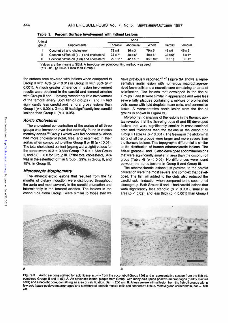
Table 3. Percent Surface Involvement with Intimal Lesions
Animalgroup
IIIIII
CoconutCoconutCoconut
Supplements
oil and cholesteroloil/fish oil (1:1) andoil/fish oil (1:3) and
cholesterolcholesterol
Thoradc
72±838±r29±11*
Aorta
Abdominal
86±358±6*42±10t
Whole
79±548±5*36±10f
Carotid
49 + 622±6t3±1t
Femoral
46±65±1f3±1f
Values are the means ± SEM. A two-observer point-counting method was used.*p<0.01; fp<0.001 less than Group I.
the surface area covered with lesions when compared toGroup II with 48% (p < 0.01) or Group III with 36% (p <0.001). A much greater difference in lesion involvementresults were obtained in the carotid and femoral arterieswith Groups II and III having remarkably little involvementof the femoral artery. Both fish-oil groups (II and III) hadsignificantly less carotid and femoral gross lesions thanGroup I (p < 0.001). Group III had significantly less carotidlesions than Group II (p < 0.05).
Aortic CholesterolThe cholesterol concentration of the aortas of all three
groups was increased over that normally found in rhesusmonkey aortas.40 Group I which was fed coconut oil alonehad more cholesterol (total, free, and esterified) in theiraortas when compared to either Group II or III (p < 0.01).The total cholesterol content (/xg/mg wet weight) values forthe aortas were 19.3 ± 0.8 for Group 1,7.6 ± 1.6 for GroupII, and 5.3 ± 0.8 for Group III. Of the total cholesterol, 34%was In the esterified form in Group I, 29%, in Group II, and15%, in Group III.
Microscopic MorphometryThe atherosclerotic lesions that resulted from the 12
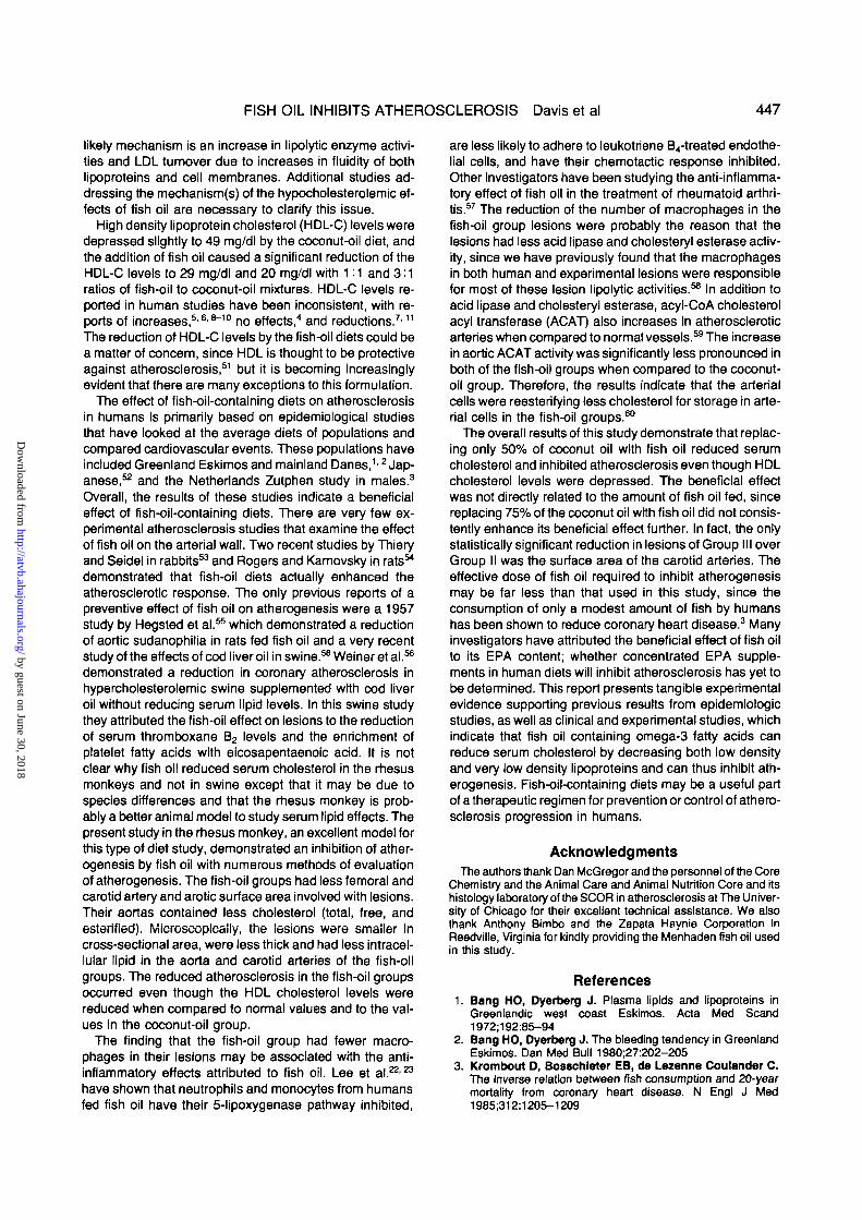
months of dietary induction were distributed throughoutthe aorta and most severely in the carotid bifurcation andintermittently in the femoral arteries. The lesions in thecoconut-oil alone Group I were similar to those that we
have previously reported.4142 Figure 3A shows a repre-sentative aortic lesion with numerous macrophage-de-rived foam cells and a necrotic core containing an area ofcalcification. The lesions that developed in the fish-oilGroups II and III were similar in appearance and were lesssevere fatty plaques containing a mixture of proliferatedcells, some with lipid droplets, foam cells, and connectivetissue. A representative aortic lesion from the fish-oilgroups is shown in Figure 3B.
Morphometric analysis of the lesions in the thoracic aor-tas revealed that the fish-oil groups (II and III) developedlesions that were significantly smaller in cross-sectionalarea and thickness than the lesions in the coconut-oilGroup I (Table 4) (p < 0.001). The lesions in the abdominalaorta of all the groups were larger and more severe thanthe thoracic lesions. This topographic differential is similarto the distribution of human atherosclerotic lesions. Thefish-oil groups (II and III) also developed abdominal lesionsthat were significantly smaller in area than the coconut-oilgroup (Table 4) (p < 0.05). No differences were foundbetween the aortic lesions in Group II and Group III.
The atherosclerotic lesions just proximal to the carotidbifurcation were the most severe and complex that devel-oped. The fish oil added to the diets also reduced thecarotid lesion induction when compared to the coconut-oilalone group. Both Groups II and III had carotid lesions thatwere significantly less stenotic (p < 0.001), smaller inarea (p < 0.02), and less thick (p < 0.001) than Group I
A B
figure 3. Aortic sections stained for add Iipase activity from the coconut-oil Group I (A) and a representative section from the fish-oil,combined Groups II and III (B). A. An advanced Intimal plaque from Group I with many acid llpase-posltive macrophages (darkly stainedcells) and a necrotic core, containing an area of calcification. Bar = 200 nm. B. A less severe intimaJ lesion from the fish-oil groups with afew add lipase-positive macrophages and a mixture of smooth muscle cells and connective tissue. Methyl green counterstain, bar = 100
by guest on June 30, 2018http://atvb.ahajournals.org/
Dow
nloaded from

FISH OIL INHIBITS ATHEROSCLEROSIS Davis et al 445
Table 4. Aortic Microscopic Data
Animalgroup Supplements
1 Coconut oil and cholesterolII Coconut oil/fish oil (1:1) and cholesterolIII Coconut oil/fish oil (1:3) and cholesterol
Lesion
Thoracic
0.67 + 0.070.20 + 0.05+0.20 + 0.04+
area (mm2)
Abdominal
1.24±0.180.52 + 0.10*0.46±0.16*
Lesion thickness (mm)
Thoracic
0.20 ±0.040.08±0.01f0.06±0.01f
Abdominal
0.26 ±0.020.13 ±0.020.15±0.06
Values are the means ± SEM.*p<0.01; +p<0.001 less than Group I.
Table 5. Carotid Microscopic Data
Animalgroup Supplements
Percent Lesionlumlnal areastenosis (mm2)
Lesionthickness
(mm)
Macrophagearea
(mm2)Percent
macrophages
Intracell-ular lipld
area (mm2)
PercentIntracell-ular lipid
Coconut oil andcholesterol
Coconut oil/fishoil (1M)andcholesterol
Coconut oil/fishoil (1:3) andcholesterol
32 ±5 0.64 ±0.22 0.28 + 0.05 0.23 ±0.06 35 ± 3
10±1t 0.13±0.02 0.08 + 0.01 + 0.03±0.01* 21 ± 5
4 ±2+ 0.06 ±0.03* 0.04 + 0.01* 0.02 ±0.01* 11 ±4*
0.21 ±0.05 35±4
0.03±0.01t 14±3*
0.02 ± 0.011 9 ±4+
Values are means ± SEM for each group.*p<0.05; tp<0.001 less than Group I.
Table 6. LIplds and Macrophages In Aortic Intlmal Lesions
Animalgroup
I
II
III
Supplements
Coconut oil andcholesterol
Coconut oil/fishoil (1 M) andcholesterol
Coconut oil/fishoil (1:3) andcholesterol
Percentular
Thoracic
43±7
36±8
30±10
intracell-lipid
Abdominal
27±4
15±5
18±9
Percentmacrophages
Thoracic Abdominal
37±5 17±4
22±5 13±5
17±7 6±4
Area intracellularliptds
Thoracic
0.28 ±0.04
0.08 ±0.03*
0.08±0.03*
(mm2)
Abdominal
0.30 ±0.06
0.10±0.05f
0.06±0.03t
Area macrophages(mm2)
Thoracic Abdominal
0.25±0.04 0.21 ±0.06
0.05 ±0.02* 0.09 ±0.04
0.04±0.02* 0.03±0.02+
Values are means ± SEM.*p<0.001; tP<0.01 less than Group I.
Table 7. Aortic Enzyme Activities
Animalgroup
I
II
III
Supplements
Coconut oil andcholesterol
Coconut oil/fishoil (1=1) andcholesterol
Coconut oil/fishoil (1:3) andcholesterol
Acid lipase*(^M/mln/g)
Thoradc Abdominal
103±23 125±56
26±7* 45±10
38±14* 39±5
CholesteryI esterase(CPM/min/g)
Thoracic
11,719±1838
2878±962+
5267±848+
Abdominal
8291±3071
1824±601
799±598+
ACAT(CPM/mln/g)
Thoracic
14,144±2576
7436±1183*
6289±1086*
Abdominal
18,744±5215
10,774±1662
8705±2119
Values are the means ± SEM.*p<0.05; +p<0.01 less than Group I.
by guest on June 30, 2018http://atvb.ahajournals.org/
Dow
nloaded from

446 ARTERIOSCLEROSIS VOL 7, No 5, SEPTEMBER/OCTOBER 1987
(Table 5). Although the carotid lesions in Group III weresmaller than in Group II, this difference was not statisticallysignificant.
The amount of lipid in the lesions was morphometricallyquantitated on a percentage of lesion and absolute cross-sectional area basis. The percentage of the aortic lesionsoccupied by intracellular lipid was less in the fish-oil groups(II and III) when compared to Group I but this differencewas not statistically significant (Table 6). The percentageof the carotid lesions occupied by intracellular lipid wassignificantly less in the fish-oil groups (II and III) whencompared to Group I (p < 0.01) (Table 5). The area occu-pied by Intracellular lipid was significantly less In both thethoracic and abdominal aortic lesions, as well as the carot-id lesions, in Groups II and III when compared to Group I(Tables 5 and 6). No differences were found betweenGroups II and III.
Macrophages were identified by staining for acid lipaseactivity, and the acid lipase-positive lesion macrophageswere quantitated in cross-sectional area and by the per-centage of the intimal lesion. The percentage of the aorticand carotid lesion areas occupied by macrophages wasless in the fish-oil groups (II and III) when compared toGroup I, but this difference was only statistically significantin the Group III carotid lesions (p < 0.001) (Tables 5 and6). The actual cross-sectional area of the lesions occupiedby acid lipase-positive macrophages was significantlysmaller in most lesions examined in Groups II and III whencompared to Group I. The macrophage involvement wasnot found to be different when Groups II and III were com-pared (Tables 5 and 6).
Aortic EnzymesAtherosclerosis-associated increases in lipid-related
enzyme activities were measured in the thoracic and ab-dominal aortas of the monkeys, and the results are seen inTable 7. Acid lipase and cholesteryl esterase hydrolyticactivities were reduced in the fish-oil groups (II and III)when compared to Group I. The difference in lipid hydroly-tic activity was significantly less in the thoracic aorta (p <0.05) and because of the large variance in the abdominalactivities, only the cholesteryl esterase of Group III wassignificantly less (p < 0.01).
Acyl-CoA cholesterol acyl transferase (ACAT) activityincreased to a lesser extent in the fish-oil groups (II and III)when compared to Group I (Table 7). These differenceswere also only statistically significant in the thoracic aorta(p < 0.05) due to the large variance in the abdominal aortaACAT activities. No differences were found between theaortic lipid-related enzyme activities of Group II when com-pared to Group III. In general, the samples with the smallerlesions had the least activity.
Discussion
This study demonstrates that the mixing of substantialquantities of fish oil with coconut oil and cholesterol re-duces both the hyperiipidemia and atherosclerotic re-sponse measured when a ration containing only coconutoil and cholesterol is fed to rhesus monkeys for 12 months.The combination of coconut oil and cholesterol has beenshown to be one of the most atherogenic diets in several
experimental models of atherosclerosis, including nonhu-man primates,41'42 rabbits,43'44 and dogs.4546 In fact,coconut oil is the only dietary fat that has been consistentlyshown to support the development of canine atherosclero-sis.45'46 The rhesus monkey is one of the best animalmodels in which to study the effects of fish oil on serumlipids and atherogenesis since their digestive system, se-rum lipoprotein pattern, and the induced atherosclerosisappear to be the most similar to humans of all the presentlytested animal models.24-47
The supplementation of Menhaden fish oil, which con-tains a large amount of long-chain polyunsaturated ome-ga-3 fatty acids,48'49 to the coconut oil/cholesterol athero-genic diet led to a marked decrease in the development ofhypercholesterolemia even though the fat content and thecaloric intake of the animals remained the same and thecholesterol consumption was substantially increased dueto the cholesterol in the fish oil (432 mg/dl). This inhibitionof the increase in serum lipids was primarily due to thereduction of the LDL cholesterol fraction in these monkeys.Many of the human dietary studies with fish oil have attrib-uted its hypocholesterolemic effect to the reduction of LDLand VLDL levels.4"12 Harris et al.4 and others56 haveshown that fish oil lowers serum cholesterol, LDL, VLDL,and triglycerides in normal humans. Phillipson et al.7 ex-tended these studies to hypertriglyceridemic patients(Type Mb and V) and found that salmon and/or Max eicosa-pentaenoic acid (EPA) reduced serum cholesterol, trigly-ceride, VLDL, and apoprotein E levels. Similar studies ofhyperiipidemics by Simon et al.8 and Sanders et al.9 dem-onstrated only a reduction in serum triglycerides, whileSaynoret al.10 reported a rapid reduction In triglycerides, aslow fall in cholesterol, and a rise in HDL cholesterol. Manyof these investigators have attributed the hypolipidemiceffects of fish oil to the reduction of VLDL synthesis in theliver, leading to a reduction in LDL synthesis.1112 In thepresent rhesus monkey study, the increase of the ratio offish oil to coconut oil from 1:1 to 3:1 did not significantlyincrease the hypolipidemic effect of the fish oil. Thisamount of fish oil consumption per day was from half of toequivalent to the amount fed in the above human studies(10 to 20 g Max EPA/day), but the monkeys only weighedbetween 4.5 and 5 kg. Potentially, a smaller dose of fish oilthat more closely mimics the human studies may be aseffective in lowering serum cholesterol levels. The mostconsistent effect of dietary fish oil in humans is the reduc-tion of serum triglyceride levels.4"12 Rhesus monkeys rare-ly increase their serum triglyceride levels in response to anatherogenic diet challenge,50 and the serum triglyceridesnever rose above 40 mg/dl in this experiment, and nodifferences were found between the groups. The mecha-nism by which the fish oil reduced serum LDL levels inrhesus monkeys may be due in part to a decrease in VLDLsynthesis,11'12 even though this is not reflected in the fast-ing serum triglyceride levels. Several other possiblemechanisms may also be operative, although not directlytested in this study. Since a majority of the serum choles-terol was in the LDL fraction, any changes in serum choles-terol would be directly reflected in the LDL levels. There-fore, fish oil could be increasing fecal steroid excretion orbile acid synthesis to reduce serum LDL levels. A more
by guest on June 30, 2018http://atvb.ahajournals.org/
Dow
nloaded from
FISH OIL INHIBITS ATHEROSCLEROSIS Davis et al 447
likely mechanism is an increase in lipolytic enzyme activi-ties and LDL turnover due to increases in fluidity of bothlipoproteins and cell membranes. Additional studies ad-dressing the mechanism(s) of the hypocholesterolemic ef-fects of fish oil are necessary to clarify this issue.
High density lipoprotein cholesterol (HDL-C) levels weredepressed slightly to 49 mg/dl by the coconut-oil diet, andthe addition of fish oil caused a significant reduction of theHDL-C levels to 29 mg/dl and 20 mg/dl with 1:1 and 3:1ratios of fish-oil to coconut-oil mixtures. HDL-C levels re-ported in human studies have been inconsistent, with re-ports of increases,5'68"10 no effects,4 and reductions.7'11
The reduction of HDL-C levels by the fish-oil diets could bea matter of concern, since HDL is thought to be protectiveagainst atherosclerosis,51 but it is becoming increasinglyevident that there are many exceptions to this formulation.
The effect of fish-oil-containing diets on atherosclerosisin humans is primarily based on epidemiological studiesthat have looked at the average diets of populations andcompared cardiovascular events. These populations haveincluded Greenland Eskimos and mainland Danes,12 Jap-anese,52 and the Netherlands Zutphen study in males.3
Overall, the results of these studies indicate a beneficialeffect of fish-oil-containing diets. There are very few ex-perimental atherosclerosis studies that examine the effectof fish oil on the arterial wall. Two recent studies by Thieryand Seidel in rabbits53 and Rogers and Karnovsky in rats54
demonstrated that fish-oil diets actually enhanced theatherosclerotic response. The only previous reports of apreventive effect of fish oil on atherogenesis were a 1957study by Hegsted et al.55 which demonstrated a reductionof aortic sudanophilia in rats fed fish oil and a very recentstudy of the effects of cod liver oil in swine.58 Weiner et al.56
demonstrated a reduction in coronary atherosclerosis inhypercholesterolemic swine supplemented with cod liveroil without reducing serum lipid levels. In this swine studythey attributed the fish-oil effect on lesions to the reductionof serum thromboxane B2 levels and the enrichment ofplatelet fatty acids with eicosapentaenoic acid. It is notclear why fish oil reduced serum cholesterol in the rhesusmonkeys and not in swine except that it may be due tospecies differences and that the rhesus monkey is prob-ably a better animal model to study serum lipid effects. Thepresent study in the rhesus monkey, an excellent model forthis type of diet study, demonstrated an inhibition of ather-ogenesis by fish oil with numerous methods of evaluationof atherogenesis. The fish-oil groups had less femoral andcarotid artery and arotic surface area involved with lesions.Their aortas contained less cholesterol (total, free, andesterifled). Microscopically, the lesions were smaller Incross-sectional area, were less thick and had less intracel-lular lipid in the aorta and carotid arteries of the fish-oilgroups. The reduced atherosclerosis in the fish-oil groupsoccurred even though the HDL cholesterol levels werereduced when compared to normal values and to the val-ues in the coconut-oil group.
The finding that the fish-oil group had fewer macro-phages in their lesions may be associated with the anti-inflammatory effects attributed to fish oil. Lee et a l . 2 2 ' a
have shown that neutrophils and monocytes from humansfed fish oil have their 5-lipoxygenase pathway inhibited,
are less likely to adhere to leukotriene B4-treated endothe-lial cells, and have their chemotactic response inhibited.Other investigators have been studying the anti-inflamma-tory effect of fish oil in the treatment of rheumatoid arthri-tis.57 The reduction of the number of macrophages in thefish-oil group lesions were probably the reason that thelesions had less acid lipase and cholesteryl esterase activ-ity, since we have previously found that the macrophagesin both human and experimental lesions were responsiblefor most of these lesion lipolytic activities.58 In addition toacid lipase and cholesteryl esterase, acyl-CoA cholesterolacyl transferase (ACAT) also increases in atheroscleroticarteries when compared to normal vessels.59 The increasein aortic ACAT activity was significantly less pronounced inboth of the fish-oil groups when compared to the coconut-oil group. Therefore, the results indicate that the arterialcells were reesterifying less cholesterol for storage in arte-rial cells in the fish-oil groups.60
The overall results of this study demonstrate that replac-ing only 50% of coconut oil with fish oil reduced serumcholesterol and inhibited atherosclerosis even though HDLcholesterol levels were depressed. The beneficial effectwas not directly related to the amount of fish oil fed, sincereplacing 75% of the coconut oil with fish oil did not consis-tently enhance its beneficial effect further. In fact, the onlystatistically significant reduction in lesions of Group III overGroup II was the surface area of the carotid arteries. Theeffective dose of fish oil required to inhibit atherogenesismay be far less than that used in this study, since theconsumption of only a modest amount of fish by humanshas been shown to reduce coronary heart disease.3 Manyinvestigators have attributed the beneficial effect of fish oilto its EPA content; whether concentrated EPA supple-ments in human diets will inhibit atherosclerosis has yet tobe determined. This report presents tangible experimentalevidence supporting previous results from epidemiologicstudies, as well as clinical and experimental studies, whichindicate that fish oil containing omega-3 fatty acids canreduce serum cholesterol by decreasing both low densityand very low density lipoproteins and can thus inhibit ath-erogenesis. Fish-oil-containing diets may be a useful partof a therapeutic regimen for prevention or control of athero-sclerosis progression in humans.
AcknowledgmentsThe authors thank Dan McGregor and the personnel of the Core
Chemistry and trie Animal Care and Animal Nutrition Core and itshistology laboratory of the SCOR in atherosclerosis at The Univer-sity of Chicago for their excellent technical assistance. We alsothank Anthony Bimbo and the Zapata Haynie Corporation inReedville, Virginia for kindly providing the Menhaden fish oil usedin this study.
References1. Bang HO, Dyerberg J. Plasma lipids and lipoproteins in
Greenlandic west coast Eskimos. Acta Med Scand1972;192:85-94
2. Bang HO, Dyerberg J. The bleeding tendency in GreenlandEskimos. Dan Med Bull 1980;27:202-205
3. Krombout D, Bosschleter EB, de Lezenne Coulandor C.The inverse relation between fish consumption and 20-yearmortality from coronary heart disease. N Engl J Med1985:312:1205-1209
by guest on June 30, 2018http://atvb.ahajournals.org/
Dow
nloaded from

448 ARTERIOSCLEROSIS VOL 7, No 5, SEPTEMBER/OCTOBER 1987
4. Harris WS, Connor WE, McMurry MP. The comparativereductions of the plasma lipids and lipoproteins by dietarypolyunsaturated fats: salmon oil vs. vegetable oils. Metabo-lism 1983;32:179-184
5. Von Lossonczy TO, Rutter A, Bronsgeest-Schoute HC, etal. The effect of a fish diet on serum lipids in healthy humansubjects. Am J Clin Nutr 1978;31:1340-1346
6. Sanders TAB, Roshanal F. The influence of different typesof w3 polyunsaturated fatty acids on blood lipids and plateletfunction in healthy volunteers. Clin Sci 1983:64:91-99
7. Phlllpson BE, Rothrock DW, Connor WE, Harris WS, II-llngworth DR. Reduction of plasma lipids, lipoproteins andapoproteins by dietary fish oils in patients with hypertriglyceri-demla. N Engl J Med 1985:312:1210-1216
8. Simons LA, Hlckle JB, Balasubramanlam S. On the effectsof dietary n-3 fatty acids (Maxepa) on plasma lipids and lipo-proteins in patients with hypertipidaemia. Atherosclerosis1985:54:75-88
9. Sanders TAB, Sullivan DR, Reeve J, Thompson GR. Tri-glyceride-lowering effect of marine polyunsaturates in pa-tients with hypertrigtyceridaemla. Arteriosclerosis 1985;5:459-465
10. Saynor R, Verel D, Qlllot T. The long-term effect of dietarysupplementation with fish lipid concentrate on serum lipids,bleeding time, platelets, and angina. Atherosclerosis1984;50:3-10
11. Nestel PJ, Connor WE, Reardon MR, Connor S, Wong S,Boston R. Suppression by diets rich in fish oil of very lowdensity llpoprotein production In man. J Clin Invest1984:74:82-89
12. Illlngworth DR, Harris WS, Connor WE. Inhibition of lowdensity llpoprotein synthesis by dietary omega-3 fatty adds inhumans. Arteriosclerosis 1984:4:270-275
13. Goodnight SH Jr, Harris WS, Connor WE. The effects ofdietary w3 fatty acids on platelet composition and function Inman: a prospective, controlled study. Blood 1981;58:880-885
14. Nagakawa Y, Orlmo H, Harasawa M, Morlta I, Yashlro K,Murota S. Effect of eicosapentaenolc acid on the plateletaggregation and composition of fatty acids in man. Athero-sclerosis 1983:47:71-75
15. Mortensen JZ, Schmidt EB, Nielsen AH, Dyerberg J. Theeffect of N-6 and N-3 polyunsaturated fatty acids on haemo-stasis, blood lipids and blood pressure. Thromb Haemost1983:50:543-546
16. Galloway JH, Cartwrlght U, Woodcock BE, et al. Effects ofdietary fish oil supplementation on the fatty acid compositionof the human platelet membrane: demonstration of selectivityin the incorporation of elcosapentaenoic add Into membranephospholipid pools. Clin Sd 1985:68:449-454
17. Knapp HR, Rellly IAG, Atessandrinl P, Fitzgerald QA. Invivo indexes of platelet and vascular function during fish oiladministration in patients with atherosclerosis. N Engl J Med1986:314:937-942
18. Hamberg M, Svenson J, Samuelson B. Thromboxane: anew group of biologically active compounds derived fromprostaglandin endoperoxides. Proc Nati Acad Sd USA1975:72:2994-2998
19. Needleman P, Raz A, Mlnkes MS, Ferrandelll JA,Sprecher H. Triene prostaglandins: prostacydin and throm-boxane biosynthesis and unique biological properties. ProcNatl Add Sd USA 1979;176:944-948
20. Corey EJ, Shlh C, Cashman JR. Docosahexaenoic add is astrong inhibitor of prostaglandin but not leukotriene biosyn-thesis. Proc Natl Acad Sd USA 1983:80:3581-3584
21. Prescott SM. The effect of eciosapentaenolc add on theleukotriene B production by human neirtrophils. J Biol Chem1984:259:7615-7621
22. Loe TH, Mencla-Hueta J-M, Shlh C, Corey EJ, Lewis RA,Austen KF. Effects of exogenous arachidonic, eisosapen-taenoic, and docosahexaenoic adds on the generation of 5-lipoxygenase pathway products by ionophore-activated hu-man neutrophils. J Clin Invest 1984:74:1922-1933
23. Lee TH, Hoover RL, Williams JD, et al. Effect of dietaryenrichment with eicosapentaenoic and docosahexaenoic
adds on in vitro neutrophil and monocyte leukotriene genera-tion and neutrophil function. N Engl J Med 1985:312:1217-1224
24. Wlssler TW, Vesaellnovitch D. Atherosclerosis in non-hu-man primates. In: Brandly CA, Cornelius CE, Simpson CF,eds. Advances In veterinary sdence and comparative medi-dne, vol 21. New York: Academic Press, 1977:351-420
25. Committee on Care and Use of Laboratory Animals of theInstitute of Laboratory Animal Resources, National Re-search Council. Guide for the care and use of laboratoryanimals. DHEW publication no (NIH) 78-23. Washington,DC: U.S. Government Printing Office, 1978
26. Vessellnovltch D, Wlssler RW, Harris L, Lusk L The rela-tionship between lipoprotein levels and xanthomas duringprogression and regression of atherosclerosis. Atherosclero-sis 1980:36:351-364
27. Howard C. Aortic atherosclerosis in normal and spontaneousdiabetic Macaca nigra. Atherosderosis 1979:33:479-493
28. Llllle RD, Ashburn LL. Supersaturated solutions of fat stainsIn dilute Isopropanolol for demonstration of acute fatty degen-eration not shown by Herzheimer technique. Arch Pathol1943:36:432-435
29. Schaffner T, Elner VM, Bauer M, Wlssler RW. Add lipase:A histochemical and biochemical study using Triton X-100-naphtylpalmrtate micelles. J Hlstochem Cytochem 1978;26:696-712
30. Schaffner T, Taylor K, Bartuccl J, et al. Arterial foam cellswith distinctive immunomorphologic and histochemical fea-tures of macrophages. Am J Pathol 1980;100:57-80
31. Qlagov S, Grande J, Vessellnovltch D, Zarlns CK. Quanti-tation of cells and fibers in hlstologic sections of arterial walls:Advantages of contour tradng on a digitizing plate. In: Mc-Donald TK, Chandler AB, eds. Connective tissues in arterialand pulmonary disease. New York: Springer-Veriag,1981:57-92
32. Wlssler RW, Vessellnovltch D, Schaffner TJ, Glagov S.Quantitating rhesus monkey atherosclerosis progression andregression with time. In: Gotto AM, Smith LC, Allen B, eds.Atherosderosis V. New York: Springer-Veriag, 1980:757-761
33. Davis HR, Vessellnovitch D, Wlssler RW. Histochemicaldetection and quantitatJon of macrophages in rhesus andcynomolgus monkey atherosderotic lesions. J HistochemCytochem 1984:32:1319-1327
34. Bllgh EG, Dyer WJ. A rapid method of total lipid extractionand purification. Can J Biochem Physiol 1959:37:911-917
35. Ishlkawa TT, MacGee J, Morrison JA, Glueck CJ. Quanti-tative analysis of cholesterol In 5 to 20 /xl of plasma. J UpldRes 1974;15:286-291
36. Bates SR, Wlssler RW. Effect of hyperilpemic serum oncholesterol accumulation in monkey aortic medial cells. Bio-chem Biophys Ada 1976;450:78-88
37. Breeder P, Yung PH, Chobanlan AV. Effect of atherosclero-sis on rysosomal cholesterol esterase activity in rabbit aorta. JUpid Res 1977;18:154-163
38. Plttman RC, Khoo JC, Steinberg D. Cholesterol esterase Inrat adipose tissue, its activation by cyclic adenosine 3,5,monophosphate-dependent protein kinase. J Biol Chem1975:250:4505-4511
39. Brecher PI, Chobanlan AV. Cholesteryl ester synthesis innormal and atherosderotic aortas of rabbit and rhesus mon-keys. Circ Res 1974;35:692-701
40. Eggen DA, Strong JP, Newman WP III, Catsulls C, Mal-colm GT, Kokatnur MG. Regression of diet-induced fattystreaks in rhesus monkeys. Lab Invest 1974:31:294-301
41. Wlssler RW, Frazler LE, Hughes RH, Rasmussen RA. Ath-erogenesis in the cebus monkey. I. A comparison of threefood fats under controlled dietary conditions. Arch Pathol1962:74:312-322
42. Wlssler RW. Recent progress in studies of experimental pri-mate atherosderosis. Progr Biochem Pharmacol 1968;4:378-392
43. Vies RO, Buller J, Gottenbos JJ, Thomasson HJ. Influ-ence of type of dietary fat on cholesterol-induced atheroscle-rosis in the rabbit. J Atheroscler Res 1964;4:170-183
by guest on June 30, 2018http://atvb.ahajournals.org/
Dow
nloaded from

For Readers in the United StatesS3 Yes, 1 want to receive my personal copy of
D Circulation• Circulation Research• StrokeD Hypertension• ArteriosclerosisD Recurring Bibliography
of HypertensionInstitutional rates available upon request
PLEASE PRINT
U.S. regular rates
$659060556020
NameAddress
In-training Rate
$325045XX)3050275030X501000
Letter from department chair-man stating post held andcompletion date is required.I am a.
ResidentInternResearch Fellow
OFFER ENDS NOVEMBER 1, 1987
City StateMy specialtyPlease send my subscription beginning with the
Advance payment required before copies are sent.
Send billCheck enclosed, payable to the American Heart Association.This is a renewal. My account number is
Zip
issue.
4707
For Readers Outside the United States, Japan, and EuropeYes, I want to receive my personal copy of
• Circulation• Circulation Research• Stroke• Hypertension• Arteriosclerosis• Recurring Bibliography
of Hypertension• Modern Concepts
PLEASE PRINT
$ 85115757575
3020
Name _Address
For readers InJapan contact:Nankodo Co., Ltd.42-6 Hongo 3-chomeBunkyo-kuTokyo 113, Japan
For readers InEurope contact:Bailliere Tindall1 St. Anne's RoadEastbourne, East SussexBN21 3UN, England
Institutional rates available upon request.
OFFER ENDS NOVEMBER 1, 1987
City State Zip CountryMy specialtyPlease send my subscription beginning with the issue.
Advance payment required before copies are sent.
Send bill
(Outside of Europe and Japan) Check enclosed, payable to the American Heart Association.This is a renewal. My account number is . 47oe
American Heart Association
Please send a sample copy of:• Arteriosclerosis• Circulation• Circulation Research
to the librarian at my institution
• HypertensionQ Stroke• Recurring Bibliography
of Hypertension
with my recommendation to subscribe.
by guest on June 30, 2018http://atvb.ahajournals.org/
Dow
nloaded from

Business Reply MailFirst Class Permit No. 16911 Dallas, Texas
Postage Will be Paid by Addressee
American HeartAssociationNational CenterSubscriptions7320 Greenville AvenueDALLAS, TX 75231-9986
NO POSTAGENECESSARY IFMAILED IN THEUNITED STATES
Business Reply MailFirst Class Permit No. 16911 Dallas, Texas
Postage Will be Paid by Addressee
American HeartAssociationNational CenterSubscriptions7320 Greenville AvenueDALLAS, TX 75231-9986
• NO POSTAGENECESSARY IFMAILED IN THEUNITED STATES
Business Reply MailFirst Class Permit No. 16911 Dallas, Texas
Postage Will be Paid by Addressee
American HeartAssociationNational CenterSubscnptions7320 Greenville AvenueDALLAS, TX 75231-9986
NO POSTAGENECESSARY IFMAILED IN THEUNITED STATES
by guest on June 30, 2018http://atvb.ahajournals.org/
Dow
nloaded from


H R Davis, R T Bridenstine, D Vesselinovitch and R W WisslerFish oil inhibits development of atherosclerosis in rhesus monkeys.
Print ISSN: 1079-5642. Online ISSN: 1524-4636 Copyright © 1987 American Heart Association, Inc. All rights reserved.
Avenue, Dallas, TX 75231is published by the American Heart Association, 7272 GreenvilleArteriosclerosis, Thrombosis, and Vascular Biology
doi: 10.1161/01.ATV.7.5.4411987;7:441-449Arterioscler Thromb Vasc Biol.
http://atvb.ahajournals.org/content/7/5/441World Wide Web at:
The online version of this article, along with updated information and services, is located on the
http://atvb.ahajournals.org//subscriptions/
at: is onlineArteriosclerosis, Thrombosis, and Vascular Biology Information about subscribing to Subscriptions:
http://www.lww.com/reprints
Information about reprints can be found online at: Reprints:
document.Permissions and Rights Question and AnswerFurther information about this process is available in theis being requested is located, click Request Permissions in the middle column of the Web page under Services.Clearance Center, not the Editorial Office. Once the online version of the published article for which permission
can be obtained via RightsLink, a service of the CopyrightArteriosclerosis, Thrombosis, and Vascular Biology Requests for permissions to reproduce figures, tables, or portions of articles originally published inPermissions:
by guest on June 30, 2018http://atvb.ahajournals.org/
Dow
nloaded from