histopathological effect of clostridium perfringens …iai.asm.org/content/12/5/1214.full.pdfwere...
TRANSCRIPT
INFECTION AND IMMUNITY, Nov. 1975, p. 1214-1218Copyright (© 1975 American Society for Microbiology
Vol. 12, No. 5Printed in USA.
Histopathological Effect of Clostridium perfringensEnterotoxin in the Rabbit Ileum
J. L. McDONEL* AND C. L. DUNCAN
Food Research Institute and Department ofBacteriology, University ofWisconsin, Madison, Wisconsin 53706
Received for publication 20 May 1975
Highly purified entertoxin from Clostridium perfringens was found to havehistopathological activity in the rabbit ileum. Unlike the action of cholera,Escherichia coli, and Shigella enterotoxins, epithelium was denuded from thetips of ileal villi at concentrations of the enterotoxin necessary to induce fluidaccumulation in the rabbit. Whether or not this observed histopathology isessential for the diarrheal syndrome associated with Clostridium perfringensfood poisoning remains unclear.
The physiology and histopathology of experi-mental diarrhea induced in the rat by Clostrid-ium perfringens recently has been studied anddescribed (8, 9). One aspect of the pathologyseen, villus epithelial desquamation, is impor-tant both in understanding the mode of actionof the enterotoxin and comparing it to otherenterotoxins currently under study (cholera,Escherichia coli, staphylococcal, and Shigellaenterotoxins). Duncan et al. (1) first reportedepithelial damage in rabbits in association withdiarrhea induced by whole cells of enteropatho-genic strains of C. perfringens, whereas Haus-child et al. (5) reported no significant lesions inlambs challenged by cells or culture filtrates.Niilo (11, 12) then reported that intravenousadministration ofcrude cell extracts from sporu-lating cells of enteropathogenic strains of C.perfringens into lambs caused partial loss ofvillus epithelium in association with diarrheaand other systemic reactions. He also reportedvariable damage in ligated loops of lambs whenthe enterotoxin was placed in the lumen of theintestine. Some sections showed sloughing ofepithelial cells and some did not, dependingupon the dosages and the susceptibility of eachanimal. Rabbits that developed diarrhea afterintravenous injection with crude cell extractsdisplayed an intact villus epithelial membrane.Guinea pigs treated similarly to the rabbitsalso showed intact epithelium but did not de-velop diarrhea.McDonel (8) showed that diarrhea in the rat
due to highly purified C. perfringens entero-toxin is associated with epithelial desquama-tion, with the degree of damage being relativeto the dosage of enterotoxin. Though diarrhea
could be developed with low doses of entero-toxin that caused only slight damage to theepithelium (8, 9), it is uncertain ifdamage is anessential part of the mechanism of diarrhea inthe case of this enterotoxin.
In the present study ileal loops of five adultwhite New Zealand rabbits were exposed induplicate to various dosages of purified entero-toxin (16) for histology studies and four rabbitswere treated similarly for fluid transport andprotein determinations. After anesthesia withsodium pentobarbital (Nembutal) and subse-quent cannulation and washing of the ileumwith warm oxygenated Ringer glucose solution(8), loops were tied in 4- to 5-cm sections andexcess fluid was removed by withdrawal with asyringe. Duplicate loops were then filled with 2ml of Ringer solution containing 1,000, 500, 250,100, or 50 erythemal units (EU) of enterotoxin.Controls consisted of loops containing 2 ml ofsolution without enterotoxin and untreated por-tions of intestine. The loops were carefullyplaced back into the abdominal cavity and theanimal was kept covered and warm throughoutthe 90-min incubation period. At timed inter-vals the loops were removed, cut open length-wise, and placed into phosphate buffered 4%formaldehyde solution prior to paraffin embed-ding and thin sectioning. Sections were stainedwith hematoxylin and eosin and with mucinstains. Fluid contents were withdrawn fromloops prior to removal from rabbits studied forfluid production and protein release. The vol-umes were measured and total protein was de-termined from trichloroacetic acid precipitatesby the method of Lowry et al. (7).
All doses of enterotoxin caused some degree
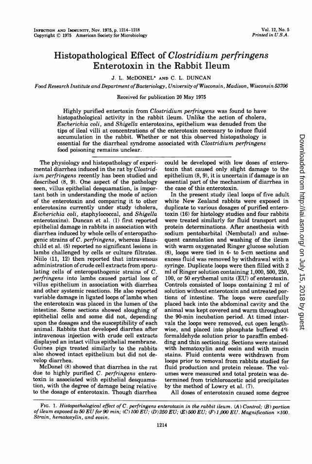
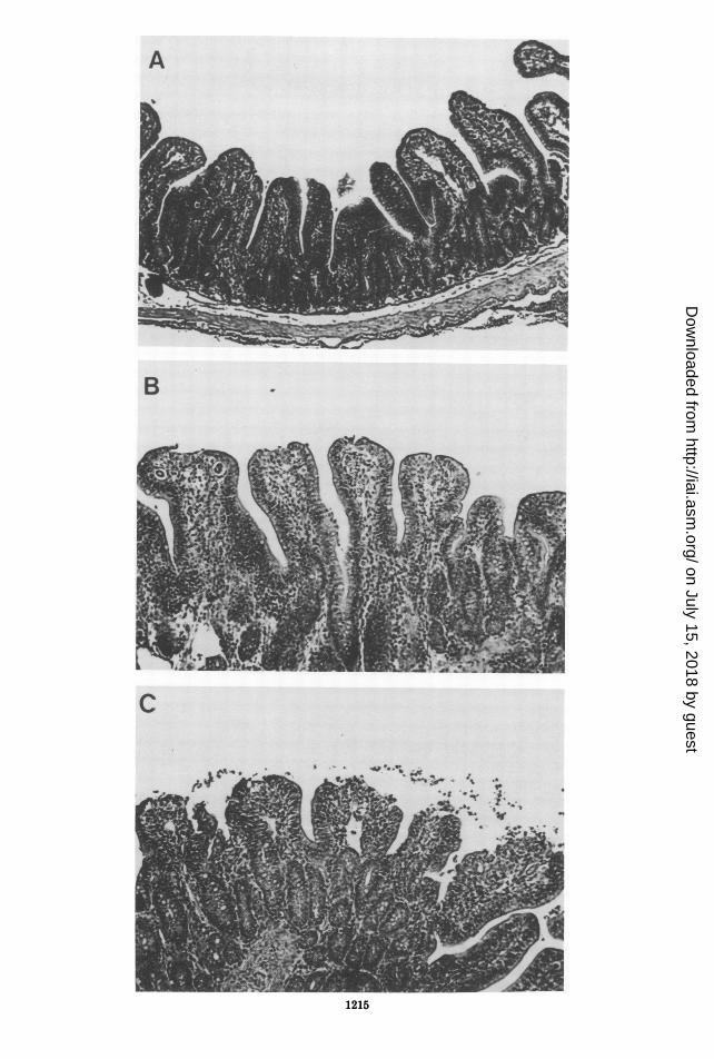
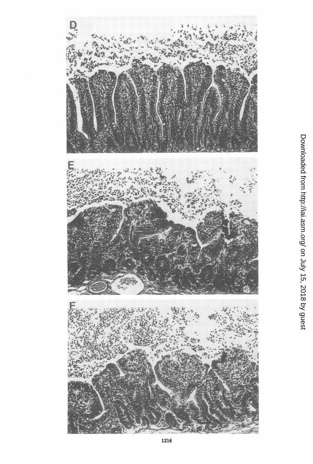
FIG. 1. Histopathological effect of C. perfringens enterotoxin in the rabbit ileum. (A) Control; (B) portionof ileum exposed to 50EU for 90 min; (C) 100 EU; (D)250 EU; (E) 500 EU; (F) 1 ,000 EU. Magnification x100X.Strain, hematoxylin, and eosin.
1214
on July 15, 2018 by guesthttp://iai.asm
.org/D
ownloaded from
A
B
C
.t, a
.&',
_ * X;~~Jvl~
|L@;$w ' {r*
1215
on July 15, 2018 by guesthttp://iai.asm
.org/D
ownloaded from
NOTES 1217
of damage to the intestinal tissue in each sac
studied. Although variations in response didoccur, the severity of reaction increased withthe dose in each animal. The sections shown inFig. 1 are representative of the damage at eachdosage. It was found that the response was dosedependent up to 500 EU, beyond which excess
enterotoxin had no noticeable augmenting ef-fect upon the histopathology described. Figure1A shows control tissue exposed to solutionwithout enterotoxin. It appeared normal, as diduntreated sections of the ileum. Figure 1Bshows slight but noticeable epithelial damagewith exposure to 50 EU of enterotoxin. Con-gested submucosa was also evident (notshown). In Fig. 1C (100 EU) a few epithelialcells are present in the lumen from the par-
tially desquamated epithelium. Mucin was alsopresent that had been expelled from the globletcells. Figure 1D (250 EU) is progressive andshows further desquamation. Intense desqua-mation, inflamatory cells (mostly lympho-cytes), and slight hyperemia in the mucosa are
seen in Fig. 1E (500 EU). Figure 1F (1,000 EU)shows a degree of damage similar to that inFig. 1E.
It can be seen (Table 1) that net fluid accumu-lation in the lumen was the characteristic re-
sponse to the enterotoxin as compared to a de-crease in fluid content in control loops. Eachdose of toxin caused a significant differencefrom controls in fluid transport (P < 0.001) as
determined by Student's t test. However, therewas no significant difference between amountsof fluid secreted at the doses studied. This sug-gests a possible threshold of response for fluidtransport above which excess stimulation haslittle or no augmenting effect. That, however,is not the case for tissue damage. The data inTable 2 support those seen in Fig. 1, in thatluminal fluid protein content increased withthe toxin dose. This would be expected sinceincreased desquamation seen with increaseddosages (Fig. 1) and subsequent leakage offluids into the lumen would cause an increasein protein to be found there. The differencesbetween protein release induced by 50 and 250EU and by 100 and 500 EU were significant (P< 0.05). It seems that an increase of nearly 200EU was needed at the time interval studied toinduce a significant increase in fluid proteincontent. The addition of more than 500 EUcaused no significant increase in protein.
Stark and Duncan (16) found the minimaldose of enterotoxin needed to induce measura-
ble fluid accumulation by the rabbit ileal looptest, as measured after an incubation period ofseveral hours, to be about 140 to 200 EU of
TABLE 1. Fluid transport in rabbit ileal loopsexposed to different doses ofClostridium perfringens
enterotoxina
Total fluid
nb Erythemal activ- transport Standardity/loop (jA/cm of intes- error Y
tine)c
7 0 (Control) 72.9 ±15.98 50 -50.1 ±22.17 100 -40.0 ±7.08 250 -61.8 ±16.68 500 -57.7 ±9.23 1,000 -66.7 ±25.5
a All loops were exposed for 90 min.bn, Number of loops.c Negative values indicate secretion into the lu-
men.
TABLE 2. Protein content offluid from rabbit ilealloops exposed to Clostridiumperfringens enterotoxina
nb Erythemal activ- Total protein Standard er-
ity/loop (mg/cm) ror Y
8 0 (control) 0.31 0.038 50 1.46 0.107 100 1.79 0.136 250 2.15 0.337 500 2.24 0.123 1,000 2.20 0.55
a All loops were exposed for 90 min.bn, Number of loops.
activity. Hauschild et al., by using a rapid de-tection procedure using a 90-min incubationperiod (4), found that as little as 2.5 EU ofenterotoxin was sufficient to prevent absorp-tion of fluid whereas between 40 and 160 EUwas needed to cause a net increase in fluidvolume of loops tested. It can be seen from theevidence presented here that the minimalrange of toxin needed to induce fluid productionis within the range needed to induce tissuedamage in the rabbit.This report establishes that a distinct histopa-
thology is produced in the rabbit intestine bythe intraluminal administration of C. perfrin-gens enterotoxin at doses needed to induce fluidaccumulation in ileal loops. This is similar toresults obtained in the rat (8). How the desqua-mation of villus epithelium is related to thetransport reversals associated with diarrheadue to this enterotoxin is not yet clear. Cer-tainly if tissue disruption is a part ofthe pathol-ogy in human cases it would contribute to fluidand electrolyte loss. However, it would not beexpected that the damage in human cases couldbe very severe as apparent recovery is oftencomplete in 24 h. The susceptibility of human
VOL. 12, 1975
on July 15, 2018 by guesthttp://iai.asm
.org/D
ownloaded from
1218 NOTES
epithelial tissue to the desquamating activity ofthe enterotoxin could be very different fromthat seen in these experimental models.The action of C. perfringens enterotoxin in
the rabbit model is in contrast to that of choleratoxin in the rabbit (13), which causes no appar-ent change in the villus epithelium. Choleratoxin does not cause noticeable damage in thecanine model (2) or human cases of the disease(3). E. coli enterotoxin also acts very similarlyto cholera toxin in the rabbit (10) and there-fore is contrasted to this enterotoxin. AlthoughShigella enterotoxin alone has no effect uponvillus morphology (6), staphylococcal entero-toxin has been shown to be capable of denudingvillus epithelium in cats (15) and dogs (12).
This research was supported by the College of Agricul-ture and Life Sciences, University of Wisconsin, Madison,Public Health Service research grant AI-11865-05 from theNational Institute of Allergy and Infectious Diseases, Pub-lic Health Service research grant FD-00203-05 from theFood and Drug Administration, and by Contributors to theFood Research Institute by member industries. J.L.M. isthe recipient of a post-doctoral award from Public HealthService grant T32-EF0715-01 of the National Institute ofEnvironmental Health Sciences. C.L.D. is the recipient ofaPublic Health Service Research Career Development awardAI-70721-02 from the National Institute of Allergy and In-fectious Diseases.We thank Joseph Lalich for assistance in interpreting
histopathological data.
LITERATURE CITED1. Duncan, C. L., H. Sugiyama, and D. H. Strong. 1968.
Rabbit ileal loop response to strains of Clostridiumperfringens. J. Bacteriol. 95:1560-1566.
2. Elliott, H. L., C. C. J. Carpenter, Jr., R. B. Sack, andJ. H. Yardley. 1970. Small bowel morphology in ex-perimental canine cholera. A light and electron mi-croscopic study. Lab. Invest. 22:112-120.
3. Fresh, J. W., P. M. Versage, and F. Reyes. 1964. Intes-
tinal morphology in human and experimental chol-era. Arch. Pathol. 77:529-537.
4. Hauschild, A. H. W., R. Hilsheimer, and C. G. Rogers.1971. Rapid detection of Clostridium perfringens en-terotoxin by a modified ligated intestinal loop tech-nique in rabbits. Can. J. Microbiol. 17:1475-1476.
5. Hauschild, A. H. W., L. Niilo, and J. Dorward. 1967.Experimental enteritis with food poisoning and classi-cal strains of Clostridium perfringens type A inlambs. J. Infect. Dis. 117:379-386.
6. Levine, M. M., H. L. DuPont, S. B. Formal, R. B.Hornick, A. Takevchi, E. J. Gangarosa, M. J. Sny-der, and J. P. Libonati. 1973. Pathogenesis of Shi-gella dysenteriae 1 (Shiga) dysentery. J. Infect. Dis.127:261-270.
7. Lowry, 0. H., N. J. Rosebrough, A. L. Farr, and R. J.Randall. 1951. Protein measurement with the Folinphenol reagent. J. Biol. Chem. 193:265-275.
8. McDonel, J. L. 1974. In vivo effects of Clostridium per-fringens enteropathogenic factors on the rat ileum.Infect. Immun. 10:1156-1162.
9. McDonel, J. L., and T. Asano. 1975. Analysis of unidi-rectional fluxes of sodium during diarrhea induced byClostridium perfringens enterotoxin in the rat termi-nal ileum. Infect. Immun. 11:526-529.
10. Moon, H. W., S. C. Whipp, and A. L. Baetz. 1971.Comparative effects of enterotoxins from Escherichiacoli and Vibrio cholerae on rabbit and swine smallintestine. Lab. Invest. 25:133-140.
11. Niilo, L. 1970. Mechanism of action of the enteropatho-genic factor ofClostridium perfringens type A. Infect.Immun. 3:100-106.
12. Niilo, L. 1973. Effect on calves of the intravenous injec-tion of the enterotoxin ofClostridium welchii Type A.J. Comp. Pathol. 83:265-269.
13. Norris, H. T., and G. Majno. 1968. On the role of theileal epithelium in the pathogenesis of experimentalcholera. Am. J. Pathol. 53:263-280.
14. Prohaska, J. V. 1963. Role of staphylococcal entero-toxin in the induction of experimental ileitis. Ann.Surg. 158:492497.
15. Prohaska, J. V., M. J. Jacobson, C. T. Drake, and T.Tan. 1959. Staphylococcus enterotoxin enteritis.Surg. Gynecol. Obstet. 109:73-77.
16. Stark, R. L., and C. L. Duncan. 1972. Purification andbiochemical properties of Clostridium perfringenstype A enterotoxin. Infect. Immun. 6:662-673.
INFECT. IMMUN.
on July 15, 2018 by guesthttp://iai.asm
.org/D
ownloaded from