
1
Hsa-miR-31-3p expression is linked to progression-free survival in patients
with KRAS wild-type metastatic colorectal cancer treated with anti-EGFR
therapy
Gilles Manceau1,4#, Sandrine Imbeaud2#, Raphaële Thiébaut3, François Liébaert3,
Karine Fontaine3, Francis Rousseau3, Bérengère Génin3, Delphine Le Corre1, Audrey
Didelot1, Marc Vincent1, Jean-Baptiste Bachet4, Benoist Chibaudel5, Olivier Bouché6,
Bruno Landi7, Frédéric Bibeau8, Karen Leroy9, Frédérique Penault-Llorca10, Jean-Luc
Van Laethem11, Pieter Demetter12, Sabine Tejpar13, Simona Rossi14, Neda
Mosakhani15, Pia Österlund16, Raija Ristamäki17, Virinder Sarhadi18, Sakari
Knuutila15,18, Valérie Boige1,19, Thierry André5, and Pierre Laurent-Puig1,20
#equally contributed to the present work
Running title: miR-31-3p expression and PFS in mCRC patients
Key words: microRNA biomarker; metastatic colorectal cancer; anti-EGFR antibody.
1Université Paris Sorbonne Cité; INSERM UMR-S775 Bases Moléculaires de la
réponse aux xénobiotiques, Paris, France
2INSERM UMR-S674 Genomique Fonctionnelle des Tumeurs, Paris, France
3Integragen S.A., Evry, France
Research. on June 30, 2018. © 2014 American Association for Cancerclincancerres.aacrjournals.org Downloaded from
Author manuscripts have been peer reviewed and accepted for publication but have not yet been edited. Author Manuscript Published OnlineFirst on April 25, 2014; DOI: 10.1158/1078-0432.CCR-13-2750

2
4Université Pierre et Marie Curie; Assistance Publique - Hôpitaux de Paris, Hôpital
Pitié Salpétrière, Paris, France
5Université Pierre et Marie Curie; Assistance Publique - Hôpitaux de Paris, Hôpital
Saint - Antoine, Paris, France
6Université de Reims Champagne-Ardenne; Centre Hospitalier Universitaire de
Reims, Reims, France
7Assistance Publique - Hopitaux de Paris, Hôpital Européen Georges Pompidou,
service d’Hépato-Gastro-Entérologie et d’Oncologie Digestive, Paris, France
8Service d’Anatomo-Pathologie, Centre Val d'Aurelle Paul-Lamarque, Montpellier,
France
9Université Paris-Est Créteil; Assistance Publique - Hôpitaux de Paris, Hôpital Henri
Mondor, Créteil, France
10Université Clermont-Ferrand, Centre Jean Perrin, Clermont-Ferrand, France
11Department of Gastroenterology, GI Cancer Unit, Erasme University Hospital,
Brussels, Belgium
12Department of Pathological Anatomy, Erasme University Hospital, Brussels,
Belgium
13Digestive Oncology Unit, University Hospital Gasthuisberg, Leuven, Belgium
14Bioinformatics Core Facility, Swiss Institute of Bioinformatics, Lausanne,
Switzerland 15Haartman Institute, University of Helsinki, Helsinki, Finland
Research. on June 30, 2018. © 2014 American Association for Cancerclincancerres.aacrjournals.org Downloaded from
Author manuscripts have been peer reviewed and accepted for publication but have not yet been edited. Author Manuscript Published OnlineFirst on April 25, 2014; DOI: 10.1158/1078-0432.CCR-13-2750
3
16Department of Oncology, Helsinki University Central Hospital and Helsinki
University, Helsinki, Finland
17Department of Oncology and Radiotherapy, Turku University Hospital, Turku,
Finland
18HUSLAB, Department of Pathology and Genetic Laboratory, Helsinki, Finland
19Department of Medicine, Institut Gustave Roussy, Villejuif, France
20Assistance Publique- Hopitaux de Paris, European Georges Pompidou,
Department of Biology, Paris, France
Authors’ contributions:
Study concept and design: R.T., F.L., F.R., B.G., P.L.P.
Acquisition of data: R.T., K.F., D.L.C., A.D., J.B.B., B.C., O.B., B.L, F.B, K.L., F.P.L.,
J.V.L., P.D., S.T., N.M., P.O., R.R., V.S., S.K., V.B., T.A., P.L.P.
Analysis and interpretation of data: G.M., S.I., R.T., F.R., B.G., M.V., F.P.L., S.R.,
S.T., N.M., P.O., S.K., P.L.P.
Drafting of manuscript: G.M., S.I., R.T., F.L., F.R., B.G., D.L.C., A.D., M.V., B.C.,
O.B., B.L., K.L., F.P.L., J.V.L., P.D., S.R., N.M., P.O., R.R., V.S., S.K., V.B., T.A.,
P.L.P.
Final approval of manuscript: G.M., S.I., R.T., F.L., K.F., F.R., B.G., D.L.C., A.D.,
M.V., J.B.B., B.C., O.B., B.L., F.B., K.L., F.P.L., J.V.L., P.D., S.R., S. T., N.M., P.O.,
R.R., V.S., S.K., V.B., T.A., P.L.P.
Research. on June 30, 2018. © 2014 American Association for Cancerclincancerres.aacrjournals.org Downloaded from
Author manuscripts have been peer reviewed and accepted for publication but have not yet been edited. Author Manuscript Published OnlineFirst on April 25, 2014; DOI: 10.1158/1078-0432.CCR-13-2750

4
Study supervision: P.L. P.
Authors’ disclosures/Conflicts of interest: Gilles Manceau: None Sandrine Imbeaud: Patents, Licenses or Royalties - Patent Title and Number: A method for predicting responsiveness to a treatment with an EGFR inhibitor. WO 2013076282 A1 Raphaële Thiébaut: Employee of IntegraGen SA; Title: Geneticist François Liebaert: Employee of IntegraGen SA; Title: VP of R&D and Medical Affairs Stock Ownership: IntegraGen SA Karine Fontaine: Employee of IntegraGen SA; Title: Lab Manager Francis Rousseau: Employee of IntegraGen SA; Title: Director of Genomics Stock Ownership: IntegraGen SA Bérengère Génin: Employee of IntegraGen SA; Title: Bioinfomatics Director Delphine Le Corre: None Audrey Didelot: None Marc Vincent: None Jean-Baptiste Bachet: Compensated Consultant or Advisory Role – Amgen and Merck Serono Other Remuneration: Merck Serono Benoist Chibaudel: Compensated Consultant or Advisory Role – Amgen Olivier Bouché: Compensated Consultant or Advisory Role – Merck Serono Honoraria: Merck Serono and Amgen Bruno Landi: Honoraria - Novartis and Ipsen Frédéric Bibeau: Honoraria – Roche and Amgen Research Funding - Roche Expert Testimony - Roche Karen Leroy: None
Research. on June 30, 2018. © 2014 American Association for Cancerclincancerres.aacrjournals.org Downloaded from
Author manuscripts have been peer reviewed and accepted for publication but have not yet been edited. Author Manuscript Published OnlineFirst on April 25, 2014; DOI: 10.1158/1078-0432.CCR-13-2750

5
Frédérique Penault-Llorca: None Jean-Luc Van Laethem: None Pieter Demetter: None Sabine Tejpar: Honoraria – Amgen and Merck Serono Simona Rossi: None Neda Mosakhani: None Pia Österlund: Compensated Consultant or Advisory Role – Amgen and Merck Other Remuneration – Amgen and Merck Raija Ristamäki: Honoraria - Amgen and Merck Serono Other Remuneration: Merck Serono Virinder Sarhadi: None Sakari Knuutila: None Valérie Boige: None Thierry André: Compensated Consultant or Advisory Role - Amgen Honoraria – Amgen and Merck Serono Pierre Laurent-Puig: Compensated Consultant or Advisory Role – Amgen, Merck Serono, and IntegraGen Honoraria – Amgen, Merck Serono, Boehringer Ingelheim Patents, Licenses or Royalties - Patent Title and Number: A method for predicting responsiveness to a treatment with an EGFR inhibitor. WO 2013076282 A1 Funding sources:
This work was funded by the Programme Hospitalier de Recherche Clinique (PHRC),
Institut National de la santé et de la recherché médicale (INSERM) through the
Biointelligence program, and by IntegraGen through the Biomos program funded by
Banque Publique d'Investissement, France. PETACC3 research on miRNAs is
supported by the Foundation Fournier-Majoie, FFMI, Belgium. Sabine Tejpar is a
Research. on June 30, 2018. © 2014 American Association for Cancerclincancerres.aacrjournals.org Downloaded from
Author manuscripts have been peer reviewed and accepted for publication but have not yet been edited. Author Manuscript Published OnlineFirst on April 25, 2014; DOI: 10.1158/1078-0432.CCR-13-2750

6
senior clinical investigator of the Fund for Scientific Research Flanders and is
supported by Belgian National Cancer Plan.
Correspondence to:
Pr. Pierre Laurent-Puig
UMR-S775 Molecular basis of response to xenobiotics, 45 Rue des Saints-Pères,
75006 Paris, France
Phone number: 33142862081 Fax number: 33142862072
Email: [email protected]
Research. on June 30, 2018. © 2014 American Association for Cancerclincancerres.aacrjournals.org Downloaded from
Author manuscripts have been peer reviewed and accepted for publication but have not yet been edited. Author Manuscript Published OnlineFirst on April 25, 2014; DOI: 10.1158/1078-0432.CCR-13-2750
7
Translational Relevance
Anti-EGFR therapy has demonstrated effectiveness in patients with metastatic
colorectal cancer. The use of these agents are limited to a population of patients for
whom the tumors show KRAS wild-type status since randomized-controlled studies
have demonstrated that only this subgroup of patients can benefit from this therapy.
Since only 40% of patients with KRAS wild-type tumors treated with anti-EGFR
agents respond to this treatment, therefore additional markers are needed to help
patient selection in order to avoid the prescription of ineffective, expensive and
potentially harmful therapy. In the present study, we identify and validate a miRNA,
hsa-miR-31-3p, as a marker for predicting survival of patients treated with anti-EGFR
monoclonal antibodies with a KRAS wild-type metastatic colorectal cancer.
Furthermore we provide information on the genes regulated by this miRNA.
Research. on June 30, 2018. © 2014 American Association for Cancerclincancerres.aacrjournals.org Downloaded from
Author manuscripts have been peer reviewed and accepted for publication but have not yet been edited. Author Manuscript Published OnlineFirst on April 25, 2014; DOI: 10.1158/1078-0432.CCR-13-2750

8
ABSTRACT
Purpose: To identify miRNAs that predict response to anti-EGFR antibodies in wild-
type KRAS metastatic colorectal cancer (mCRC) patients.
Experimental Design: microRNA profiling was performed in a training set of
87mCRC patients refractory to chemotherapy treated with anti-EGFR antibodies.
This included 33 fresh frozen (FF) and 35 formalin fixed paraffin embedded (FFPE)
samples retrospectively collected and 19 prospectively collected FF samples. An
independent validation cohort consisting of 19 FF and 26 FFPE prospectively
collected samples from mCRC patients treated with anti-EGFR antibodies was used
to confirm our findings.
Results: After screening the expression of 1,145 miRNAs in FF samples from the
training set, we identified that hsa-miR-31-3p expression level was significantly
associated with progression-free survival (PFS). Statistical models based on miRNA
expression discriminated between high and low risk of progression for both FF and
FFPE samples. These models were confirmed in the validation cohort for both FF
(HR: 4.1 CI95%[1.1-15.3] P<0.04) and FFPE samples (HR=2.44 CI95%[1.1- 5.4] P=
0.028). The percentage of variation of RECIST criteria in the validation series was
significantly associated with the expression level of hsa-miR-31-3p (r2=0.49
p=0.0035) and risk status determined by hsa-miR-31-3p expression level (p=0.02,
Kruskal-Wallis rank test). Nomograms were built and validated to predict PFS
depending onhsa-miR-31-3p expression level. Following in vitro studies, we identified
47 genes regulated by hsa-miR-31-3p.
Research. on June 30, 2018. © 2014 American Association for Cancerclincancerres.aacrjournals.org Downloaded from
Author manuscripts have been peer reviewed and accepted for publication but have not yet been edited. Author Manuscript Published OnlineFirst on April 25, 2014; DOI: 10.1158/1078-0432.CCR-13-2750
9
Conclusion: Hsa-miR-31-3p appears to be a new mCRC biomarker whose
expression level allows for the identification of patients with wild-type KRAS mCRC
who are more likely to respond to anti-EGFR therapy.
Research. on June 30, 2018. © 2014 American Association for Cancerclincancerres.aacrjournals.org Downloaded from
Author manuscripts have been peer reviewed and accepted for publication but have not yet been edited. Author Manuscript Published OnlineFirst on April 25, 2014; DOI: 10.1158/1078-0432.CCR-13-2750

10
INTRODUCTION
With more than 1.2 million new cases in 2008, colorectal cancer (CRC) is the third
most common form of cancer worldwide (1,2). Nearly 20% of patients are diagnosed
at an advanced stage with metastatic disease (1) and over half will ultimately develop
metachronous metastases. The recent development of targeted therapies has
improved outcomes in patients with advanced CRC. Cetuximab and panitumumab,
two monoclonal antibodies, which neutralize the extracellular domain of epidermal
growth factor receptor (EGFR), have demonstrated effectiveness in terms of both
response and survival when used as first, second or third-line chemotherapy. While
anti-EGFR agents are routinely utilized in clinical practice, their use is limited to 60%
of patients who have KRAS wild-type tumors (2) since randomized-controlled studies
have shown activating mutations of this oncogene were predictive of resistance to
these agents (3-12).
Since response to anti-EGFR therapy is seen in less than 40% of patients with KRAS
wild-type tumors (13), additional factors are needed to help patient selection for this
therapy in order to avoid the prescription of ineffective, expensive and potentially
harmful treatments. Many studies have evaluated other potential biomarkers,
typically using a candidate gene approach focused on different intracellular pathways
downstream of EGFR or a genome-wide approach. At the present time, however, the
routine use of these biomarkers is not warranted. Indeed, BRAF V600E mutation
appears to be a marker of poor prognosis (9, 10, 14, 15). Furthermore, studies that
examined PIK3CA mutations, loss of PTEN expression, EGFR gene copy number
and polymorphisms of fragment c gamma receptors have reported conflicting results
(14, 16-27). Finally, mRNA expression level of amphiregulin and epiregulin seems to
Research. on June 30, 2018. © 2014 American Association for Cancerclincancerres.aacrjournals.org Downloaded from
Author manuscripts have been peer reviewed and accepted for publication but have not yet been edited. Author Manuscript Published OnlineFirst on April 25, 2014; DOI: 10.1158/1078-0432.CCR-13-2750

11
be of limited clinical interest because of the inability to set an appropriate threshold
limit (28, 29).
MicroRNAs (miRNAs) are small 19 to 25 nucleotide, non-coding RNAs that
negatively regulate 30% of gene expression post-transcriptionally by inhibiting
translation and degrading mRNAs. miRNAs control biological processes such as cell
proliferation, differentiation, angiogenesis and apoptosis (30) while also acting as
oncomiR or suppressor miRs depending of their target mRNA. MiRNAs deregulation,
through expression modification and point mutations, is involved in the occurrence of
many types of cancer including CRC (31). These abnormalities are of major interest
and could serve for cancer diagnosis, prognosis, and chemotherapy response
prediction.
This study aimed to develop a miRNA expression-based model for predicting survival
of patients with KRAS wild-type mCRC treated with anti-EGFR monoclonal
antibodies.
Research. on June 30, 2018. © 2014 American Association for Cancerclincancerres.aacrjournals.org Downloaded from
Author manuscripts have been peer reviewed and accepted for publication but have not yet been edited. Author Manuscript Published OnlineFirst on April 25, 2014; DOI: 10.1158/1078-0432.CCR-13-2750

12
PATIENTS AND METHODS
Patients
Four cohorts of mCRC patients were included in this study (n=132). Three were used
as training sets and one as a validation set. All samples were obtained from studies
which had previously received appropriate ethical committee approvals. Samples for
the first training series (TS-Frozen1) were obtained from a retrospective collection of
33 patients, some of whom were reported on in a previously published series (13,
32). All patients were refractory to a FOLFOX or FOLFIRI regimen, refractory to
these regimens plus anti-EGFR antibodies, or received panitumumab as a single
agent.
Samples for the second training series (TS-Frozen2) were obtained from a
prospective collection of patients treated by anti-EGFR antibodies alone or in
combination with an irinotecan-based chemotherapy regimen. All of these patients
were considered refractory to FOLFIRI regimen.
The third training set (TS-FFPE) was made up of samples from a retrospective series
of pooled Belgian and Finnish mCRC patients. Some of these patients were a part of
previously published study (33). All patients in this training set were considered
refractory to a 5-fluorouracil-based regimen combined with irinotecan and oxaliplatin.
The last cohort of patients (validation set: VS) was comprised of patients from a
randomized phase II trial sponsored by GERCOR (Groupe Cooperateur
Multidisciplinaire en Oncologie). From the 65 patients eligible for this phase II study,
50 KRAS wild-type patients were considered for subsequent analysis. Among them,
45 patients had enough tumor material to be included (84%) with 26 having only
Research. on June 30, 2018. © 2014 American Association for Cancerclincancerres.aacrjournals.org Downloaded from
Author manuscripts have been peer reviewed and accepted for publication but have not yet been edited. Author Manuscript Published OnlineFirst on April 25, 2014; DOI: 10.1158/1078-0432.CCR-13-2750

13
FFPE samples available, 6 with FF samples only, and 13 having both FFPE and FF
samples available. All patients in this cohort were treated with third-line therapy using
a combination of irinotecan and panitumumab after a demonstration of progression to
oxaliplatin and irinotecan-based chemotherapy regimens.(34) This validation cohort
was divided in two (Figure 1A & 1B). The first cohort (VS-Frozen) consisted of
patients with FF samples (n=19) with the second cohort (VS-FFPE) consisting of
patients with FFPE samples without FF samples available (n=26) or with FF samples
(n=39).
DNA and RNA extraction
DNA and RNA extraction and mutation analysis was only performed on specimens
with greater than 50% tumor component. DNAs were extracted from frozen matched
tumor and non-tumor tissue samples using the QIAamp DNA mini kit (Qiagen,
Courtaboeuf, France). Total RNAs were extracted from frozen tumors, cells or FFPE
using the mirVanamiRNA Isolation Kit (Ambion, Austin, TX), miRNeasy and
miRNeasy FFPE extraction kit (Qiagen) respectively (see supplementary methods).
KRAS and BRAF mutation
Methods of KRAS and BRAF mutation detection are described in the supplementary
methods.(13, 35)
Screening of the microRNAs
Research. on June 30, 2018. © 2014 American Association for Cancerclincancerres.aacrjournals.org Downloaded from
Author manuscripts have been peer reviewed and accepted for publication but have not yet been edited. Author Manuscript Published OnlineFirst on April 25, 2014; DOI: 10.1158/1078-0432.CCR-13-2750
14
Global miRNA profiling was performed by labeling and hybridizing 750ng of extracted
RNA from 43 tumor frozen samples randomly chosen from TS-Frozen1 and TS-
Frozen2 on Illumina Human v2 microRNA Expression Beadchips. Beadchips were
scanned with the Illumina I-Scan Reader and data were imported into GenomeStudio
data analysis software (Illumina), quantile-normalized and log2-transformed. For
expression analysis on Taqman (Applied Biosystems) of the 11 selected miRNAs,
see supplementary methods.
Cell culture and transfection
Three colorectal adenocarcinoma cell lines (HTB-37, CCL-222, CCL220-1) from the
American Type Culture Collection (ATCC, Manassas, CA) were selected and utilized
in the three month following reception of the cells for cell culture studies. The
authentication of the cells has been provided by ATCC using STR profiling method.
All cells were transfected with miRVanamiRNA (Ambion) mimic negative control or
hsa-miR-31-3p miRVanamiRNA mimic (for details see supplementary methods).
In silico analysis
We developed a database integrating contemporary miRNA target predictions from
six individual databases (PITA, picTar 5-way Targetscan microRNA.org,
MicroCosmandmiRDB). These databases allowed for the determination of miRNAs
co-targeted by candidate genes taking into account the number of miRNA prediction
databases which predicted each miRNA/target relationship and the rank of this
prediction. This database was current as of November 2012 when our analysis was
performed.
Research. on June 30, 2018. © 2014 American Association for Cancerclincancerres.aacrjournals.org Downloaded from
Author manuscripts have been peer reviewed and accepted for publication but have not yet been edited. Author Manuscript Published OnlineFirst on April 25, 2014; DOI: 10.1158/1078-0432.CCR-13-2750

15
Statistical analyses
Prediction of survival risks utilized the miRNA expression data in a supervised
principal component (SuperPC) approach analysis, a modification of the conventional
PCA as described by Bair and colleagues (36, 37). To evaluate the predictive value
of the method, a leave-one-out cross-validation was performed and permutation
analysis (N=100) estimated the statistical significance of separation of the Kaplan-
Meier survival curves. A prognostic index is provided for each patient with a log
expression profile given a vector x; a high value of the prognostic index
corresponding to a high value of hazard of death.
Survival statistical analysis was performed using the R packages ‘survival’ and ‘rms’.
Univariate and multivariate analyses used a Cox proportional regression hazard
model generating a hazard ratio (HR) and a performance index (area under the
curve, AUC). Nomograms were developed based on Cox proportional regression
hazard models which predict the probability of PFS based on a training set of
patients. Internal validation relied on 150 bootstrap resamples, then independent
validation. The VS series and whole series (Training + Validation series) were
submitted to a multivariate Cox model analysis including ECOG performance status
and the level of LDH when available. These two variables were obtained at the time
of the first administration of anti-EGFR therapy. The analysis was performed by
STATA version 11 (College Station, TX). The patients were dichotomized in two
groups, high and low risk, as determined above. The ECOG performance status was
Research. on June 30, 2018. © 2014 American Association for Cancerclincancerres.aacrjournals.org Downloaded from
Author manuscripts have been peer reviewed and accepted for publication but have not yet been edited. Author Manuscript Published OnlineFirst on April 25, 2014; DOI: 10.1158/1078-0432.CCR-13-2750

16
obtained in 108 patients. The LDH have been prospectively recorded for the patients
included in the phase II PIMABI study.
Gene and miRNA expression value comparison analyses were performed using non-
parametric test (Kruskal-Wallis tests) with the pairwise Wilcox test function in R using
the stats library.
False-discovery rate (FDR)-adjusted p-values were calculated using the Benjamini
and Hochberg procedure for multiple testing correction. The cor.test function was
used to calculate Pearson correlations between expression values together with
matching p-values. Statistical significance was set at p<0.05 for all analyses.
RESULTS
Hsa-miR-31-3p survival analysis
Patient characteristics of the different sets are described in Table 1. Overall, 132
patients with mCRC were included in this study. All patients were wild-type KRAS
based on screening of exon 2, 3 and 4. From training sets 1 and 2, 43 RNAs were
extracted from frozen tumor samples and analyzed by Illumina Beadchips. Using
supervised principal component (SuperPC) analysis, univariate association between
each miRNA and PFS allowed us to select a set of 11 miRNAs, for which Cox score
statistics were found significant (p<0.01) (supplementary table S2 and supplementary
figure S1).
All miRNAs were tested for in the first and second training sets using Taqman
probes. Only one miRNA, hsa-miR-31-3p, showed a significant association with PFS
Research. on June 30, 2018. © 2014 American Association for Cancerclincancerres.aacrjournals.org Downloaded from
Author manuscripts have been peer reviewed and accepted for publication but have not yet been edited. Author Manuscript Published OnlineFirst on April 25, 2014; DOI: 10.1158/1078-0432.CCR-13-2750

17
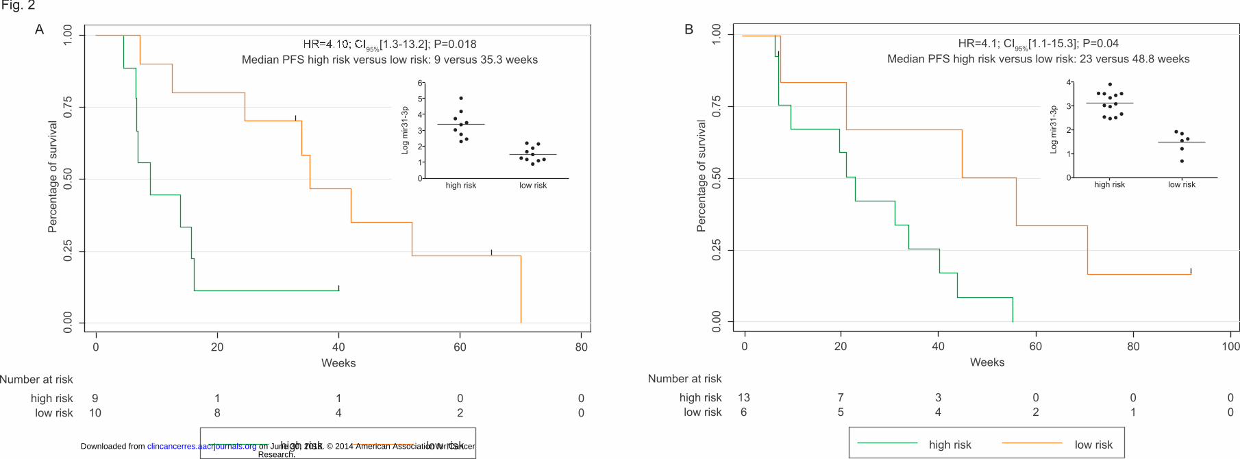
on TS-Frozen1 and TS-Frozen2 with HRs being 1.8, CI95%[1.1-2.9], P=0.01 and 2.5,
CI95%[1.3-4.5], P=0.002, respectively. In addition, a Super-PC analysis combining
Cox’s proportional hazard model and principal component analysis performed on TS-
Frozen1 using the Taqman expression level of miR-31-3p allowed us to define a
prognostic score (PS), computed by the formula: 0.178 x - 1.36 where x is the
Taqman log transformed expression of hsa-miR-31-3p, and a cutoff value of -0.031
dividing patients into 2 groups with a high and low risk of progression
(Supplementary figure S2). The HR was 2.6; CI95%[1.15-5.8]; P=0.021; the PFS of
high and low risk patients was 13 versus 31.4 weeks respectively. This PS was
calculated and the cutoff value was applied on TS-Frozen2. The PFS of high and low
risk patients was 9 versus 35.3 weeks respectively (HR: 4.1; CI95%[1.3-13.2];
P=0.018) (Figure 2A). The model was further validated for patients with FF sample in
the PIMABI phase II study (Validation Set Frozen, n=19). The PFS of high and low
risk patients was 23 versus 48.8 weeks respectively (HR: 4.1; CI95% [1.1-15.3];
P=0.02) (Figure 2B).
To be independent of a dichotomized variable, we built a nomogram based on Cox’s
proportional hazards regression modeling patients PFS probability with the log
transformed hsa-miR-31-3p expression on the TS-Frozen1 (Figure 3). BRAF status,
a known prognostic marker in mCRC, was added into the model. The nomogram was
then tested on TS-Frozen2 and VS-Frozen. The nomogram demonstrated the ability
to predict PFS with an AUC superior to 0.70 whatever the time threshold tested
(supplementary figure S3).
In anticipation of the use of this marker in clinical practice, we evaluated its
performance on RNA extracted from FFPE tissues. To validate the prognostic value
Research. on June 30, 2018. © 2014 American Association for Cancerclincancerres.aacrjournals.org Downloaded from
Author manuscripts have been peer reviewed and accepted for publication but have not yet been edited. Author Manuscript Published OnlineFirst on April 25, 2014; DOI: 10.1158/1078-0432.CCR-13-2750
18
of the expression of hsa-miR-31-3p on FFPE samples, we used two sets of patients
where FFPE tissue samples were available (TS-FFPE, n=35 and VS-FFPE, n=26).
Patients from TS-FFPE were divided into 2 groups according to expression of hsa-
miR-31-3p, as was done for frozen samples using a Cox model associated with a
principal component analysis to calculate a new PS (0.093x -0.407 where x is the
FFPE Taqman log transformed expression of hsa-miR-31-3p) and a new cutoff value
(-0.058). A HR for PFS of 2.79; CI95%[1.12- 6.96]; P=0.027 was observed between
high and low risk group of patients. This threshold was then validated on VS-FFPE
from the PIMABI trial for patients where only FFPE samples were available. A HR for
PFS of 5.1; CI95%[1.48-14.44], P=0.01 was observed.
We then added patients from VS-Frozen for whom we also had FFPE samples
(n=13). The PFS HR between high and low risk patients was of 2.44; CI95%[1.1- 5.4];
P= 0.028. When we added 6 patients to this cohort for whom only FF tissues was
available in order to test whole series of PIMABI, the PFS HR between high and low
risk patients was of 2.46; CI95% [1.22-4.94]; P=0.012 (Figure 4). Finally, we observed
a significant inverse correlation between the percentage variation of the RECIST
criteria and hsa-miR-31-3p expression (r2=0.49;p=0.0035). This result was confirmed
by the significant association between patient risk status and the percentage of
variation of RECIST criteria (p=0.02; Kruskal-Wallis rank test, Figure 3). Multivariate
analyses were performed including the BRAF status (i.e. mutated versus non
mutated), the LDH level and the ECOG performance status at time of inclusion in
PIMABI phase 2 study in RAS wild-type population. The status of patients
determined by the level of expression of the hsa-miR-31-3p remains significant. The
PFS HR and OS HR between high and low risk patients were 2.6; CI95%[1.1-5.8];
P=0.023 and 3.2; CI95%[1.4-7.5]; P=0.008.
Research. on June 30, 2018. © 2014 American Association for Cancerclincancerres.aacrjournals.org Downloaded from
Author manuscripts have been peer reviewed and accepted for publication but have not yet been edited. Author Manuscript Published OnlineFirst on April 25, 2014; DOI: 10.1158/1078-0432.CCR-13-2750

19
Finally, when we pooled the different series (n=132) taking into account the
classification based on the expression of hsa-miR-31-3p (supplementary figure S4).
The odds ratio of non-response in the high risk patients were OR:4.9; CI95%[2-
12.5];P<0.001). We performed a multivariate analysis including ECOG performance
status and the BRAF status in RAS wild-type patients (n=104). The PFS HR and OS
HR between high and low risk patients were 2.3; CI95%[1.4-3.8]; P<0.001 and
1.9;CI95%[1.2-3.1];P=0.008.
Hsa-miR-31-3p targets
Three CRC cell lines were transfected with hsa-miR-31-3p mimic or with a mimic
control. The transfection efficacy was demonstrated by a 1,500 times average rise of
hsa-miR-31-3p levels. Expression profile analysis of transfected cells allowed us to
identify 47 genes significantly down-regulated (fc<0.7; p<0.05), and 27 genes
significantly up-regulated by hsa-miR-31-3p (fc<1.3; p<0.05) (supplementary table
S3).
As the role of a miRNA includes degradation of its transcript target, we studied if our
database predicted the 47 down-regulated genes as a hsa-miR-31-3p putative target.
Twenty-five of the genes were predicted to be putative direct targets of hsa-miR-31-
3p and displayed a good rank in the prediction database. This number and the
ranking of genes were significant (P<0.0001 for both by permutation test).Twenty-six
of the up-regulated genes were not predicted to be direct targets of hsa-miR-31-3p.
While one of the up-regulated genes was predicted to be a direct target, it was the
last ranked target in the prediction database.
Research. on June 30, 2018. © 2014 American Association for Cancerclincancerres.aacrjournals.org Downloaded from
Author manuscripts have been peer reviewed and accepted for publication but have not yet been edited. Author Manuscript Published OnlineFirst on April 25, 2014; DOI: 10.1158/1078-0432.CCR-13-2750

20
The 25 putative direct and 27 indirect target genes were validated on qRT-PCR. Out
of these 52 genes, 45 displayed an expression level comparable to the level obtained
in the array. When the expression of these genes was analyzed in 39 tumor samples,
we found a correlation between hsa-miR-31-3p expression pattern and PFS for
DBNDD2 (p=0.02) and EPB41L4B (p=0.009). Interestingly, both genes displayed a
negative correlation with hsa-miR-31-3p expression levels: DBNDD2 (-0.5;p=0.001)
and EPB41L4B (-0.3;p=0.04) (supplementary figureS5).
DISCUSSION
By using an assumption-free approach in order to identify miRNAs associated with
PFS in KRAS wild-type mCRC patients treated by anti-EGFR therapy, we
established for the first time a link between hsa-miR-31-3p expression and the risk of
progression. All patients in our cohorts were refractory to the chemotherapy regimen
used with anti-EGFR therapy. We were able to define a robust threshold by dividing
patients in two groups according to their risk of progression after anti-EGFR therapy.
Our results independently confirm those of a Finnish group (33) who demonstrated a
significant differential expression of hsa-miR-31-3p between patients with stable and
progressive disease, even though we used a part of their patient set in TS-FFPE to
determine the threshold of the hsa-miR-31-3p in FFPE tissues.
The availability of multiple drugs for mCRC (e.g. panitumumab, cetuximab, and
bevacizumab) underlines the need for better decision-making tools. The survival
improvement of patients with mCRC often results from the use of these different
drugs via a personalized approach or precise manner. The nomogram we developed
which predicts PFS on an individualized basis provides such a decision making tool.
Research. on June 30, 2018. © 2014 American Association for Cancerclincancerres.aacrjournals.org Downloaded from
Author manuscripts have been peer reviewed and accepted for publication but have not yet been edited. Author Manuscript Published OnlineFirst on April 25, 2014; DOI: 10.1158/1078-0432.CCR-13-2750

21
The accuracy of this nomogram on frozen samples was assessed by the calculation
of the AUC that gives a value superior to 0.70, which can be considered as a good
performance for a predictor. There is also a good correlation between hsa-miR-31-3p
expression levels measured in frozen samples and those measured in FFPE
samples, confirming the interest to use miRNAs as surrogate for wider gene
expression signatures. Nevertheless, we needed to readjust the threshold of the hsa-
miR-31-3p expression in order to divide the high and low risk progression groups for
FFPE samples. Furthermore, although PFS can be considered a good surrogate
marker of efficacy in well-designed randomized clinical trials, its value per se in
retrospective series is unclear. We also investigated the impact of hsa-miR-31-3p
with other critical variables such as response rate and overall survival. There was a
significant link between response status (Non-Responder versus Responder) and the
classification in high and low risk patients according hsa-miR-31-3p expression as
shown in the waterfall plot of the patients included in the PIMABI phase II study. We
also validated this association in the whole series, the risk of non-response in 5 times
more frequent in the high-risk patient group as compared to low-risk patient group
Finally, we also showed a significant link between the overall survival and the hsa-
miR-31-3p expression level in the whole series adjusted on the other prognostic
variables (ECOG and BRAF mutational status). The high-risk patient group has a
significant shorter survival than the patients belonging to the low-risk group.
Several miRNAs have been shown to be associated with response to chemotherapy
in different types of cancers (38-40). To our knowledge only one group has reported
an association between the expression of a miRNA (hsa-miR-143) with PFS and
cancer-specific survival in mCRC patients treated by anti-EGFR alone or in
combination with chemotherapy in refractory setting (41). The authors, however,
Research. on June 30, 2018. © 2014 American Association for Cancerclincancerres.aacrjournals.org Downloaded from
Author manuscripts have been peer reviewed and accepted for publication but have not yet been edited. Author Manuscript Published OnlineFirst on April 25, 2014; DOI: 10.1158/1078-0432.CCR-13-2750

22
failed to identify a link between the expression of this miRNA and response to anti-
EGFR therapy, suggesting this biomarker is more a prognostic marker than
predictive, even if some relation with 5FU sensitivity have been described(42)
The specific role of the hsa-miR-31-3p has been investigated through in silico and in
vitro approaches and the list of genes is not easy to interpret. The 25 genes
putatively directly down-regulated by the hsa-miR-31-3p do not give a clear picture to
a regulatory mechanism of response to anti-EGFR therapy. A recent link between
some specific regulation of miRNAs maturation and EGFR kinase activity has been
emphasized and could enlighten our results(43). In hypoxic conditions the maturation
of pre-miRNAs with long loop structures are dependent on the phosphorylation of
AGO2 protein by EGFR. The phosphorylated AGO2 protein reduced its interaction
with DICER protein leading to decrease significantly the loading of pre-miRNA or the
expression of mature miRNA. Among the miRNAs regulated by this mechanism, the
premature mir-31 is one of the most likely candidates. The absence of EGFR induced
increased expression of mir-31 and inhibition of EGFR by tyrosine kinase inhibitors
such as gefitinib. The high level of hsa-miR-31-3p expression in our patient’s tumors
could be the result of an absence of EGFR response to hypoxia(44) and thus
reduced benefit from anti-EGFR therapy. Among the genes de-repressed by the
inhibition of maturation of miRNA by EGFR response to hypoxia,(43) two genes were
found in common with our list of down-regulated genes by the hsa-miR-31-3p; AMFR
and EPB41L4B, the latter being also associated with PFS.
We were unable to demonstrate the predictive role of hsa-miR-31-3p expression in
our study since there was no control arm consisting of patients without anti-EGFR
therapy. In an attempt to assess the predictive nature of hsa-miR-31-3p, we first
Research. on June 30, 2018. © 2014 American Association for Cancerclincancerres.aacrjournals.org Downloaded from
Author manuscripts have been peer reviewed and accepted for publication but have not yet been edited. Author Manuscript Published OnlineFirst on April 25, 2014; DOI: 10.1158/1078-0432.CCR-13-2750

23
tested its prognostic value in the PETACC3 cohort(45, 46) and did not find a
correlation between hsa-miR-31-3p expression and survival after relapse
(HR=0.93,p=0.48,data not shown). These data were based on 124 KRAS and NRAS
wild-type patients. Furthermore, in a recent study by Shen et al,(43) the expression of
phosphorylated AGO2 protein has been shown to be associated with worse
prognosis in breast cancer patients, suggesting that maturation inhibition of miRNAs
controlled by EGFR is a marker of aggressiveness in at least breast cancer patients.
Our results are exactly at the opposite suggesting that high expression of hsa-miR-
31-3p is rather a predictive than a prognostic marker.
Altogether, our results suggest that the expression hsa-miR-31-3p could be of help in
the decision to add anti-EGFR therapy to common chemotherapy regimens in the
population of KRAS wild type patients by giving a probability of PFS, which will be of
interest with the development of several targeted therapy in mCRC.
ACKNOWLEDGEMENTS
We wish to thank Céline Vazart and Virginie Decaulne for their expertise and
contribution associated with the extraction and quantification of miRNA for this study.
This work was funded by the Programme Hospitalier de Recherche Clinique (PHRC),
the Institut National du Cancer (INCa) through the Biointelligence program, and by
IntegraGen through the Biomos program funded by Banque Publique
d'Investissement, France.PETACC3 research on miRNAs is supported by the
Foundation Fournier-Majoie, FFMI, Belgium. Sabine Tejpar is a senior clinical
Research. on June 30, 2018. © 2014 American Association for Cancerclincancerres.aacrjournals.org Downloaded from
Author manuscripts have been peer reviewed and accepted for publication but have not yet been edited. Author Manuscript Published OnlineFirst on April 25, 2014; DOI: 10.1158/1078-0432.CCR-13-2750

24
investigator of the Fund for Scientific Research Flanders and is supported by Belgian
National Cancer Plan.
Research. on June 30, 2018. © 2014 American Association for Cancerclincancerres.aacrjournals.org Downloaded from
Author manuscripts have been peer reviewed and accepted for publication but have not yet been edited. Author Manuscript Published OnlineFirst on April 25, 2014; DOI: 10.1158/1078-0432.CCR-13-2750
25
REFERENCES
1. van der Pool AE, Damhuis RA, Ijzermans JN, de Wilt JH, Eggermont AM, Kranse R, et al. Trends in incidence, treatment and survival of patients with stage IV colorectal cancer: a population-based series. Colorectal Dis. 2012;14:56-61. 2. Forbes SA, Bindal N, Bamford S, Cole C, Kok CY, Beare D, et al. COSMIC: mining complete cancer genomes in the Catalogue of Somatic Mutations in Cancer. Nucleic Acids Res. 2011;39:D945-50. 3. Amado RG, Wolf M, Peeters M, Van Cutsem E, Siena S, Freeman DJ, et al. Wild-type KRAS is required for panitumumab efficacy in patients with metastatic colorectal cancer. J Clin Oncol. 2008;26:1626-34. 4. Karapetis CS, Khambata-Ford S, Jonker DJ, O'Callaghan CJ, Tu D, Tebbutt NC, et al. K-ras mutations and benefit from cetuximab in advanced colorectal cancer. N Engl J Med. 2008;359:1757-65. 5. Tol J, Koopman M, Cats A, Rodenburg CJ, Creemers GJ, Schrama JG, et al. Chemotherapy, bevacizumab, and cetuximab in metastatic colorectal cancer. N Engl J Med. 2009;360:563-72. 6. Peeters M, Price TJ, Cervantes A, Sobrero AF, Ducreux M, Hotko Y, et al. Randomized phase III study of panitumumab with fluorouracil, leucovorin, and irinotecan (FOLFIRI) compared with FOLFIRI alone as second-line treatment in patients with metastatic colorectal cancer. J Clin Oncol. 2010;28:4706-13. 7. Ocvirk J, Brodowicz T, Wrba F, Ciuleanu TE, Kurteva G, Beslija S, et al. Cetuximab plus FOLFOX6 or FOLFIRI in metastatic colorectal cancer: CECOG trial. World J Gastroenterol. 2010;16:3133-43. 8. Douillard JY, Siena S, Cassidy J, Tabernero J, Burkes R, Barugel M, et al. Randomized, phase III trial of panitumumab with infusional fluorouracil, leucovorin, and oxaliplatin (FOLFOX4) versus FOLFOX4 alone as first-line treatment in patients with previously untreated metastatic colorectal cancer: the PRIME study. J Clin Oncol. 2010;28:4697-705. 9. Bokemeyer C, Bondarenko I, Hartmann JT, de Braud F, Schuch G, Zubel A, et al. Efficacy according to biomarker status of cetuximab plus FOLFOX-4 as first-line treatment for metastatic colorectal cancer: the OPUS study. Ann Oncol. 2011;22:1535-46. 10. Van Cutsem E, Kohne CH, Lang I, Folprecht G, Nowacki MP, Cascinu S, et al. Cetuximab plus irinotecan, fluorouracil, and leucovorin as first-line treatment for metastatic colorectal cancer: updated analysis of overall survival according to tumor KRAS and BRAF mutation status. J Clin Oncol. 2011;29:2011-9. 11. Allegra CJ, Jessup JM, Somerfield MR, Hamilton SR, Hammond EH, Hayes DF, et al. American Society of Clinical Oncology provisional clinical opinion: testing for KRAS gene mutations in patients with metastatic colorectal carcinoma to predict response to anti-epidermal growth factor receptor monoclonal antibody therapy. J Clin Oncol. 2009;27:2091-6. 12. Van Cutsem E, Dicato M, Arber N, Berlin J, Cervantes A, Ciardiello F, et al. Molecular markers and biological targeted therapies in metastatic colorectal cancer: expert opinion and recommendations derived from the 11th ESMO/World Congress on Gastrointestinal Cancer, Barcelona, 2009. Ann Oncol. 2010;21 Suppl 6:vi1-10. 13. Lievre A, Bachet JB, Boige V, Cayre A, Le Corre D, Buc E, et al. KRAS mutations as an independent prognostic factor in patients with advanced colorectal cancer treated with cetuximab. J Clin Oncol. 2008;26:374-9.
Research. on June 30, 2018. © 2014 American Association for Cancerclincancerres.aacrjournals.org Downloaded from
Author manuscripts have been peer reviewed and accepted for publication but have not yet been edited. Author Manuscript Published OnlineFirst on April 25, 2014; DOI: 10.1158/1078-0432.CCR-13-2750

26
14. Tol J, Dijkstra JR, Klomp M, Teerenstra S, Dommerholt M, Vink-Borger ME, et al. Markers for EGFR pathway activation as predictor of outcome in metastatic colorectal cancer patients treated with or without cetuximab. Eur J Cancer. 2010;46:1997-2009. 15. Bokemeyer C, Cutsem EV, Rougier P, Ciardiello F, Heeger S, Schlichting M, et al. Addition of cetuximab to chemotherapy as first-line treatment for KRAS wild-type metastatic colorectal cancer: Pooled analysis of the CRYSTAL and OPUS randomised clinical trials. Eur J Cancer. 2012;48:1466-75. 16. Lievre A, Bachet JB, Le Corre D, Boige V, Landi B, Emile JF, et al. KRAS mutation status is predictive of response to cetuximab therapy in colorectal cancer. Cancer Res. 2006;66:3992-5. 17. Prenen H, De Schutter J, Jacobs B, De Roock W, Biesmans B, Claes B, et al. PIK3CA mutations are not a major determinant of resistance to the epidermal growth factor receptor inhibitor cetuximab in metastatic colorectal cancer. Clin Cancer Res. 2009;15:3184-8. 18. Sartore-Bianchi A, Martini M, Molinari F, Veronese S, Nichelatti M, Artale S, et al. PIK3CA mutations in colorectal cancer are associated with clinical resistance to EGFR-targeted monoclonal antibodies. Cancer Res. 2009;69:1851-7. 19. De Roock W, Claes B, Bernasconi D, De Schutter J, Biesmans B, Fountzilas G, et al. Effects of KRAS, BRAF, NRAS, and PIK3CA mutations on the efficacy of cetuximab plus chemotherapy in chemotherapy-refractory metastatic colorectal cancer: a retrospective consortium analysis. Lancet Oncol. 2010;11:753-62. 20. Laurent-Puig P, Cayre A, Manceau G, Buc E, Bachet JB, Lecomte T, et al. Analysis of PTEN, BRAF, and EGFR status in determining benefit from cetuximab therapy in wild-type KRAS metastatic colon cancer. J Clin Oncol. 2009;27:5924-30. 21. Frattini M, Saletti P, Romagnani E, Martin V, Molinari F, Ghisletta M, et al. PTEN loss of expression predicts cetuximab efficacy in metastatic colorectal cancer patients. Br J Cancer. 2007;97:1139-45. 22. Loupakis F, Pollina L, Stasi I, Ruzzo A, Scartozzi M, Santini D, et al. PTEN expression and KRAS mutations on primary tumors and metastases in the prediction of benefit from cetuximab plus irinotecan for patients with metastatic colorectal cancer. J Clin Oncol. 2009;27:2622-9. 23. Sartore-Bianchi A, Moroni M, Veronese S, Carnaghi C, Bajetta E, Luppi G, et al. Epidermal growth factor receptor gene copy number and clinical outcome of metastatic colorectal cancer treated with panitumumab. J Clin Oncol. 2007;25:3238-45. 24. Moroni M, Veronese S, Benvenuti S, Marrapese G, Sartore-Bianchi A, Di Nicolantonio F, et al. Gene copy number for epidermal growth factor receptor (EGFR) and clinical response to antiEGFR treatment in colorectal cancer: a cohort study. Lancet Oncol. 2005;6:279-86. 25. Italiano A, Follana P, Caroli FX, Badetti JL, Benchimol D, Garnier G, et al. Cetuximab shows activity in colorectal cancer patients with tumors for which FISH analysis does not detect an increase in EGFR gene copy number. Ann Surg Oncol. 2008;15:649-54. 26. Bibeau F, Lopez-Crapez E, Di Fiore F, Thezenas S, Ychou M, Blanchard F, et al. Impact of Fc{gamma}RIIa-Fc{gamma}RIIIa polymorphisms and KRAS mutations on the clinical outcome of patients with metastatic colorectal cancer treated with cetuximab plus irinotecan. J Clin Oncol. 2009;27:1122-9.
Research. on June 30, 2018. © 2014 American Association for Cancerclincancerres.aacrjournals.org Downloaded from
Author manuscripts have been peer reviewed and accepted for publication but have not yet been edited. Author Manuscript Published OnlineFirst on April 25, 2014; DOI: 10.1158/1078-0432.CCR-13-2750

27
27. Park SJ, Hong YS, Lee JL, Ryu MH, Chang HM, Kim KP, et al. Genetic polymorphisms of FcgammaRIIa and FcgammaRIIIa are not predictive of clinical outcomes after cetuximab plus irinotecan chemotherapy in patients with metastatic colorectal cancer. Oncology. 2012;82:83-9. 28. Khambata-Ford S, Garrett CR, Meropol NJ, Basik M, Harbison CT, Wu S, et al. Expression of epiregulin and amphiregulin and K-ras mutation status predict disease control in metastatic colorectal cancer patients treated with cetuximab. J Clin Oncol. 2007;25:3230-7. 29. Jacobs B, De Roock W, Piessevaux H, Van Oirbeek R, Biesmans B, De Schutter J, et al. Amphiregulin and epiregulin mRNA expression in primary tumors predicts outcome in metastatic colorectal cancer treated with cetuximab. J Clin Oncol. 2009;27:5068-74. 30. Iorio MV, Croce CM. MicroRNAs in cancer: small molecules with a huge impact. J Clin Oncol. 2009;27:5848-56. 31. Corte H, Manceau G, Blons H, Laurent-Puig P. MicroRNA and colorectal cancer. Dig Liver Dis. 2012;44:195-200. 32. Laurent-Puig P, Cayre A, Manceau G, Buc E, Bachet JB, Lecomte T, et al. Analysis of PTEN, BRAF, and EGFR Status in Determining Benefit From Cetuximab Therapy in Wild-Type KRAS Metastatic Colon Cancer. J Clin Oncol. 2009;27:5924-30. 33. Mosakhani N, Lahti L, Borze I, Karjalainen-Lindsberg ML, Sundstrom J, Ristamaki R, et al. MicroRNA profiling predicts survival in anti-EGFR treated chemorefractory metastatic colorectal cancer patients with wild-type KRAS and BRAF. Cancer Genet. 2012;205:545-51. 34. Andre T, Blons H, Mabro M, Chibaudel B, Bachet JB, Tournigand C, et al. Panitumumab combined with irinotecan for patients with KRAS wild-type metastatic colorectal cancer refractory to standard chemotherapy: a GERCOR efficacy, tolerance, and translational molecular study. Ann Oncol. 2013;24:412-9. 35. Didelot A, Le Corre D, Luscan A, Cazes A, Pallier K, Emile JF, et al. Competitive allele specific TaqMan PCR for KRAS, BRAF and EGFR mutation detection in clinical formalin fixed paraffin embedded samples. Exp Mol Pathol. 2012;92:275-80. 36. Bair E, Hastie T, Paul D, Tibshirani R. Prediction by supervised principal components. J Am Stast Ass 2006;101:119-37. 37. Bair E, Tibshirani R. Semi-supervised methods to predict patient survival from gene expression data. PLoS Biol. 2004;2:E108. 38. Wei J, Gao W, Zhu CJ, Liu YQ, Mei Z, Cheng T, et al. Identification of plasma microRNA-21 as a biomarker for early detection and chemosensitivity of non-small cell lung cancer. Chin J Cancer. 2011;30:407-14. 39. Odenthal M, Bollschweiler E, Grimminger PP, Schroder W, Brabender J, Drebber U, et al. MicroRNA profiling in locally advanced esophageal cancer indicates a high potential of miR-192 in prediction of multimodality therapy response. Int J Cancer. 2013;133:2454-63. 40. Ali S, Ahmad A, Banerjee S, Padhye S, Dominiak K, Schaffert JM, et al. Gemcitabine sensitivity can be induced in pancreatic cancer cells through modulation of miR-200 and miR-21 expression by curcumin or its analogue CDF. Cancer Res. 2010;70:3606-17.
Research. on June 30, 2018. © 2014 American Association for Cancerclincancerres.aacrjournals.org Downloaded from
Author manuscripts have been peer reviewed and accepted for publication but have not yet been edited. Author Manuscript Published OnlineFirst on April 25, 2014; DOI: 10.1158/1078-0432.CCR-13-2750
28
41. Pichler M, Winter E, Stotz M, Eberhard K, Samonigg H, Lax S, et al. Down-regulation of KRAS-interacting miRNA-143 predicts poor prognosis but not response to EGFR-targeted agents in colorectal cancer. Br J Cancer. 2012;106:1826-32. 42. Borralho PM, Kren BT, Castro RE, da Silva IB, Steer CJ, Rodrigues CM. MicroRNA-143 reduces viability and increases sensitivity to 5-fluorouracil in HCT116 human colorectal cancer cells. FEBS J. 2009;276:6689-700. 43. Shen J, Xia W, Khotskaya YB, Huo L, Nakanishi K, Lim SO, et al. EGFR modulates microRNA maturation in response to hypoxia through phosphorylation of AGO2. Nature. 2013;497:383-7. 44. Franovic A, Gunaratnam L, Smith K, Robert I, Patten D, Lee S. Translational up-regulation of the EGFR by tumor hypoxia provides a nonmutational explanation for its overexpression in human cancer. Proc Natl Acad Sci U S A. 2007;104:13092-7. 45. Van Cutsem E, Labianca R, Bodoky G, Barone C, Aranda E, Nordlinger B, et al. Randomized phase III trial comparing biweekly infusional fluorouracil/leucovorin alone or with irinotecan in the adjuvant treatment of stage III colon cancer: PETACC-3. J Clin Oncol. 2009;27:3117-25. 46. Roth AD, Tejpar S, Delorenzi M, Yan P, Fiocca R, Klingbiel D, et al. Prognostic role of KRAS and BRAF in stage II and III resected colon cancer: results of the translational study on the PETACC-3, EORTC 40993, SAKK 60-00 trial. J Clin Oncol. 2010;28:466-74.
Research. on June 30, 2018. © 2014 American Association for Cancerclincancerres.aacrjournals.org Downloaded from
Author manuscripts have been peer reviewed and accepted for publication but have not yet been edited. Author Manuscript Published OnlineFirst on April 25, 2014; DOI: 10.1158/1078-0432.CCR-13-2750

29
Table 1: Baseline Patient Characteristics
Patient data Training set 1N=33
Training set 2N=19
Training set 3N=35
Validation set 1aN=19
Validation set 1bN=26
Gender Male 24 (72.7%) 11 (57.9%) 16 (45.7%) 12 (63.2%) 18 (69.2%)
Female 9 (27.3%) 8 (42.1%) 19 (54.3%) 7 (21.2%) 8 (30.8%)
Age Mean 55.6 65.0 63.3 61.8 60.2
Range (22.0-78.1) (47.3-77.9) (37-82) (33.9-84.5) (34-82)
Chemotherapy
regimen
cetuxiumab 0 0 2 (5.7%) 0 0
cetuximab+irinotecan 24 (72.7%) 11 (57.9%) 16 (45.7%) 0 0
cetuximab+Xeloda 1 (3.0%) 0 0 0 0
cetuximab+FOLFIRI 5 (15.2%) 4 (21.1%) 4 (11.4%) 0 0
cetuximab+FOLFOX 2 (6.0%) 0 1 (2.9%) 0 0
panitumumab 0 4 (21.1%) 8 (22.9%) 0 0
panitumumab+XELIRI 0 0 1 (2.9%) 0 0
panitumumab+irinotecan 1 (3.0%) 0 3 (8.6%) 19 (100%) 26 (100%)
Number of
treatment lines
prior to anti-EGFR
therapy
Median 2 2 3 2 2
Range (1-5) (1-6) (1-6) (1-6) (1-6)
Complete response 2 (6.0%) 0 0 1 (5.3%) 2 (7.7%)
Response
according to
RECIST Criteria
Partial response 10 (30.3%) 4 (21.1%) 13 (37.1%) 7 (36.8%) 8 (30.8%)
Stable disease 11 (33.3%) 9 (47.4%) 10 (28.6%) 6 (31.6%) 8 (30.8%)
Progressive disease 10 (30.3%) 6 (31.6%) 12 (34.3%) 5 (26.3%) 7 (26.9%)
Research.
on June 30, 2018. © 2014 A
merican A
ssociation for Cancer
clincancerres.aacrjournals.org D
ownloaded from
Author m
anuscripts have been peer reviewed and accepted for publication but have not yet been edited.
Author M
anuscript Published O
nlineFirst on A
pril 25, 2014; DO
I: 10.1158/1078-0432.CC
R-13-2750

30
Figure 1: Flow chart
Research. on June 30, 2018. © 2014 American Association for Cancerclincancerres.aacrjournals.org Downloaded from
Author manuscripts have been peer reviewed and accepted for publication but have not yet been edited. Author Manuscript Published OnlineFirst on April 25, 2014; DOI: 10.1158/1078-0432.CCR-13-2750
31
Figure 2: Kaplan-Meier curves for risk groups obtained from the second training set
(A) and the validation set 1a (B).
Research. on June 30, 2018. © 2014 American Association for Cancerclincancerres.aacrjournals.org Downloaded from
Author manuscripts have been peer reviewed and accepted for publication but have not yet been edited. Author Manuscript Published OnlineFirst on April 25, 2014; DOI: 10.1158/1078-0432.CCR-13-2750

32
Figure 3: Nomogram to predict PFS survival using log transformed hsa-miR-31-3p
expression levels (miR) and BRAF mutational status (brafm). To use the nomogram,
locate patient’s variable on the corresponding axis; draw a line to the points axis,
sum the points, and draw a line from the total points axis to the PFS probability axis.
Research. on June 30, 2018. © 2014 American Association for Cancerclincancerres.aacrjournals.org Downloaded from
Author manuscripts have been peer reviewed and accepted for publication but have not yet been edited. Author Manuscript Published OnlineFirst on April 25, 2014; DOI: 10.1158/1078-0432.CCR-13-2750
33
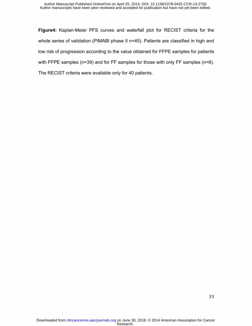
Figure4: Kaplan-Meier PFS curves and waterfall plot for RECIST criteria for the
whole series of validation (PIMABI phase II n=45). Patients are classified in high and
low risk of progression according to the value obtained for FFPE samples for patients
with FFPE samples (n=39) and for FF samples for those with only FF samples (n=6).
The RECIST criteria were available only for 40 patients.
Research. on June 30, 2018. © 2014 American Association for Cancerclincancerres.aacrjournals.org Downloaded from
Author manuscripts have been peer reviewed and accepted for publication but have not yet been edited. Author Manuscript Published OnlineFirst on April 25, 2014; DOI: 10.1158/1078-0432.CCR-13-2750

Published OnlineFirst April 25, 2014.Clin Cancer Res Gilles Manceau, Sandrine Imbeaud, Raphaele Thiebaut, et al. treated with anti-EGFR therapyin patients with KRAS wild-type metastatic colorectal cancer Hsa-miR-31-3p expression is linked to progression-free survival
Updated version
10.1158/1078-0432.CCR-13-2750doi:
Access the most recent version of this article at:
Material
Supplementary
http://clincancerres.aacrjournals.org/content/suppl/2014/04/29/1078-0432.CCR-13-2750.DC1
Access the most recent supplemental material at:
Manuscript
Authoredited. Author manuscripts have been peer reviewed and accepted for publication but have not yet been
E-mail alerts related to this article or journal.Sign up to receive free email-alerts
Subscriptions
Reprints and
To order reprints of this article or to subscribe to the journal, contact the AACR Publications
Permissions
Rightslink site. Click on "Request Permissions" which will take you to the Copyright Clearance Center's (CCC)
.http://clincancerres.aacrjournals.org/content/early/2014/04/25/1078-0432.CCR-13-2750To request permission to re-use all or part of this article, use this link
Research. on June 30, 2018. © 2014 American Association for Cancerclincancerres.aacrjournals.org Downloaded from
Author manuscripts have been peer reviewed and accepted for publication but have not yet been edited. Author Manuscript Published OnlineFirst on April 25, 2014; DOI: 10.1158/1078-0432.CCR-13-2750


