antitumorefficacyofacombinationofcmc...
TRANSCRIPT

Antitumor Efficacy of a Combination of CMC-544 (InotuzumabOzogamicin), a CD22-Targeted Cytotoxic Immunoconjugateof Calicheamicin, and Rituximab againstNon-Hodgkin’s B-Cell LymphomaJohn F. DiJoseph,1Maureen M. Dougher,1Lyka B. Kalyandrug,1DouglasC. Armellino,1Erwin R. Boghaert,1
Philip R. Hamann,2 Justin K. Moran,3 and Nitin K. Damle1
Abstract Purpose: CMC-544 is a CD22-targeted cytotoxic immunoconjugate, currently being evaluatedin B-cell non-Hodgkin’s lymphoma (B-NHL) patients. Rituximab is a CD20-targeted antibodycommonly used in B-NHL therapy. Here, we describe antitumor efficacy of a combination ofCMC-544 and rituximab against B-cell lymphoma (BCL) in preclinical models.Experimental Design:BCLs were cultured in vitro with CMC-544, rituximab, or their combina-tion. BCLs were injected either s.c. or i.v. to establish localized s.c. BCL in nude mice or dissemi-nated BCL in severe combined immunodeficient mice, respectively. I.p. treatment with CMC-544or rituximab was initiated at various times either alone or in combination and its effect on s.c. BCLgrowth or survival of mice with disseminated BCL was monitored.Results: In vitro growth-inhibitory activity of CMC-544 combined with rituximab was additive.Rituximab but not CMC-544 exhibited effector functions, such as antibody-dependent cellularcytotoxicity and complement-dependent cytotoxicity. Rituximab was less effective in inhibitinggrowth of established BCL xenografts than developing xenografts. In contrast, CMC-544 wasequally effective against both developing and established BCL xenografts. Although CMC-544and rituximab individually caused partial inhibition of the growth of BCL xenografts at suboptimaldoses examined, their combination suppressed xenograft growthby >90%. In a disseminated BCLmodel, 60% of CMC-544-treated mice and 20% of rituximab-treated mice survived for125 days.In contrast, 90% of mice treated with the combination of CMC-544 and rituximab survived forlonger than125 days.Conclusion:The demonstrationof superior antitumor activity of a combinationof CMC-544 andrituximab described here provides the preclinical basis for its clinical evaluation as a treatmentoption for B-NHL.
CMC-544 (inotuzumab ozogamicin) is a CD22-targetedcytotoxic agent composed of a humanized IgG4 anti-CD22antibody covalently linked to N-acetyl-g-calicheamicin dimeth-yl hydrazide (CalichDMH) via the acid-labile 4-(4V-acetylphe-noxy)butanoic acid linker (1–3). CD22 is a B-lymphoidlineage–specific differentiation antigen expressed on bothnormal and malignant B cells. CMC-544 binds CD22 withsubnanomolar affinity, and, upon binding, is rapidly internal-
ized delivering the conjugated CalichDMH inside the cells. Thispreferential intracellular delivery of CalichDMH causes DNAdamage resulting in B-cellular apoptosis. CalichDMH is aderivative of g-calicheamicin, a natural product produced byMicromonospora echinospora and is significantly more potent thancytotoxic chemotherapeutic agents used in cancer therapy.It binds in the minor groove of DNA and causes double-strand DNA breaks in a relatively sequence-specific and thiol-dependent manner leading to apoptotic response in cells (4–6).
CMC-544 exerts potent and CD22-selective growth inhibito-ry activity against CD22+ B-cell lymphoma (BCL) cell linesin vitro and causes regression of developing (minimal disease),small established (palpable disease), and large BCL xenografts,with a high therapeutic index (1). In addition, CMC-544protects severe combined immunodeficient (SCID) miceagainst hind limb paralysis and death caused by systemicallydisseminated BCL (7). In the absence of the conjugatedCalichDMH, G5/44, the targeting monoclonal antibody(mAb) in CMC-544, is ineffective in vivo as an antitumor agentin various preclinical models (1, 7) and, thus, CMC-544 isregarded as an antibody-targeted chemotherapy agent ratherthan an immunotherapeutic agent like rituximab. Largely due
Cancer Therapy: Preclinical
Authors’Affiliations: 1Oncology Discovery Research, 2Chemical and ScreeningSciences, and 3Chemical and Process Development,Wyeth Research, Pearl River,NewYorkReceived 8/30/05; revised10/14/05; accepted10/20/05.The costs of publication of this article were defrayed in part by the payment of pagecharges.This article must therefore be hereby marked advertisement in accordancewith18 U.S.C. Section1734 solely to indicate this fact.Note: All authors are employed by Wyeth Research whose investigational agent,CMC-544, was studied in the present work. CMC-544 is being jointly developedbyWyeth Research and UCBCelltech (Slough, United Kingdom).Requests for reprints: Nitin K. Damle, Oncology Discovery Research, WyethResearch, 200/4604, 401North Middletown Road, Pearl River, NY10965. Phone:845-602-3984; Fax: 845-602-5557; E-mail: [email protected].
F2006 American Association for Cancer Research.doi:10.1158/1078-0432.CCR-05-1905
www.aacrjournals.orgClin Cancer Res 2006;12(1) January 1, 2006 242
Research. on June 24, 2018. © 2006 American Association for Cancerclincancerres.aacrjournals.org Downloaded from

to its tumor-targeted, drug-delivery capability, CMC-544 islikely to have a better therapeutic index than that ofconventional chemotherapeutic agents. Based on its potentantitumor activity in preclinical models, CMC-544 is currentlybeing evaluated for safety and tolerability in patients with B-cellnon-Hodgkin lymphoma (B-NHL) in phase I clinical trials.
Rituximab is a chimeric human IgG1 antibody targeted toanother B-lymphoid lineage–specific molecule, CD20. Itrepresents a major therapeutic advance in B-NHL therapy(8–11). Rituximab mediates its antitumor activity by multiplemechanisms that include complement-dependent cytotoxicity(CDC), antibody-dependent cellular cytotoxicity (ADCC), anddirect induction of apoptosis in BCL (12–14). Despite theantitumor efficacy and safety shown by rituximab in B-NHLpatients, only a small subset of patients achieve completeresponses and the disease eventually relapses. Due to itsclinically shown safety, rituximab is widely used in combina-tion with various cytotoxic agents. Cycle treatment using acombination of rituximab with cytoreductive combinationchemotherapy, such as CHOP (a combination of cyclophos-phamide, doxorubicin, vincristine, and prednisone), hassignificantly improved clinical responses and durations ofdisease remission (15–18). However, such combinations alsosuffer from various systemic toxicities associated with thenontargeted nature of CHOP chemotherapy.
Because of the widespread therapeutic use of rituximab inB-NHL patients in the United States and Europe, initial clinicalevaluation of any experimental agent intended for B-NHL islikely to be carried out in B-NHL patients that have been treatedwith rituximab with or without chemotherapy. Because B-NHLcells consistently express both CD20 and CD22, it is reasonableto combine rituximab and CMC-544 in an attempt to enhancethe therapeutic advantage of either agent. This study evaluatedantitumor activities of CMC-544, rituximab, and their combi-nation and shows that a therapeutic combination of rituximaband CMC-544 is able to significantly inhibit the growth of s.c.human BCL xenografts and protects against systemicallydisseminated BCL. These results support the evaluation of acombination of CMC-544 and rituximab in patients withB-NHL with the ultimate goal of improving the therapeuticindex of the combination.
Materials andMethods
MaterialsA humanized anti-CD22 mAb (G5/44, an IgG4 isotype) was derived
by CDR grafting from murine anti-CD22 mAb m5/44 by Celltech(Slough, United Kingdom; ref. 19) and expressed at Wyeth Biopharma(Andover, MA). G5/44 was conjugated to CalichDMH with an acid-labile 4-(4V acetylphenoxy)butanoic acid linker and the resultingconjugate was termed CMC-544. The quantity of CalichDMH presentin CMC-544 was 73 Ag/mg G5/44. Chimeric human IgG1 anti-CD20mAb, rituximab (Rituxan, Biogen-Idec Pharmaceuticals, San Diego, CA,and Genentech, South San Francisco, CA), was purchased from MedWorld Pharmacy (Chestnut Ridge, NY). All conjugates were endotoxinfree (<5.0 endotoxin units/mL) by a modified Limulus amebocyteassay test (Biowhittaker, Walkersville, MD). Doses of calicheamicinconjugates are expressed as equivalents of CalichDMH and that ofunconjugated antibody are expressed as antibody protein.
The Burkitt lymphoma cell lines Ramos (CRL-1923), Raji (CCL-86),Daudi (CCL-213), and a NHL line RL (CRL-2261) were obtained from
the American Type Culture Collection (Manassas, VA) and determinedto be Mycoplasma free by a PCR Mycoplasma detection assay. Cell lineswere maintained as suspension cultures in RPMI medium plus 10%FCS, 10 mmol/L HEPES, 1 mmol/L sodium pyruvate, 4.5 g/L glucose,1.5 g/L sodium bicarbonate, penicillin G sodium 100 units/mL, andstreptomycin sulfate 100 Ag/mL. Before use, viable cells were isolated bycentrifugation (30 minutes at 1,000 � g) using Lymphoprep(Nycomed, Oslo, Norway) density gradient.
MiceFemale, BALB/c nu/nu (nude) mice (18-23 g) and 6- to 8-week-old
male SCID mice (CB17 SCID, body weight 20-25 g) were obtainedfrom Charles River Laboratories (Wilmington, MA). All mice werehoused in microisolator units and provided with sterile food and waterad libitum throughout the studies. All procedures involving mice wereapproved by the Wyeth Animal Care and Use Committee and carriedout according to established guidelines.
MethodsFlow cytometric detection of CD22 expression. The expression of
CD22 by various BCLs treated with or without rituximab was confirmedusing both direct and indirect immunofluorescence analyses asdescribed before (19). Fluorescein-conjugated murine anti-humanCD22 antibody, RFB4 (Santa Cruz Biotechnology, Santa Cruz, CA)was used to assess the surface expression of CD22 on various BCL.Alternatively, murine anti-CD22 antibody, m5/44, was used duringindirect immunofluorescence analysis of CD22 expression by BCL (19).RFB4 and m5/44 recognize distinct epitopes on human CD22 (19).
Cytotoxicity assays. A vital dye (MTS cell proliferation assay kit,
Promega, Madison, WI) stain was used to determine the number of
surviving cells following exposure to various drug treatments. BCLs
were seeded in 96-well dishes at a density of either 5,000 cells per well
(Ramos, Raji, and RL) or 10,000 cells per well (Daudi). Immediately
after seeding, the cells were exposed to various concentrations of CMC-
544 (0.0001 to 10 ng CalichDMH equivalents/mL), rituximab (20 or
200 Ag/mL), or a combination of the above. The number of cells
surviving 96 hours after drug exposure was determined. Percent survival
of cells in these cultures was calculated using the following equation:
percent survival = 100 � (no. viable cells in treated cultures / no. viable
cells in control cultures). IC50 values were calculated based on a four-
variable logistic model or, to obtain a better fit in some cases, a three-
variable model.
The Bliss independence model was used to define the interaction of
the drugs in combination (20). In the Bliss model, the expected
percentage of control of a combination of two drugs is the product of
the percentage of control for each drug alone at the same concentration
used in the combination. The medium-alone value was used as the
control and was subtracted from each well (background subtraction),
and then the mean of the (background adjusted) values for cells alone
wells were used to calculate the percentage of control. Statistical
significance was assessed using ANOVA to calculate 95% confidence
intervals on the observed mean percentage of control for each
combination. If the expected percentage of control for the combination
was outside of the 95% confidence interval for the observed mean, the
difference between the observed and expected growth inhibition was
noted as statistically significant. Separate ANOVAs were run for each cell
line. In each ANOVA, CMC-544 and rituximab concentrations were the
independent variables.Assessment of ADCC and CDC. Nude mouse peripheral blood was
allowed to clot for 60 minutes at 4jC after which the tubes werecentrifuged to collect serum, used as a source of murine complement.For CDC, Ramos B-lymphoma cells were used as target cells. Typically,10,000 or 50,000 Ramos B cells were mixed in 96-well microtiter plateswith increasing protein concentrations (range of 0.1-10 Ag/mL) of G5/44, CMC-544, or rituximab in the presence or absence of mouse serum(1:100 final dilution). The microtiter plates were incubated at 37jC for4 hours after which the activity of lactate dehydrogenase (LDH) in the
CombinationTherapy of B-NHLwith CMC-544 and Rituximab
www.aacrjournals.org Clin Cancer Res 2006;12(1) January 1, 2006243
Research. on June 24, 2018. © 2006 American Association for Cancerclincancerres.aacrjournals.org Downloaded from

cell-free supernatants was assessed using the Cytotox-1 homogeneousmembrane integrity kit (Promega). Ramos cells were lysed using lysisbuffer to assess the maximum LDH releasable from these cells. LDHactivity observed with Ramos cells maintained in RPMI 1640 in theabsence of antibody or conjugate, and complement represented thebackground release.
The percent cytotoxicity was calculated by the following equation:
%Lysis ¼ 100 � ðexperimental release � background releaseÞðmaximum release � background releaseÞ
Mononuclear cells (MNC) isolated from the spleen or peripheralblood of nude mice were used as effector cells in the experimentsassessing the ADCC activity of G5/44, CMC-544, and rituximab.Peripheral blood was collected in heparinized collection tubes (BDBiosciences, San Jose, CA) and MNC were separated from theheparinized blood or single-cell suspension of splenocytes usingOptiprep by floatation mixture technique. MNCs were washed in TBSand resuspended in RPMI 1640 supplemented with 10% fetal bovineserum and used as effector cells in ADCC assays.
For ADCC activity, 5,000 Ramos B cells were mixed with 250,000MNC (effector cell to target cell ratio of 50:1) in the presence ofincreasing protein concentrations of G5/44, CMC-544, or rituximab(range of 0.1-10 Ag/mL) and incubated at 37jC for 4 hours. The releaseof LDH in the cell-free culture medium was assessed using theCytotox-1 homogeneous membrane integrity kit as described above.The negative controls included Ramos cells alone, MNC alone, amixture of Ramos cells and MNC in the absence of antibodies, Ramoscells and antibodies without MNC, and MNC and antibodies withoutRamos cells. Maximum release of LDH was derived from cells treatedwith the lysis buffer. Percent lysis was calculated as described above.
Subcutaneous xenografts. Female, athymic nude mice were exposedto total body irradiation (400 rad) to suppress their residual immunesystem and facilitate the establishment of BCL xenografts. Three dayslater, the irradiated mice were injected s.c. with 1 � 107 Ramos or RLcells in Matrigel (Collaborative Biomedical Products, Belford, MA,diluted 1:1 in RPMI 1640) in the dorsal, left flank. Therapeutic agentswere administered i.p. at 0.2 mL/dose volume. CMC-544 (16 or 160Ag/kg), rituximab (2 or 20 mg/kg) or a combination of each wasadministered (1, 7). Three doses of CMC-544 were administered 4days apart (Q4D�3), whereas rituximab was either administeredaccording to the same schedule as CMC-544 (Q4D�3) or as six doses,twice a week for 3 weeks (Q3D�6). The initial day of drugadministration was considered day 1. For the established xenograftmodels, therapy was initiated when tumors reached the average tumormass of >150 mg. Doses of CMC-544 were based on the quantity ofCalichDMH (calicheamicin equivalents). Tumors were measured atleast weekly and their mass was calculated as follows: tumor mass (g)= 0.5 � (tumor width2) (tumor length). Mean (FSE) tumor mass foreach treatment group was calculated and compared with the vehicle-
treated group for statistical significance using ANOVA and subsequentpairwise comparisons to the vehicle-treated group by a one-tailed ttest with the error term for the t test based on the pooled varianceacross all treatment groups. Statistical significance was declared at thetraditional level of 0.05.
Disseminated BCL xenografts. Male SCID mice were injected i.v.with 1 � 106 Ramos BCLs in a volume of 0.2 mL in the tail vein.Dissemination of BCL was allowed to occur for 3 days before theinitiation of drug therapy (7). Mice with disseminated BCL wereadministered vehicle, CMC-544 (0.4 Ag/kg), rituximab (1 mg/kg), or acombination of both drugs on days 3, 7, and 11 (Q4D�3, 10 mice pertreatment group). Mice were monitored daily for the presence of hindlimb paralysis or death. Mice exhibiting hind limb paralysis wereeuthanized by CO2 asphyxiation according to institutional regulations.The average survival time (days F SD) was calculated for each group.The percentage of mice surviving throughout the observation periodwas recorded. The difference in survival distributions between groupswas determined by using the log-rank test. Multiple comparisons weredone using the rank transformation procedure proposed by Conoverand Iman (21). The rank transformation procedure consists of replacingthe survival times with their ranks and performing an ANOVA test onthe ranks. This approach enabled us to perform multiple comparisonsusing Tukey’s procedure on the ranks. Statistical significance wasdeclared at the traditional 0.05 level. The survival curves wereconstructed using the Kaplan-Meier method.
Results
In vitro effect of a combination of CMC-544 and rituximab onBCL growth. To determine the effect of combining CMC-544and rituximab on the growth of CD20+ CD22+ Daudi, Raji, RL,and Ramos BCL, the sensitivity of these cell lines to rituximabwas first assessed. Rituximab at 20 Ag/mL caused a modestinhibition (ffi20%) in growth of these BCL. Increasing therituximab concentration up to 200 Ag/mL did not furtherincrease the inhibition of BCL growth. Consistent with ourprevious observations, CMC-544 caused a potent and dose-dependent inhibition of growth of each BCL with IC50 valuesranging between 12 pmol/L and 2.2 nmol/L of conjugatedCalichDMH (Table 1). Addition of rituximab at either 20 or200 Ag/mL to CMC-544 caused reduction in the IC50 values ofCMC-544 for each of the cell lines. The results of the Blissanalysis of the drug interactions suggested that the interactionbetween CMC-544 and rituximab in vitro was additive in eachof the BCLs studied. Human IgG1 used as an isotype-matchedcontrol for rituximab did not alter the IC50 values of CMC-544against these BCLs (data not shown). Thus, rituximab can cause
Table1. Effect of rituximab on the sensitivity of BCLs to CMC-544
Treatment CMC-544 IC50 [CalichDMH (pmol/L), 95% confidence interval]
Ramos* Ramos* Daudi RL Raji
CMC-544 100 (60-160) 73 (40-147) 40 (33-60) 12 (6-24) 2,200 (1,660-2,860)CMC-544 + rituximab 20 Ag/mL 66 (40-100) 53 (33-80) 20 (13-33) 5 (2-10) 600 (413-893)CMC-544 + rituximab 200 Ag/mL 40 (20-73) 40 (20-87)
NOTE: Ramos, Raji, RL, or Daudi BCLs were cultured for 4 days with increasing concentrations ofCMC-544 in the presence of either 20 or 200 Ag/mLof rituximab, afterwhich thenumber of viable cells in each culture was quantified. IC50 values (with 95% confidence intervals) were calculatedusing a three- or four-variable logistic modelwith the lowerasymptote fixed at 0. Rituximab, whenused at either 20 or 200 Ag/mL concentration in the absence of CMC-544, caused V20% inhibitionof BCL growth.Addition of nonbinding human IgG1at 20 or 200 Ag/mL to CMC-544 did not change IC50 of CMC-544.*Two independent experiments.
Cancer Therapy: Preclinical
www.aacrjournals.orgClin Cancer Res 2006;12(1) January 1, 2006 244
Research. on June 24, 2018. © 2006 American Association for Cancerclincancerres.aacrjournals.org Downloaded from

a modest increase in the potency of the in vitro anti-BCL activityof CMC-544.
Rituximab has been shown in vitro to slightly increase thesurface expression of CD22 on BCL (22). We thereforeexamined whether the increased sensitivity of rituximab-treatedBCL was related to their enhanced expression of CD22.Individual BCLs were cultured for 18, 44, or 66 hours with10 Ag/mL of either rituximab or human IgG1 (isotype-matchedcontrol) after which the binding of fluorescein-conjugatedmurine anti-human CD22 mAb (RFB4) to treated BCL wasexamined by flow cytometry. Rituximab failed to causemeaningful changes in the expression of CD22 on the BCLsat any of the time frames examined (data not shown).Occasionally, there was an incremental increase in theexpression of CD22 of these cultured BCLs but these changeswere not consistently observed.
Effect of CMC-544 or rituximab on the growth of developingand established BCL xenografts. The effect of CMC-544 andrituximab on the growth of both the developing andestablished Ramos and RL BCL xenografts was evaluated.Rituximab was able to prevent the establishment of bothRamos and RL BCL developing xenografts (P < 0.05 versusvehicle; Fig. 1A and B). However, upon discontinuation of therituximab treatment (days 13 and 21 in the developing Ramosand RL models, respectively), tumors began to grow, suggesting
that the inhibitory effect of rituximab was principally man-ifested during the period of drug administration. In contrast,the inhibitory effect of rituximab on the growth of establishedBCL xenografts was inconsistent, either moderate antitumoractivity (Fig. 1C and D) or, in a number of experiments, noantitumor activity (see below Fig. 2). In Fig. 1C and D,rituximab showed modest but statistically significant activity. Incontrast, CMC-544 at the doses evaluated caused potentinhibition of growth (P < 0.05 versus vehicle) of bothdeveloping and established BCL xenografts (Fig. 1A, C, D).
The influence of prior exposure of BCL to rituximab on thesubsequent antitumor efficacy of CMC-544 was also examined.The developing RL BCL xenografts were treated with rituximab(20 mg/kg Q3D�6) resulting in the inhibition of RL BCLgrowth (P < 0.05 rituximab versus vehicle on days 19-38;Fig. 2A). However, discontinuation of rituximab treatmentallowed regrowth of the RL xenografts. When these RL BCLregrew to an average tumor mass of 750 mg, they were furthertreated with CMC-544. CMC-544 caused regression of RL BCLthat had regrown after the discontinuation of rituximab treat-ment. In contrast, rituximab had no effect on vehicle-treatedand now established RL BCL xenografts (Fig. 2A). Similarly,rituximab failed to inhibit growth of established RL BCL xeno-grafts (Fig. 2B). However, CMC-544 treatment caused regressionof the same rituximab-refractory RL xenografts (P < 0.05 CMC-544 treated on days 51 and 59 versus rituximab treated on
Fig. 1. Effect of rituximab and CMC-544 on the growth of developing (A and B)or established (C and D) B-lymphoma xenografts. Ramos or RL B-lymphomacells were injected s.c. in nude mice to establish s.c. xenografts. Treatmentwith vehicle (n = 8 to 10 per treatment group), CMC-544 (160 Ag/kg; A, n = 12),or 80 Ag/kg (C, n = 8 and D, n = 9; Q4D�3), or rituximab Q3D�3 (20 mg/kg;A, n = 11; C, n = 8; and D, n = 7) or Q3D�6 (B, n = 8) was initiated i.p. either4 to 5 days after the s.c. injection of lymphoma cells (developing tumor) or afterthe establishment of B lymphomas to an average mass of 300 mg. Tumor growthwas monitored for up to 40 days postinitiation of the treatment. Error bars, averagetumor mass F SE.
Fig. 2. BCL relapsed after rituximab treatment retain their sensitivity to CMC-544.RL BCLs were injected s.c. to initiate development of xenografts. Rituximab(20 mg/kg, n = 8) or vehicle (A, n = 8 and B, n = 7) was administered i.p. Q3D�6either 5 days after the s.c. injections of RL cells to assess its effect on theestablishment of RL xenografts (developing tumor; A) or after the RL xenograftswere allowed to grow to an average mass of 400 mg (established tumor; B).Rituximab-treated RL xenografts were further treated with CMC-544 (160 Ag/kg)i.p. Q4D�3 beginning on day 39 (developing tumor, n = 5) or day 22 (establishedtumor, n = 6).Vehicle-treated developing tumors were also treated with rituximab(20 mg/kg Q4D�3, n = 4) beginning on day 39. Subsequent tumor growth inthese various treatment groups was monitoreduntil 62 days. Arrows, initiationof thetreatments.
CombinationTherapy of B-NHLwith CMC-544 and Rituximab
www.aacrjournals.org Clin Cancer Res 2006;12(1) January 1, 2006245
Research. on June 24, 2018. © 2006 American Association for Cancerclincancerres.aacrjournals.org Downloaded from
day 22). These results indicate that BCL that are refractory to therituximab treatment can still be sensitive to the antitumoractivity of CMC-544.
Effector functional capabilities of CMC-544 and rituximab.Rituximab inhibited the growth of developing BCL xenograftsbut was poorly active against established BCL xenografts innude mice. The in vivo antitumor efficacy of rituximab (IgG1)is thought to be largely dependent on its capabilities tomediate ADCC and CDC against BCL targets (12, 13). Theability of CMC-544 and unconjugated G5/44 (both humanIgG4 isotype) and rituximab (human IgG1) to mediateADCC and CDC against CD20+CD22+ Ramos B-lymphomacells was explored using murine effector cells and comple-ment. MNCs isolated from spleens of nude mice were usedas a source of effector cells in the ADCC assays and freshlyprepared murine serum from nude mice was used as a sourceof complement.
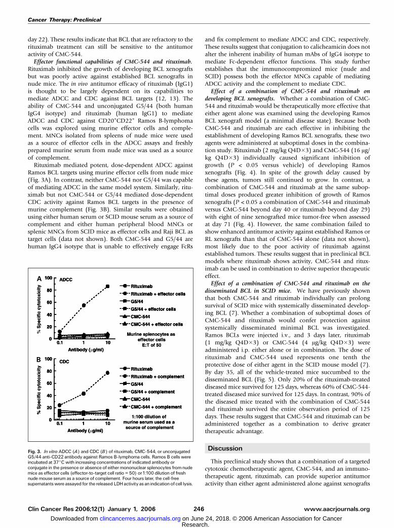
Rituximab mediated potent, dose-dependent ADCC againstRamos BCL targets using murine effector cells from nude mice(Fig. 3A). In contrast, neither CMC-544 nor G5/44 was capableof mediating ADCC in the same model system. Similarly, ritu-ximab but not CMC-544 or G5/44 mediated dose-dependentCDC activity against Ramos BCL targets in the presence ofmurine complement (Fig. 3B). Similar results were obtainedusing either human serum or SCID mouse serum as a source ofcomplement and either human peripheral blood MNCs orsplenic MNCs from SCID mice as effector cells and Raji BCL astarget cells (data not shown). Both CMC-544 and G5/44 arehuman IgG4 isotype that is unable to effectively engage FcRs
and fix complement to mediate ADCC and CDC, respectively.These results suggest that conjugation to calicheamicin does notalter the inherent inability of human mAbs of IgG4 isotype tomediate Fc-dependent effector functions. This study furtherestablishes that the immunocompromized mice (nude andSCID) possess both the effector MNCs capable of mediatingADCC activity and the complement to mediate CDC.
Effect of a combination of CMC-544 and rituximab ondeveloping BCL xenografts. Whether a combination of CMC-544 and rituximab would be therapeutically more effective thateither agent alone was examined using the developing RamosBCL xenograft model (a minimal disease state). Because bothCMC-544 and rituximab are each effective in inhibiting theestablishment of developing Ramos BCL xenografts, these twoagents were administered at suboptimal doses in the combina-tion study. Rituximab (2 mg/kg Q4D�3) and CMC-544 (16 Ag/kg Q4D�3) individually caused significant inhibition ofgrowth (P < 0.05 versus vehicle) of developing Ramosxenografts (Fig. 4). In spite of the growth delay caused bythese agents, tumors still continued to grow. In contrast, acombination of CMC-544 and rituximab at the same subop-timal doses produced greater inhibition of growth of Ramosxenografts (P < 0.05 a combination of CMC-544 and rituximabversus CMC-544 beyond day 40 or rituximab beyond day 29)with eight of nine xenografted mice tumor-free when assessedat day 71 (Fig. 4). However, the same combination failed toshow enhanced antitumor activity against established Ramos orRL xenografts than that of CMC-544 alone (data not shown),most likely due to the poor activity of rituximab againstestablished tumors. These results suggest that in preclinical BCLmodels where rituximab shows activity, CMC-544 and ritux-imab can be used in combination to derive superior therapeuticeffect.
Effect of a combination of CMC-544 and rituximab on thedisseminated BCL in SCID mice. We have previously shownthat both CMC-544 and rituximab individually can prolongsurvival of SCID mice with systemically disseminated develop-ing BCL (7). Whether a combination of suboptimal doses ofCMC-544 and rituximab would confer protection againstsystemically disseminated minimal BCL was investigated.Ramos BCLs were injected i.v., and 3 days later, rituximab(1 mg/kg Q4D�3) or CMC-544 (4 Ag/kg Q4D�3) wereadministered i.p. either alone or in combination. The dose ofrituximab and CMC-544 used represents one tenth theprotective dose of either agent in the SCID mouse model (7).By day 35, all of the vehicle-treated mice succumbed to thedisseminated BCL (Fig. 5). Only 20% of the rituximab-treateddiseased mice survived for 125 days, whereas 60% of CMC-544-treated diseased mice survived for 125 days. In contrast, 90% ofthe diseased mice treated with the combination of CMC-544and rituximab survived the entire observation period of 125days. These results suggest that CMC-544 and rituximab can beadministered together as a combination to derive greatertherapeutic advantage.
Discussion
This preclinical study shows that a combination of a targetedcytotoxic chemotherapeutic agent, CMC-544, and an immuno-therapeutic agent, rituximab, can provide superior antitumoractivity than either agent administered alone against xenografts
Fig. 3. In vitro ADCC (A) and CDC (B) of rituximab, CMC-544, or unconjugatedG5/44 anti-CD22 antibody against Ramos B-lymphoma cells. Ramos B cells wereincubated at 37jC with increasing concentrations of indicated antibody orconjugate in the presence or absence of either mononuclear splenocytes from nudemice as effector cells (effector-to-target cell ratio = 50) or1:100 dilution of freshnude mouse serum as a source of complement. Four hours later, the cell-freesupernatants were assayed for the released LDHactivity as an indicationof cell lysis.
Cancer Therapy: Preclinical
www.aacrjournals.orgClin Cancer Res 2006;12(1) January 1, 2006 246
Research. on June 24, 2018. © 2006 American Association for Cancerclincancerres.aacrjournals.org Downloaded from

of human BCL. CMC-544 is a CD22-targeted cytotoxicchemotherapeutic agent devoid of any effector capability,whereas rituximab is a CD20-targeted immunotherapeuticagent capable of mediating both ADCC and CDC (12, 13).This combination makes use of the CD22-specific cytoreductivecapability of CMC-544 in addition to the effector functionalityof rituximab as well as its direct antiproliferative and apoptoticactivity (23, 24). Because both CMC-544 and rituximab exertantitumor effects by distinct mechanisms, it is reasonable toanticipate that CMC-544 could be combined with rituximab toderive greater therapeutic advantage.
In vitro, rituximab caused f20% inhibition of growth of fourdistinct BCLs. Because the in vitro conditions lacked variouscellular and humoral components required to mediate ADCCand CDC, the observed growth inhibitory effect of rituximabwas most likely due to its direct but weak antiproliferativeactivity against these BCLs (25, 26). In contrast, CMC-544exerted a potent growth-inhibitory effect against the same BCLsattributed to the potent cytotoxic activity of CalichDMHconjugated to anti-CD22 mAb. Combining rituximab withCMC-544 produced an ‘‘additive’’ growth-inhibitory effect asanalyzed by the bliss independence model.
When evaluated in vivo , rituximab was able to significantlyinhibit growth of developing BCL xenografts. However, upondiscontinuation of the rituximab treatment, these tumorsrelapsed again. Unlike its potent inhibitory effect againstdeveloping s.c. BCL xenografts, the activity of rituximab againstestablished s.c. BCL xenografts was modest. In contrast, CMC-544 was able to mediate potent antitumor activity against boththe developing and established BCL xenografts in nude mice(ref. 1 and this study). More importantly, CMC-544 was able tocause regression of BCL that were refractory to prior rituximabtreatment (Fig. 2). Similarly, rituximab was able to inhibitgrowth of disseminated BCLs when administered early duringthe disseminated phase of the disease but not when thedisseminated BCL disease was already established (7). The
preclinical demonstration of antitumor efficacy of CMC-544against rituximab-refractory BCL is relevant to the ongoingclinical trials of CMC-544 as most patients being treated withCMC-544 are likely to have had rituximab-refractory BCL.
We investigated whether the weak activity of rituximabagainst established tumors was related to the lack of host-derived effector activity in immunocompromized mice. Sple-nocytes from both nude and SCID mice were able to mediaterituximab-mediated ADCC (refs. 27, 28 and present study)and murine complement was able to mediate CDC againstrituximab-treated ramos BCL targets. In contrast, CMC-544 wasunable to mediate either of these activities. The lack of ADCCand CDC activities by CMC-544 is consistent with its humanIgG4 isotype that is a poor mediator of either activity.Rituximab-mediated ADCC and CDC activities against theBCL may contribute toward its in vivo antitumor activityobserved in both the s.c. xenograft and disseminated BCLmodels. If the therapeutic effect of rituximab in these models isprimarily dependent on the participation of effector cells, thenit is likely that, with increasing BCL burden in the establisheddisease, the available effector cells are unable to effectivelyeliminate all BCL and, as a result, allow progression of theestablished disease (7). Consistent with this hypothesis is thestudy reported by Bertolini et al. (29), wherein rituximab waseffective in inhibiting the development of BCL but was inactiveagainst established or ‘‘bulky’’ disease. Because the antitumoractivity of rituximab could only be consistently shown againstthe developing BCL xenografts, we evaluated the activity of acombination of CMC-544 and rituximab against developing s.c.or disseminated ramos BCL xenografts. The results of thesestudies show enhanced antitumor activity when the com-pounds were used in combination.
Both the in vitro and in vivo results described above supportthe combined use of CMC-544 with rituximab in B-NHL. Therewas no indication of antagonism between CMC-544 andrituximab in any of the studies. Both the antiproliferative effect
Fig. 4. Effect of a combination of CMC-544 and rituximab on s.c. growth ofdeveloping Ramos BCL xenografts. Ramos BCLs were injected s.c. in nude mice(n = 9 per group) and 5 days later, i.p. treatment with CMC-544 (16 Ag/kg),rituximab (2 mg/kg), or a combination of CMC-544 and rituximab at the abovedoses was initiated and repeated twice 4 days apart (Q4D�3).Tumor growth wasmonitored for up to 72 days.There were no tumor-free mice in the rituximabtreatment group, whereas four of nine mice were tumor-free in the CMC-544treatment group when assessed at day 50. Error bars, average tumor mass F SE.
Fig. 5. Effect of a combination of CMC-544 and rituximab at suboptimal doses onthe survival of SCID mice with developing disseminated Ramos BCL. SCID mice(n = 10 per group) were injected i.v. with Ramos BCL to cause their disseminationand 3 days later, CMC-544 (0.4 Ag/kg), rituximab (1mg/kg), or a combination ofCMC-544 and rituximab at the above doses was administered.The same treatmentswere repeated twice 4 days apart (Q4D�3). Mice in each group were monitored upto125 days for hind limb paralysis or death due to the disseminated disease.
CombinationTherapy of B-NHLwith CMC-544 and Rituximab
www.aacrjournals.org Clin Cancer Res 2006;12(1) January 1, 2006247
Research. on June 24, 2018. © 2006 American Association for Cancerclincancerres.aacrjournals.org Downloaded from

observed in vitro as well as the antitumor activity exerted inboth of the tumor models suggest that combining these twoagents may produce, at minimum, an additive inhibitory effecton BCL growth. The dose-limiting adverse event profile ofCMC-544 is currently being determined in ongoing phase Iclinical trials. Combining subtherapeutic doses of CMC-544with rituximab may derive the same antilymphoma effectachievable with higher doses of CMC-544 without precipitatingdose-limiting toxicity. How rituximab enhances the activity ofCMC-544 in vivo is not known. Rituximab binds CD20 on BCLand causes its rapid segregation into lipid rafts (30). What effectrituximab-mediated CD20 segregation into lipid rafts has onthe internalization of CMC-544 by BCL has not been studied. Ifrituximab binding to BCL resulted in the increased expressionof CD22, it may allow quantitatively greater accumulation ofCMC-544 on the surface of BCL leading to its internalization.Such an effect could conceivably result in the increasedintracellular delivery of the conjugated calicheamicin, leadingto higher degree of cytotoxicity. However, we were unable toreproducibly show in vitro any meaningful increase in thesurface expression of CD22 on rituximab-treated BCL.4
Several studies have shown that rituximab can be combinedwith cytotoxic agents and induce a synergistic antiproliferativeeffect in vitro. Rituximab sensitized B-NHL lines to variouslympholytic agents, such as paclitaxel, dexamethasone, fludar-abine, and other cytotoxic agents, causing either synergistic oradditive growth inhibitory activity (10, 26, 31–38). The abilityof rituximab to sensitize BCL to cytotoxic agents has been partlyattributed to the nullification of the apoptosis-resistingmolecular components of the Bcl2/Bcl-Xl family of molecules(31). In addition, rituximab is also used in radioimmunother-apy with radiolabeled anti-CD20 ibritumomab ([90Y]ibritumo-mab tiuxetan/Zevalin; ref. 39). Given its unique mechanism(s)of antitumor activity, rituximab can be combined with variousagents to improve their combined therapeutic potential. Theantitumor activity of a combination of CMC-544 and rituximabas shown in the current study is consistent with this rationale.Of interest in this context is the recent demonstration ofsignificant clinical activity of a combination of a CD22-targetedantibody, epratuzumab, and rituximab in B-NHL patients (40).
Epratuzumab and the targeting antibody in CMC-544, G5/44,recognize distinct epitopes on human CD22 (19). G5/44 is ahumanized IgG4 antibody and as shown in this study is devoidof ADCC and CDC activities. Epratuzumab is a humanizedIgG1 antibody and, like rituximab, may possess both theseeffector capabilities (41). Unconjugated G5/44 is devoid of anyantitumor activity against either s.c. or disseminated BCLxenografts (1, 7, 19) and the strong antitumor activity of CMC-544 can thus be attributed to the targeted delivery of conjugatedcalicheamicin (1, 7, 19). Irrespective of the molecular detailsunderlying the supra-additive therapeutic effect of a combina-tion of suboptimal CMC-544 and rituximab, its demonstrationhere provides preclinical justification for its clinical evaluation.
A therapeutic combination of rituximab and CHOP isextensively used in the treatment of B-NHL (15–18). A majorreason that rituximab is used extensively in B-NHL therapy is itssafety profile that allows it to be combined with various cytotoxicchemotherapeutic agents without increasing the adverse eventprofile of the drug combination. CHOP is a first-line cytor-eductive combination chemotherapy used extensively in thetreatment of both low-grade indolent and intermediate-gradeaggressive B-NHL. Thus, CMC-544, as a targeted chemothera-peutic, may be added to the existing treatment regimen of CHOPdepending on the nature of the disease. The CHOP combinationincludes two DNA-targeted agents, cyclophosphamide anddoxorubicin, that are the two most active agents against diffuselarge BCL. It may be advantageous to add a targeted chemother-apeutic agent, such as CMC-544, to the CHOP combination togain additional therapeutic benefit, especially during thetreatment of aggressive BCL. Wider application of CMC-544 inB-NHL will largely depend on its clinical efficacy and safetyprofile. The addition of rituximab may not change the safetyprofile of the treatment combination of CMC-544 and rituximabwhile anticipating greater antitumor activity. This preclinicaldemonstration of therapeutically advantageous combination ofCMC-544 and rituximab strongly supports its clinical evaluationin patients with B-NHL.
Acknowledgments
We thank Jorge Quiroz and Fred Immerman from Wyeth Biometric Researchfor statistical analysis; Michael Cinque for his assistance with in vivo studies; andDrs. Janis Upeslacis, Ken Jacobs, and Eliel Bayever for discussions, suggestions,and encouragement.4 D. Armellino, unpublished observation.
Cancer Therapy: Preclinical
www.aacrjournals.orgClin Cancer Res 2006;12(1) January 1, 2006 248
References1. DiJosephJF, Armellino DC, Boghaert ER, et al. Anti-
body-targeted chemotherapy with CMC-544: aCD22-targeted immunoconjugate of calicheamicin forthe treatment of B lymphoid malignancies. Blood2004;103:1807^14.
2. Hamann P, Hinman L, Beyer C, et al. An anti-CD33antibody-calicheamicin conjugate for treatment ofacute myeloid leukemia. Choice of linker. BioconjugChem 2002;3:40^6.
3. Damle NK. Tumor-targeted chemotherapy usingimmunoconjugates of calicheamicin. Expert Opin BiolTher 2004;4:1445^52.
4. Lee M, DunneT, Chang C, et al. Calicheamicins, anovel family of antibiotics. 4. Structural elucidations ofcalicheamicins. J Am Chem Soc1992;114:985^7.
5. Zein N, Sinha A, McGahrenW, Ellestad G. Calichea-micin g1
I: an antitumor antibiotic that cleaves double-stranded DNA site specifically. Science 1988;240:1198^201.
6. Thorson J, Sievers E, Ahlert J, et al. Understandingand exploiting nature’s chemical arsenal: the past,present and future of calicheamicin research. CurrPharm Des 2000;6:1841^79.
7. DiJoseph JF, Goad ME, Dougher MM, et al. Potentand specific anti-tumor efficacy of CMC-544, aCD22-targeted immunoconjugate of calicheamicin,against systemically disseminated B-cell lymphoma.Clin Cancer Res 2004;10:8620^9.
8. Grillo-Lopez A. Rituximab (Rituxan/MabThera): thefirst decade (1993-2003). Expert RevAnticancerTher2003;3:767^79.
9. Coiffier B. Effective immunochemotherapy for ag-gressive non-Hodgkin’s lymphoma. Semin Oncol2004;31:7^11.
10. Jazirehi AR, Bonavida B. Cellular and molecularsignal transduction pathways modulated by rituxi-mab (rituxan, anti-CD20 mAb) in non-Hodgkin’slymphoma: implications in chemosensitization
and therapeutic intervention. Oncogene 2005;24:2121^43.
11. BoyeJ, ElterT, Engert A. An overview of the currentclinical use of the anti-CD20 monoclonal antibodyrituximab. Ann Oncol 2003;14:520^35.
12. Uchida J, Hamaguchi Y, Oliver JA, et al. The innatemononuclear phagocyte network depletes B lympho-cytes through Fc receptor-dependent mechanismsduring anti-CD20 antibody immunotherapy. J ExpMed 2004;199:1659^69.
13. Gaetano ND, Cittera E, Nota R, et al. Com-plement activation determines the therapeutic ac-tivity of rituximab in vivo. J Immunol 2003;171:1581^7.
14. Manches O, Lui G, Chaperot L, et al. In vitro mech-anisms of action of rituximab on primary non-Hodgkinlymphomas. Blood 2003;101:949^54.
15. Czuczman MS, Grillo-Lopez AJ, White CA, et al.Treatment of patients with low-grade B-cell lymphoma
Research. on June 24, 2018. © 2006 American Association for Cancerclincancerres.aacrjournals.org Downloaded from

with the combinationof chimeric anti-CD20monoclo-nal antibody and CHOP chemotherapy. J Clin Oncol1999;17:268^76.
16. Non-Hodgkin’s lymphoma. Natl Compr CancerNetw Clin Pract Guidel Oncol 2005;1:1^62.
17. Coiffier B, Herbrecht R, Tilly H, et al. GELAstudy comparing CHOP and R-CHOP in elderlypatients with DLCL: 3-year median follow-upwith an analysis according to co-morbidity factors[abstract 2395]. Proc Am Soc Clin Oncol 2003;22:596.
18. Pfreundschuh MG,Trumper L, Ma D, et al.Random-ized intergroup trial of first-line treatment for patientsV60 years with diffuse large B-cellnon-Hodgkin’s lym-phoma (DLBCL) with a CHOP-like regimen with orwithout the anti-CD20 antibody rituximabOearlystopping after the first interim analysis [abstract6500]. ProcAmSocClinOncol 2004;23:556.
19. DiJoseph JF, Popplewell AG,Tickle S, et al. Anti-body-targeted chemotherapy of B-cell lymphomausing calicheamicin conjugated to murine or human-ized antibody against CD22. Cancer Immunol Immun-other 2005;54:11^24.
20. GrecoWR, Bravo G, ParsonsJC.The search for syn-ergy: a critical review from a response surface per-spective. Pharmacol Rev1995;47:331^85.
21. Conover WJ, Iman RL. Rank transformation as abridge between parametric and nonparametric statis-tics. Am Stat1981;35:124^9.
22. Stein R, Qu Z, Chen S, et al. Characterization of anew humanized anti-CD20 monoclonal antibody,IMMU-106, and its use in combination with the hu-manized anti-CD22 antibody, epratuzumab, for thetherapy of non-Hodgkin’s lymphoma. Clin Cancer Res2004;10:2868^78.
23. Hernandez-Ilizaliturri FJ, JupudyV, OstbergJ, et al.Neutrophils contribute to the biological antitumoractivity of rituximab in a non-Hodgkin’s lymphoma
severe combined immunodeficiency mouse model.Clin Cancer Res 2003;9:5866^73.
24. Clynes RA,TowersTL,Tresta LG, RavetchJV. Inhib-itory Fc receptors modulate in vivo cytotoxicityagainst tumor targets. Nat Med 2000;6:443^6.
25. Shan D, Ledbetter JA, Press OW. Apoptosis ofmalignant human B cells by ligation of CD20 withmonoclonal antibodies. Blood 1998;91:1644^52.
26. Rose AL, Smith BE, Maloney DG. Glucocorti-coids and rituximab in vitro : synergistic direct anti-proliferative and apoptotic effects. Blood 2002;100:1765^73.
27. Dorshkind K, Pollack SB, Bosma MJ, Phillips RA.Natural killer cells are present in mice with severe com-bined immunodeficiency (scid). J Immunol 1985;134:3798^801.
28. Greiner DL, Hesselton RA, Schultz LD. SCIDmousemodels of human stem cell engraftment. Stem Cells1998;16:166^77.
29. Bertolini F, Fusetti L, Manciso P, et al. Endostatin,an antiangiogenic drug, induces tumor stabilization af-ter chemotherapy or anti-CD20 therapy in NOD/SCIDmouse model of humanhigh-grade non-Hodgkin lym-phoma. Blood 2000;96:282^7.
30. Semac I, Palomba C, Kulangara K, et al. Anti-CD20therapeutic antibody rituximab modifies the functionalorganization of rafts/microdomains of B lymphomacells. Cancer Res 2003;63:534^40.
31. Jazirehi AR, Gan X-H, De Vos S, et al. Rituximab(anti-CD20) selectively modifies Bcl-XL and apopto-sis protease activating factor-1 (APAF-1) expressionand sensitizes human non-Hodgkin’s lymphoma B celllines to paclitaxel-induced apoptosis. Mol CancerTher2003;2:1183^93.
32. Alas S, Bonavida B, Emmanouilides C. Potentiationof fludarabine cytotoxicity on non-Hodgkin’s lympho-ma by pentoxifylline and rituximab. Anticancer Res2000;20:2961^6.
33. Alas S, Ng CP, Bonavida B. Rituximab modifies thecisplatin-mitochondrial signaling pathway, resulting inapoptosis in cisplatin-resistant non-Hodgkin’s lym-phoma. Clin Cancer Res 2002;8:836^45.
34. Gaetano ND, XiaoY, Erba E, et al. Synergism be-tween fludarabine and rituximab revealed in a follicularlymphoma cell line resistant to the cytotoxic activity ofeither drug alone. BrJHaematol 2001;114:800^11.
35. Chow KU, Sommerlad WD, Boehrer S, et al. Anti-CD20 antibody (IDEC-C2B8, rituximab) enhances ef-ficacy of cytotoxic drugs on neoplastic lymphocytesin vitro : role of cytokines, complement, and caspases.Haematologica 2002;87:33^43.
36. Emmanouilides C, Jazirehi AR, Bonavida B. Rituxi-mab-mediated sensitization of B-non-Hodgkin’s lym-phoma (NHL) to cytotoxicity induced by paclitaxel,gemcitabine, and vinorelbine. Cancer Biother Radio-pharm 2002;17:621^30.
37. Gopal AK, Pagel JM, Hedin N, Press OW. Fenreti-nide enhances rituximab-induced cytotoxicity againstB-cell lymphoma xenografts through a caspase-dependent mechanism. Blood 2004;103:3516^20.
38. Ghetie M, Bright H,Vitetta ES. Homodimers but notmonomers of Rituxan (chimeric anti-CD20) induceapoptosis in human B-lymphoma cells and synergizewith a chemotherapeutic agent and an immunotoxin.Blood 2001;97:1392^8.
39. White CA. Rituxan immunotherapy and zevalinradioimmunotherapy in the treatment of non-Hodg-kin’s lymphoma. Curr Pharm Biotechnol 2003;4:221^38.
40. LeonardJP, Coleman M, KetasJ, et al. Combinationantibody therapy with epratuzumab and rituximab inrelapsed or refractory non-Hodgkin’s lymphoma.JClin Oncol 2005;23:5044^51.
41. Coleman M, Goldenberg DM, Siegel AB, et al. Epra-tuzumab: targeting B malignancies through CD22.Blood 2003;9:3991^4s.
CombinationTherapy of B-NHLwith CMC-544 and Rituximab
www.aacrjournals.org Clin Cancer Res 2006;12(1) January 1, 2006249
Research. on June 24, 2018. © 2006 American Association for Cancerclincancerres.aacrjournals.org Downloaded from

2006;12:242-249. Clin Cancer Res John F. DiJoseph, Maureen M. Dougher, Lyka B. Kalyandrug, et al. Non-Hodgkin's B-Cell LymphomaImmunoconjugate of Calicheamicin, and Rituximab against(Inotuzumab Ozogamicin), a CD22-Targeted Cytotoxic Antitumor Efficacy of a Combination of CMC-544
Updated version
http://clincancerres.aacrjournals.org/content/12/1/242
Access the most recent version of this article at:
Cited articles
http://clincancerres.aacrjournals.org/content/12/1/242.full#ref-list-1
This article cites 39 articles, 20 of which you can access for free at:
Citing articles
http://clincancerres.aacrjournals.org/content/12/1/242.full#related-urls
This article has been cited by 13 HighWire-hosted articles. Access the articles at:
E-mail alerts related to this article or journal.Sign up to receive free email-alerts
Subscriptions
Reprints and
To order reprints of this article or to subscribe to the journal, contact the AACR Publications
Permissions
Rightslink site. (CCC)Click on "Request Permissions" which will take you to the Copyright Clearance Center's
.http://clincancerres.aacrjournals.org/content/12/1/242To request permission to re-use all or part of this article, use this link
Research. on June 24, 2018. © 2006 American Association for Cancerclincancerres.aacrjournals.org Downloaded from