dna the universal code of life. early history 1869: friederich mieschner isolates “nuclein” from...
TRANSCRIPT
DNAThe Universal Code of Life
Early History
• 1869: Friederich Mieschner isolates “nuclein” from nuclei of cells. His student Richard Altman later renames the substance “nucleic acid.”
• Mid 1800s: Biochemists identify two distinct nucleic acids.
• 1929: Phoebus Levine identifies four distinct bases in DNA.
Heredity as a Science
• Genetics arose as a new science in the late 19th and early 20th centuries, spurred by questions raised by Darwin’s On the Origin of Species:
• Are there patterns to inheritance?
• Are traits handed on intact (particle theory) or blended together in each generation (blending theory)?
Mendel’s Answers
• Gregor Mendel’s work was rediscovered in 1900, answering both questions:
• Inheritance of many traits follows predictable patterns.
• Traits are handed on intact via some kind of particle: “elementen.”
Hereditary Molecule?
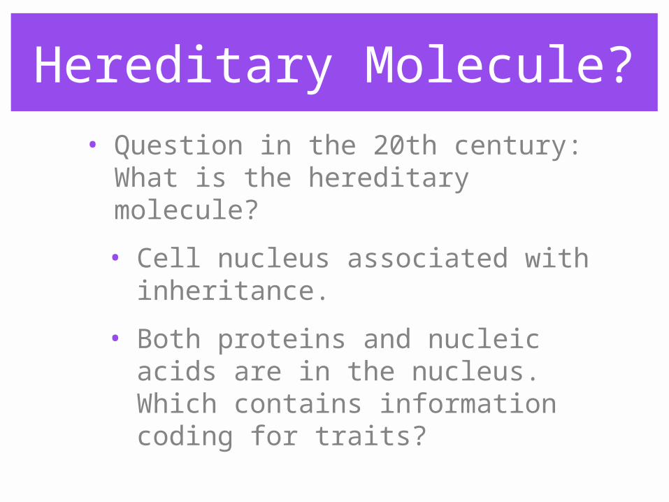
• Question in the 20th century: What is the hereditary molecule?
• Cell nucleus associated with inheritance.
• Both proteins and nucleic acids are in the nucleus. Which contains information coding for traits?
Protein or DNA?
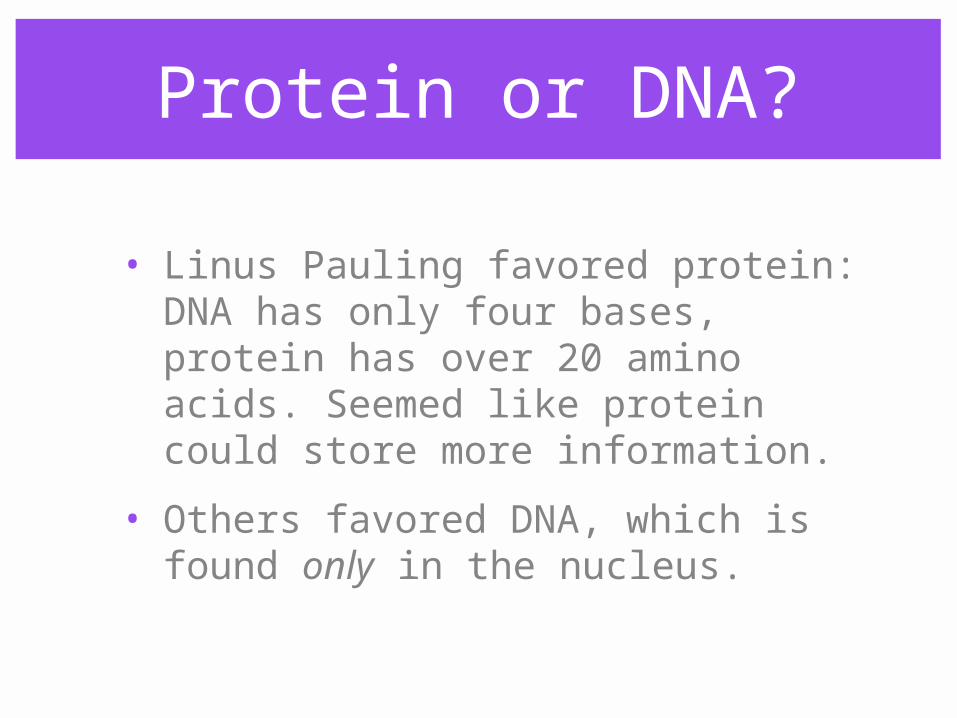
• Linus Pauling favored protein: DNA has only four bases, protein has over 20 amino acids. Seemed like protein could store more information.
• Others favored DNA, which is found only in the nucleus.
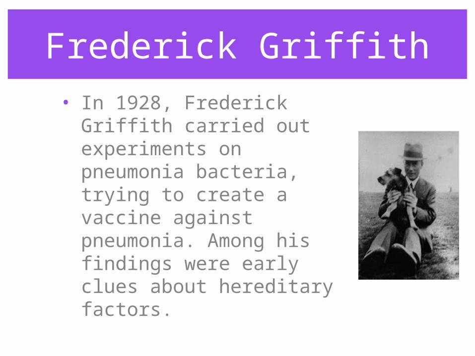
Frederick Griffith
• In 1928, Frederick Griffith carried out experiments on pneumonia bacteria, trying to create a vaccine against pneumonia. Among his findings were early clues about hereditary factors.
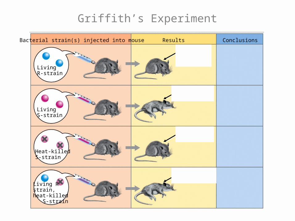
LivingR-strain
LivingS-strain
Heat-killedS-strain
Living Rstrain,heat-killed S-strain
Bacterial strain(s) injected into mouse Results Conclusions
Griffith’s Experiment
Oswald Avery
• Avery learned of Griffith’s experiment and thought it might hold a clue to the identity of the hereditary molecule.
• Avery isolated carbohydrates, proteins, lipids, and nucleic acids from the bacteria to discover which, if any, would transform the non-virulent R-strain bacteria.
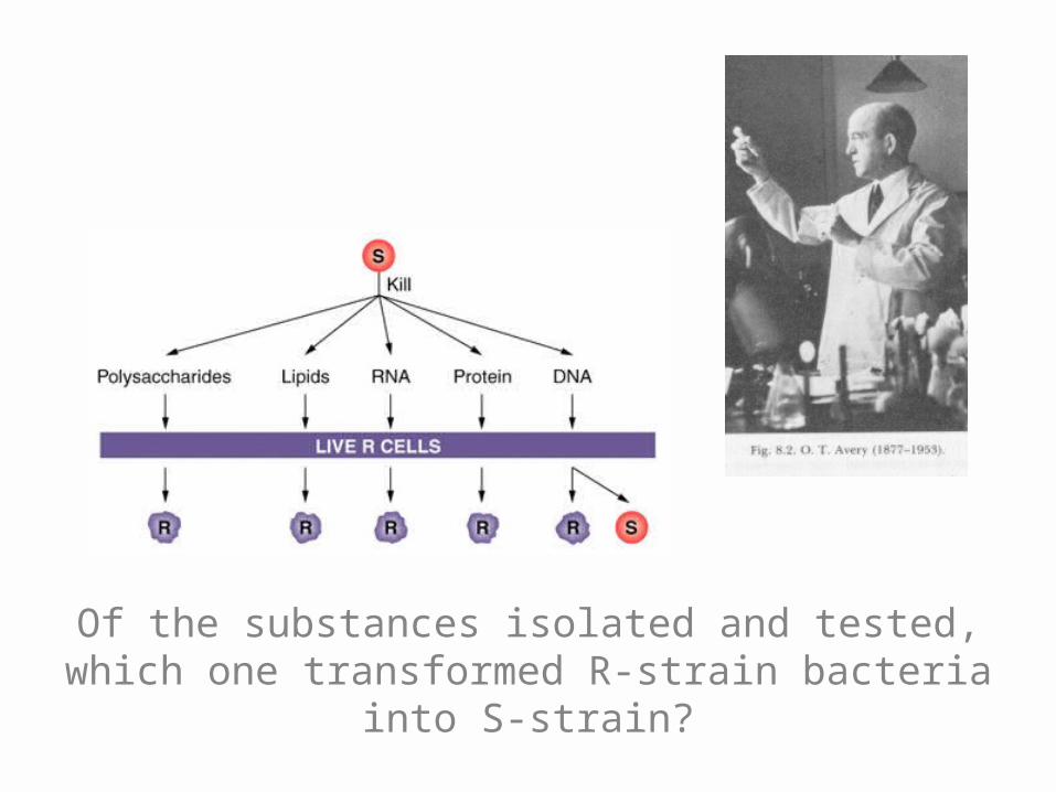
Of the substances isolated and tested, which one transformed R-strain bacteria into S-strain?
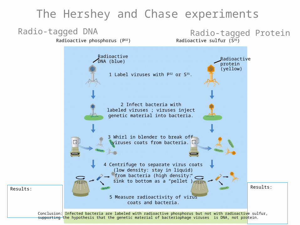
Hershey & Chase
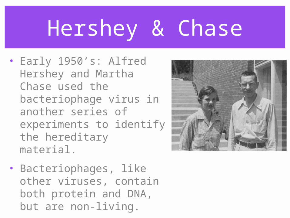
• Early 1950’s: Alfred Hershey and Martha Chase used the bacteriophage virus in another series of experiments to identify the hereditary material.
• Bacteriophages, like other viruses, contain both protein and DNA, but are non-living.
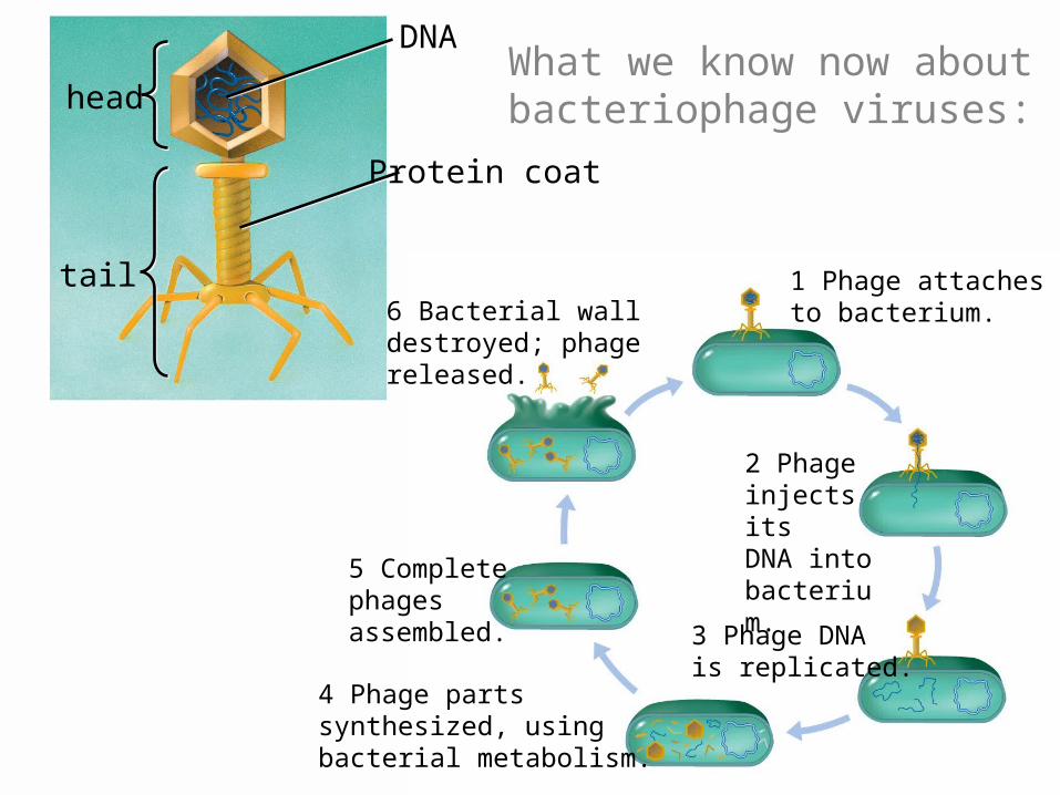
DNA
tail
head
Protein coat
What we know now about bacteriophage viruses:
1 Phage attachesto bacterium.
2 Phage injects itsDNA intobacterium.
3 Phage DNAis replicated.
4 Phage partssynthesized, usingbacterial metabolism.
5 Completephages assembled.
6 Bacterial walldestroyed; phagereleased.
Results:
5 Measure radioactivity of viruscoats and bacteria.
1 Label viruses with P32 or S35.
2 Infect bacteria withlabeled viruses ; viruses injectgenetic material into bacteria.
3 Whirl in blender to break offviruses coats from bacteria.
4 Centrifuge to separate virus coats(low density: stay in liquid)from bacteria (high density:sink to bottom as a “pellet”)
Results:
Radioactive sulfur (S35)Radioactive phosphorus (P32)
RadioactiveDNA (blue) Radioactive
protein(yellow)
Conclusion: Infected bacteria are labeled with radioactive phosphorus but not with radioactive sulfur,supporting the hypothesis that the genetic material of bacteriophage viruses is DNA, not protein.
Radio-tagged DNA Radio-tagged Protein
The Hershey and Chase experiments
DNA Structure?
• While many research teams were trying to discover the hereditary molecule, other researchers were working to discover the nature of DNA.
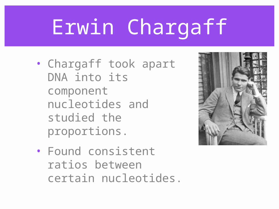
Erwin Chargaff
• Chargaff took apart DNA into its component nucleotides and studied the proportions.
• Found consistent ratios between certain nucleotides.
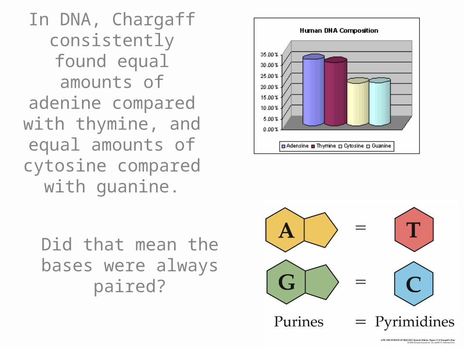
In DNA, Chargaff consistently found equal amounts of adenine compared with thymine, and equal amounts of
cytosine compared with guanine.
Did that mean the bases were always
paired?
Franklin and Wilkins
• Rosalind Franklin worked in Maurice Wilkins’ lab in the late 1940s, using X-ray crystalography to find clues about the structure of DNA.
• Franklin’s images were the first to suggest a helical structure.

The X-shape on the radiograph was characteristic of helical molecules. Franklin also measured distances
between bases and other dimensions using her images.
Watson and Crick
• James Watson and Francis Crick worked at the same time as Franklin and Wilkins.
• Applying Chargaff’s rule, they concluded that A pairs with T, C with G.
• Used their knowledge of molecular geometry to try to discover the structure of DNA.
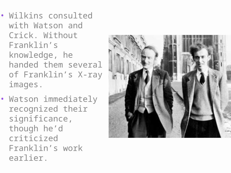
• Wilkins consulted with Watson and Crick. Without Franklin’s knowledge, he handed them several of Franklin’s X-ray images.
• Watson immediately recognized their significance, though he’d criticized Franklin’s work earlier.
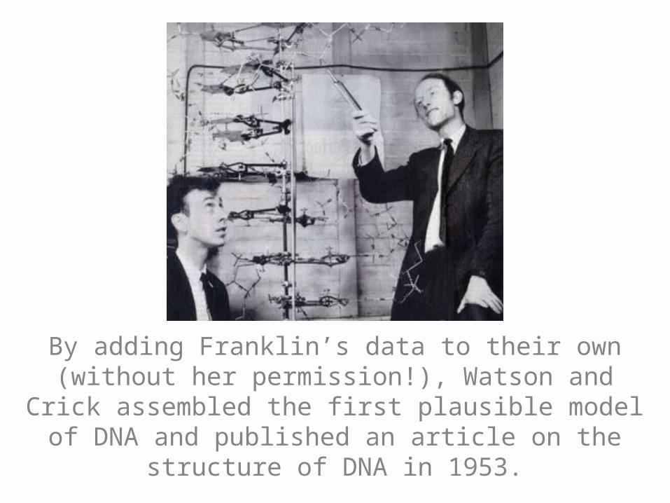
By adding Franklin’s data to their own (without her permission!), Watson and Crick assembled
the first plausible model of DNA and published an article on the structure of DNA in 1953.
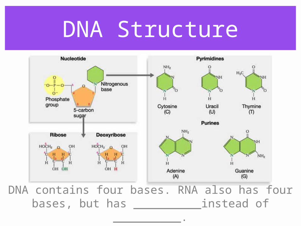
DNA Structure
DNA contains four bases. RNA also has four bases, but has __________instead of __________.
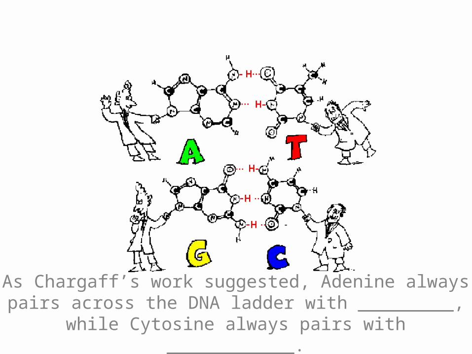
As Chargaff’s work suggested, Adenine always pairs across the DNA ladder with _________, while Cytosine
always pairs with ____________.
5’ end
3’ end
1’
2’3’
4’
5’1 2
345
6
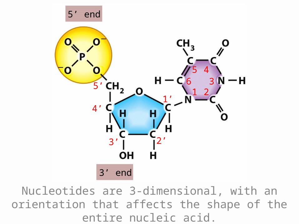
Nucleotides are 3-dimensional, with an orientation that affects the shape of the entire nucleic acid.
5’ end
3’ end
1’
2’3’
4’
5’
1 23
456
789
1’
2’3’
4’
5’ 1 23
456
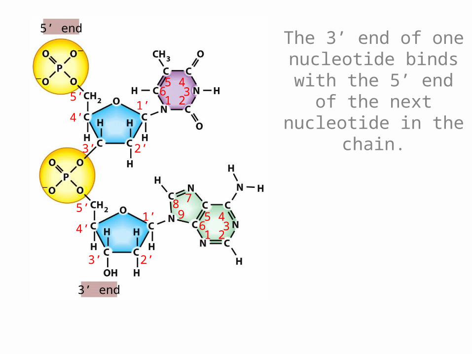
The 3’ end of one nucleotide binds with
the 5’ end of the next nucleotide in
the chain.
5’end
3’end
3’ end
5’end
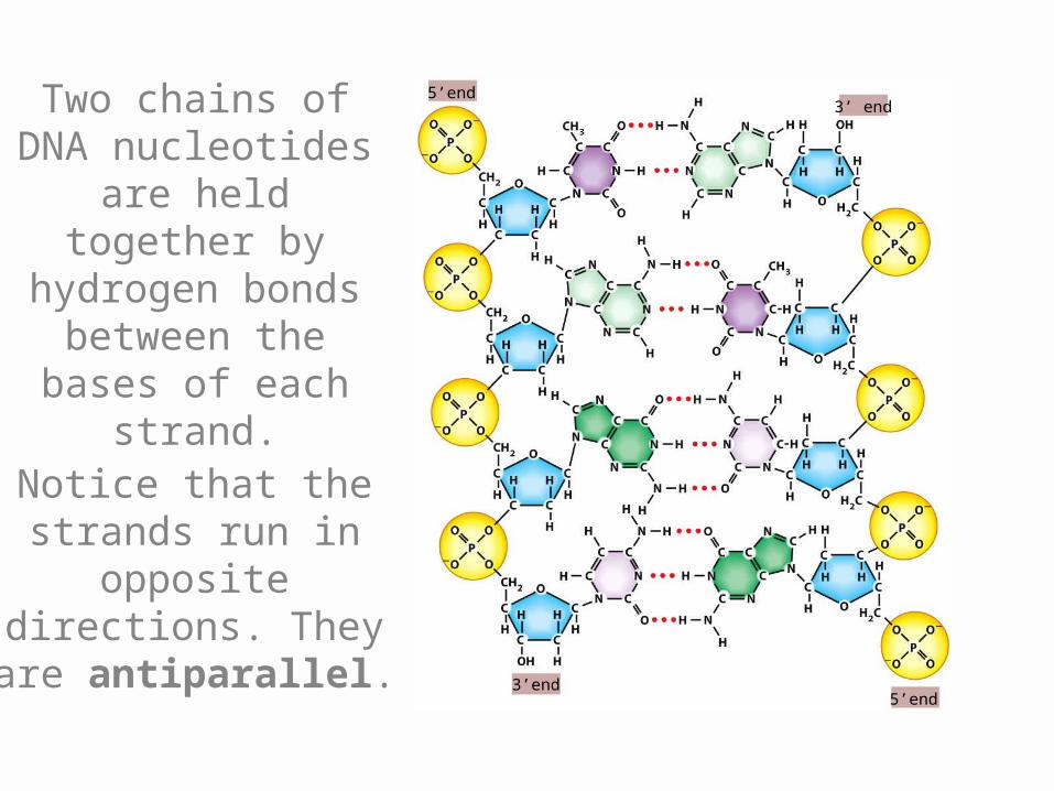
Two chains of DNA nucleotides are held together
by hydrogen bonds between
the bases of each strand.
Notice that the strands run in
opposite directions. They are
antiparallel.
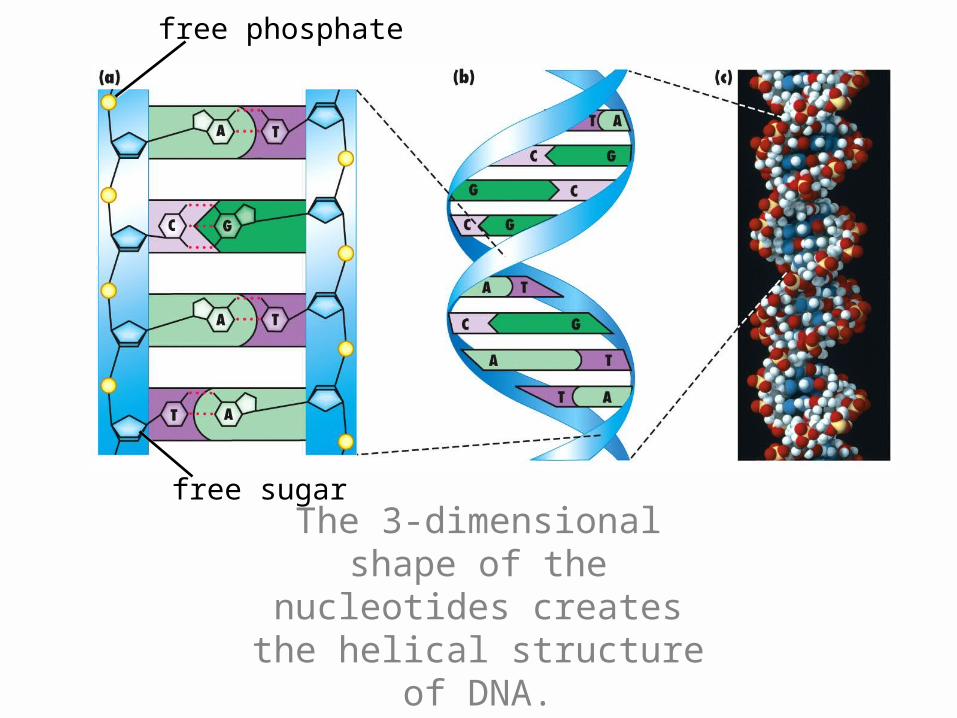
free phosphate
free sugar
The 3-dimensional shape of the nucleotides creates
the helical structure of DNA.
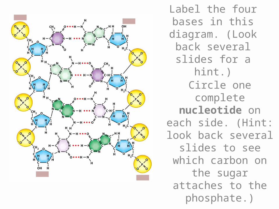
Label the four bases in this diagram.
(Look back several slides for a hint.)
Circle one complete nucleotide on
each side. (Hint: look back several
slides to see which carbon on the sugar
attaches to the phosphate.)
DNA Replication
• When cells divide, the two resulting daughter cells must have exactly the same DNA as the original cell.
• Therefore, before cell division happens, the cell must replicate (copy) its DNA.
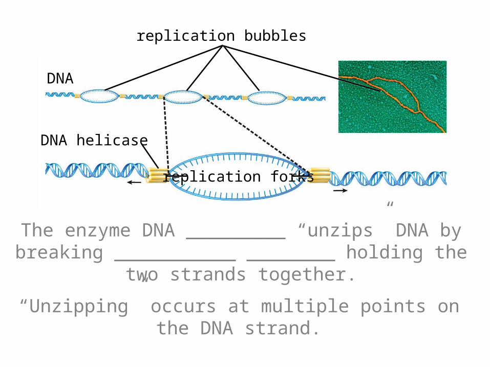
replication forks
replication bubbles
DNA
DNA helicase
The enzyme DNA _________ “unzips” DNA by breaking ___________ ________ holding the two
strands together.
“Unzipping” occurs at multiple points on the DNA strand.
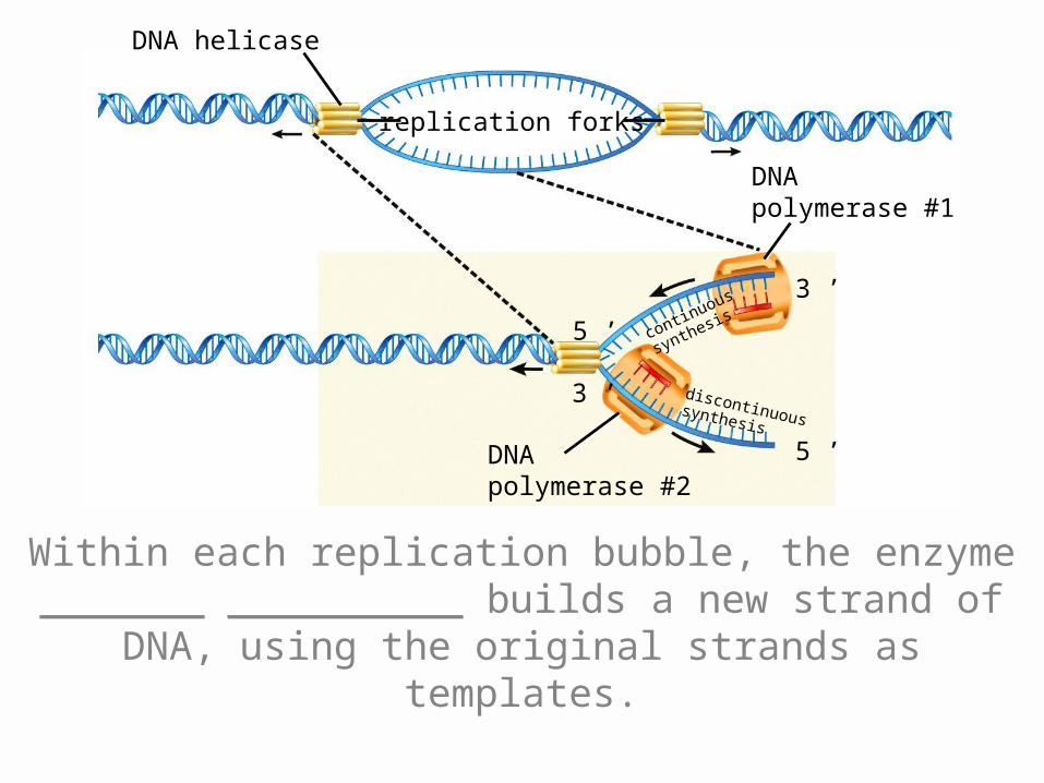
replication forks
DNA helicase
DNApolymerase #2
DNApolymerase #1
5 ’
3 ’
5 ’
3 ’
continuous
synthesis
discontinuoussynthesis
Within each replication bubble, the enzyme _______ __________ builds a new strand of DNA, using the
original strands as templates.
DNApolymerase #2
DNApolymerase #1
5 ’
3’5 ’
3 ’
continuous
synthesis
discontinuoussynthesis
5 ’3 ’
5 ’3 ’
3 ’
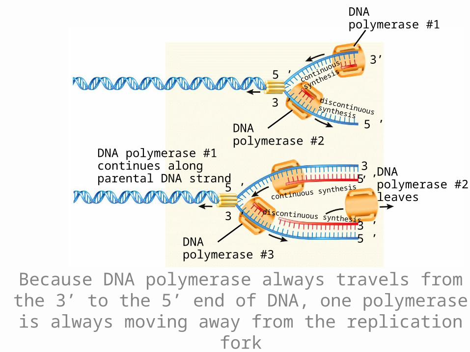
5 ’continuous synthesis
discontinuous synthesis
DNApolymerase #2leaves
DNApolymerase #3
DNA polymerase #1continues alongparental DNA strand
Because DNA polymerase always travels from the 3’ to the 5’ end of DNA, one polymerase is always
moving away from the replication fork
5 ’3 ’
5 ’3 ’
3 ’
5 ’continuous synthesis
discontinuous synthesis
DNApolymerase #2leaves
DNApolymerase #3
DNA polymerase #1continues alongparental DNA strand
5 ’3 ’
5 ’3 ’
3 ’
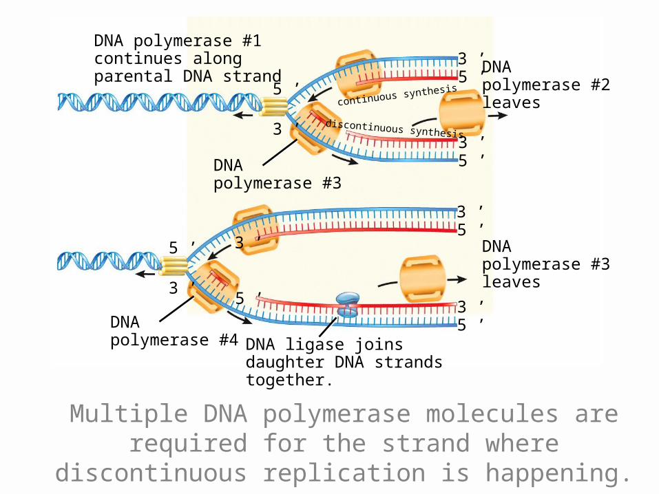
5 ’ DNApolymerase #3leaves
DNApolymerase #4
3 ’
5 ’
DNA ligase joinsdaughter DNA strandstogether.
Multiple DNA polymerase molecules are required for the strand where discontinuous replication is
happening.
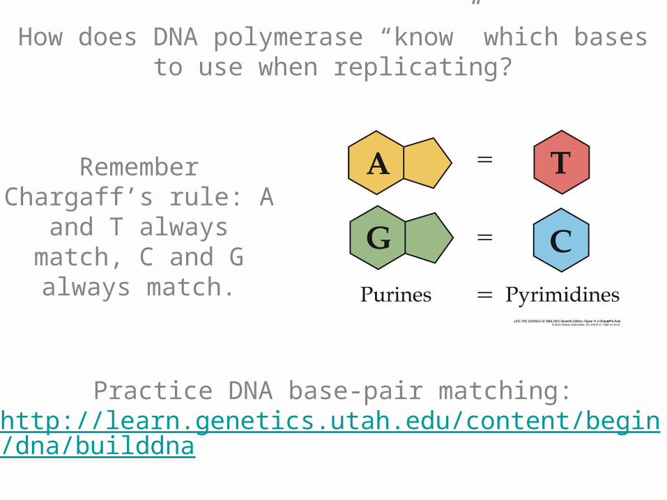
How does DNA polymerase “know” which bases to use when replicating?
Remember Chargaff’s rule: A and T always
match, C and G always match.
Practice DNA base-pair matching:http://learn.genetics.utah.edu/content/begin/dna/builddna
Mutations
• Though many enzymes patrol your DNA, looking for replication errors, some errors do creep in.
• Most cells with a DNA error will die. A few may turn cancerous.
• If mutated cells are sex cells, the mutation can be passed on and will affect all cells in the offspring.
• Mutations may be harmful, helpful, or neutral.
• Harmful mutations result in genetic disease or death.
• Helpful mutations increase evolutionary “fitness” (i.e. having offspring).
• Neutral mutations neither help nor harm at the present.
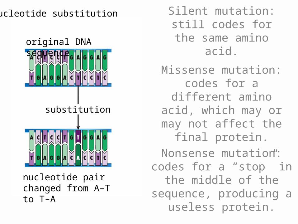
original DNA sequence
Nucleotide substitution
nucleotide pair changed from A–T to T–A
substitution
Silent mutation: still codes for the same
amino acid.
Missense mutation: codes for a different
amino acid, which may or may not affect the
final protein.
Nonsense mutation: codes for a “stop” in the middle
of the sequence, producing a useless
protein.
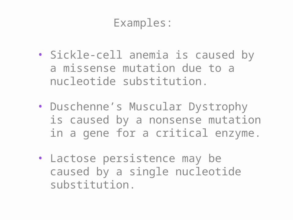
Examples:
• Sickle-cell anemia is caused by a missense mutation due to a nucleotide substitution.
• Duschenne’s Muscular Dystrophy is caused by a nonsense mutation in a gene for a critical enzyme.
• Lactose persistence may be caused by a single nucleotide substitution.
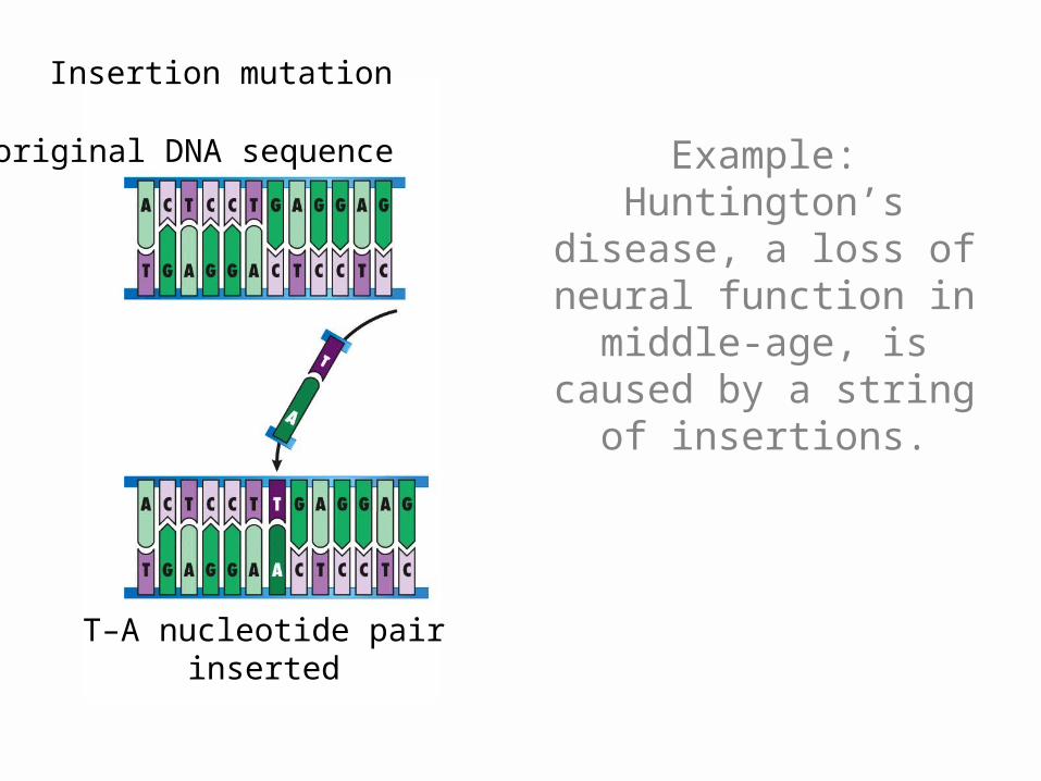
original DNA sequence
T–A nucleotide pairinserted
Insertion mutation
Example: Huntington’s disease, a loss of neural function in middle-age, is caused by a string of
insertions.
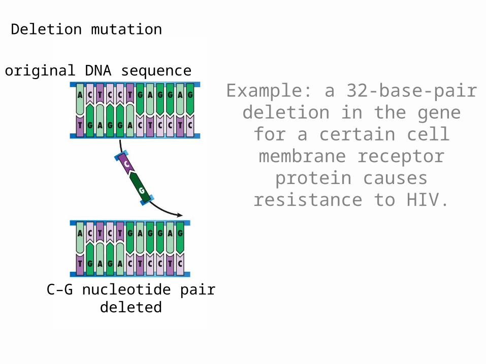
Deletion mutation
C–G nucleotide pairdeleted
original DNA sequence
Example: a 32-base-pair deletion in the gene for a
certain cell membrane receptor protein causes
resistance to HIV.
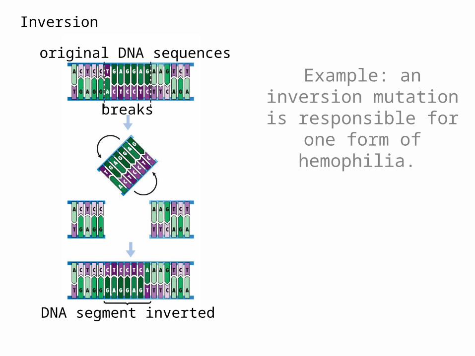
original DNA sequences
DNA segment inverted
Inversion
breaks
Example: an inversion mutation is responsible
for one form of hemophilia.
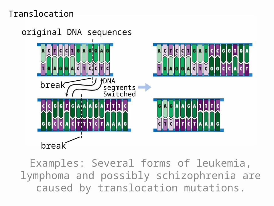
original DNA sequences
break
Translocation
break DNAsegmentsSwitched
Examples: Several forms of leukemia, lymphoma and possibly schizophrenia are
caused by translocation mutations.
What causes mutations?
http://learn.genetics.utah.edu/archive/sloozeworm/mutationbg.html
Recap
• DNA is a nucleic acid which contains coded hereditary information.
• The base-pairing rule helps the information in DNA accurate.
• All cells in the body have the same DNA containing the same information. DNA must be replicated before cell division.