dissertation identification and characterization of kif1bβ
TRANSCRIPT

Dissertation
Identification and characterization of Kif1bβ and hsSASS-6 as
novel disease genes for two hereditary neurological disorders
submitted by
Verena RUPP, BSc. MSc.
for the Academic Degree of
Doctor of Philosophy (PhD)
at the
Medical University of Graz
Institute of Human Genetics
under the Supervision of
Assoz. Prof. Priv.-Doz. Mag. Dr. Christian WINDPASSINGER
2018
I
Statutory Declaration
I hereby declare that this dissertation is my own original work and that I have fully
acknowledged by name all of those individuals and organizations that have
contributed to the research for this dissertation. Due acknowledgement has been
made in the text to all other material used. Throughout this dissertation and in all
related publications I followed the “Standards of Good Scientific Practice and
Ombuds Committee at the Medical University of Graz”.
Please note that parts of this thesis have been published in ‘Human Molecular
Genetics’ (2014).
KHAN, M.A.*, RUPP, V.M.*, ORPINELL, M.*, HUSSAIN, M.S., ALTMULLER, J.,
STEINMETZ, M.O., ENZINGER, C., THIELE, H., HOHNE, W., NURNBERG, G.,
BAIG, S.M., ANSAR, M., NURNBERG, P., VINCENT, J.B., SPEICHER, M.R.,
GONCZY, P. and WINDPASSINGER, C., 2014. A missense mutation in the PISA
domain of HsSAS-6 causes autosomal recessive primary microcephaly in a large
consanguineous Pakistani family. Human molecular genetics, 23(22). pp. 5940-
5949.
* Contributed equally
This work was only possible with the contribution of following people:
Institute of Human Genetics, Medical University of Graz:
Christian Windpassinger, Peter Kroisel, Michael Speicher: recruitment of
patients, supervision and project leaders
Department of Neurology, Medical University of Graz:
Christian Enzinger: analysis of MRI data from microcephaly patients

II
Institute of Molecular Biology and Biochemistry, Medical University of Graz:
Ernst Malle, Chintan Navinchandra Koyani: Western Blot and Pull-down assays
for Kif1B
Roland Malli: Kif1B microscopy
UPGON, EPFL Lausanne:
Pierre Gönczy and Meritxell Orpinell Mercadé: supervision and cellular
localization studies for hsSAS-6
Gomal Centre of Biochemistry and Biotechnology, Gomal University D.I.Khan:
Muzammil Khan: Recruitment of microcephaly patients
Department of Biochemistry, Quaid-i-Azam University:
Muhammad Ansar: Recruitment of microcephaly patients
Laboratory of Bimolecular Research, Paul Scherrer Institute Villigen:
Michel Steinmetz: generation of protein models for SAS-6
Cologne Center of Genomics:
Peter Nürnberg, Janine Altmüller, Holger Thiele, Gudrun Nürnberg: NGS analysis
of microcephaly family
Human molecular Genetics Laboratory, NIBGE Faisalabad:
Shahid M. Baig: NGS analysis of microcephaly family
MiND Lab, The Campbell Family Brain Research Institute, CAMH, Toronto:
John Vincent: Discussion of results
Graz, 15.03.2018

III
Acknowledgment
At this point, I want to say thank you to all the people who have supported me
throughout my time as a PhD student at the Medical University of Graz. I want to
express my special gratitude to my principle investigators and mentors, Christian
Windpassinger and Michael Speicher, who gave me the opportunity to work at this
institute, supported my ideas and helped me finding my own niche in science.
Although experiments or projects sometimes turned out to be not as successful as
expected, you never gave me the feeling that I screwed it up. I could have not wished
for better mentors.
Special thanks goes also towards Ellen Heitzer, who always had an open ear for my
scientific questions and problems. Despite lots of other work you came up with
several ideas on how to improve my workflow. You are such an inspiring researcher!
I am very thankful for the support of Sasa Frank from the Institute of Molecular
Biology and Biochemistry who was part of my PhD committee.
Not to forget my wonderful colleagues at the Institute of Human Genetics who are a
warmhearted and supportive crowed of people. Apart from work I found real friends
and this is more than one could wish for. Especially, I want to mention Christine,
who jumped in for me whenever I was sick or on holiday. Also many thanks to Peter
who helped me with questions concerning bioinformatics, to Tom for discussing the
trickiest problems with me, and to Erwin, who was very supportive in terms of
administrative and study-related questions.
During my PhD I had the chance to visit extraordinarily good research institutes,
namely the EPFL in Lausanne and the Stanford University in California. This would
not have been possible without the invitation and supervision of Pierre Gönczy and
Vittorio Sebastiano - thank you for the wonderful experiences in your labs. Special
thanks goes to Meritxell Orpinell Mercadé. You have been taking good care of me
at the EPFL – I am so grateful for your time and patience.
Finally, I want to mention my friends and my wonderful family who were so incredibly
supportive throughout the years. Whenever I was down, you put me up again and
reminded me of why I initially wanted to pursue a career in science. You were always
there for me, believed in me, and were happy for me whenever I achieved something
new. I want to especially mention Katharina. You initiated my invitation to Stanford

IV
where I was trained the generation of iPSCs. You vouched for me and I highly
appreciate this.
I am truly grateful to have you all in my life.
Verena Rupp received funding from the ÖNB Jubiläumsfond, Project Nr. 15093, the
FFG Project, Number 8507000, and the Medical University of Graz through the PhD
Program ‘Molecular Medicine’ (MolMed).

V
Table of Contents
Statutory Declaration……………………………………………………………….......I
Acknowledgements…………………………………………………………….………III
Abbreviations…………………………………………………………………..…...….VII
Zusammenfassung………………………………………………………..……...…..XIII
Abstract………………………………...……………………………………………….XV
1. Introduction .................................................................................................... 1
1.1 Development of the neocortex................................................................... 2
1.2 Neuronal stem cells in the fetal brain ........................................................ 3
1.3 Neuronal establishment of the cortex ........................................................ 5
1.4 Autosomal recessive primary microcephaly .............................................. 9
1.5 Agenesis of the corpus callosum ............................................................. 16
1.6 Consanguinity and the inheritance of rare diseases ................................ 19
2. Material and Methods .................................................................................. 21
2.1 Sample collection .................................................................................... 21
2.2 Karyotyping ............................................................................................. 21
2.3 Genotyping and LOD score calculation ................................................... 22
2.4 Whole-Exome sequencing ...................................................................... 24
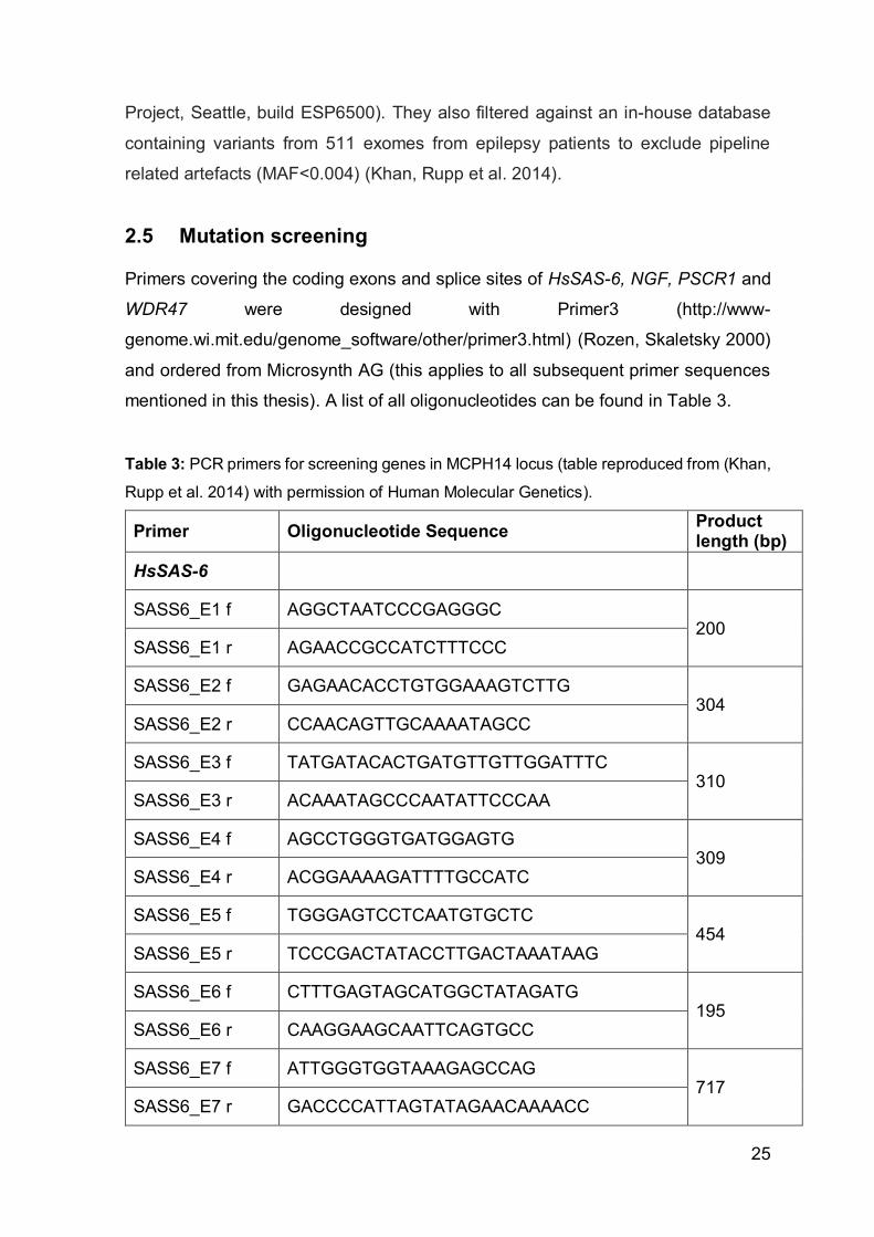
2.5 Mutation screening .................................................................................. 25
2.6 RT-PCR and qPCR ................................................................................. 29
2.7 HsSAS-6 homology analysis in Jalview ................................................... 30
2.8 Kif1bβ splice site prediction ..................................................................... 31
2.9 Cloning .................................................................................................... 31
2.9.1 Kif1bβ ............................................................................................... 31
2.9.2 HsSass-6 .......................................................................................... 33
2.10 Cell culture and transfection .................................................................... 34
2.10.1 Kif1bβ ............................................................................................... 34
2.10.2 HsSASS-6......................................................................................... 35
2.11 Cell-extract preparation and biochemical assays .................................... 36
2.11.1 HsSAS-6 ........................................................................................... 36
2.11.2 Kif1Bβ ............................................................................................... 36
2.12 Immunofluorescence and microscopy for hsSASS-6 transfected cells .... 37
2.13 Confocal Imaging for Kif1Bβ and p75 transfected cells ........................... 37

VI
3. Results ......................................................................................................... 39
3.1 Autosomal recessive syndromal intellectual disability with agenesis of the
corpus callosum ....................................................................................... 39
3.2 HsSass-6 is mutated in autosomal recessive primary microcephaly ....... 62
4. Discussion ................................................................................................... 76
5. Bibliography ................................................................................................ 88

VII
Abbreviations
°C Celsius
A Adenosine
AA Amino acid
ABI Applied Biosystems
ACC Agenesis of the corpus callosum
ACLSM Array confocal laser scanning microscope
aIP Apical intermediate progenitor
AD Autosomal dominant
ALFY WD Repeat and FYVE Domain Containing 3 (=WDFY3)
ANKLE2 Ankyrin Repeat And LEM Domain Containing 2
AR Autosomal recessive
ASPM Abnormal spindle microtubule assembly
ATP Adenosine triphosphate
bIP Basal intermediate progenitor
bp Base pair
BWA Burrows-Wheeler aligner
C Cysteine (amino acid)
C Cytosine (nucleotide)
Ca2+ Calcium
CaM Calmodulin
CASC5 Cancer susceptibility candidate 5
CC Corpus callosum
CDK5RAP2 CDK5 regulatory subunit associated protein 2
cDNA Complementary DNA
CDK6 Cyclin Dependent Kinase 6
cen Centrin
CEP Centrosomal protein
CENPE or J Centromere Protein E or J
Chr Chromosome
CIT Citron Rho-Interacting Serine/Threonine Kinase
cm Centimeter
VIII
cM Centimorgan
CMT Charcot-Marie Tooth
CNS Central nervous system
coMIP Codon optimized miniplasmid
ConDel CONsensus DELeteriousness score of missense SNVs
CPAP Centrosomal P4.1-Associated Protein (=CENPJ)
crSAS-6 Clamydomonas reinhardtii spindle assembly protein 6
CrispR-Cas9 Clustered Regularly Interspaced Short Palindromic
Repeats-CrispR associated endonuclease 9
Ct Carboxy-termianl or C-terminal
CT Computed tomography
dbSNP Database of single nucleotide polymorphisms
DMEM Dulbecco’s Modified Eagle medium
DNA Deoxyribonucleic acid
Dox Doxycycline
DrSAS-6 Danio rerio spindle assembly protein 6
E plus number Embryonic day
EDTA Ethylenediaminetetraacetic acid
EGFP Enhanced green fluorescence protein
ESP6500 NHLBI Exome Sequencing Project 6500
EtOH Ethanol
ExPASy Expert Protein Analysis System
F Phenylalanine
FBS Fetal bovine serum
Fg Femtogram
FHA Forkhead-associated domain
g Gram
G Guanosine
GABA γ-aminobutyric- acid
GATK Genome analysis tool kit
Gb Giga bite
GE Ganglionic eminence
GFP Green fluorescence protein
Gsx GS Homeobox
IX
GW Gestational week
H2O Water
HFB Human fetal brain
Hg17/19 Human genome version 17 or 19
HGMD Human genome mutation database
HRP Horse reddish peroxidase
hsSAS-6/ hsSASS-6 Homo sapiens spindle assembly protein 6
I Iseoleucine
IBD Identical by decent
ID Intellectual development
IDACC Intellectual disability with agenesis of the corpus
callosum
IKM Interkinetic migration
Ile Isoleucine
IP Intermediate progenitor cells
iPSC Induced pluripotent stem cells
IQ Intelligence quotient
250K 250 000
KBP Kinesin binding protein
KCL Kalium chloride
Kif/KIF Kinesin family member
kg Kilogram
L Leucine
LBC Lymphoblastoid cells
Leu Leucine
LOD Logarithm of odds
LPC Lysophosphatidylcholine
MAF Minor allele frequency
MAfftWS Multiple Alignment using fast furrier transform web
service
Mb Mega bite
MB Mega base
Mbp Myelin binding protein
MCPH Microcephaly primary hereditary

X
MFSD2A Major Facilitator Superfamily Domain Containing 2A
min Minutes
mRNA Messenger RNA
MRI Magnet resonance imaging (=MRT)
MRT Magnetic resonance tomography
MS Multiple sclerosis
MST Mitotic somal translocations
Mut Mutant, mutated
MZ Marginal zone
N Asparagine
NaCl Sodium chloride
NE Neuroepithelial cell
NHLBI National Heart, Lung, and Blood Institute
NSC Neuronal stem cell
NT Amino-terminal or N-terminal
Olig2 Oligodendrocyte Transcription Factor 2
OMIM Online Mendelian Inheritance in Men
oRG Outer radial glial cell
oSVZ Outer subventricular zone
p75NTR p75 neurotrophin receptor
PBS Phosphate-Buffered Saline
PCR Polymerase chain reaction
Pen/Strep Penicillin / streptomycin
PH domain Pleckstrin homology domain
PHC1 Polyhomeotic Homolog 1
Phe Phenylalanine
PISA Present in SAS-6
PLK4 Polo-like kinase 4
PNS Peripheral nervous system
PolyPhen Polymorphism Phenotyping v2
PP Preplate
p-ter End of short chromosome arm
qPCR Quantitative polymerase chain reaction
Q Glutamine

XI
q-ter End of long chromosome arm
R Arginine
RFP Red fluorescence protein
RG Radial glial cell
RNA Ribonucleic acid
rRNA Risbosomal RNA
RQ Relative quantification
rs + number Reference single nucleotide polymorphism
identification number
RTA Realtime analysis
RT-PCR Reverse transcription PCR
SCG10 Superior cervical ganglion-10 protein
SCKL Seckl Syndrome
SD Standard deviation
Ser Serine
siRNA Small interfering ribonucleic acid
SDS-PAGE Sodium dodecyl sulfate polyacrylamide gel
electrophoresis
SE (buffer) Sodium EDTA
sec Seconds
SCKL Seckel syndrome
SNP Short neural precursors
SNP Single nuclear polymorphism
SP Subplate
SSC (buffer) Saline-sodium citrate
STIL SCL/TAL1 interrupting locus
STR Small tandem repeat
SV2 Synaptic vesicle glycoprotein 2
SVZ Subventricular zone
T Threonine (amino acid)
T Thyrosine (nucleotide)
t0-t4 Timepoint 1-4
TBST Tris-buffered saline containing Tween-20
TE (buffer) Tris-EDTA

XII
Thr Threonine
UCSC browser University of California Sana Cruz genome browser
UTR Untranslated region
Val Valine
VZ Ventricular zone
vRG Ventricular radial glial cell
WDFY3 WD Repeat And FYVE Domain Containing 3 (=ALFY)
WDR WD-repeat
WES Whole exome sequencing
WT Wildtype
ZNF335 Zink finger protein 335

XIII
Zusammenfassung
Die Entwicklung des menschlichen Gehirns wird durch die Abfolge hochregulierter
Prozesse gewährleistet. Bereits geringe Abweichungen können schwere
neurologische Entwicklungsschäden zur Folge haben. Im Zuge dieser Arbeit
wurden zwei konsanguine Familien mit autosomal rezessiven neurologischen
Erkrankungen untersucht. Während Patienten aus der einen Familie mit einem
bekannten Krankheitsbild diagnostiziert wurden (autosomal rezessive primäre
Mikrocephalie [MCPH]), zeigten die Betroffenen aus der zweiten Familie eine bis
dato unbekannte syndromale Form einer mentalen Entwicklungsstörung mit
Balkenagenesie. Aufgrund des konsanguinen Familienhintergrunds ist die
Wahrscheinlichkeit einer Homozygotie des betroffenen Allels bei autosomal
rezessiven Erkrankungen stark erhöht. Mit Hilfe eines SNP-Arrays war es uns
möglich zwei neue homozygote Loci auf Chromosom 1 für die besagten
Erkrankungen zu identifizieren. Durch nachfolgende Sanger Sequenzierung und
Next Generation Sequencing konnten die potentiell kausalen Mutationen entdeckt
werden. Im bis dato unbekannten MCPH14 Lokus auf Chromosom 1p21.3-p13.1
wurde bei allen Betroffenen der Familie die homozygote Fehlsinn-Mutation
(c.185T>C) in der hoch-konservierten PISA-Domäne des hsSAS-6 Proteins
nachgewiesen. SAS-6 ist ein essentieller Faktor für die Generierung der
Prozentriole. Über Knock-down und Co-Lokalisierungsstudien in U2OS Zellen, war
es uns möglich eine durch die Mutation induzierte Häufung an monopolaren
Spindeln, als auch einen Anstieg der Zellen mit weniger als 4 Zentriolen
nachzuweisen. Die Anzahl der letztendlich im Gehirn vorhandenen Neuronen wird
durch die Etablierung eines ausreichend großen Pools an neuronalen Stammzellen
bestimmt. Unsere Ergebnisse zeigen jedoch, dass die Zellteilung durch die Mutation
zum Teil gestört ist, was einen möglichen Pathomechanismus bei der Entstehung
einer primären Mikrozephalie darstellt. Durch molekulargenetische Untersuchungen
des homozygoten Bereichs auf Chromosom 1p36.31-p36.21 konnten wir in der
zweiten Familie eine Splice-site Mutation (c.5270+2T>G) im C-Terminus der β-
Isoform des Kif1b Gens als potentiell kausale Mutation identifizieren. Durch den
Verlust dieser Splice Donor Stelle kommt es zum „skipping“ des vorletzten Exons
und einem nachfolgenden Frameshift in der restlichen Sequenz von Kif1Bβ. Kif1B

XIV
gehört zur Familie der Kinesin-3 Proteine und ist für den Transport von Vorläufern
von synaptischen Vesikeln verantwortlich. Im Zuge dieser Arbeit gelang es uns zu
beweisen, dass durch den partiellen Verlust der PH-Domäne, welche für die
Vesikelinteraktion notwendig ist, die Lokalisation von Kif1Bβ in der Zelle verändert
und eine Interaktion mit p75NTR Vesikeln nicht mehr möglich ist. Es konnte somit ein
eindeutiger Funktionsverlust des mutierten Kif1Bβ Proteins nachgewiesen werden.
Mit unseren funktionellen Studien konnte die Bedeutung von Kif1B während der
Entwicklung des zentralen Nervensystems nachgewiesen werden.

XV
Abstract
Normal corticogenesis is guaranteed by a sequence of tightly regulated processes
and interruption of any of these can eventually lead to the development of severe
neurological disorders. In this thesis, two consanguineous families with autosomal
recessive primary microcephaly (MCPH) and a novel, autosomal recessive
syndromal form of intellectual disability with agenesis of the corpus callosum are
described. Such rare autosomal recessive diseases occur more frequently in
consanguineous families, since the likelihood of inheriting two of the diseased
alleles is strongly increased. By homozygosity mapping, we identified potential
candidate loci on chromosome 1p21.3-p13.1 in the MCPH family, and on
chromosome 1p36.31-p36.21 in the family with the syndromal form of mental
disability. By Next Generation Sequencing and Sanger Sequencing, we identified
mutations in two novel genes for each disease. For the novel MCPH14 locus, a
single missense mutation (c.185T>C), located in a highly conserved domain of the
hsSASS-6 gene, was found. This gene encodes for a protein required for
procentriole formation. By performing a variety of knock-down and co-localization
studies, we were able to prove the reduced function of the mutated protein, depicted
by the higher incidence of monopolar spindles in U2OS cells, as well as the
increased amount of cells with less than 4 centrioles. Since cell divisions in neuronal
stem cells are essential for establishing a sufficient pool of progenitors, we suggest
that this malfunction of hsSAS-6 has led to the described disease phenotype. In the
second family with the novel syndrome, we discovered a splice site mutation
(c.5270+2T>G) in the β-isoform of Kif1b resulting in skipping of the second to last
exon and a thus induced frameshift in the remaining gene sequence. Kif1B is a
kinesin-3 member highly important for the transport of synaptic vesicle precursors.
Here we show that the partial loss of the vesicle-interacting PH-domain induced by
the alternative splicing, alters the cellular localization of Kif1Bβ and completely
abrogates the co-localization with p75NTR-containing vesicles. As many previous
studies have proven the need for Kif1Bβ in neuronal outgrowth, our discoveries
confirm the importance of Kif1Bβ during embryonic brain development.

1
1. Introduction
Although the human brain is not the largest across mammalian species, it is said to
be the most complex with the highest level of intelligence (Borrell, Calegari 2014).
With a total number of around 170 billion cells (half neurons / half glial cells) and
weighing approximately 1,5kg it is larger than would be expected for the average
human body mass (Andrade-Moraes, Oliveira-Pinto et al. 2013). Hence, a highly
sophisticated gyrification system is required to densely pack all the neurons in a
1320-1500cm3 sized skull (Luders, Steinmetz et al. 2002, Mota, Herculano-Houzel
2015). This small volume is essential, as giving birth to babies with larger brain
circumferences would have otherwise been impossible in times before C-section
(Stiles, Jernigan 2010). Generally considered as the control center of our body, the
brain harbors together with the spinal cord the central nervous system (CNS), one
of the two main parts of the nervous system. It receives and processes all
information coming in from the body itself as well as external. The second part, the
so called ‘peripheral nervous system’ (PNS), consists of all nerves in the remaining
body, connecting the peripheral regions with the CNS (Garzorz 2009). Thus, in order
to guarantee constant and flawless neuronal signal transduction it is of essential,
that the neuronal system remains functional throughout our life. Due to its highly
complex architecture and several tightly regulated developmental steps, it is also
prone to severe disorders (Silbereis, Pochareddy et al. 2016). The ‘National Institute
of Neurological Disorders and Stroke’ provides a list of 447 known neurological
disorders and syndromes, potential treatment options, prognosis and current clinical
trials (National Institute of Neurological Disorders and Stroke 2017).
In the course of a larger ongoing disease gene-identification project we
characterized numerous families with complex neurological disorders. For the
majority of families, known disease genes were found to be causative during the
screening process. However, in two consanguineous families from Pakistan and
Austria (latter one with Turkish background) with autosomal recessive brain
disorders, namely ‘autosomal recessive primary microcephaly’ (MCPH) and a novel,
previously undescribed syndromal form of intellectual disability that will be hereafter
referred to as ‘autosomal recessive syndromal intellectual disability with agenesis
of the corpus callosum’ (IDACC), we were able to identify two novel candidate
genes.

2
However, in order to fully understand these disorders, it is important to gain an
overview of how the brain develops.
1.1 Development of the neocortex
Neuronal development in the embryo is initiated by the emergence of neural stem
cells (NSC) during gastrulation in the 3rd gestational week (Stiles, Jernigan 2010,
Gotz, Huttner 2005). They are located in the ectoderm of the developing embryo
and are often referred to as ‘neuroepithelial cells’ (NE) (Stiles, Jernigan 2010). The
final number of neurons, oligodendrocytes and astrocytes in the brain is already
determined at this stage. This requires the production of a large enough pool of
progenitor cells by generating identical NSC clones through symmetric cell divisions
(Homem, Repic et al. 2015, Gotz, Huttner 2005). Around E20 (embryonic day), the
neuroepithelial cells can be found aligned to both sides of the neuronal plate which
starts to fold inwards due to expansion of the progenitor cells, eventually forming
the neuronal tube (Stiles, Jernigan 2010, Sun, Hevner 2014). Upon completion, NEs
have moved to the hollow center of the neuronal tube aligning as a single cellular
layer along the anlage of the future ventricular system. This so called ‘ventricular
zone’ (VZ) is the apical layer of the future cerebral cortex (Stiles, Jernigan 2010,
Homem, Repic et al. 2015). Neuroepithelial cells are attached to both, the basal
lamina and the apical surface of the VZ, having a highly apical-basal polarity (Gotz,
Huttner 2005, Laguesse, Peyre et al. 2015). Due to the active nuclear movement
(interkinetic migration, IKM) in the cytoplasm of NEs throughout the cell cycle, the
layer exhibits a so called ‘pseudostratified’ character, allowing a high number of
neuronal stem cells to accumulate in the VZ (Dehay, Kennedy 2007, Florio, Huttner
2014, Gotz, Huttner 2005, Takahashi, Nowakowski et al. 1993).
When the neuronal tube expands along the rostro-caudal axis, it starts to form the
three primary brain vesicles, developing later into the fore-, mid-, and hindbrain
(pros-, mes- and rhombencephalon, respectively), while the posterior region gives
rise to the anlage of the spinal cord. The forebrain segment will further divide into
the tel- and diencephalon, while met- and myelencephalon are derived from a
subdivision of the rhombencephalon (Silbereis, Pochareddy et al. 2016, Stiles,
Jernigan 2010). The cleavage of the prosencephalon around gestational week (GW)
5-6 also gives rise to the paired optic vesicles, the olfactory bulbs and the 2

3
hemispheres of the brain (Volpe 2000). When the neuronal tube closes around E30,
the fluid pressure in the ventricles increases and the brain starts to expand rapidly
(Budday, Steinmann et al. 2015). Eventually, during the continuous corticogenesis,
a 6-layered cortex will be established (Mountcastle 1997).
1.2 Neuronal stem cells in the fetal brain
Once at the neuronal tube, neurogenesis starts (around E42) and division of NEs
switches gradually from symmetric to asymmetric (Stiles, Jernigan 2010). To ensure
asymmetric divisions, the radially orientated cleavage plane from NEs has to tilt,
resulting in a slightly oblique or horizontal position (Laguesse, Peyre et al. 2015,
Gotz, Huttner 2005). This so called ‘neurogenic’ division will eventually give rise to
one identical progenitor cell and one radial glial cell (RG) or other kinds of progenitor
cells, such as intermediate progenitor cells (IP) (Gao, Postiglione et al. 2014,
Homem, Repic et al. 2015). RGs are a more specialized type of progenitor cells with
features similar to NEs but lack tight junctions and start to express astroglial
markers, such as GFAP or PAX6 (therefore they are often instead referred to as
‘radial glial progenitor’ cells) (Malatesta, Hartfuss et al. 2000, Gotz, Huttner 2005,
Lui, Hansen et al. 2011). Their divisions are now both, neurogenic (asymmetric) and
proliferative (symmetric) (Gotz, Huttner 2005, Gao, Postiglione et al. 2014). In
contrast to NEs, interkinetic migration in RGs is now restricted to the part of the cell
located in the ventricular and the above located subventricular zone, but not to the
more basal areas (Gotz, Huttner 2005). While those RGs that reside within the VZ
(vRG) remain attached to both, the lower apical and upper pial surface, throughout
the cell cycle and continue proliferating at the ventricular zone (Noctor, Flint et al.
2001), outer radial glial cells (oRG) migrate into a more superficial layer, the outer
subventricular zone (oSVZ), where the apical connections are lost (Lui, Hansen et
al. 2011). This latter RG type is generated during asymmetric, self-renewing mitotic
divisions of vRGs due to the shift from vertical to horizontal cleavage plane
orientation. While the daughter cells inherit the basal processes and exit the VZ to
become oRGs (also called ‘basal radial glial cells’ (Ronan, Fletcher 2015, Florio,
Huttner 2014)), the daughter vRGs retain the ventricular processes and start to re-
grow new pial processes (LaMonica, Lui et al. 2013, Hansen, Lui et al. 2010). It is
to note that oRGs are highly abundant in the developing human but not in the rodent
4
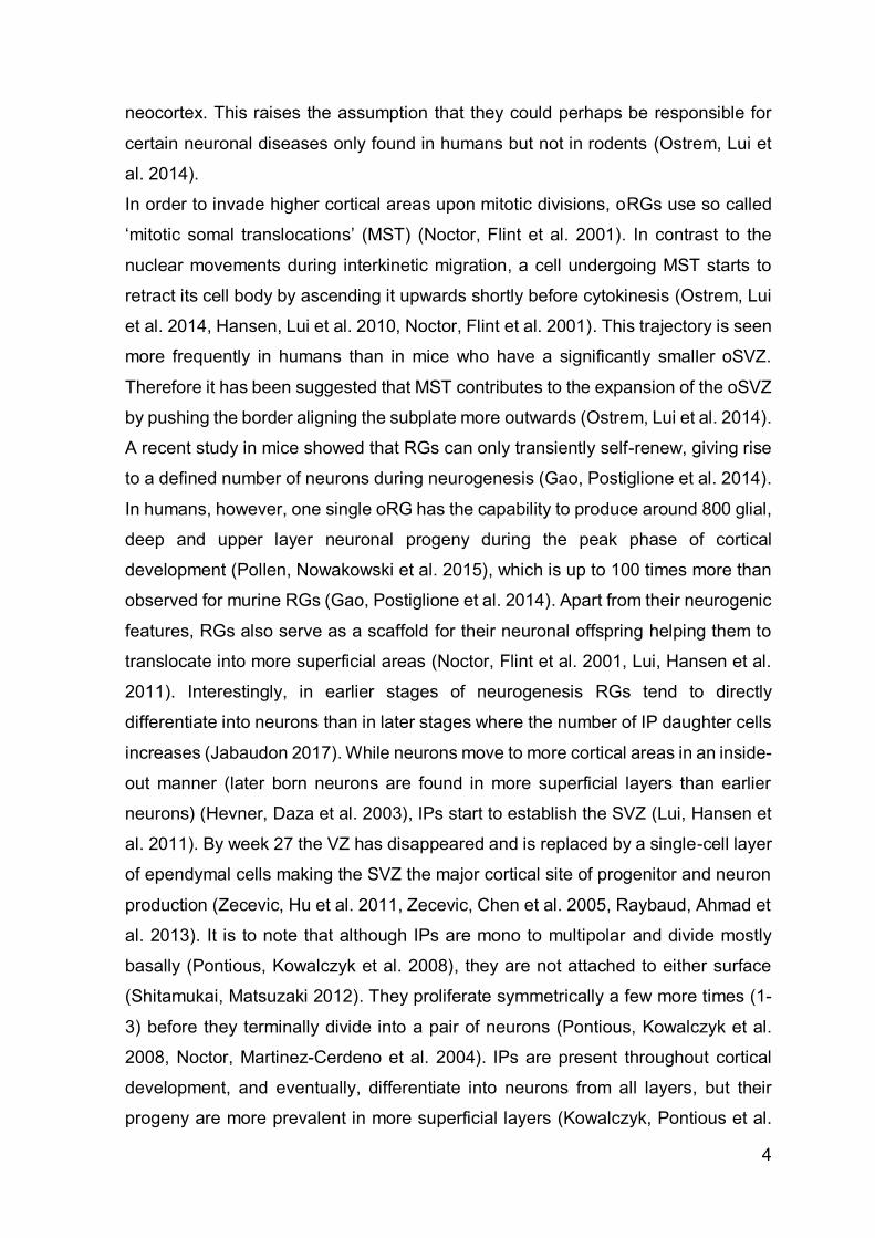
neocortex. This raises the assumption that they could perhaps be responsible for
certain neuronal diseases only found in humans but not in rodents (Ostrem, Lui et
al. 2014).
In order to invade higher cortical areas upon mitotic divisions, oRGs use so called
‘mitotic somal translocations’ (MST) (Noctor, Flint et al. 2001). In contrast to the
nuclear movements during interkinetic migration, a cell undergoing MST starts to
retract its cell body by ascending it upwards shortly before cytokinesis (Ostrem, Lui
et al. 2014, Hansen, Lui et al. 2010, Noctor, Flint et al. 2001). This trajectory is seen
more frequently in humans than in mice who have a significantly smaller oSVZ.
Therefore it has been suggested that MST contributes to the expansion of the oSVZ
by pushing the border aligning the subplate more outwards (Ostrem, Lui et al. 2014).
A recent study in mice showed that RGs can only transiently self-renew, giving rise
to a defined number of neurons during neurogenesis (Gao, Postiglione et al. 2014).
In humans, however, one single oRG has the capability to produce around 800 glial,
deep and upper layer neuronal progeny during the peak phase of cortical
development (Pollen, Nowakowski et al. 2015), which is up to 100 times more than
observed for murine RGs (Gao, Postiglione et al. 2014). Apart from their neurogenic
features, RGs also serve as a scaffold for their neuronal offspring helping them to
translocate into more superficial areas (Noctor, Flint et al. 2001, Lui, Hansen et al.
2011). Interestingly, in earlier stages of neurogenesis RGs tend to directly
differentiate into neurons than in later stages where the number of IP daughter cells
increases (Jabaudon 2017). While neurons move to more cortical areas in an inside-
out manner (later born neurons are found in more superficial layers than earlier
neurons) (Hevner, Daza et al. 2003), IPs start to establish the SVZ (Lui, Hansen et
al. 2011). By week 27 the VZ has disappeared and is replaced by a single-cell layer
of ependymal cells making the SVZ the major cortical site of progenitor and neuron
production (Zecevic, Hu et al. 2011, Zecevic, Chen et al. 2005, Raybaud, Ahmad et
al. 2013). It is to note that although IPs are mono to multipolar and divide mostly
basally (Pontious, Kowalczyk et al. 2008), they are not attached to either surface
(Shitamukai, Matsuzaki 2012). They proliferate symmetrically a few more times (1-
3) before they terminally divide into a pair of neurons (Pontious, Kowalczyk et al.
2008, Noctor, Martinez-Cerdeno et al. 2004). IPs are present throughout cortical
development, and eventually, differentiate into neurons from all layers, but their
progeny are more prevalent in more superficial layers (Kowalczyk, Pontious et al.
5
2009, Vasistha, Garcia-Moreno et al. 2015). Moreover, it has been shown that every
type of projections neuron can be derived from IP progenitors (Mihalas, Elsen et al.
2016) .
In addition to the above mentioned cells, another type of progenitor derived from
radial glial cells has been found in the murine brain, so called short neural precursors
(SNP) (Gal, Morozov et al. 2006). Together with vRGs they are located in the
ventricular zone, giving, like RGs, rise to either stellate or pyramidal neurons that
immediately leave the VZ after birth (Stancik, Navarro-Quiroga et al. 2010, Tyler,
Haydar 2013). Compared to RGs SNPs undergo only one terminal division (Tyler,
Haydar 2013). Therefore, the amount of their neuronal output is much lower. SNP-
derived neurons contribute to the establishment of lower cortical layers (studies in
mice mapped its localization to layer IV) while neurons derived from RGs rather
translocate to upper-layer II/III (Stancik, Navarro-Quiroga et al. 2010). Since both
secondary progenitors are derived from radial glial cells, SNPs were renamed
‘apical intermediate progenitors’ (aIP) and IPs in the SVZ were called ‘basal
intermediate progenitors’ (bIP). However, Tyler et al highlighted that aIPs are an
independent subtype of progenitor cells (Tyler, Haydar 2013).
Although neurogenesis completes around GW21, postnatal differentiation and
migration of progenitor cells will continue to a certain degree (Stiles, Jernigan 2010).
1.3 Neuronal establishment of the cortex
During neurogenesis two main types of neurons are generated: inhibitory
interneurons and excitatory glutamatergic projection neurons (Kwan, Sestan et al.
2012, Florio, Huttner 2014). Excitatory neurons are further divided into pyramidal
projection and spiny stellate interneurons, both using glutamate as their
neurotransmitter (Kwan, Sestan et al. 2012, Markram, Toledo-Rodriguez et al.
2004). The inhibitory interneuronal neurotransmitter ᵞ-aminobutyric-acid (GABA) is
a derivate of the excitatory neurotransmitter, synthesized by decarboxylation of
glutamate (National Institute of Neurological Disorders and Stroke 2017).
Pyramidal neurons owe their name to their triangle shaped soma and make up to
80% of all neurons in the neocortex (Kwan, Sestan et al. 2012, National Institute of
Neurological Disorders and Stroke 2017). While projection neurons have their origin
in the neocortex, interneurons are mainly produced in a thelencephalic area called
6
ganglionic eminence (GE), but can also be detected in SVZ of the cortex or in the
subpial granular layer starting from mid-term (Zecevic, Hu et al. 2011). Their radial
glial progenitors can be distinguished by the expression of Pax6 and Emx1
(neocortical progenitors) and Gsx1, Gsx2 and Olig2 (telencephalic progenitors)
(Tan, Shi 2013). In contrast to interneurons that interact with more local sites,
projection neurons have long axons enabling them to intervene far distant areas
(Hevner, Daza et al. 2003). Upon birth, projection neurons start to move upwards to
establish the neocortex, the thin, gray layer on the surface of the brain (Stiles,
Jernigan 2010). Interestingly, newborn neurons do not directly translocate to their
final destination. They rather rest for another 24 hours in the SVZ in a multipolar
state before the majority of neuronal progeny becomes again bipolar, reverses their
direction and extends their fibers back towards the ventricle. Only after touching and
moving closer to the apical membrane, they eventually ascend to the cortical plate.
About one third of all post-mitotic neurons however skip retrograde migration but do
remain in the SVZ for 24h before their final translocation (Noctor, Martinez-Cerdeno
et al. 2004). In contrast to the radial migration of excitatory glutamatergic neurons,
inhibitory GABAergic neurons invade the cortex tangentially from the GE (Dwyer,
Chen et al. 2016, Budday, Steinmann et al. 2015). Once there, they also switch to
a radial migration mode and translocate to their final cortical layer (Zechel,
Nakagawa et al. 2016).
The very first neurons to be found in the early phase of neurogenesis are Cajal-
Retzius cells. Cajal Retzius cells produce reelin, an important factor controlling the
right positioning, branching and growth of newborn neurons in the developing
cortical plate (Stiles, Jernigan 2010, Olson 2014). It is to note that reelin as a marker
for Caja Retzius cells is only applicable until birth, as after birth reelin is additionally
produced by cortical interneurons (Hevner, Daza et al. 2003).
Together with the subplate neurons, Cajal Retzius cells establish the preplate (PP)
right above the VZ (Toma, Hanashima 2015). The preplate harbors early born cells
that either only have a transient function and die or become cortical or subcortical
inhibitory neurons (Budday, Steinmann et al. 2015). While underneath the
intermediate and subventricular zones arise (first detectable in GW9), which start to
produce pyramidal projection neurons, an event called ‘preplate splitting’ initiates
the formation of a well-organized cortical plate that divides the PP into the marginal
zone (MZ) and the subplate (SP) around GW8 (Zecevic, Chen et al. 2005, Olson
7
2014, O'Dell, Cameron et al. 2015). Induced by the dendritic outgrowth of later layer
VI neurons, the event itself is of significant importance for proper brain development
(Olson 2014). Eventually, the marginal zone will later transform into layer I. The
subplate, however, is only a transient layer important for axon guidance and
connecting intra- and extracortical areas (Hoerder-Suabedissen, Molnar 2015,
Perkins, Hughes et al. 2008, Kwan, Sestan et al. 2012). By week 28 it has reached
its maximum size before it starts to degrade around GW35 and will be undetectable
by the age of 6 months (Hoerder-Suabedissen, Molnar 2015, Raybaud, Ahmad et
al. 2013). The cortical plate will expand until gestational week 18 when finally well-
defined layers I-VI have been formed (Budday, Steinmann et al. 2015). The layers
are divided in two subgroups: Layer V and VI are known as the subgranular or deep
layers (Kwan, Sestan et al. 2012) whereas layers II-IV represent the upper or
superficial layers (Gao, Postiglione et al. 2014). In contrast to the deep layer,
neurons that can also target subcortical regions such as the thalamus
(corticothalamic neurons: mainly layer VI and to a certain extent also layer V
neurons), the brain stem and the spinal cord (subcerebral projection neurons are
layer V neurons), connections of neurons of the superficial layers are restricted to
cortical areas (Gerfen, Economo et al. 2016, Molyneaux, Arlotta et al. 2007).
During establishment of the cortex another important cell type is produced, namely
glial cells, including astrocytes, oligodendrocytes and microglial cells (Budday,
Steinmann et al. 2015). Differentiation of astrocytes is induced around E108 when
radial glia cells switch from neurogenesis to gliogenesis (Stiles, Jernigan 2010,
Raybaud, Ahmad et al. 2013). In the postnatal human brain, astrocytes are the most
abundant cell type and play a primary role in neuronal synapse formation as well as
in controlling their function (Clarke, Barres 2013). Oligodendrocyte progenitor
development starts much earlier, around GW 10-15 but they do not become mature
oligodendrocytes until GW30 (Raybaud, Ahmad et al. 2013, Budday, Steinmann et
al. 2015). They ensure rapid impulse conduction by forming insulating myelin sheets
around axons of neurons (Peferoen, Kipp et al. 2014). It is to note that the generation
of oligodendrocytes will continue after birth until the age of 9 (Yeung, Zdunek et al.
2014). In contrast to astro- and oligodendrocytes which are derived from radial glial
cells, microglial cell originate from yolk sac macrophages that invade the brain and
spinal cord anlagen concomitantly with or shortly before the development of the
8
blood-brain-barrier and serve as the CNS’ innate immune system (Waisman,
Ginhoux et al. 2015).
Almost simultaneously with the start of gliogenesis and the increasing tangential
expansion of the cortex another important developmental step is induced (Yoshida,
Ishizu et al. 2017). Due to the constant division, differentiation, migration and axon
branching of cells and the concurrent limited space in the scull, around GW 20-21
the cortex starts to fold itself resulting in its typical wrinkled architecture composed
of gyri and sulci (Sun, Hevner 2014, Bayly, Taber et al. 2014, Raybaud, Ahmad et
al. 2013). They are built in 3 waves, establishing primary (evident around GW31),
secondary (mainly present around GW 27) and tertiary sulci, which evolve together
with some secondary sulci until after term (Raybaud, Ahmad et al. 2013). While in
humans the primary folds are more or less the same, the latter two do vary (Yoshida,
Ishizu et al. 2017, Holland, Miller et al. 2015).
Eventually, the intermediate zone will transform into the white matter, harboring
mostly long axons wrapped with myelin, responsible for its characteristic white
appearance (Tallinen, Chung et al. 2014, Raybaud, Ahmad et al. 2013, Stiles,
Jernigan 2010). The neocortex, on the other hand, contains the cell bodies of
neurons and therefore appears gray (Stiles, Jernigan 2010) (for easier
understanding, see Figure 1).
9
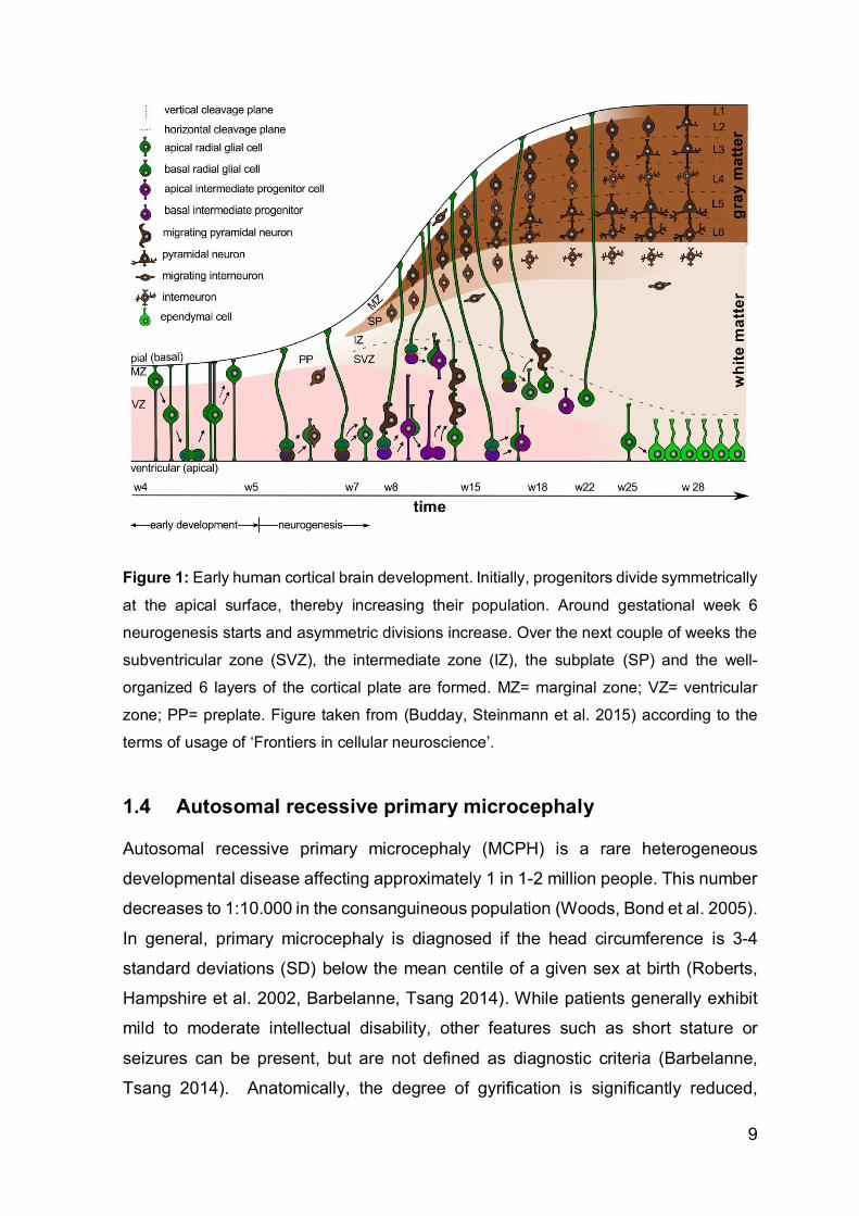
Figure 1: Early human cortical brain development. Initially, progenitors divide symmetrically
at the apical surface, thereby increasing their population. Around gestational week 6
neurogenesis starts and asymmetric divisions increase. Over the next couple of weeks the
subventricular zone (SVZ), the intermediate zone (IZ), the subplate (SP) and the well-
organized 6 layers of the cortical plate are formed. MZ= marginal zone; VZ= ventricular
zone; PP= preplate. Figure taken from (Budday, Steinmann et al. 2015) according to the
terms of usage of ‘Frontiers in cellular neuroscience’.
1.4 Autosomal recessive primary microcephaly
Autosomal recessive primary microcephaly (MCPH) is a rare heterogeneous
developmental disease affecting approximately 1 in 1-2 million people. This number
decreases to 1:10.000 in the consanguineous population (Woods, Bond et al. 2005).
In general, primary microcephaly is diagnosed if the head circumference is 3-4
standard deviations (SD) below the mean centile of a given sex at birth (Roberts,
Hampshire et al. 2002, Barbelanne, Tsang 2014). While patients generally exhibit
mild to moderate intellectual disability, other features such as short stature or
seizures can be present, but are not defined as diagnostic criteria (Barbelanne,
Tsang 2014). Anatomically, the degree of gyrification is significantly reduced,

10
associated with a thinner than normal cortex but no other brain malformations. To
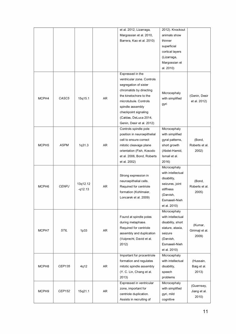
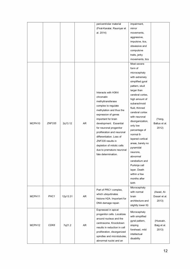
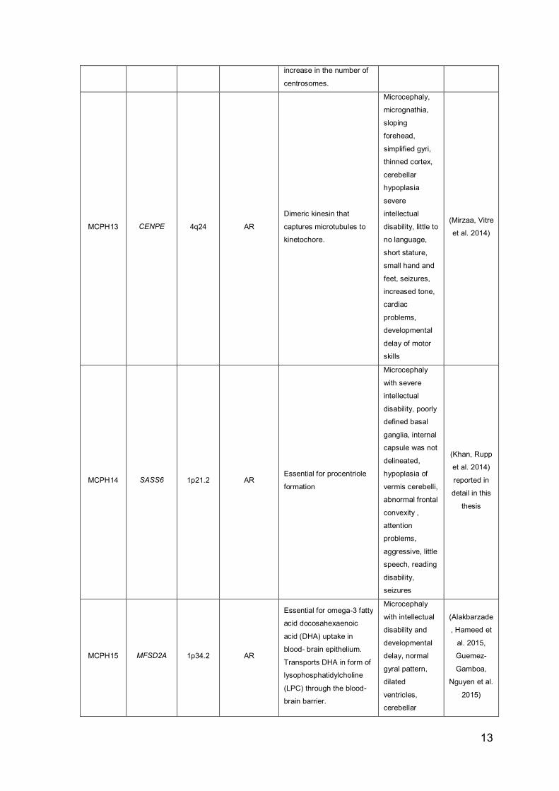
date, 18 MCPH loci and the corresponding genes have been identified (see Table
1)
Table 1: Primary microcephaly genes
Locus Gene
Chrom.
location
(hg38)
Inheritance
mode Gene function Phenotype
Gene first
published
MCPH1 MCPH1 8p23.1 AR
Responsible for
chromosome
condensation and
decondensation.
Regulates cdk1 activation
and thus the entry into
mitosis. Late
neurogenesis and
symmetric cell divisions
are negatively affected
upon knock-down (Gruber,
Zhou et al. 2011)
Mild- to
moderate
intellectual
disability,
microcephaly
(Jackson,
Eastwood et
al. 2002)
MCPH2 WDR62 19q13.12 AR
Expressed in ventricular
and subventricular zone.
Assembles kinetochore
microtubules to control
mitotic spindle orientation
(Miyamoto, Akutsu et al.
2017)
Microcephaly,
intellectual
disability,
thickened
cortex, no
boarder
between white
and gray matter,
pachygyria,
polymicrogyria,
lissencephaly,
schizencephaly,
subcortical
heterotopia,
brainstem
atrophy,
abnormal
corpus
callosum,
cerebellar
hypoplasia,
seizures
(Yu, Mochida
et al. 2010,
Bilguvar,
Ozturk et al.
2010)
MCPH3 CDK5RAP2 9q33.2 AR
Controls orientation of
mitotic spindles in
neuronal progenitors and
Cdk5rap2 knockout cells
show premature cell cycle
exit (Pagnamenta, Murray
Microcephaly
with
sensorineural
deafness
(Pagnamenta,
Murray et al.
(Bond,
Roberts et al.
2005)
11
et al. 2012, Lizarraga,
Margossian et al. 2010,
Barrera, Kao et al. 2010)
2012). Knockout
animals show
thinner
superficial
cortical layers
(Lizarraga,
Margossian et
al. 2010)
MCPH4 CASC5 15q15.1 AR
Expressed in the
ventricular zone. Controls
segregation of sister
chromatids by directing
the kinetochore to the
microtubule. Controls
spindle assembly
checkpoint signaling
(Caldas, DeLuca 2014,
Genin, Desir et al. 2012)
Microcephaly
with simplified
gyri
(Genin, Desir
et al. 2012)
MCPH5 ASPM 1q31.3 AR
Controls spindle pole
position in neuroepithelial
cell to ensure correct
mitotic cleavage plane
orientation (Fish, Kosodo
et al. 2006, Bond, Roberts
et al. 2002)
Microcephaly
with simplified
gyral patterns,
short growth
(Abdel-Hamid,
Ismail et al.
2016)
(Bond,
Roberts et al.
2002)
MCPH6 CENPJ 13q12.12
-q12.13 AR
Strong expression in
neuroepithelial cells.
Required for centriole
formation (Kohlmaier,
Loncarek et al. 2009)
Microcephaly
with intellectual
disability,
seizures, joint
stiffness
(Darvish,
Esmaeeli-Nieh
et al. 2010)
(Bond,
Roberts et al.
2005)
MCPH7 STIL 1p33 AR
Found at spindle poles
during metaphase.
Required for centriole
assembly and duplication
(Vulprecht, David et al.
2012)
Microcephaly
with intellectual
disability, short
stature, ataxia,
seizure
(Darvish,
Esmaeeli-Nieh
et al. 2010)
(Kumar,
Girimaji et al.
2009)
MCPH8 CEP135 4q12 AR
Important for procentriole
formation and regulates
mitotic spindle assembly
(Y. C. Lin, Chang et al.
2013)
Microcephaly
with intellectual
disability,
speech
problems
(Hussain,
Baig et al.
2013)
MCPH9 CEP152 15q21.1 AR
Expressed in ventricular
zone, important for
centriole duplication.
Assists in recruiting of
Microcephaly
with simplified
gyri, mild
cognitive
(Guernsey,
Jiang et al.
2010)
12
pericentriolar material
(Firat-Karalar, Rauniyar et
al. 2014)
impairment,
mirror
movements,
aggressive,
impulsive, tics,
obsessive and
compulsive
traits, jerky
movements, tics
MCPH10 ZNF335 2q13.12 AR
Interacts with H3K4
chromatin
methyltransferase
complex to regulate
methylation and thus the
expression of genes
important for brain
development. Essential
for neuronal progenitor
proliferation and neuronal
differentiation. Loss of
ZNF335 results in
depletion of mitotic cells
due to premature neuronal
fate determination.
Most severe
form of
microcephaly
with extremely
simplified gyral
pattern, skull
larger than
cerebral cortex,
high amount of
subarachnoid
fluid, thinned
cerebral cortex
with neuronal
disorganization,
only low
percentage of
normal 6-
layered cortical
areas, barely no
pyramidal
neurons,
abnormal
cerebellum and
Purkinje cell
layer. Death
within a few
months after
birth
(Yang,
Baltus et al.
2012)
MCPH11 PHC1 12p13.31 AR
Part of PRC1 complex,
which ubiquitinates
histone H2A. Important for
DNA damage repair.
Microcephaly
with normal
brain
architecture and
slightly lower IQ
(Awad, Al-
Dosari et al.
2013)
MCPH12 CDK6 7q21.2 AR
Expressed in apical
progenitor cells. Localizes
around nucleus and the
centrosome. Knockdown
results in reduction in cell
proliferation, disorganized
spindles and microtubules,
abnormal nuclei and an
Microcephaly
with simplified
gyral pattern,
sloping
forehead, mild
intellectual
disability
(Hussain,
Baig et al.
2013)
13
increase in the number of
centrosomes.
MCPH13 CENPE 4q24 AR
Dimeric kinesin that
captures microtubules to
kinetochore.
Microcephaly,
micrognathia,
sloping
forehead,
simplified gyri,
thinned cortex,
cerebellar
hypoplasia
severe
intellectual
disability, little to
no language,
short stature,
small hand and
feet, seizures,
increased tone,
cardiac
problems,
developmental
delay of motor
skills
(Mirzaa, Vitre
et al. 2014)
MCPH14 SASS6 1p21.2 AR Essential for procentriole
formation
Microcephaly
with severe
intellectual
disability, poorly
defined basal
ganglia, internal
capsule was not
delineated,
hypoplasia of
vermis cerebelli,
abnormal frontal
convexity ,
attention
problems,
aggressive, little
speech, reading
disability,
seizures
(Khan, Rupp
et al. 2014)
reported in
detail in this
thesis
MCPH15 MFSD2A 1p34.2 AR
Essential for omega-3 fatty
acid docosahexaenoic
acid (DHA) uptake in
blood- brain epithelium.
Transports DHA in form of
lysophosphatidylcholine
(LPC) through the blood-
brain barrier.
Microcephaly
with intellectual
disability and
developmental
delay, normal
gyral pattern,
dilated
ventricles,
cerebellar
(Alakbarzade
, Hameed et
al. 2015,
Guemez-
Gamboa,
Nguyen et al.
2015)

14
hypoplasia,
thinned corpus
callosum,
hypoplastic
brainstem, loss
of white matter
in posterior
regions,spastic
gait, some have
talopes
eqinovarus,
limited to no
speech,
pulmonary
insufficiency
and dysphagia,
spastic
quadriparesis,
lethal in case of
inactivating
mutation
MCPH16 ANKLE2 12q24.33 AR
Important for neuroblast
production. Mutations do
not affect mitotic spindle
orientation, centriole
formation or centriole
number but has a negative
impact on cell proliferation.
Important for nuclear
envelope reassembly after
cell division (Kaufmann,
Kukolj et al. 2016).
Extremely
sloping
forehead,
enlarged extra
axial space,
extremely
reduced
gyrification,
agenesis of the
corpus
callosum,
ptosis, slightly
thicker cortex,
small jaw,
hyper- and
hypopigmented
macules,
spastic
quadriplegia,
anemia,
glaucoma,
seizures
(Yamamoto,
Jaiswal et al.
2014)
MCPH17 CIT 12q24.23 AR
Localized at central
spindle and cleavage
furrow. Interacts with
KIF14 to promote
chromosome segregation
during cytokinesis
Microcephaly
with mild-to-
moderate
intellectual
disability,
simplified gyral
pattern, extra-
(Basit, Al-
Harbi et al.
2016)
15
(Gruneberg, Neef et al.
2006).
axial space
enlarged
MCPH18 ALFY/
WDFY3 4q21.23 AD
Autophagy mediating
scaffold protein that
controls neuronal
progenitor differentiation.
Microcephaly
with mild-to-
moderate
intellectual
disability
(Kadir, Harel
et al. 2016)
MCPH19 Kif14 1q32.1 AR
Interacts with CIT to
promote cytokinesis
(Gruneberg, Neef et al.
2006).
Reduced cortex
with simplified
gyral pattern,
enlarged lateral
ventricles,
sloping
forehead and
moderate to
severe
intellectual
disability,
impaired
speech, spastic
tetraparesis,
total agenesis of
corpus
callosum,
thicker cortex,
small kidneys
(Moawia,
Shaheen et
al. 2017)
AR=autosomal recessive; AD=autosomal dominant
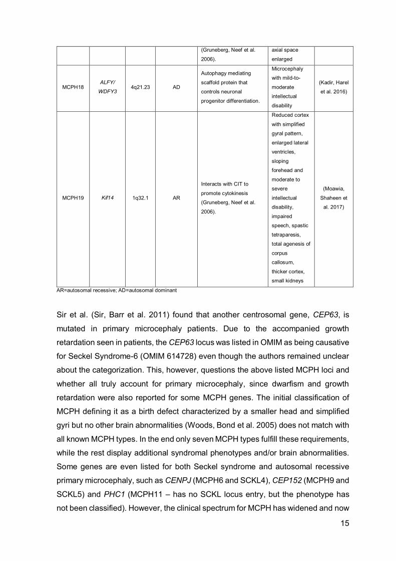
Sir et al. (Sir, Barr et al. 2011) found that another centrosomal gene, CEP63, is
mutated in primary microcephaly patients. Due to the accompanied growth
retardation seen in patients, the CEP63 locus was listed in OMIM as being causative
for Seckel Syndrome-6 (OMIM 614728) even though the authors remained unclear
about the categorization. This, however, questions the above listed MCPH loci and
whether all truly account for primary microcephaly, since dwarfism and growth
retardation were also reported for some MCPH genes. The initial classification of
MCPH defining it as a birth defect characterized by a smaller head and simplified
gyri but no other brain abnormalities (Woods, Bond et al. 2005) does not match with
all known MCPH types. In the end only seven MCPH types fulfill these requirements,
while the rest display additional syndromal phenotypes and/or brain abnormalities.
Some genes are even listed for both Seckel syndrome and autosomal recessive
primary microcephaly, such as CENPJ (MCPH6 and SCKL4), CEP152 (MCPH9 and
SCKL5) and PHC1 (MCPH11 – has no SCKL locus entry, but the phenotype has
not been classified). However, the clinical spectrum for MCPH has widened and now

16
includes seizures, slight growth retardations (up to -3SD), mild spasticity as well as
hypoplasia of the cerebellum and hindbrain (Verloes, Drunat et al. 1993).
An interesting aspect of microcephaly genes is that most of them play an essential
role during cell division and cell cycle progression. As already discussed in detail in
chapter 1.2, a large enough neuronal progenitor pool has to be generated via
symmetric cell divisions before neurogenesis takes place. A premature switch from
vertical to horizontal division plane orientation during mitosis can prevent the
establishment of the adequately sized neuronal stem cell pool and leads to
premature neuronal differentiations (Homem, Repic et al. 2015). The recent
epidemic Zika virus outbreak on the American continent depicts an elegant example
which highlights the importance of an undisturbed symmetric progenitor division.
This Arbovirus is transferred to humans via Aedes mosquitoes and causes
symptoms such as fever, headache, painful joint movements, a skin rash,
inflammation of the eyes and diarrhea within a few days (Javed, Manzoor et al.
2018). While it is not life-threatening for the general population, fetuses of infected
pregnant women have an increased danger of developing microcephaly (Al-
Qahtani, Nazir et al. 2016). Li et al. (C. Li, Xu et al. 2016) found that the virus
targets only neuroepithelial cells, which divide at high rates in the developing brain
of fetuses, thus suppressing further cell divisions and thereby leading to
microcephaly in the embryo. This neurovirulent feature is untypical for the original
wildtype strain and was only recently associated with a specific mutation (S139N) in
the contemporary Cam/2010 virus (F. Zhang, Wang et al. 2017) exhibiting an
enhanced replication rate in human neuronal progenitor cells, which is eventually
responsible for an increase in neuronal apoptosis (Yuan, Huang et al. 2017).
Furthermore, studies on brain organoids have provided proof of interference with
centrosomal protein recruitment and shown that spindles in apical progenitor cells
are tilted, thus inducing premature asymmetric divisions (Gabriel, Ramani et al.
2017).
1.5 Agenesis of the corpus callosum
While the left hemisphere of the brain is the center for language, which controls our
speech and auditory perception, the right half of the brain is responsible for
processing what we see and supports spatial awareness (Wolman 2012).
17
Connection of these two by sending axons via the corpus callosum to the opposite
hemisphere enables us to develop higher cognitive skills (Edwards, Sherr et al.
2014). The corpus callosum (CC) is a large tract of commissural fibers comprised
of around 200 million axons of excitatory and, to a lesser extent, inhibitory neurons
that are vital for the integration of sensory and motor information, support the
learning of languages, and have an impact on emotions and the development of
social skills (Paul 2011, Bloom, Hynd 2005). It is the largest known structure of white
matter only evident in placental mammals (van der Knaap, van der Ham 2011).
However, the CC is not the only axonal connection between the two cerebral
hemispheres. Beside this primary interhemispheric tract, anterior, posterior,
habenular and hippocampal commissures also exist (Palmer, Mowat 2014, Bloom,
Hynd 2005).
The earliest precursor of this connective bridge, the commissural plate, arises
shortly after the prosencephalic cleavage in GW5-6 (Dobyns 1996, Achiron, Achiron
2001). Navigated by guidance- and repulsive molecules expressing glial cells in the
midline, the first callosal pioneering axons from the cingulate cortex start to cross
the midline around gestational week 13-14 (Edwards, Sherr et al. 2014, Ren,
Anderson et al. 2006, Unni, Piper et al. 2012). It has been shown that the remaining
axons of the CC then simply follow the pioneer axons through this glial zipper after
they have finally made it to the other hemisphere (Klose, Bentley 1989, Kuwada
1986, Tovar-Moll, Moll et al. 2007). Although recent studies suggest a bidirectional
development of the CC, enhanced growth of the anterior part is evident in the first
weeks (Paul 2011). The corpus callosum as a structure can first be visualized
around GW15, but its final structure is not manifested before GW17 (embryonic day
115) (H. Huang, Xue et al. 2009, Achiron, Achiron 2001). It mainly constitutes of
axons of superficial layers II and III neurons, but harbors projection fibers from layer
V and VI neurons as well (O'Leary, Koester 1993, Lavado, Ware et al. 2014). By
term, the total amount of callosal axons is established, but due to pruning and
myelination, the CC undergoes most of its maturation within the first 4 years of life.
However, it will evolve until early adulthood before it starts to slightly decrease in
size by the age of 40 (Luders, Thompson et al. 2010, Fitsiori, Nguyen et al. 2011).
The mature CC consists of four different regions, starting in the front with the
rostrum, followed by the genu, the body (anterior, posterior body and isthmus) and
finally the splenium at the posterior end (Filippi, Cauley 2014). Studies on adult split-

18
brain patients that have undergone callosotomy to treat epileptic seizures have shed
most light onto the function of the corpus callosum (Wolman 2012). Additionally,
lesions in different callosal areas have helped to unravel regional callosal functions:
among other abnormalities, injuries in the anterior sections of the CC impair
movements and speech, while patients exhibiting posterior lesions have reading
disabilities, but interestingly maintain writing skills (Filippi, Cauley 2014).
Nevertheless, it has been suggested that subcortical commissures take over some
integrative tasks after transection of the CC, which has to be kept in mind when
studying the connective role of CC in these patients (Bloom, Hynd 2005).
In a few cases, the formation of the CC is disturbed during embryogenesis. There
are four forms of aberrant callosal morphologies, including hypoplasia (thinner than
usual CC), dysplasia (structurally malformed CC), hypoplasia with dysplasia and
complete agenesis of the corpus callosum (ACC indicates a total absence of the
CC) (Hanna, Marsh et al. 2011). The frequency of the latter can be as rare as 0.5-1
affected in 10.000 and increases to 2-2.3% in people with additional intellectual
disabilities (Jeret, Serur et al. 1985, Fratelli, Papageorghiou et al. 2007). A list of
syndromal and non-syndromal inherited diseases that are associated with ACC can
be found in the ‘Online Mendelian Inheritance in Man’ Database (McKusick-Nathans
Institute of Genetic Medicine, Johns Hopkins University (Baltimore, MD) 2012).
Entering the term ‘corpus callosum agenesis’ will reveal 849 OMIM entries of
syndromes associated with callosal abnormalities (, OMIM Clinical Synopsis Search
- corpus callosum agenesis). In general, there are two types of ACC distinguished
by the presence or absence of so-called ‘Probst bundles’. These are longitudinal
fibers, which have not crossed the midline and accumulate diffusely along the
medial wall on their hemisphere of origin. In ACC patients lacking these Probst
bundles, either the formation of commissural neurons or the extension of projecting
axons is disturbed (Dobyns 1996, Schell-Apacik, Wagner et al. 2008). A recent
study revealed that patients with Probst bundles actually exhibited significantly
better social and adaptive functions (Al-Hashim, Blaser et al. 2016). Complete
absence does not indicate a concurrent bad or worse neurodevelopmental outcome
than in partial agenesis. Sotiriadis and Makrydimas (Sotiriadis, Makrydimas 2012)
showed that children with prenatally diagnosed isolated ACC (partial and complete
ACC without additional brain abnormalities) developed normally in 75% of cases
with a favorable development in 80% in infants with complete ACC. Interestingly, in

19
their study partial ACC was associated with a slightly higher rate of severe outcome
than observed for complete ACC. However, due to the small number of participating
partial ACC patients with severe outcome, this finding was not statistically
significant. In addition, the researchers noted that prognosis worsens with the
parallel occurrence of other brain malformations.
Preterm delivery, viral infections, maternal phenylketonuria, alcohol abuse during
pregnancy or genetic predispositions may contribute to developmental callosal
abnormalities (Jo, Cho et al. 2012, Riley, Mattson et al. 1995, Schell-Apacik,
Wagner et al. 2008, Paul, Brown et al. 2007). These factors can affect neuronal
progenitor proliferation, neuronal faith, neuronal translocation, midline patterning,
the formation of the two hemispheres as well as the extension, projection and
navigation of axons to their target cells in the contralateral hemisphere (Edwards,
Sherr et al. 2014). Interestingly, only 32-37% of ACC patients have an identifiable
underlying genetic cause with 48% of them carrying mutations in single genes and
30% showing chromosomal aberrations. While the remaining 63-68% of ACC
patients present with known clinical syndromes, these have as yet not been link to
causative genes (Al-Hashim, Blaser et al. 2016, Schell-Apacik, Wagner et al. 2008).
This indicates that the genetic testing for syndromal ACC remains challenging even
in times of Next Generation Sequencing.
1.6 Consanguinity and the inheritance of rare diseases
As mentioned above, disturbance of any of these tightly regulated developmental
steps during cortical development can lead to severe disorders. In the course of this
dissertation I came across two of such rare developmental disorders, which have a
major impact on the patients’ architecture of the brain, their mental performance,
social skills and behaviors.
In Europe, a disease is by definition considered as ‘rare’ if the probability of
inheritance is 1:2000 (information taken from https://www.eurordis.org), while the
US has set the threshold to less than 200.000 affected (information taken from
National Institute of Health https://rarediseases.info.nih.gov). The majority of all
known rare conditions (around 80%) is due to inherited genetic defects (Institute of
Medicine (US) Committee on Accelerating Rare Diseases Research and Orphan
Product Development 2010). Especially when it comes to consanguineous

20
marriages, the chance of giving birth to children suffering from extremely rare
autosomal recessive traits increases significantly (A. Bittles 2001, Zlotogora 1997).
In North African, Arab and some Western and Southern Asian countries, the rate of
such marriages is above 20% and increases to over 50% in Pakistan and Sudan (A.
H. Bittles, Black 2015). Despite the common knowledge that their genomes share a
larger number of homologous regions, 1/3rd of all consanguineous couples are not
aware of the fact that they thus face higher reproductive risks (Teeuw, Loukili et al.
2014). While it is rather difficult to find the causative gene in affected children of
unrelated parents, children of consanguineous parents share blocks of homozygous
(in this case called ‘autozygous’) regions, which are identical by decent (IBD). Thus,
homozygosity mapping is the method of choice when it comes to identifying
autosomal recessive disease loci in consanguineous families (Hildebrandt,
Heeringa et al. 2009). The easy - and nowadays affordable - massive parallel
sequencing of the target loci allows the detailed screening of all genes located in
the filtered regions (Alkuraya 2010).
In this thesis two consanguineous families with inherited neurodevelopmental
diseases were screened for the genetic cause of their autosomal recessive disorder.
Applying a positional cloning strategy we were able to identify two novel disease-
causing genes suspected to play a major role during brain development.

21
2. Material and Methods
Some passages have been quoted verbatim from the following source: ‘A missense
mutation in the PISA domain of HsSAS-6 causes autosomal recessive primary
microcephaly in a large consanguineous Pakistani family’ published in Human
Molecular Genetics (Khan, Rupp et al. 2014).
2.1 Sample collection
After obtaining informed consent, blood was collected into EDTA vacutainer tubes
(BD Bioscience) from the two affected IDACC patients, the healthy brother of VI:1
and their parents. For the microcephaly family, blood was drawn from 3 affected
patients, 7 healthy family members and 116 healthy Pakistani individuals. DNA was
isolated according to our in-house desalting protocol. Briefly, whole blood was
overlaid with 4-5 volumes of blood lysis buffer (16,58g NH4Cl, 2g KHCO3, 0,074g
EDTA [all from Sigma] dissolved in 2l dH2O) and incubated on ice for 30min followed
by a second lysis step for another 10min in lysis buffer before the resulting pellet
was subjected to protein digestion (5ml 1xSE buffer supplemented with 250µl of
20% SDS and 25µl of a 10mg/ml Proteinase K [Qiagen] stock) overnight at 37°C.
The following day DNA was precipitated with 6M NaCl, washed with 100% EtOH
and dissolved in 1xTE buffer.
For cDNA analysis, blood from both IDACC patients and MCPH patients V-3, IV-7
and two healthy controls was collected into PAXgene RNA blood tubes
(PreAnalytiX) and RNA was isolated using PAXgene RNA Kit.
The study on the IDACC family was approved by the ethics committee of the Medical
University of Graz (approval number 24-421 ex 11/12) and conducted according to
the Declaration of Helsinki. The study on the MCPH family was approved by
institutional ethical review boards of Gomal University, Dera Ismail Khan, and
Quaid-i-Azam University, Islamabad, Pakistan.
2.2 Karyotyping
Metaphases of lymphoblastoid cells (see 2.10.1) were prepared according to our in-
house protocol. Briefly, lymphoblastoid cells were resuspended in 10ml of fresh
22
media. 2ml were supplemented with 100µl of KaryoMAX Colcemid Solution
(10µg/ml stock solution from Gibco, ThermoFisher) and incubated for 1h at 37°C
and 5%CO2. After 1h the remaining 8ml of cell suspension were added and
incubated for another 30min at 37°C. Subsequently cells were pelleted at 2000rpm
for 8min. The pellet was resuspended in 300µl supernatant and 5ml of pre-warmed
0,075M KCl (37°C) were added dropwise. Cells were left at 37°C for 20min and
harvested at 1200rpm for 8min. The pellet was carefully resuspended in 300µl
supernatant and ice-cold fixative (3:1 ratio of methanol:glacial acetic acid) was
added dropwise. After adding a total of 5ml, cells were centrifuged (1200rpm, 8min)
and washed for three more times with fixative solution. Eventually, cells were
resuspended in fixative and dropped from about 20cm on pre-cooled glass-slides
which were stored in H2O. Slides were dried and aged for 2 days at room
temperature before they were transferred into SSC-buffer (35,06g NaCl, 17,648g
tri-sodium citrate dehydrate in 2L H2O) and 60°C for 7h. Subsequently, they were
transferred into 4% Giemsa solution for 8 min and rinsed with water afterwards.
Slides were scanned with a scanner from Metasystems and analyzed with Metafer
4 program.
2.3 Genotyping and LOD score calculation
All affected individuals were genotyped on an Affymetrix GeneChip Human Mapping
250K NspI Array at the ‘Center for Medical Research’ at the Medical University of
Graz. Data were analyzed with dChip software (M. Lin, Wei et al. 2004) available
from http://www.hsph.harvard.edu/cli/complab/dchip/. For haplotype analysis of the
IDACC family, a total of 3 highly polymorphic STR markers covering the autozygous
region of interest were selected for fine mapping and segregation analysis, including
D1S214 (6.962 Mb), D1S450 (9.585 Mb) and D1S2667 (11.486 Mb) from the ABI
Prism Linkage Mapping Set v2.5 (Applied Biosystems).
To genotype the MCPH family six highly polymorphic STR markers were selected,
including D1S206 (101,6MB) and D1S2726 (111,18MB) from the ABI Prism Linkage
Mapping Set v2.5, as well as D1S2719 (96,81Mb), D1S2739 (98,93Mb), D1S2671
(101,67Mb), and D1S495 (102,56MB) from the UCSC browser mapping track (build
37/ hg19) (Khan, Rupp et al. 2014, Kent, Sugnet et al. 2002). PCRs were performed

23
with ABI Prism True Allele PCR Premix (Applied Biosystems) according to the
program listed in Table 2.
Table 2: Temperature profile for microsatellite marker PCR
1 95°C 12min
2 94°C 15sec
3 55°C 15sec
4 72°C 30sec
Go to step 2 for 10x
5 89°C 15sec
6 55°C 15sec
7 72°C 30sec
Go to step 5 for 20x
8 72°C 10min
9 6°C ∞
Amplicons were denaturated using HiDi Formamide supplemented with Gene Ruler
500-Liz Size Standard (both from Applied Biosystems). Genome scan data were
generated on the ABI3130xl and analyzed with Peak Scanner Software v1.0
(Applied Biosystems).
For the MCPH family a genome wide linkage analysis with 20,044 SNP markers
from the Affymetrix array was conducted and LOD scores were calculated with
ALLEGRO (Gudbjartsson, Jonasson et al. 2000). Data handling, evaluation and
statistical analysis were performed as described previously (Hussain, Baig et al.
2013). For linkage analysis using STR marker results an autosomal recessive trait
with full penetrance and a disease allele frequency of 0,001 were assumed. The two
point LOD score was calculated using Superlink, for the multipoint LOD score
calculation Merlin (Abecasis, Cherny et al. 2002) was used. Sex-averaged
recombination rates between markers were obtained from Rutgers map (build 37,
patch 4) (Matise, Chen et al. 2007).
24
2.4 Whole-Exome sequencing
Whole exome sequencing of the two IDACC patients was performed at Macrogen
Inc. DNA was enriched using EZ Human Exome Library v2.0 kit from NimbleGen
(Roche) and 76bp paired end reads were generated on Illumina Sequencing
machines resulting in a mean sequencing depth of 142,4. We filtered the received
excel data files for the target loci, splice site mutations, deletions, insertions, non-
synonymous-, and stop gain mutations. Subsequently data were filtered for high-
quality (coverage = PASS) rare (MAF<0.005) homozygous variants (dbSNP build
135).
Exom sequencing of the affected MCPH family members was performed at the
Cologne Center for Genomics in Cologne, Germany. Briefly, 1 μg of DNA was
fragmented using sonification technology (Covaris). The fragments were end-
repaired and adaptor-ligated including incorporation of sample index barcodes. After
size selection, the library was subjected to the enrichment process. For that SeqCap
EZ Human Exome Library v2.0 kit from NimbleGen (Roche NimbleGen) was
chosen. The enriched library was subsequently sequenced on an Illumina HiSeq
2000 sequencing instrument using a paired end 2 × 100 bp protocol.
This resulted in 8.4 Gb of mapped sequences, a mean coverage of 89-fold, a 30x
coverage of 87%, and a 10x coverage of 97% of target sequences. For data
analysis, the Varbank pipeline v.2.3 and filter interface was used (unpublished,
https://varbank.ccg.uni-koeln.de/). Primary data were filtered according to signal
purity by the Illumina Realtime Analysis (RTA) software v1.8. Subsequently, the
reads were mapped to the human genome reference build hg19 using the BWA (H.
Li, Durbin 2009) alignment algorithm. GATK v1.6 (McKenna, Hanna et al. 2010) was
used to mark duplicated reads, to do a local realignment around short insertion and
deletions, to recalibrate the base quality scores and to call SNPs and short indels.
Scripts developed in-house at the Cologne Center for Genomics were applied to
detect protein changes, affected donor and acceptor splice sites, and overlaps with
known variants. Acceptor and donor splice site mutations were analysed with a
Maximum Entropy model (Yeo, Burge 2004) and filtered for effect changes. In
particular, we filtered for high-quality (coverage >15; quality >25) rare (MAF<0.005)
homozygous variants (dbSNP build 135, the 1000 Genomes database build
20110521, and the public Exome Variant Server, NHLBI Exome Sequencing
25
Project, Seattle, build ESP6500). They also filtered against an in-house database
containing variants from 511 exomes from epilepsy patients to exclude pipeline
related artefacts (MAF<0.004) (Khan, Rupp et al. 2014).
2.5 Mutation screening
Primers covering the coding exons and splice sites of HsSAS-6, NGF, PSCR1 and
WDR47 were designed with Primer3 (http://www-
genome.wi.mit.edu/genome_software/other/primer3.html) (Rozen, Skaletsky 2000)
and ordered from Microsynth AG (this applies to all subsequent primer sequences
mentioned in this thesis). A list of all oligonucleotides can be found in Table 3.
Table 3: PCR primers for screening genes in MCPH14 locus (table reproduced from (Khan,
Rupp et al. 2014) with permission of Human Molecular Genetics).
Primer Oligonucleotide Sequence Product length (bp)
HsSAS-6
SASS6_E1 f AGGCTAATCCCGAGGGC 200
SASS6_E1 r AGAACCGCCATCTTTCCC
SASS6_E2 f GAGAACACCTGTGGAAAGTCTTG 304
SASS6_E2 r CCAACAGTTGCAAAATAGCC
SASS6_E3 f TATGATACACTGATGTTGTTGGATTTC 310
SASS6_E3 r ACAAATAGCCCAATATTCCCAA
SASS6_E4 f AGCCTGGGTGATGGAGTG 309
SASS6_E4 r ACGGAAAAGATTTTGCCATC
SASS6_E5 f TGGGAGTCCTCAATGTGCTC 454
SASS6_E5 r TCCCGACTATACCTTGACTAAATAAG
SASS6_E6 f CTTTGAGTAGCATGGCTATAGATG 195
SASS6_E6 r CAAGGAAGCAATTCAGTGCC
SASS6_E7 f ATTGGGTGGTAAAGAGCCAG 717
SASS6_E7 r GACCCCATTAGTATAGAACAAAACC
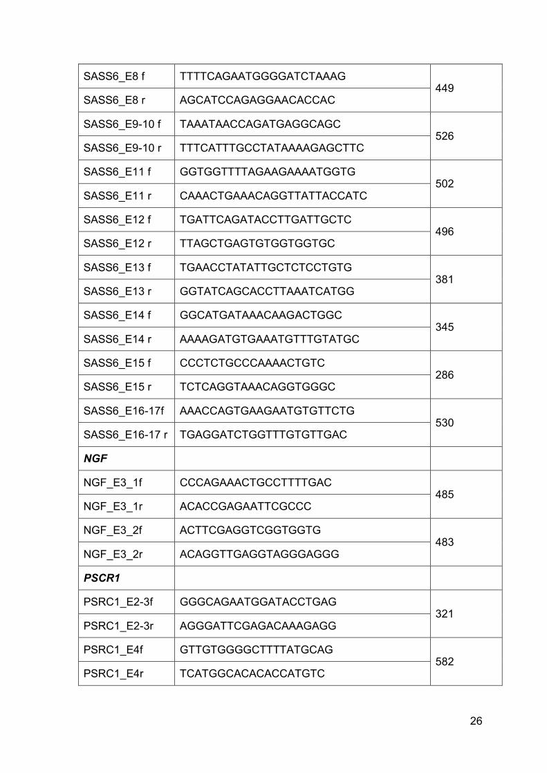
26
SASS6_E8 f TTTTCAGAATGGGGATCTAAAG 449
SASS6_E8 r AGCATCCAGAGGAACACCAC
SASS6_E9-10 f TAAATAACCAGATGAGGCAGC 526
SASS6_E9-10 r TTTCATTTGCCTATAAAAGAGCTTC
SASS6_E11 f GGTGGTTTTAGAAGAAAATGGTG 502
SASS6_E11 r CAAACTGAAACAGGTTATTACCATC
SASS6_E12 f TGATTCAGATACCTTGATTGCTC 496
SASS6_E12 r TTAGCTGAGTGTGGTGGTGC
SASS6_E13 f TGAACCTATATTGCTCTCCTGTG 381
SASS6_E13 r GGTATCAGCACCTTAAATCATGG
SASS6_E14 f GGCATGATAAACAAGACTGGC 345
SASS6_E14 r AAAAGATGTGAAATGTTTGTATGC
SASS6_E15 f CCCTCTGCCCAAAACTGTC 286
SASS6_E15 r TCTCAGGTAAACAGGTGGGC
SASS6_E16-17f AAACCAGTGAAGAATGTGTTCTG 530
SASS6_E16-17 r TGAGGATCTGGTTTGTGTTGAC
NGF
NGF_E3_1f CCCAGAAACTGCCTTTTGAC 485
NGF_E3_1r ACACCGAGAATTCGCCC
NGF_E3_2f ACTTCGAGGTCGGTGGTG 483
NGF_E3_2r ACAGGTTGAGGTAGGGAGGG
PSCR1
PSRC1_E2-3f GGGCAGAATGGATACCTGAG 321
PSRC1_E2-3r AGGGATTCGAGACAAAGAGG
PSRC1_E4f GTTGTGGGGCTTTTATGCAG 582
PSRC1_E4r TCATGGCACACACCATGTC

27
PSCR1_E5f ATTTCTTAATTCCTTAAGCCACAG 614
PSCR1_E5r ACAGCTCTGGGAGGGTGAG
PSCR1_E6-7f ACTAATCCCACACTCGCTCC 527
PSCR1_E6-7r CAGAATCTGCTGGAGTCAGG
WDR47
WDR47_E2f ATGTGTAGTTGCCAGGCAGT 354
WDR47_E2r CATAGCCTTTTCATGATTATAAAGTTC
WDR47_E3f TGCTGTTTTGTCATCTGTTCTG 271
WDR47_E3r TTCAAGGCCAGTGGAGAAAG
WDR47_E4f GGCTTCAATGTGATAAGAAGCCTA 300
WDR47_E4r TGCTAATCCTTCGTTTTAGTGAC
WDR47_E5af TCGTTTTCATATTTCATAGCATGT 494
WDR47_E5ar TGGAAGATTCTGAAGCCATGA
WDR47_E5bf GAAAGCGAAGTGCTTCTTGG 527
WDR47_E5br ACAGGCATAAGCCACTGCAC
WDR47_E6f GTCGGGGCTACAGAGAGAGA 357
WDR47_E6r TTTCCTACTCCCAAATATCAGAAAA
WDR47_E7f TGTTTCCGTGGAATAAAGCA 498
WDR47_E7r TTCAAATGCGTAAAAAGCCTTA
WDR47_E8f TGTGCATATTTTTACCTTTTCACAC 435
WDR47_E8r AAGGCTCCAAAGACAAGTTCA
WDR47_E9f GAAAAATTAGTGTTTGAAAGGGTAAA 240
WDR47_E9r TTCTTTGAATTGAAAATTTCTTAGCA
WDR47_E10f GGCAGTAAACCTAACATGATCCA 359
WDR47_E10r GGCAATTCTGAAAAAGGAACC
WDR47_E11f TGAAAACTATTAATAAGCTGTTGTCTG 383

28
WDR47_E11r AAGACAATACAGAAATACAACATGGAG
WDR47_E12f AAACCATTATTCTTGACCAGTTTTG 380
WDR47_E12r TGGACAGTAAGAGCAAGACCC
WDR47_E13f TATAGGCATGAGCCACCTTG 392
WDR47_E13r CAAATGCAAGTATTGTGAAAGAAC
WDR47_E14f ATTTTGTTTAGGCCTTGGGC 610
WDR47_E14r AAACCTAACAATAAGACTATTCACTGC
WDR47_E15f CAATCTATACATATACTTATTTTGCATTGTA 290
WDR47_E15r TGGACACTATGTTCTTCAGATTTGT
CAPZA1_cDNA_forw 5’GGAAGTTCACCATCACACCA and CAPZA1_cDNA_rev
5’GGCCTTGAATGTGGTATCTGA primers were used to investigate the effect of the
splice site mutation in exon 8 of CAPZA1 in the IDACC patients on mRNA level. To
verify the splice site mutation detected in Kif1bβ PCR primers Kif1b_mut_f (5’-
ATTGCTGTCTCTGTAGTAACTTTCTTG) and Kif1b_mut_r (5’-
AAGACCAAGCCACTGCACTT) were designed. All PCRs were performed using
HotStarTaq Master Mix Kit (Qiagen) with the following cycling conditions: 94°C for
15 min, followed by 35 cycles of 95°C for 25 s, 57°C for 30 s and 72°C for 1 min
with no final elongation step (subsequently referred to as ‘HS57’ program). The
sequencing reaction was set up with the Big Dye v3.1 cycling sequencing kit
(Applied Biosystems) according to the manufacturers’ protocol. Prior to sequence
analysis, dye remnants were removed with Centri-SepTM columns (Applied
Biosystems) and samples were bidirectionally sequenced on the ABI3130xl (Applied
Biosystems). Resulting data were analyzed with ChromasLite software
(Technelysium Pty Ltd.) and the UCSC browser (Kent, Sugnet et al. 2002, Fujita,
Rhead et al. 2011).
The impact of the mutation on splicing was investigated on mRNA level. For cDNA
generation, blood derived total RNA was transcribed with Omniscript RT Kit
(Qiagen). The region of interest was amplified by PCR (with KIF1b_Isocheck_f (5’-
TGAAGAAATTAGACCAAGCTCAGTG) and KIF1b_cDNA_r (5’-
TGTATTTCGACTGGCTCGG) using HotStarTaq Master Mix Kit (Qiagen) with the

29
following cycling conditions: 94°C for 15 min, followed by 35 cycles of 95°C for 25
s, 55°C for 30 s and 72°C for 1 min with no final elongation step. Sequencing was
performed as described above.
To check the presence or absence of exon 25 in the mRNA we used primers
kif1b_E25_check_f (5’- tggatttgatgcgagagatgt) and Kif1b_E25_check_r (5’-
acttcacctttctcactgacga) to amplify the region of interest with HotStarTaq Master Mix
(Qiagen) applying the same cycle conditions as mentioned above.
2.6 RT-PCR and qPCR
To measure absolute quantities of Kif1A, Kif1bα and Kif1bβ expressed in human
fetal brain total RNA (Clontech), U2OS cells and lymphoblastoid cells of patients
and a healthy control, 1µg RNA was transcribed with QuantiTecht reverse
transcription kit (Qiagen). To generate templates for the corresponding standard
curves, first, a conventional PCR using HotStar Taq Mastermix and HS57 program
was performed to amplify Kif1bβ, Kif1bα and Kif1A target sequences using following
primer pairs: Kif1b_alpha_cDNA_f (5’- TCTTCTTTTGGAGCAGCAGAG) and
Kif1b_alpha_cDNA_r (5’- TTTCCAGCTCTCTTCACACG), Kif1b_beta_cDNA_f (5’-
CTCACCCACACTTTCAACAGAG) and Kif1b_beta_cDNA_r (5’-
TGCTGAAACTGGACTCAGAGG) as well as KIF1A_RTf (5’-
CGGACATAGGCCACAGATG) and KIF1A_RTr (5’-
CGTGTGACTGGTGTGTACGA). Subsequently, the PCR products were run on a
1% agarose gel for 30min at 130V, and corresponding lanes were cut out and
extracted using GenElute Gel Extraction Kit (Sigma). DNA amounts were measured
with Qubit 2.0 Fluorometer and pre-diluted to 1pg. To create standard curves 1:5
(for KIF1A: 1000, 200, 40, 8, 1.6, 0.32, 0.064, 0.0128fg) and 1:3 (for Kif1bα and
Kif1bβ: 1000, 333.3, 111.1, 37.04, 12.34, 4.12, 1.37, 0.457fg) serial dilution series
of the corresponding template DNAs were prepared. Sample cDNAs were applied
either diluted or undiluted (according to Qubit (Invitrogen, ThermoFisher)
concentrations) to make sure that concentrations were within the standard curve.
To measure relative quantities of Kif1b isoform expression in brain and muscle, RNA
from muscle tissue was isolated as described recently (Y. Xue, Schoser et al. 2016).
cDNAs were transcribed as mentioned above and levels were measured relative to
30
the expression of 18s rRNA (primer 18s_f 5’GAGACTCTGGCATGCTAACTAG and
18S_r 5’GGACATCTAAGGGCATCACAG).
All reactions were set up in triplicates with 10µl Fast SYBR Green master mix
(Applied Biosystems), 8µl sterile water, 1µl cDNA and 1µl of a 10µM primer mix on
the StepOne Plus Real-time PCR System (Applied Biosystems) according to the
manufacturer’s instructions. PCR was performed using following cycling conditions:
a holding stage of 95°C for 20min, followed by a cycling stage of 40 cycles at 95°C
for 3 sec and 60°C for 30sec. Finally, the melt curve was generated at 95°C for
15sec, 60°C for 1min and a final increase of 0.3°/sec from 60 to 95°C where the
temperature was held for another 15sec. Data were analyzed with the StepOnePlus
Software v.2.3 and quantities, fold changes and diagrams were then calculated and
generated in Excel according to the guidelines from applied biosystems.
2.7 HsSAS-6 homology analysis in Jalview
HsSAS-6 Ile62 homology among nine different species (from NCBI Homo sapiens
(AAI01027), Danio rerio (AAI65167), Pan troglodytes (JAA36473), Aligator
mississippienis (XP_006277337), Apis melifera (XP_395972), Mus musculus
(NP_082625), Caenorhabditis elegans (CAA16384), Drosophila melanogaster
(AAF56983), Chlamydomonas reinhardtii (BAF94334)) was investigated with the
desktop version of Jalview2.8 (Waterhouse, Procter et al. 2009). For Kif1bβ, PH
domain amino acid sequences of following species were aligned in Jalview 2.10.3
(accession numbers according to UniProt): Homo sapiens (O60333), Rattus
norvegicus (O88656), Mus musculus (Q60575), Danio Rerio (C0LSN9_DANRE),
Drosophila melanogaster (A1ZAJ2), Alligator sinensis (A0A1U8DAW3_ALLSI),
Erinaceus europaeus (A0A1S3WSN7_ERIEU), Gallus gallus (E1C889_CHICK) and
and Xenopus tropicalis (F7DRH5_XENTR). Sequences were aligned with MafftWS
alignment using the default settings (Katoh, Kuma et al. 2005). Regions that were
highly conserved in terms of their hydrophobicity are highlighted in red and blue.
31
2.8 Kif1bβ splice site prediction
We analyzed the mutagenic potential of the splice site variant with the ‘Berkley
Drosophila Genome Project: Splice Site Prediction by Neural Network’ online tool
(available at http://www.fruitfly.org/seq_tools/splice.html) (Reese, Eeckman et al.
1997). We applied 0.40 as the donor and acceptor score cutoff value.
2.9 Cloning
2.9.1 Kif1bβ
WT Kif1bβ cDNA was amplified from Human Fetal Brain Marathon Ready cDNA
(Clontech) with Phusion Hot Start II High-Fidelity Polymerase (Thermo Scientific)
using the GC buffer and kif1b_cDNAf_long (5’-
TAAAATGTCGGGAGCCTCAGTGAAG) and kif1b_cDNAr_long (5’-
TGTATTTCGACTGGCTCGGGC) in a two-step protocol: 98°C for 30 sec, followed
by 40 cycles of 98°C for 10sec and 72°C for 3min28sec, and a final elongation step
at 72°C for 10min. We derived multiple PCR products which were separated on a
0.8% agarose gel. The correct band was extracted with the GenElute Gel Extraction
Kit (Sigma) and eluted in 30µl H2O. Subsequent A-tailing of 25µl of the eluted
product was performed with Taq DNA Polymerase with Standard Taq Buffer, 1.5
mM MgCl2 (all from New England Biolabs) and 200µM dATP (Roche) for 14min at
72°C. 4µl of the A-tailing mix was used for cloning into a eukaryotic expression
vector with the pcDNA™3.1 vector⁄ CT-GFP-fusion TOPO® Expression kit
(Invitrogen, Life Technologies) in order to create pcDNA3.1-Kif1Bβ-WT-GFP.
For pcDNA3.1-kif1Bβ-MUT-GFP, we first amplified an extended version of Kif1b
beta (up to the new Stop codon, resulting from the frame shift) from human fetal
brain cDNA using the same cycle conditions as for the WT construct and the
following primer pair: kif1b_cDNAf_long and kif1b_Mut_cDNAr (5’-
CCAAAGAGAAATGAGAGGTAACGCTTTC). After A-tailing, the product was
cloned into pcDNA™3.1 vector⁄ CT GFP-fusion TOPO®. Clones with the same
isoform as the WT construct were selected for in-vitro mutagenesis. The
mutagenesis reaction was set up with Ultra Pfu HF DNA Polymerase (Stratagen)
and 5’ phosphorylated primers kif1b_mut_rev (5’-
CTTCACCATGGCCTGCTGGTCCTCACT) and kif1b_mut_f (5’-

32
GTCAAAGCTTTCCCGCAGATGCCCGA). An initial step of 2min at 92°C was
followed by 10 cycles at 92°C for 10sec, 59°C for 30sec and 68°C for 23min with a
subsequent 20 cycles at 92°C for 10sec, 59°C for 30sec and 68°C for
23min+10sec/cycle. The 11kb lane was isolated from a 0,8% agarose gel with
GenElute Gel Extraction kit (Sigma), and the linear PCR products were ligated with
Quick ligase (New England Biolabs) and transformed into TOP10 chemical
competent cells (LifeTechnologies). For the cloning of NT-GFP-Kif1bbeta_CtailWT
and NT-GFP-Kif1b_CtailMUT, the Kif1bβ tail region was amplified from the
corresponding pcDNA3.1-Kif1bβ-WT-GFP or pcDNA3.1-kif1bBβ-mut-GFP plasmid,
respectively, with primers Kif1b_PH_NTf (5’-
ATGGTCCCAGCTGTGGAAACACCATATT) and and Kif1b_PH_NTr (5’-
TTATTTCGACTGGCTCGGGC) or Kif1b_deltaPH_NTr (5’-
TTACAAAGAGAAATGAGAGGTAACGCTTTC) using HotStarTaq Mastermix
(Qiagen). The following cycling conditions were used: initial 15min at 94°C followed
by 35 cylces of 95°C for 25sec, 57°C for 30sec and 72°C for 1min. Resulting PCR
products were directly used in the cloning reaction with pcDNA™3.1 vector⁄ NT-GFP
TOPO® vector (LifeTechnologies). Clones containing correctly orientated
sequences were identified by colony PCR using primers T7f (5´-
TAATACGACTCACTATAGGG) and Kif_fulllength_r (5’-
GATCTTCGGGTGAGGTATGAG) for full-length Kif1bβ WT and MUT constructs or
GFP_f (5´-CGACACAATCTGCCCTTTCG) and Kif1b_PH_NTr for WT and MUT C-
terminal constructs. To cover all exons by sequencing, in addition to T7f and GFP_f
primers, the following primers listed in Table 4 were used.
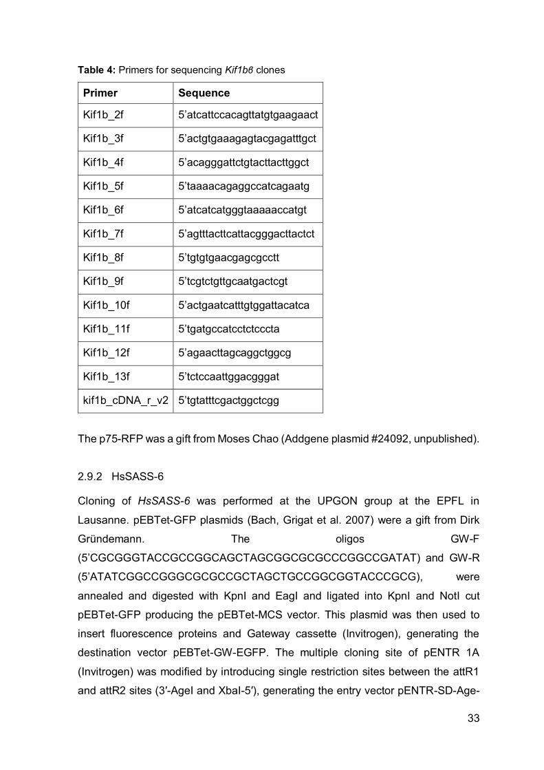
33
Table 4: Primers for sequencing Kif1bβ clones
Primer Sequence
Kif1b_2f 5’atcattccacagttatgtgaagaact
Kif1b_3f 5’actgtgaaagagtacgagatttgct
Kif1b_4f 5’acagggattctgtacttacttggct
Kif1b_5f 5’taaaacagaggccatcagaatg
Kif1b_6f 5’atcatcatgggtaaaaaccatgt
Kif1b_7f 5’agtttacttcattacgggacttactct
Kif1b_8f 5’tgtgtgaacgagcgcctt
Kif1b_9f 5’tcgtctgttgcaatgactcgt
Kif1b_10f 5’actgaatcatttgtggattacatca
Kif1b_11f 5’tgatgccatcctctcccta
Kif1b_12f 5’agaacttagcaggctggcg
Kif1b_13f 5’tctccaattggacgggat
kif1b_cDNA_r_v2 5’tgtatttcgactggctcgg
The p75-RFP was a gift from Moses Chao (Addgene plasmid #24092, unpublished).
2.9.2 HsSASS-6
Cloning of HsSASS-6 was performed at the UPGON group at the EPFL in
Lausanne. pEBTet-GFP plasmids (Bach, Grigat et al. 2007) were a gift from Dirk
Gründemann. The oligos GW-F
(5’CGCGGGTACCGCCGGCAGCTAGCGGCGCGCCCGGCCGATAT) and GW-R
(5’ATATCGGCCGGGCGCGCCGCTAGCTGCCGGCGGTACCCGCG), were
annealed and digested with KpnI and EagI and ligated into KpnI and NotI cut
pEBTet-GFP producing the pEBTet-MCS vector. This plasmid was then used to
insert fluorescence proteins and Gateway cassette (Invitrogen), generating the
destination vector pEBTet-GW-EGFP. The multiple cloning site of pENTR 1A
(Invitrogen) was modified by introducing single restriction sites between the attR1
and attR2 sites (3′-AgeI and XbaI-5′), generating the entry vector pENTR-SD-Age-

34
AGT. Full length HsSASS-6 (NM_194292.1) was amplified using the primers Age-
Ko-HsSAS6-F (5’CGCGACCGGTACCATGAGCCAAGTGCTGTTCCAC) and Xba-
noST-S6-R (5’CGCGTCTAG ATAACTGTTTGGTAACTGCCCA), and cloned into
pENTR-SD-Age vector by restriction digest with AgeI and XbaI.
Mutations of the I62 residue in HsSAS-6 were generated by site-directed
mutagenesis on pENTR-SD-Age-HsSAS-6 using the following primers: S6-I62T-
fwd, ACATCTGAGGAAGATTTTCAAAGT and S6-I62T-rev,
AACAAGGTTATATAAAAAAAATGG (mutated codon is italicized). Gateway
reaction was then performed according to the manufacturer's protocol to generate
the expression plasmids pEBTet-HsSAS-6-GFP and pEBTet-HsSAS-6-I62T, which
were sequence verified’ (Khan, Rupp et al. 2014).
All plasmids were isolated with EndoFreePlasmid Maxi Kit (Qiagen).
2.10 Cell culture and transfection
2.10.1 Kif1bβ
The human choriocarcinoma cell line JAr (ATCC) was cultured as previously
described. U-2 OS cells were kindly provided by the ‘Cell Culture Core Facility’ of
the Medical University of Graz. They were maintained in DMEM with 10% FBS and
1% Pen/Strep (all from Life Technologies) at 37°C and 5% CO2. Cells were
transfected with pcDNA3.1-kif1bβ-WT and FuGENE HD Transfection Reagent
(Promega) at 50% confluence with a ratio of 3,5:1 (FuGENE:DNA) according to the
manufacturer’s protocol. For co-transfection with p75-RFP, cells were first
transfected with the pcDNA3.1-kif1bβ-WT plasmid applying a 4:1 ratio. The day
after, cells were transfected with p75-RFP (addgene #24092) applying a 3:1 ratio of
FuGENE:DNA. 24h later, cells were monitored under an array confocal laser
scanning microscope (ACLSM) built on a fully automated inverse microscope (Axio
Observer.Z1, Zeiss) at the Institute of Biochemistry of the Medical University of
Graz.
For electroporation 4x10⁶ cells were transfected with 12µg plasmid DNA using the
NEON transfection system (Invitrogen). Cells were co-transfected with 500ng p75-
RFP and 13µg pcDNA3.1-GFP-Kif1bβ-delta-PH or pcDNA3.1-GFP- kif1bβ-PH. The
following conditions were used: 1230V, 10ms, 4 Pulse. Cells were analyzed 18h
later.
35
To generate lymphoblastoid cell lines, lymphocytes were isolated from heparinized
peripheral blood by density gradient centrifugation using Lymphocyte Separation
Medium LSM 1077 (PAA) according to the manufacturers protocol. Lymphozytes
were further resuspended in 2ml of blood media (RPMI 1640 with hepes, 10%FBS,
1%Pen/Strep; all from Gibco, Life Technologies) and supplemented by the same
volume of filtered B-95-8 culture supernatant. After 24h at 37°C half of the media
was removed and new media with 3-4 drops of cyclosporine (Sandoz) was added
to the cells. Cells were maintained in cyclosporine supplemented media for 3 more
weeks. When cells had finally reached a high division rate, media was changed to
normal blood media (see above).
2.10.2 HsSASS-6
Transfection and co-localization studies were performed at the UPGON group in
Lausanne. U2OS cells were obtained from the EACC and maintained in McCoy's
5A GlutaMAX medium (Invitrogen) supplemented with 10% fetal bovine serum
(FBS) for U2OS cells or tetracycline-negative FBS (Brunschwig) for the inducible
episomal cell lines (iU2OS). To generate such iU2OS lines, U2OS cells were
transfected with pEBTet-HsSAS-6-GFP or pEBTet-HsSAS-6[I62T]-GFP using
Lipofectamine2000 (Invitrogen). Transfected cells were selected with 1 μg/ml
puromycin one day after transfection and amplified. Early passage cells were used,
inducing expression with 1ug/ml doxycycline for 48 hr.
Endogenous HsSAS-6 was depleted using a Stealth RNAi siRNA (Invitrogen)
targeting the 3′UTR of HsSAS-6 (5-GAGCUGUUAAAGACUGGAUACUUUA-3).
Stealth RNAi siRNA negative control LO GC (Invitrogen) was used as a control.
siRNA transfection was performed using Lipofectamine RNAiMax (Invitrogen)
according to the manufacturer's protocol, and cells were analyzed 48-72 hr after
siRNA treatment (Khan, Rupp et al. 2014).
36
2.11 Cell-extract preparation and biochemical assays
2.11.1 HsSAS-6
Cells were collected, washed in PBS, and lysed on ice for 1 hour in lysis buffer
(50 mM Tris-HCl [pH 7.5], 150 mM NaCl, 0.5 mM EDTA, 0.5% NP-40, Complete
Mini Protease Inhibitor Cocktail [Roche Diagnostics]). Lysates were cleared by
centrifugation for 10 min at 12,000 × g at 4°C and the supernatant collected. SDS-
PAGE was performed using 10% polyacrylamide gels (BioRad), followed by transfer
on nitrocellulose membrane (Amersham). The membrane was probed with mouse
HsSAS-6 antibody (Santa Cruz, 1:1000) or mouse Tubulin antibody (Sigma,
1:10000), followed by incubation with their respective HRP-conjugated secondary
(Promega) and the signal detected with chemiluminescence. Western Blots were
performed at the UPCON group at the EPFL in Lausanne.
2.11.2 Kif1Bβ
For protein isolation, cells were washed twice with ice cold PBS and lysed with 100µl
lysis buffer (50 mM HEPES, 150 mM NaCl, 1 mM EDTA, 10 mM Na4P2O7, 2 mM
Na3VO4, 10 mM NaF, 1% (v/v) Triton X-100, 10% (v/v) glycerol and Complete Mini
protease inhibitor cocktail tablets; pH 7.4) for 10 min on ice (Semlitsch, Shackelford
et al. 2011). Scraped cells were centrifuged at 13,000 rpm and 4°C for 10 min to
remove insoluble cell debris. Protein contents of cell lysates were determined using
the BCATM Protein Assay Kit (Thermo Scientific) according to the manufacturer’s
instructions.
Fifty µg of total protein was added to 10 µl of 4x NuPAGE LDS sample buffer
(Invitrogen) containing 2 µl sample reducing agent (Invitrogen) and heated (70°C,
10 min). Proteins were separated by electrophoresis on 6% Bis-Tris gel and
transferred to nitrocellulose (Invitrogen, 40 µM). Membranes were blocked with 5%
(w/v) non-fat milk in TBST (Tris-buffered saline containing Tween-20) (25°C, 2 h)
and incubated with polyclonal rabbit anti-KIF1Bβ (1:2000, Bethyl, A301-055A)
overnight at 4°C. After washing, membranes were incubated with HRP-conjugated
goat anti-rabbit IgG (Biomol-8101102, 1:100,000 or Pierce, 1:50,000) at 25°C for
2h. Immunoreactive bands were visualized using Super Signal West Pico
Chemiluminescent substrate (Thermo Scientific) and developed using Bio-Rad
ChemiDoc MP Imaging System.

37
Co-immunoprecipitation was performed by incubating protein lysates (1 mg) from
WT, V:1, VI:2, and VI:4, respectively; with anti-KIF1Bβ antibody (1 µg) according to
the manufacturer’s suggestions (GE healthcare). Immunoprecipitates were
separated by 6% (KIF1Bβ) or 15% (CaM) SDS-PAGE Bis-Tris gels and transferred
to nitrocellulose membranes. Immunoreactive bands were visualized using anti-
KIF1Bβ or rabbit polyclonal anti-CaM (1:500, Santa Cruz, sc-5537) as the primary
antibody followed by HRP-labelled secondary antibody as described above. This
work was conducted at the Institute of Biochemistry at the Medical University of
Graz.
2.12 Immunofluorescence and microscopy for hsSASS-6
transfected cells
Centriole and monopolar spindle quantification was performed at the UPCON group
at the EPFL in Lausanne. U2OS cells grown on glass coverslips were fixed for 7–
10 min in –20°C methanol, washed in PBS, and blocked in 1% bovine serum
albumin and 0.05% Tween-20 in PBS. Cells were incubated 2 hr at room
temperature with primary antibodies, washed three times for 10 min in PBST (0.05%
Tween-20 in PBS), incubated 45 min at room temperature with secondary
antibodies, stained with ∼1 μg/ml Hoechst 33258, washed three times in PBST, and
mounted. Primary antibodies were 1:4000 mouse centrin (20H5; gift from Jeffrey L.
Salisbury) and 1:500 rabbit GFP (gift from Viesturs Simanis). Secondary antibodies
were 1:1000 goat anti-rabbit coupled to Alexa 488 and 1:1000 goat anti-mouse
coupled to Alexa 568. For quantification of centrioles, mitotic cells (prophase to
metaphase) with similar cytoplasmic GFP expression were used; highly expressing
cells that often harbored GFP aggregates were not retained for analysis. Imaging
was done on a Zeiss LSM710 confocal microscope. Optical sections were acquired
every 0.12μm, and planes containing centrioles were projected together. Images
were processed using ImageJ and Adobe Photoshop, preserving the relative image
intensities within a series.
2.13 Confocal Imaging for Kif1Bβ and p75 transfected cells
Co-Localization of p75 and KIF1Bβ was investigated by recording GFP and RFP
signals using the array confocal laser scanning microscope (ACLSM) built on a fully

38
automatic inverse microscope (Axio Observer.Z1, Zeiss) equipped with VoxCell
Scan® (VisiTech, Visitron Systems) using a 100 × objective (Plan-Fluor 100 × /1.45
oil, Zeiss) at the Institute of Biochemistry of the Medical University of Graz. Live cells
were cultured and transfected on glass slides (see 2.10.1) and excited with diode
lasers (Visitron Systems); GFP was excited at 473nm (50mW) and RFP was excited
at 561nm (50mW). Emitted light was collected with filters ET525/50m for GFP and
ET480/40m for RFP (Chroma Technologies Corporation). Pictures were captured
using a photometrics CCD camera (CoolSnap HQ2, Photometrics), Images were
processed with ImageJ (Schindelin, Arganda-Carreras et al. 2012, Schneider,
Rasband et al. 2012) and movies were subsequently generated with MetaMorph
7.7.0.0 software (Visitron) (Bischof, Rehberg et al. 2017, Waldeck-Weiermair,
Bischof et al. 2015).
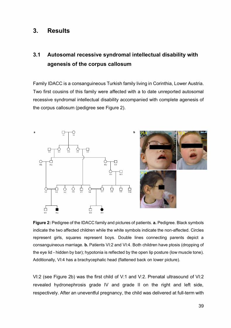
39
3. Results
3.1 Autosomal recessive syndromal intellectual disability with
agenesis of the corpus callosum
Family IDACC is a consanguineous Turkish family living in Corinthia, Lower Austria.
Two first cousins of this family were affected with a to date unreported autosomal
recessive syndromal intellectual disability accompanied with complete agenesis of
the corpus callosum (pedigree see Figure 2).
Figure 2: Pedigree of the IDACC family and pictures of patients. a. Pedigree. Black symbols
indicate the two affected children while the white symbols indicate the non-affected. Circles
represent girls, squares represent boys. Double lines connecting parents depict a
consanguineous marriage. b. Patients VI:2 and VI:4. Both children have ptosis (dropping of
the eye lid - hidden by bar); hypotonia is reflected by the open lip posture (low muscle tone).
Additionally, VI:4 has a brachycephalic head (flattened back on lower picture).
VI:2 (see Figure 2b) was the first child of V:1 and V:2. Prenatal ultrasound of VI:2
revealed hydronephrosis grade IV and grade II on the right and left side,
respectively. After an uneventful pregnancy, the child was delivered at full-term with

40
a birth weight of 2,610 g (date of birth: October 2007). The lab values were within
the normal range, beside a cirrhosis of the right kidney associated with
vesicoureteral reflux grade IV-V and a grade III reflux on the left side. At the age of
three months, bilateral ptosis and nystagmus were diagnosed. Subsequent brain
magnetic resonance tomography (MRT) at the age of four months revealed a
complete agenesis of the corpus callosum, a hypoplastic gyrus cinguli, deformation
of both frontal horns and slightly enlarged occipital horns (colpocephaly).
Furthermore, the third ventricle was slightly enlarged and reached cranial to the
interhemispheric fissure between the lateral ventricles and communicated with the
interhemispheric fissure anteriorly. Basal ganglia, posterior cranial fossa and
myelination were normal (see Figure 3). Developmental delay, hypotonia with
delayed motor development and some mild dysmorphic features were diagnosed
after the first month of life. Chromosome analysis was inconspicuous showing a
normal male karyotype (46,XY). At four months of age he had 5.5 kg, and his head
circumference was 40.5 cm. At 10 months of age his growth parameters were at the
3rd percentile. Around his first birthday his weight was 7.8 kg, and the head
circumference was 45 cm. In addition to the known bilateral ptosis ophthalmologic
examination at the age of two (26 months) revealed strabismus that could be due to
oculomotor nerve palsy. At four years of age, a neurologic examination diagnosed
an atactic gait with an external rotation of the right leg. There was no evidence for a
peripheral nerve disorder, confirmed by a nerve conduction study, which showed no
evidence for an axonal or demyelinating disorder. His fine motoric skills are
impaired, spoken language and expressive speech development is severely
retarded. He has issues maintaining eye contact, exhibits significant attention and
concentration problems and is reported to be a restless child.
V:6 had four pregnancies, including one still birth in the 20th week of gestation. A
second pregnancy revealed bilateral hydronephrosis and megacystis during an
ultrasound examination. Due to these findings the pregnancy was terminated
despite showing a normal male karyotype (46, XY) during prenatal testing.
The pregnancy with VI:4 was normal. The girl (Figure 2b) was delivered at full term
(date of birth: February 2004), with a birth weight of 3,730 g and lab values that were
within the normal range. Chromosome analysis showed a normal female karyotype
(46,XX). Within the first year of life severe developmental delay with a congenital
left facial nerve paresis was diagnosed. As for patient V:2, brain MRTs revealed
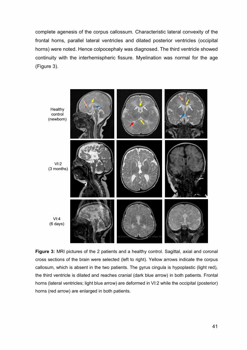
41
complete agenesis of the corpus callossum. Characteristic lateral convexity of the
frontal horns, parallel lateral ventricles and dilated posterior ventricles (occipital
horns) were noted. Hence colpocephaly was diagnosed. The third ventricle showed
continuity with the interhemispheric fissure. Myelination was normal for the age
(Figure 3).
Figure 3: MRI pictures of the 2 patients and a healthy control. Sagittal, axial and coronal
cross sections of the brain were selected (left to right). Yellow arrows indicate the corpus
callosum, which is absent in the two patients. The gyrus cingula is hypoplastic (light red),
the third ventricle is dilated and reaches cranial (dark blue arrow) in both patients. Frontal
horns (lateral ventricles; light blue arrow) are deformed in VI:2 while the occipital (posterior)
horns (red arrow) are enlarged in both patients.
42
At two months of age she weighted 5,500 g (>97th percentile), was 60 cm tall (>97th
percentile), and had a head circumference of 36.5 cm (25th – 50th percentile). At 8.2
kg (>25th percentile) and 74 cm (>50th percentile), her weight and size were still
within the normal range by the age of 10 months. However, her head circumference
was clearly below the 3rd percentile (41cm). Though her vision was normal, at one
year and two months of age, ophthalmologic examination revealed bilateral ptosis,
strabismus, and a congenital oculomotor nerve palsy. At one year and seven
months she was still not able to walk and her motor movements were reduced. At 3
years and 7 months her weight and height were still within the normal range for the
given age (12.5 kg [3rd - 10th percentile], 98 cm [>50th percentile]), whereas her head
circumference was significantly smaller compared with children her age (44cm). She
showed a hypoton-atactic gait which was slightly asymmetric. She understood and
followed instructions relatively well but had problems paying attention. A
neurological examination by the age of 11 ½ found pronounced dysmorphic facial
features and aggressive behavior. Due to ocular motor apraxia she exhibited
dystonic movements of her head. Her coordinative functions were strongly reduced,
intellectual development was severely delayed and cognitive functions were
impaired. She was reported to show motoric clumsiness, speech and language
delay. There was no evidence for a peripheral neurologic disorder.
The clinical data of both patients are summarized in Table 5.

43
Table 5: Clinical data of IDACC patients
Pedigree ID VI:2 VI:4
Chromosomes 46,XY 46,XX
Kidneys hydronephrosis normal
Delivery Full-term Full-term
Apgar Normal Normal
Birth weight 2,610 g 3,730 g
Eyes Bilateral ptosis
Strabismus
Ophtalmoplegia
Bilateral ptosis
Strabismus
Brain Complete agenesis of the
corpus callossum
3rd ventricle enlarged reaching
to the interhemispheric fissure
Deformation of both frontal
horns
Enlarged occipital horns
Mild microcephaly/
brachycephaly
Complete agenesis of the
corpus callossum
3rd ventricle enlarged reaching
to the interhemispheric fissure
Deformation of both frontal
horns
Enlarged occipital horns
Cranial nerves Congenital oculomotor nerve
palsy
Congenital oculomotor nerve
palsy
Congenital left facial nerve
paresis
Growth parameters All at the 3rd percentile Head circumference below 3rd
percentile
Dysmorphic facial
features
Mild More pronounced
Motor development Severely delayed
Hypoton
Severely delayed
Hypoton
Gait Atactic Atactic
ID Severely delayed Severely delayed
EEG Normal Normal
Speech 5 words 5 words
Sense of hearing Normal Hearing aid on left ear
ID=intellectual development; EEG= electroencephalography

44
We collected blood from both affected children (VI:2 and VI:4), the mother (V:1) and
the brother (VI:1) of VI:2, as well as from the parents (V:6 and V:7) of VI:4. In addition
to DNA and RNA isolation, lymphoblastoid cell lines were generated from the blood
of both affected and from V:1. As mentioned above, karyotyping revealed no
chromosomal abnormalities (Figure 4).
Figure 4: Chromosome analysis of lymphoblastoid cells shows a normal karyotype
(chromosomes of VI:2)
The affected children’s consanguineous background suggested the disease-
causing gene to be located in an autozygous region of the genome. Hence a SNP
250K array was conducted for both affected. Analysis of the data in dChip revealed
four homozygous stretches with identical haplotypes on chromosome 1p36.31-
p36.32 (6,775 MB), 2q22.1-q23.3 (8,534MB), 3p14.1 (2.86MB) and 9q21.13-q21.2
(positions according to chromosome build hg17) (Figure 5).

45
Figure 5: 250K NspI SNP array of patients VI:2 and VI:4. Blue and yellow bars represent
homozygous and heterozygous regions, respectively. Identical red and blue SNP
haplotypes depict autozygous regions. Red is homozygous for SNP A, blue is homozygous
for SNP B, yellow is heterozygous for SNP A and B. SNPs restricting the homozygous
stretch are noted as rs numbers with their corresponding location on the chromosomes
(according to genome build hg17).
Due to the large number of genes located in these loci, we decided to send the DNA
for whole exome sequencing (WES). Since data received from WES were matched
with the hg19 build, positions of our loci changed to 1p36.31-p36.21 (7,141,204-
14,105,298bp), 2q22.2-q23.3 (142,498,632-151,041,667bp), 3p14.1 (66,486,380-
69,346,680bp) and 9q21.13-q21.2 (77,535,512-80,906,946bp). Filtering for rare and

46
high quality variants revealed only three candidate genes all located in the locus on
chromosome 1, including two nonsynonymous mutations with reported rs numbers
in PRAMEF11 (c.A308G; p.E103G; rs2994114) and LOC440563 (c.A434G;
p.H145R; rs28713604) and an unknown splice site mutation c.5270+2T>G in the
donor splice site of exon 47 in the β isoform of the KIF1b gene (NM_015074.3; exon
number according to the longest isoform of Kif1bβ). The RNA expression data on
UCSC clearly showed that PRAMEF11 and LOC440563 were both only very weakly
expressed in brain tissue whereas KIF1b displayed an extremely high expression
pattern in diverse brain regions and in skeletal muscle. Moreover, we were not able
to find the mutation in the NHLBI Exome Variant Server
(http://evs.gs.washington.edu/EVS/). Zhao et al found that Kif1Bβ is a kinesin
required for synaptic vesicle precursor transport in neurons (Zhao, Takita et al.
2001). Most interestingly, in the same paper they associated this gene with an
autosomal dominant hereditary motoneuropathy, Charcot-Marie-Tooth disease type
2A. Brain sections of the Kif1b knock-out mouse generated by this research group
showed an impaired development of the commissural fibers, which is in accordance
with the MRI data from our patients. With regards to these background data our
hypothesis that Kif1bβ was our candidate gene was therefore strengthened.
To check whether the mutation indeed affects splicing, we carried out several
experiments to confirm its mutagenic potential. First, we used the online splice site
prediction program on http://www.fruitfly.org. By entering the last 2382bp’s of the
genomic wildtype and mutant sequence of Kif1bβ, the program calculated the donor
and acceptor splice site probabilities. In the wildtype sequence the donor splice site
achieved the highest maximum score of 1 while the donor splice site was lost in the
mutated sequence (Figure 6). The predicted acceptor splice sites, however,
remained unaffected.

47
Figure 6: Splice site prediction using www.fruitfly.org. The maximum score for a splice site
prediction in the wildtype (WT) sequence was achieved for the position between 1162 and
1176bp’s containing our target splice site (red square). In the mutant (MUT), however, the
splice site was predicted to be lost.
To verify the mutation, we performed Sanger Sequencing on the DNA of all affected
and unaffected family members. Only the patients were homozygous for the splice
site mutation, while the unaffected were all heterozygous carriers (Figure 7).

48
Figure 7: Sanger sequencing confirmed the c.5270+2T>G transition in the long isoform of
Kif1b, Kif1bβ. The red arrow marks the mutation site. Exons in orange are present in the
longest known isoform of Kif1bβ but are not included in the refseq sequence of
NM_015074.3. The affected are homozygous for the mutation; the unaffected are
heterozygous.
In parallel, we analyzed the segregation of the disease locus on chromosome 1
within the IDACC family using highly polymorphic markers D1S214 (6.962 Mb),
D1S450 (9.585 Mb) and D1S2667 (11.486 Mb) (positions according to genome build
hg19). This confirmed the disease-specific autosomal recessive inheritance pattern
of the red haplotype, as only the affected were homozygous for the mutation and
had two of the red alleles, while the healthy family members were heterozygous for
the disease haplotype (Figure 8).

49
Figure 8: Segregation analysis. Highly polymorphic microsatellite markers confirm that only
patients VI:2 and VI:4 are homozygous for the same haplotype (red) including the
c.5270G+2T>G transition, while the non-affected family members are all heterozygous
carriers.
To investigate how the mutation affects splicing, we analyzed the cDNA of VI:2 and
VI:4. Primers located within exon 45 and 48 to span the entire exon 47 were
selected. No alternative splice variants are known for this region. Therefore, all PCR
products were expected to show the same size if the mutation has no impact on
splicing. Already in the gel it was visible that the amplified product of both patient
cDNAs was significantly smaller in size than the band derived from the human fetal
brain control PCR (Figure 9A). Sanger sequencing confirmed the loss of exon 47
and the fusion of exon 46 with exon 48 (Figure 9B).

50
Figure 9: cDNA analysis of VI:2 and VI:4. a. PCR from human fetal brain (HFB) cDNA
reveals a band at the expected size around 371bp while the PCR product of both patients
is approximately 100bp smaller. Exon 47 is 119bp in size, which would be in line with the
observed shift in size. No remaining WT splicing can be recognized on the gel. b. Sanger
sequencing confirmed the fusion of exon 46 and 48 upon splicing. Sequence alignment
using BLAT in the UCSC browser displays the missing exon (alignment of cDNA sequences
of VI:4 are indicated in black).
Frameshift mutations eventually might lead to mRNA degradation after transcription.
We thus questioned, if this alteration has a potential impact on mRNA stability. We
therefore measured the total quantities of both Kif1b isoforms in lymphoblastoid cells
derived from patients and an unrelated control LBC line and calculated its relative
quantities to the amount of Kif1bβ expressed in brain (Figure 10a).

51
As the shorter α isoform is not affected by the mutation, we expected its expression
profile to remain consistent in all samples tested. qPCR data revealed equal mRNA
levels in patients and the healthy controls. Moreover, this experiment also confirmed
previously published data showing the much higher expression level of Kif1bβ in the
brain compared with Kif1bα (Conforti, Dell'Agnello et al. 2003).
We further wanted to exclude a nonsense-mediated decay on protein level, as
protein and mRNA expression levels do not always correlate. To test this, we
performed a Western Blot in the laboratory of Ernst Malle comparing the expression
in lymphoblastoid cells of both affected, the heterozygous mother of VI: 2, V:I, and
two cancer cell lines (Hela and Jar). For this, we chose an antibody from Bethyl
derived against the amino acid residues between 1575 and 1625 amino acids
(CAC16629.1; GeneID 23095), targeting specifically the β isoform but not the
mutated tail area. While KIF1Bβ levels in the lymphoblastoid cells were equal, the
cancer cell lines the amount of KIF1Bβ protein varied, as expected (Figure 10b).
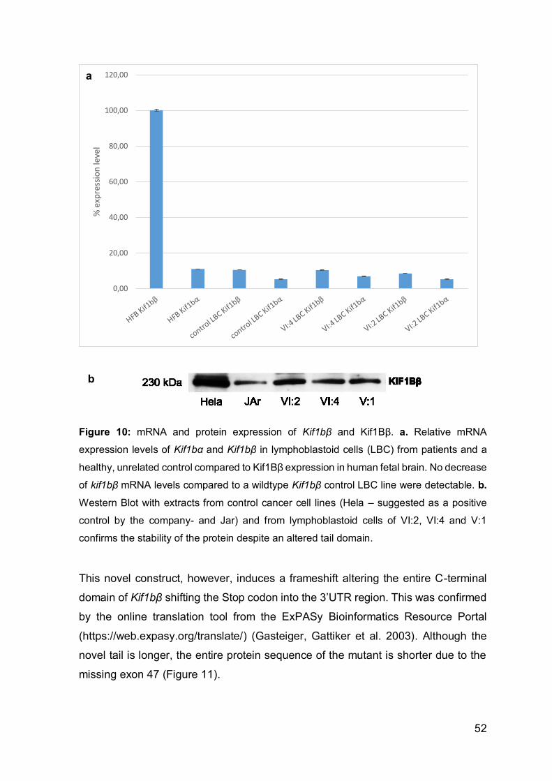
52
Figure 10: mRNA and protein expression of Kif1bβ and Kif1Bβ. a. Relative mRNA
expression levels of Kif1bα and Kif1bβ in lymphoblastoid cells (LBC) from patients and a
healthy, unrelated control compared to Kif1Bβ expression in human fetal brain. No decrease
of kif1bβ mRNA levels compared to a wildtype Kif1bβ control LBC line were detectable. b.
Western Blot with extracts from control cancer cell lines (Hela – suggested as a positive
control by the company- and Jar) and from lymphoblastoid cells of VI:2, VI:4 and V:1
confirms the stability of the protein despite an altered tail domain.
This novel construct, however, induces a frameshift altering the entire C-terminal
domain of Kif1bβ shifting the Stop codon into the 3’UTR region. This was confirmed
by the online translation tool from the ExPASy Bioinformatics Resource Portal
(https://web.expasy.org/translate/) (Gasteiger, Gattiker et al. 2003). Although the
novel tail is longer, the entire protein sequence of the mutant is shorter due to the
missing exon 47 (Figure 11).
0,00
20,00
40,00
60,00
80,00
100,00
120,00
% e
xpre
ssio
n le
vel
a
b

53
Figure 11: Predicted changes in DNA translation upon skipping exon 47. The blue
sequence in the red square indicates the amino acid sequence which is lost in the mutated
isoform upon splicing (lower protein sequence block). However, the sequence following the
ablated exon is not identical with the C-terminal domain of the mutated protein (indicated in
red letter).
To investigate the influence of the altered carboxy terminus on cellular localization,
we cloned the full-length versions of wildtype and mutant Kif1bβ C-terminally fused
to GFP into the pcDNA3.1 expression vector (pcDNA3.1-Kif1Bβ-WT-GFP and
pcDNA3.1-Kif1Bβ-MUT-GFP) and overexpressed these in U2OS cells. For cloning
we chose the 46 exon long isoform of Kif1bβ (NM_015074 with 1770 amino acids)
(construct displayed in Figure 7 without the orange exons), which is associated with
synaptic vesicle precursor transport. Since the motor domain that interacts with the
microtubules remained intact we did not expect major changes in localization.
Indeed, a brief first look did not reveal any interesting aberrations, but upon more
detailed analysis we observed accumulated GFP granules in the wildtype Kif1bβ
expressing cells. The mutant, however, showed a diffuse distribution of the GFP
signal throughout the cytoplasm leaving cell organelles empty (Figure 12).

54
Figure 12: Overexpression of pcDNA3.1-Kif1Bβ-WT-GFP and pcDNA3.1-Kif1Bβ-MUT-
GFP in U2OS cells 48 hours after transfection. Dense GFP expressing spots were
recognized in the KIF1Bβ WT expressing cells, while they were undetectable in the cells
overexpressing the mutated isoform.
Skipping of the second to last exon and the thus induced frameshift depletes a third
of the highly conserved Pleckstrin homology (PH) domain of Kif1bβ required for the
vesicle transport in neurons (see Figure 13).
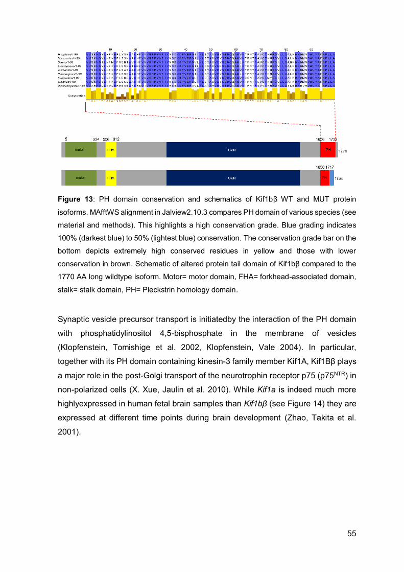
55
Figure 13: PH domain conservation and schematics of Kif1bβ WT and MUT protein
isoforms. MAfftWS alignment in Jalview2.10.3 compares PH domain of various species (see
material and methods). This highlights a high conservation grade. Blue grading indicates
100% (darkest blue) to 50% (lightest blue) conservation. The conservation grade bar on the
bottom depicts extremely high conserved residues in yellow and those with lower
conservation in brown. Schematic of altered protein tail domain of Kif1bβ compared to the
1770 AA long wildtype isoform. Motor= motor domain, FHA= forkhead-associated domain,
stalk= stalk domain, PH= Pleckstrin homology domain.
Synaptic vesicle precursor transport is initiatedby the interaction of the PH domain
with phosphatidylinositol 4,5-bisphosphate in the membrane of vesicles
(Klopfenstein, Tomishige et al. 2002, Klopfenstein, Vale 2004). In particular,
together with its PH domain containing kinesin-3 family member Kif1A, Kif1Bβ plays
a major role in the post-Golgi transport of the neurotrophin receptor p75 (p75NTR) in
non-polarized cells (X. Xue, Jaulin et al. 2010). While Kif1a is indeed much more
highlyexpressed in human fetal brain samples than Kif1bβ (see Figure 14) they are
expressed at different time points during brain development (Zhao, Takita et al.
2001).

56
Figure 14: Quantification of Kif1bβ and Kif1A cDNA in human fetal brain (HFB) RNA.
P75NTR is known to interact with all neurotrophins (NGF, BDNF, NT-3, NT-4)
(Escudero, Lazo et al. 2014). It controls apoptosis in developing neurons and is
essential for axonal outgrowth (E. J. Huang, Reichardt 2001). Thus we asked
whether p75NTR vesicle trafficking was affected. Therefore we simultaneously co-
expressed p75-RFP (Addgene plasmid #24092, unpublished) with either wildtype or
the mutated Kif1bβ. In pcDNA3.1-Kif1bβ-WT-GFP expressing cells, Kif1Bβ was
clearly accumulating around vesicles containing p75-RFP (Figure 15a). Timelapse
experiments allowed us to track vesicle movements within a co-expressed cell over
several seconds. This demonstrated very clearly, that p75NTR granules move along
with KIF1Bβ. For the pcDNA3.1-Kif1Bβ-MUT-GFP transfected cells, however, co-
expression could not be observed. In these cells vesicle membranes were solely
visualized by p75-RFP expression (Figure 15b).
0,00
50,00
100,00
150,00
200,00
250,00
300,00
350,00
400,00
450,00
Kif1A Kif1B
pg/
µl R
NA
in H
FB

57
Figure 15: Co-expression of pcDNA3.1-Kif1Bβ-WT-GFP and pcDNA3.1-Kif1Bβ-MUT-GFP
with p75-RFP. a. KIF1Bβ-WT accumulates around p75NTR containing vesicles. In KIF1B-
MUT expressing cells co-localization is impaired. b. Recording of vesicle movement in co-
expressed WT cells. P75NTR vesicles transported by KIF1Bβ-WT move synchronously
through the cell. t0-t4 indicate time points. Yellow and white arrows mark two recorded
vesicles for easier tracking of movements.
As we wanted to confirm that this phenotype is caused by the impairment of the PH-
domain, we cloned the wildtype and mutant tail domain into the pcDNA3.1 vector
containing an N-terminal GFP fusion protein (starting with amino acid 1669) (Figure
16a). Figure 16b illustrates the overexpression of the recombinant proteins in U2OS
cells. Although the GFP expression was now detected across the entire cell (even
in the nucleus), we observed the same differences as when expressing the full-
length constructs. Co-localization occured when the wildtype tail domain was
simultaneously overexpressed with p75-RFP. In cells transfected with the novel
alternatively spliced tail domain, p75NTR vesicles failed to co-localize with the
mutated Kif1bβ tail.

58
Figure 16: Co-expression of WT and MUT C-terminal domains with p75-RFP. a. Schematic
presentation of KIF1Bβ WT and MUT tail domains cloned into pcDNA3.1-NT-GFP. Numbers
indicate amino acid positions in the full-length reference protein. Motor= motordomain,
FHA= forkhead-associated domain, Stalk= stalk domain, PH= Pleckstrin homology domain
b. GFP-expressing granules of the wildtype KIF1Bβ tail domain are overlapping with p75
expression. Expression of the mutated KIF1Bβ tail construct is found throughout the cell
with no preferential localization.
As before, we recorded the movements of the expressed KIF1Bβ-tail domains.
Pronounced GFP-expressing areas in cells expressing the WT tail domain show full-
length construct-like movements while upon expression of the mutated carboxy
terminus, the GFP signal remains diffusely distributed within the cell with no
recognizable movements. We therefore concluded that p75NTR vesicle transport is
severely impaired upon loss of the second to last exon of Kif1bβ.
Recently, the C-terminal part (the last 552 amino acids) of KIF1Bβ was reported to
interact with calmodulin (CaM), the major receptor for intracellular calcium
(Charalambous, Pasciuto et al. 2013). In addition to the fact, that CaM is known to
play a crucial role in axon extension and guidance through the midline by controlling
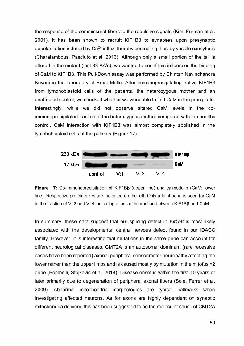
59
the response of the commissural fibers to the repulsive signals (Kim, Furman et al.
2001), it has been shown to recruit KIF1Bβ to synapses upon presynaptic
depolarization induced by Ca2+ influx, thereby controlling thereby vesicle exocytosis
(Charalambous, Pasciuto et al. 2013). Although only a small portion of the tail is
altered in the mutant (last 33 AA’s), we wanted to see if this influences the binding
of CaM to KIF1Bβ. This Pull-Down assay was performed by Chintan Navinchandra
Koyani in the laboratory of Ernst Malle. After immunoprecipitating native KIF1Bβ
from lymphoblastoid cells of the patients, the heterozygous mother and an
unaffected control, we checked whether we were able to find CaM in the precipitate.
Interestingly, while we did not observe altered CaM levels in the co-
immunoprecipitated fraction of the heterozygous mother compared with the healthy
control, CaM interaction with KIF1Bβ was almost completely abolished in the
lymphoblastoid cells of the patients (Figure 17).
Figure 17: Co-immunoprecipitation of KIF1Bβ (upper line) and calmodulin (CaM; lower
line). Respective protein sizes are indicated on the left. Only a faint band is seen for CaM
in the fraction of VI:2 and VI:4 indicating a loss of interaction between KIF1Bβ and CaM.
In summary, these data suggest that our splicing defect in Kif1bβ is most likely
associated with the developmental central nervous defect found in our IDACC
family. However, it is interesting that mutations in the same gene can account for
different neurological diseases. CMT2A is an autosomal dominant (rare recessive
cases have been reported) axonal peripheral sensorimotor neuropathy affecting the
lower rather than the upper limbs and is caused mostly by mutation in the mitofusin2
gene (Bombelli, Stojkovic et al. 2014). Disease onset is within the first 10 years or
later primarily due to degeneration of peripheral axonal fibers (Sole, Ferrer et al.
2009). Abnormal mitochondria morphologies are typical hallmarks when
investigating affected neurons. As for axons are highly dependent on synaptic
mitochondria delivery, this has been suggested to be the molecular cause of CMT2A

60
(Chen, Chan 2006). Kif1Bα but not β is known to be associated with mitochondrial
transport (Conforti, Dell'Agnello et al. 2003). As shown, in the brain, the α isoform is
expressed at significant lower levels than the β isoform. Thus, we wondered if the
expression pattern of Kif1bα and β in muscle is reversed. By relative quantification
of both isoforms in human fetal brain and muscle RNA we saw that compared to the
longer Kif1bβ the α isoform was indeed higher expressed in muscle tissue (Figure
18). The CMT2A mutation is located in the motor region of the gene thus affecting
both isoforms. This could therefore offer an explanation for the influence of the Q98L
mutation on peripheral neuron degeneration and why we do not see neuropathy-
related signs in our patients and the heterozygous, non-affected family members.
Figure 18: Quantification of Kif1bβ and Kif1bα levels between human fetal brain and muscle
tissue relative to Kif1bβ levels in human fetal brain. The Kif1bβ isoform is expressed at
significantly higher levels in brain than Kif1bα. However, in muscle tissue of 2 different
donors, the α isoform is more abundant compared to Kif1bβ. HFB= human fetal brain; RQ=
relative quantification level
More recently homozygous Kif1bβ mice deleterious for the second last exon of
Kif1bβ were generated by David Keays’ research group at the Research Institute of
Molecular Pathology (IMP) in Vienna. Detailed phenotypic characterizations are still
pending, but all homozygous littermates died shortly after delivery and showed

61
severe brain abnormalities (personal communication), which is in line with previous
observations in Kif1b complete knockout mice (Zhao, Takita et al. 2001). These
results confirm that Kif1Bβ is fundamental during brain development and that the
homozygous splice site mutation is causative for the brain developmental disorder
detected in our patients.

62
3.2 HsSASS-6 is mutated in autosomal recessive primary
microcephaly
The following findings have been published in Human Molecular Genetics in 2014
(Khan, Rupp et al. 2014). Pictures and tables are identical with those in the
publications; some text passages may also overlap.
As mentioned in chapter 1.5, MCPH is a rare disease in the non-consanguineous
population. In Pakistan, however, first-cousin marriages are still common, with a
frequency of 54% in rural areas (Agha 2016). We here report about a Pakistani
family from Dera Ismail Khan, a city in the Khyber Pakhtunkhwa province, with four
individuals affected with autosomal recessive primary microcephaly (MCPH),
including two girls at the age of three and six (V:3 and V:1) and two adult brothers
(VI:7 and VI:8). All MCPH patients were progeny from consanguineous marriages
(Figure 19).
Figure 19: Pedigree and pictures of the MCPH family. a. Affected are children of
consanguineous parents. b. Patients VI:7, V1 and V:3. Due to the small head ears, nose
and eyes (not shown here) appear larger than normal (pictures reproduced from (Khan,
Rupp et al. 2014) with permission of Human Molecular Genetics).
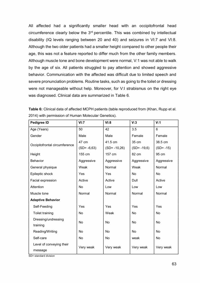
63
All affected had a significantly smaller head with an occipitofrontal head
circumference clearly below the 3rd percentile. This was combined by intellectual
disability (IQ levels ranging between 20 and 40) and seizures in VI:7 and VI:8.
Although the two older patients had a smaller height compared to other people their
age, this was not a feature reported to differ much from the other family members.
Although muscle tone and bone development were normal, V:1 was not able to walk
by the age of six. All patients struggled to pay attention and showed aggressive
behavior. Communication with the affected was difficult due to limited speech and
severe pronunciation problems. Routine tasks, such as going to the toilet or dressing
were not manageable without help. Moreover, for V:I strabismus on the right eye
was diagnosed. Clinical data are summarized in Table 6.
Table 6: Clinical data of affected MCPH patients (table reproduced from (Khan, Rupp et al.
2014) with permission of Human Molecular Genetics).
Pedigree ID VI:7 VI:8 V:3 V:1
Age (Years) 50 42 3.5 6
Gender Male Male Female Female
Occipitofrontal circumference 47 cm
(SD= -6,63)
41.5 cm
(SD= -10,26)
35 cm
(SD= -19,6)
38.5 cm
(SD= -15)
Height 155 cm 157 cm 82 cm 95 cm
Behavior Aggressive Aggressive Aggressive Aggressive
General physique Weak Normal Weak Normal
Epileptic shock Yes Yes No No
Facial expression Active Active Dull Active
Attention No Low Low Low
Muscle tone Normal Normal Normal Normal
Adaptive Behavior
Self-Feeding Yes Yes Yes Yes
Toilet training No Weak No No
Dressing/undressing
training No No No No
Reading/Writing No No No No
Self-care No No weak No
Level of conveying their
message Very weak Very weak Very weak Very weak
SD= standard division

64
Computed tomography (CT) of patient V:3 confirmed a microcephalic brain with
poorly defined ganglia. Additionally, delineation of the internal capsule was missing,
infratentorial abnormalities, a hypoplastic vermis cerebellar and deformation of
lateral ventricles were noted (Figure 20).
Figure 20: Computed tomography of V:3. Selected axial slices of cranial computed
tomography. Notwithstanding compromised image quality, some distinct features can be
noted: diminished cranial circumference with symmetrically poorly developed frontal lobes
(arrows; also note deformed shape of the skull), poorly confined basal ganglia (oval) with
missing delineation of the internal capsule, dysmorphic lateral ventricles (curved arrows;
slit-like frontal horns), and infratentorial abnormality with hypoplasia of the vermis cerebelli
(images not shown) (figure and text reproduced from (Khan, Rupp et al. 2014) with
permission of Human Molecular Genetics).
Due to the aggressive behavior of patient IV:8 DNA could only be collected from
patients IV:7, V:1 and V:3 as well as from several other unaffected family members.
Assuming that the disease causing gene was located within an autozygous area of
the genome, SNP genotyping was performed on individuals IV:7, V:3 and V1 using
a 250K array from Affymetrix. Most interestingly, only one homozygous locus with
identical haplotype patterns was shared by all three spanning a 18,53Mb region on
chromosome 1p21.3-p13.1. This interval was restricted by SNPs rs555557
(98,912,075bp) and rs2251406 (117,445,365bp) and did not harbor any known
MCPH genes (Figure 21a). This suggested the discovery of a novel MCPH locus,
namely MCPH14. Full scale linkage analysis calculated a maximum multipoint LOD
score of 3.9 for this locus (Figure 21b).

65
Figure 21: Homozygosity mapping and linkage analysis. a. 250K NspI SNP array reveals
a homozygous 18,53Mb stretch (blue) with identical haplotype on chromosome 1p21.3-
p13.1 restricted by SNP rs555557 and rs2251406 in all three patients. b. Parametric linkage
analysis of the family. LOD scores (y-axis) are plotted over genetic distance in cM (centi
Morgan) across the genome (x-axis), where chromosomes are concatenated from p-ter to
q-ter from left to right. A single LOD score peak at 3.9 cM (red arrow) marks the locus on
chromosome 1 (figures and text reproduced from (Khan, Rupp et al. 2014) with permission
of Human Molecular Genetics).
To confirm intrafamilial segregation we conducted STS marker analysis using 6
highly polymorphic microsatellite markers, including D1S2719 (96,8Mb), D1S2739
(98,9Mb), D1S2671 (101,6Mb), D1S206 (101,6MB), D1S495 (102,56MB) and
D1S2726 (111,18MB) (Figure 22). All affected were homozygous for the same
haplotype between marker D1S2671 and D1S2726 whereas the healthy family
members did either carry one copy of the green haplotype or none (as for V:2).
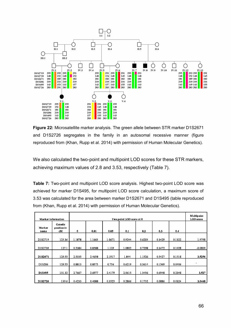
66
Figure 22: Microsatellite marker analysis. The green allele between STR marker D1S2671
and D1S2726 segregates in the family in an autosomal recessive manner (figure
reproduced from (Khan, Rupp et al. 2014) with permission of Human Molecular Genetics).
We also calculated the two-point and multipoint LOD scores for these STR markers,
achieving maximum values of 2.8 and 3.53, respectively (Table 7).
Table 7: Two-point and multipoint LOD score analysis. Highest two-point LOD score was
achieved for marker D1S495, for multipoint LOD score calculation, a maximum score of
3.53 was calculated for the area between marker D1S2671 and D1S495 (table reproduced
from (Khan, Rupp et al. 2014) with permission of Human Molecular Genetics).

67
Next, we screened the target locus for potential disease genes that either play a role
during cell division or cell cycle control as for most of the 13 known MCPH genes
have a major impact on either of these (see table Table 1: Primary microcephaly
genes We selected four genes for subsequent Sanger Sequencing, including
WDR47, PSRC1, NGF and SASS6 (hereafter referred to as HsSASS-6). Only in
HsSASS-6, a well-characterized gene encoding for a protein involved in procentriole
formation during cell division (Gonczy 2012, Rodrigues-Martins, Bettencourt-Dias et
al. 2007, Leidel, Delattre et al. 2005, Strnad, Leidel et al. 2007, van Breugel, Hirono
et al. 2011, Gopalakrishnan, Guichard et al. 2010), we eventually detected a
homozygous missense mutation, c.185T>C (NM_194292.1;
Chr.1:100588787A>G). Lying within a highly conserved region in exon 3, the
transition leads to an amino acid replacement of the non-polar and hydrophobic
isoleucine with a hydrophilic and polar threonine (p.Ile62Thr). All available family
members were further screened for the mutation. Results were in line with the STR
marker analysis. While all affected were homozygous for the mutation, the healthy
family members were either heterozygous carriers or displayed the wildtype
sequence (V:2) (Figure 23).
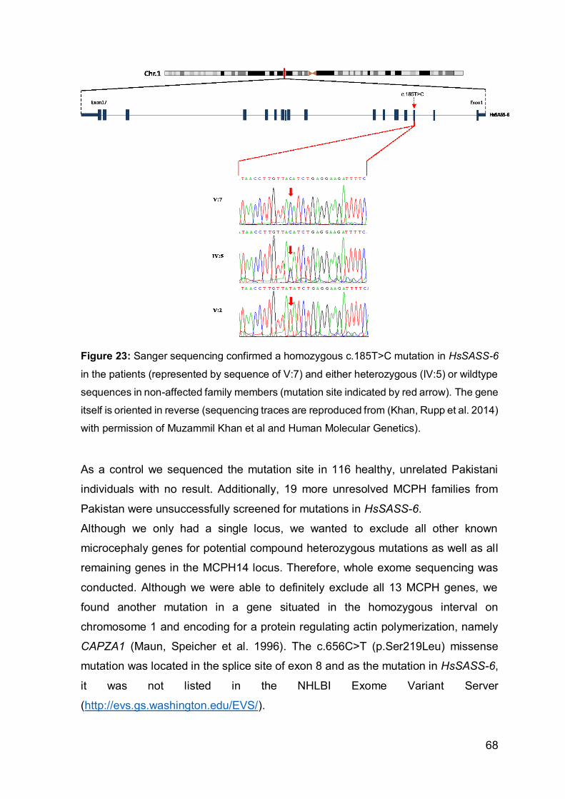
68
Figure 23: Sanger sequencing confirmed a homozygous c.185T>C mutation in HsSASS-6
in the patients (represented by sequence of V:7) and either heterozygous (IV:5) or wildtype
sequences in non-affected family members (mutation site indicated by red arrow). The gene
itself is oriented in reverse (sequencing traces are reproduced from (Khan, Rupp et al. 2014)
with permission of Muzammil Khan et al and Human Molecular Genetics).
As a control we sequenced the mutation site in 116 healthy, unrelated Pakistani
individuals with no result. Additionally, 19 more unresolved MCPH families from
Pakistan were unsuccessfully screened for mutations in HsSASS-6.
Although we only had a single locus, we wanted to exclude all other known
microcephaly genes for potential compound heterozygous mutations as well as all
remaining genes in the MCPH14 locus. Therefore, whole exome sequencing was
conducted. Although we were able to definitely exclude all 13 MCPH genes, we
found another mutation in a gene situated in the homozygous interval on
chromosome 1 and encoding for a protein regulating actin polymerization, namely
CAPZA1 (Maun, Speicher et al. 1996). The c.656C>T (p.Ser219Leu) missense
mutation was located in the splice site of exon 8 and as the mutation in HsSASS-6,
it was not listed in the NHLBI Exome Variant Server
(http://evs.gs.washington.edu/EVS/).

69
Analysis in PolyPhen2 and ConDel predicted the mutation in Hs-SASS6, but not the
mutation in CAPZA1 to be potentially pathogenic (PolyPhen2: HsSAS-6 p.Ile62Thr:
score = 1.0; CAPZA1: p.Ser219Leu: score = 0.014; and ConDel: HsSAS-6
p.Ile62Thr: score = 0.847; CAPZA1: p.Ser219Leu: score = 0.001). Alternative
splicing variants caused by the CAPZA1 mutation were excluded by RNA analysis.
For this we picked primers spanning the region between exon 7 and the last exon
10. However, gel electrophoresis of the PCR products already clearly highlighted
that no alternative transcripts were present as we obtained the same band sizes as
in the analysis of mRNAs from two healthy and unrelated controls (Figure 24).
Sanger sequencing confirmed normally spliced wildtype CAPZA1 transcripts.
Additionally, knock-out experiments in mice did not show any association of this
gene with any cell division defects (Neumann, Walter et al. 2010). Thus, we
excluded CAPZA1 as a potential MCPH disease gene.
Figure 24: cDNA analysis of CAPZA1. cDNA analysis of the two patients V:3 and IV:7
reveal the same 242bp PCR products as the amplicons from the unaffected controls.
Relevant 100bp standard sizes are shwon on the left side (figure reproduced from (Khan,
Rupp et al. 2014) with permission of Human Molecular Genetics).
HsSAS-6, however, has already been studied in detail and is known to be essential
to ensure proper cell division (Strnad, Leidel et al. 2007, Leidel, Delattre et al. 2005,
Nakazawa, Hiraki et al. 2007). The gene itself consists of 17 exons coding for a 657
amino acid long protein. Upon procentriole formation, HsSAS-6 dimerizes and

70
interacts via the head domain with nine more homodimers to form the hub and
spokes of a cartwheel like ring structure and initiates the establishment of the
daughter centriole (Kitagawa, Vakonakis et al. 2011, Hilbert, Erat et al. 2013,
Gonczy 2012). The Ile62 residue is located within the highly conserved PISA domain
supporting the formation of the N-terminal head domain of HsSAS-6 (Figure 25).
Figure 25: Conservation and location of the Ile62 residue in the DrSAS-6 head domain. a.
Conservation of the SAS-6 PISA domain in nine different organisms is highlighted by
MAfftWS alignment analysis in Jalview2.8. Residues are colored according to the
hydrophobicity table of Kyte and Doolittle (Kyte, Doolittle 1982). Red indicates conserved
hydrophobic, blue the conserved hydrophilic residues. Shading intensities represent the
conservation grade of the hydrophobicity (dark=very conserved; white= not conserved)
(figure and text reproduced from (Khan, Rupp et al. 2014) with permission of Human
Molecular Genetics).
So far, crystal structures of the human HsSAS-6 are not available since recombinant
human SAS-6 proteins are not soluble (own, unpublished experiments). Thanks to
existing high resolution crystal structure of the head of the Danio rerio homolog,
DrSAS-6, the exact position of the mutated residue was determinable. This
highlighted that the Ile62 residue (green spheres) is located within the hydrophobic
core of the head together with the highly conserved Leu44, Leu60, Leu70, Phe80,
Phe83 and Leu139 as shown by Figure 26.
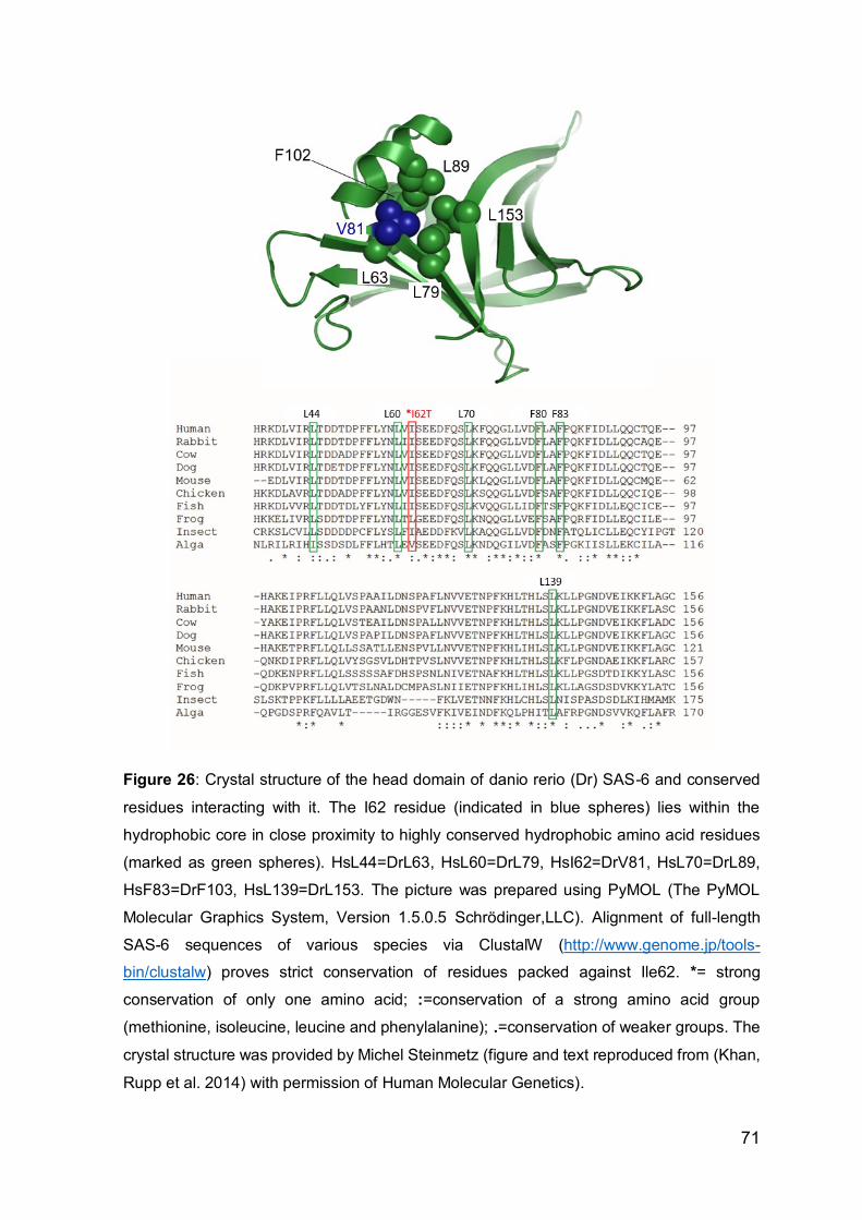
71
Figure 26: Crystal structure of the head domain of danio rerio (Dr) SAS-6 and conserved
residues interacting with it. The I62 residue (indicated in blue spheres) lies within the
hydrophobic core in close proximity to highly conserved hydrophobic amino acid residues
(marked as green spheres). HsL44=DrL63, HsL60=DrL79, HsI62=DrV81, HsL70=DrL89,
HsF83=DrF103, HsL139=DrL153. The picture was prepared using PyMOL (The PyMOL
Molecular Graphics System, Version 1.5.0.5 Schrödinger,LLC). Alignment of full-length
SAS-6 sequences of various species via ClustalW (http://www.genome.jp/tools-
bin/clustalw) proves strict conservation of residues packed against Ile62. *= strong
conservation of only one amino acid; :=conservation of a strong amino acid group
(methionine, isoleucine, leucine and phenylalanine); .=conservation of weaker groups. The
crystal structure was provided by Michel Steinmetz (figure and text reproduced from (Khan,
Rupp et al. 2014) with permission of Human Molecular Genetics).

72
These data suggested an impaired or weakened folding of the N-terminal head
domain due to the exchange of the hydrophobic isoleucine with the hydrophilic
threonine. A model of the affected domain in the clamydomonas reinhardtii homolog
crSAS-6, were the corresponding residue crVal81 is highlighted in yellow, shows
that this core is in direct proximity to the head-head interaction site between two
homodimers (Figure 27).
Figure 27: SAS-6 location in procentriole and composition. Nine homodimers form a ring-
like structure via N-terminal head domain interactions. On their tail they are associated
through A, B and C-tubulin. A model of the head domain of the chlamydomonas reinhardtii
homolog crSAS-6 depicts the location of the mutated residue (in this case crSAS-6 V81) at
the end of the β4 sheet directly before the α1 helix. Two interacting crSAS-6 head domains
are presented- a lighter shaded domain on the left and the darker colored domain on the
right (green box).
To therefore study the potential effect of the HsSASS-6 mutation on localization, we
transfected U2OS cells with either doxycycline-inducible wildtype or c.185T>C
mutant constructs fused to EGFP. This was performed at the EPFl in Lausanne in
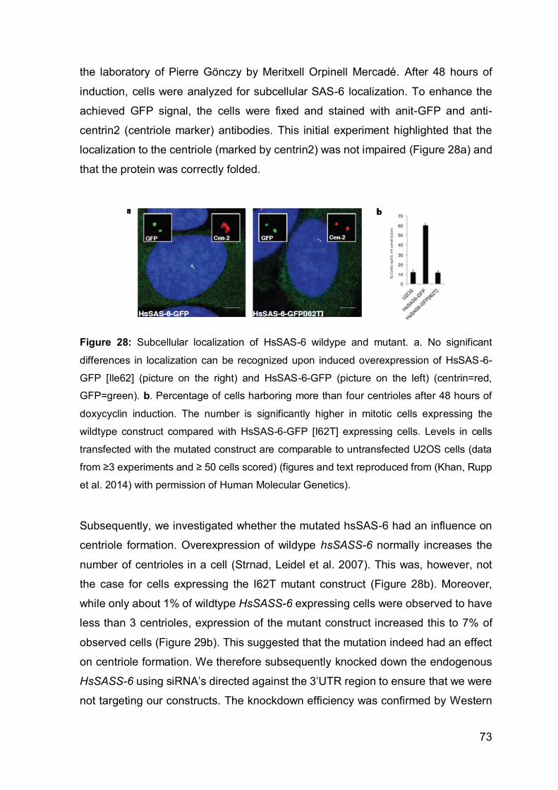
73
the laboratory of Pierre Gönczy by Meritxell Orpinell Mercadé. After 48 hours of
induction, cells were analyzed for subcellular SAS-6 localization. To enhance the
achieved GFP signal, the cells were fixed and stained with anit-GFP and anti-
centrin2 (centriole marker) antibodies. This initial experiment highlighted that the
localization to the centriole (marked by centrin2) was not impaired (Figure 28a) and
that the protein was correctly folded.
Figure 28: Subcellular localization of HsSAS-6 wildype and mutant. a. No significant
differences in localization can be recognized upon induced overexpression of HsSAS-6-
GFP [Ile62] (picture on the right) and HsSAS-6-GFP (picture on the left) (centrin=red,
GFP=green). b. Percentage of cells harboring more than four centrioles after 48 hours of
doxycyclin induction. The number is significantly higher in mitotic cells expressing the
wildtype construct compared with HsSAS-6-GFP [I62T] expressing cells. Levels in cells
transfected with the mutated construct are comparable to untransfected U2OS cells (data
from ≥3 experiments and ≥ 50 cells scored) (figures and text reproduced from (Khan, Rupp
et al. 2014) with permission of Human Molecular Genetics).
Subsequently, we investigated whether the mutated hsSAS-6 had an influence on
centriole formation. Overexpression of wildype hsSASS-6 normally increases the
number of centrioles in a cell (Strnad, Leidel et al. 2007). This was, however, not
the case for cells expressing the I62T mutant construct (Figure 28b). Moreover,
while only about 1% of wildtype HsSASS-6 expressing cells were observed to have
less than 3 centrioles, expression of the mutant construct increased this to 7% of
observed cells (Figure 29b). This suggested that the mutation indeed had an effect
on centriole formation. We therefore subsequently knocked down the endogenous
HsSASS-6 using siRNA’s directed against the 3’UTR region to ensure that we were
not targeting our constructs. The knockdown efficiency was confirmed by Western

74
Blot analysis of the iU2OS protein extracts (Figure 29a). hsSAS-6 levels were
increased upon doxycycline induction while endogenous hsSAS-6 expression was
concomitantly inhibited upon siRNA knock-down (see Figure 29a, e & f).
Figure 29: siRNA knock down of endogenously expressed HsSASS-6. a. Western Blot
showing the expression of HsSAS-6 WT and mutant constructs upon doxycycline induction,
which highlights the knockdown efficiency of the siRNA compared to negative control siRNA
(NC). b & c. Frequency of cells with less than four centrioles and monopolar spindles in
untransfected control U2OS, HsSAS-6-GFP or HsSAS6-GFP[I62T] cells, transfected with
either negative control siRNA (siNC) or siRNa knocking-down endogenous HsSAS-6
(3’UTR) (data from ≥3 experiments and ≥ 50 cells scored). d-i. U2OS (d-f), iU2OS HsSAS-
6-GFP (g) or iU2OS HsSAS-6-GFP[I62T] (h & i) cells in mitosis, stained for GFP (green),
Centrin-2 (red) and DNA (blue). In (g-i), doxycycline induction for 48h was concomitant with
siHsSAS-6 3’UTR (3’UTR) treatment. Monopolar spindles were found in f and i. Scale bars
represent 5µm. (Figures and text reproduced from (Khan, Rupp et al. 2014) with permission
of Human Molecular Genetics).

75
As we wanted to see if the number of centrioles changed upon endogenous
HsSASS-6 knockdown we only concentrated on mitotic cells. Knockdown in U2OS
cells was extremely efficient (Figure 29e) with up to 95% of cells having less than
four centrin foci whereas the number of foci for cells transfected with negative
control siRNA was equal to untreated cells (see Figure 28 versus Figure 29b). We
also noted a large increase in monopolar cells in these control cells upon HsSAS-6
depletion (Figure 29f). By trying to rescue this phenotype through overexpression of
either HsSAS-6-GFP or HsSAS-6-GFP[I62T] we found significant differences. While
we successfully rescued the amount of monopolar spindles (less than 10%) and
increased the number of cells having four foci (~80%) by expressing the wildtype
construct the results for the construct containing the p.Ile62Thr mutation were less
promising. In terms of centriole numbers, the mutant construct was only able to
reduce the number of cells having less than four foci in 27% of cells. Moreover,
monopolar spindle formation was still observed in nearly 30% of transfected cells.
These experiments clearly provide evidence that the mutation has an impact on cell
division and especially centriole formation, though it must be noted that HsSAS-6
function is not completely impaired. As altered cell division is a major cause of
autosomal recessive primary microcephaly, we conclude that the c.185T>C
transition in HsSASS-6 is most likely pathogenic and causes the development of
MCPH14.

76
4. Discussion
In this thesis, we report the discovery of two genes, Kif1bβ and hsSASS-6,
associated with an autosomal recessive inherited brain disorder. Moreover, we
describe for the first time a yet unreported syndromal intellectual disability with
agenesis of the corpus callosum, which we found appears to be linked with a
mutation in Kif1bβ. While Kif1bβ has already been identified as a causative gene for
CMT2A (Zhao, Takita et al. 2001) and has furthermore been associated with the
development of multiple sclerosis (Aulchenko, Hoppenbrouwers et al. 2008),
hsSASS-6 has previously not been established as causative gene for any known
syndromes or diseases. Interestingly, both genes identified in our experiments have
different functions: while the plays a major role in cell division (hsSASS-6), the other
(Kif1bβ) codes for a transport molecule in neurons (Zhao, Takita et al. 2001, Leidel,
Delattre et al. 2005).
In chapter 3.1 we discussed the discovery of a single base pair exchange in the
donor splice site of the second to last exon of Kif1bβ. As a result, this exon is
skipped, causing a frameshift in the remaining C-terminal sequence. Accordingly,
more than a third of the Pleckstrin homology region and the entire remaining tail
domain are replaced by the novel frame generated by the single base pair
exchange, which subsequently encodes a different amino acid sequence and thus
for a completely different tail.
Kinesins are motor proteins that transport different cargos through the cell. Their
movement along microtubules is driven by ATP binding to the motor. Forty-two
genes coding for kinesins are known and all are assigned to one of 14 different
superfamilies (Wang, Cao et al. 2015). Together with Kif1A, Kif1C, Kif13A, Kif13B,
KIF14 and Kif16B, Kif1B is member of the kinesin-3 family (Hirokawa, Noda 2008,
Siddiqui, Straube 2017). In the unbound state, kinesin-3 motors reside as monomers
within the cells, but dimerize upon cargo binding to guarantee vesicle transport in
neurons (Hammond, Cai et al. 2009, Soppina, Norris et al. 2014). Kif1b gives rise
to two major isoforms, namely KIF1bβ and KIF1bα. As depicted in Figure 7, we
found that Sanger Sequencing confirmed the c.5270+2T>G transition in Kif1bβ. The
longer β isoform is the main isoform expressed in the brain (see Figure 10b),
encoding a kinesin interacting with synaptic vesicle membranes containing

77
synaptotagmin, SV2 and synaptophysin (Zhao, Takita et al. 2001), whereas the
shorter α isoform has a mitochondrial binding site not present in the β variant
(Conforti, Dell'Agnello et al. 2003). Despite the general assumption that Kif1bβ is
implemented solely in anterograde vesicle transport in axons, Niovi Santama ’s
research group has observed mRNA (Arc and CaM) transporting Kif1Bβ molecules
within dendrites moving in both directions, antero- and retrogradely (Charalambous,
Pasciuto et al. 2013). In general, kinesin-3 members share an amino terminal motor
and a FHA domain (fork head associated domain), which allows kinesins to interact
with phosphorylated proteins or peptides and interacts with another Kinesin-3
domain (CC2) to control intramolecular autoinhibition (J. R. Lee, Shin et al. 2004,
Westerholm-Parvinen, Vernos et al. 2000)). However, only KIF1A, KIF1bβ and
KIF16B possess a C-terminal PH domain (Hirokawa, Noda 2008). As mentioned in
chapter 3.1, this region is responsible for the interaction with phosphatidylinositol
4,5-bisphosphate in vesicle membranes in order to transport its cargos
(Klopfenstein, Tomishige et al. 2002, Klopfenstein, Vale 2004). It has been shown,
that kinesin-3 motors remain in an autoinhibited state until they bind to a vesicle.
Only upon binding are they able to localize to microtubules and membranes (Niwa,
Lipton et al. 2016, Hammond, Cai et al. 2009) which could explain the more cytosolic
distribution we observed in cells overexpressing the defective PH domain molecule
when compared with wildtype pcDNA3.1-KIF1bβ-GFP transfected cells. As neither
movements of GFP expressing KIF1bβ mutant molecules nor a co-localization of
the mutated isoform with p75NTR containing vesicles could be seen in cells, we
conclude that the mutated kinesin remains inactivated as monomers in the cell and
even in the presence of its cargo is not able to dimerize, bind to and move along
microtubules (Soppina, Norris et al. 2014) (Figure 30).

78
Figure 30: Hypothetic model of Kif1Bβ loss-of-function due to altered tail domain. Orange
part of dimeric kinesin indicates tails with PH domains interacting with p75NTR transporting
vesicle membrane. In the mutated molecule it is altered. Thus vesicle-kinesin binding,
subsequent kinesin activation, dimerization and its recruitment to microtubules is inhibited.
The suggested pathomechanism according to the kinesin-3 activity model was first
proposed by Soppina et al. (Soppina, Norris et al. 2014). The GFP molecule is connected
to kinesin via the brown linker. RFP-expressing p75NTR is anchored in vesicle membrane.
Blue = stalk domain; yellow = FHA domain; green= motor domain. Black lines depict
microtubules.
Looking at the phenotype, neurological diseases are frequently related to mutations
in kinesins or genes interacting with them (Hirokawa, Niwa et al. 2010). For
example, mutations in Kif1a, which is highly homologous to KIF1bβ, have been
linked to three neurological diseases, including autosomal dominant mental
retardation syndrome 9 (OMIM 614255), autosomal recessive hereditary sensory
neuropathy type 2C (OMIM 614213) and autosomal recessive spastic paraplegia 30
(OMIM 610357) (Erlich, Edvardson et al. 2011, Hamdan, Gauthier et al. 2011,
Riviere, Ramalingam et al. 2011). Interestingly, in addition to other neurological
abnormalities brain MRIs of patients with mutations in Kif1c, Kif2a, Kif5c, Kif7 or
Kif14 revealed total loss or thinning of the corpus callosum (Moawia, Shaheen et al.
2017, Dor, Cinnamon et al. 2014, Michels, Foss et al. 2017, Putoux, Thomas et al.
2011). This highlights that some kinesins are indeed essential for neuronal and
corpus callosum development. The importance of Kif1bβ during neuronal
development may already be suggested by its Pax6 dependent activation (Xie, Yang
et al. 2013). Pax6 is one of the first genes to be specifically expressed in stem cells
upon differentiation into the neuronal lineage in humans (X. Zhang, Huang et al.
79
2010) and is essential for proper brain development (Xie, Yang et al. 2013, Glaser,
Jepeal et al. 1994, X. Zhang, Huang et al. 2010, Mo, Zecevic 2008). Just like Kif1b
knock-out mice, homozygous Pax6 mutant mice die shortly after birth due to
neurological defects (Favor, Bradley et al. 2009, Estivill-Torrus, Vitalis et al. 2001).
There has been one report on a compound heterozygous mutation in Pax6, which
was associated with facial dysmorphisms, no development of the eyes, an absent
corpus callosum, hypotonia, missing optic nerves and chiasm as well as severe
CNS defects. The patient died within eight days after birth (Glaser, Jepeal et al.
1994). Additional evidence for the importance of Kif1bβ in neuronal development
comes from detailed studies in zebrafish where Kif1bβ, but not Kif1bα, was identified
as essential for axonal outgrowth (Lyons, Naylor et al. 2009). Compared with
wildtype animals, long axons in Kif1b transgenic fish with a mutation in the motor
domain were shortened by 50-70% (Drerup, Lusk et al. 2016). In addition to
defective axonal outgrowth, Lyons et al (Lyons, Naylor et al. 2009) also noted a 30%
reduction in the total number of neurons in Kif1b mutant fish. These data correlate
with studies conducted in p75NTR knockdown neurons. Marco Canossa’s group
provided evidence that neuronal polarization is dependent on p75NTR since depletion
of p75NTR prevents axongenesis (Zuccaro, Bergami et al. 2014). As p75NTR transport
is not solely dependent on Kif1Bβ but also on Kif1A (X. Xue, Jaulin et al. 2010),
axonal outgrowth is perhaps not completely impaired in all cells. The loss of
anterograde commissural fibers in Kif1Bβ knock out mice (Zhao, Takita et al. 2001),
however, lets us assume that Kif1Bβ plays an important role in axonal development
of long projection fibers. As the longest commissural fibers in the brain are found in
the corpus callosum (Paul, Brown et al. 2007), we therefore suggest a high
correlation between the C-terminal loss-of-function mutation in our patients with
reduced p75NTR vesicle transport and the absence of this fiber tract.
Zebrafish models of Kif1B also exhibited myelination defects due to abberant myelin
binding protein (mbp) transport (Lyons, Naylor et al. 2009). The Kif1b variant
rs10492972 was formerly linked to a higher susceptibility for the demyelinating
disease multiple sclerosis (MS) (Aulchenko, Hoppenbrouwers et al. 2008).
However, Sombekke et al. later rebutted this claim (Sombekke, Jafari et al. 2011).
As myelination was normal in our patients and no cases of MS have been reported
in the healthy family members, we assume that mutations in human Kif1bβ are
unlikely related to myelination defects.

80
It is to note that the Kif1B binding protein, KBP, has been reported to be causative
for brain developmental disorders associated with intellectual disability,
microcephaly, polymicrogyria, dilated ventricles and hypoplastic corpus callosum
development (Valence, Poirier et al. 2013, Brooks, Bertoli-Avella et al. 2005), and
that similar brain abnormalities that were seen in the MRIs of our IDACC patients.
Furthermore, depletion of KBP in zebrafish revealed reduced outgrowth of long
axons, axonal degeneration but also disorganization of the axonal cytoskeleton
(Lyons, Naylor et al. 2008). These effects of KBP depletion were suggested to result
in impaired SCG10 transport to extending axonal growth cones, as it is required to
stabilize microtubules (Drerup, Lusk et al. 2016).
Additionally, we have shown that the interaction between calmodulin and Kif1Bβ is
disrupted in lymphoblastoid cells of our patients (Figure 17). This interaction has
been suggested to support Ca2+-dependent recruitment of Kif1Bβ to synapses upon
depolarization, triggering synaptic vesicle transport when needed (Charalambous,
Pasciuto et al. 2013). In addition to the fact that vesicle binding seems to be
prevented by the altered tail of Kif1Bβ, the impaired interaction between CaM and
Kif1Bβ may also have a negative impact on the translocation to the growing axons.
The most interesting aspect, however, is that electrophysiological studies in our
patients were normal, not matching those with other reported CMT2A patients. The
severity of the mutation in our homozygous mouse models seems to be in line with
results seen in the Kif1b knock-out mice from Zhao (Zhao, Takita et al. 2001). Also,
as our mutation was shown to cause a loss of function according to our cellular co-
expression studies, it is therefore surprising that a heterozygous mutation in the ATP
binding site of the motor domain can cause a phenotype but that neither our homo-
nor heterozygous affected individuals show signs of peripheral neuropathies. Of
course, depending on the location of each mutation, phenotypic development can
vary (for example mutations in Kif1A account for three different phenotypes). The
mutation in the CMT2A family published by Zhao et al (Zhao, Takita et al. 2001) is
located in the ATP binding site of the motor domain shared by both isoforms. Zhao
et al suggested that the neurological phenotypes are caused by the impairment of
the β isoform, as rescue experiments in Kif1B-depleted neurons succeeded only
upon overexpression of Kif1bβ but not α. This is interesting as our mutation
specifically impairs the longer β isoform resulting in a totally different phenotype. A
study published in Neurology by Carvill et al also raised the question if CMT2A is

81
truly associated with Kif1B. Here, during the course of whole exome sequencing in
patients with Dravet syndrome, a disease characterized by infantile epileptic
seizures combined with progressive intellectual disability, the groups of Ingrid
Scheffer and Heather Mefford found a compound heterozygous mutation (p.T606I
and p.R942C) in Kif1B (Carvill, Weckhuysen et al. 2014). The researchers did not
report any CMT-related phenotypical abnormalities. While the T606 residue is
shared by both Kif1B isoforms, the latter R942 amino acid is specific for the α
isoform, located in its putative mitochondria binding site (Conforti, Dell'Agnello et al.
2003). Therefore, only the shorter alpha isoform is affected by both mutations. Some
studies have linked Dravet syndrome to mitochondrial dysfunction and
abnormalities (reviewed in (Castro-Gago, Perez-Gay et al. 2011)). Although not
discussed in their paper, the Carvill mutations could perhaps be linked to defective
Kif1Bα-dependent mitochondria transport. As mentioned in the results section of this
thesis, CMT2A is frequently associated with abnormal mitochondria and defective
transport along peripheral nerves. Taking into account that Kif1bα has a slightly
higher abundance in muscle than Kif1bβ and that no signs of peripheral
neuropathies were observed in our patients, one could therefore suggest that the
CMT phenotype is more likely caused by mutations in the alpha but not the beta
isoform of Kif1B. Since the mutation in the report of Zhao et al (Zhao, Takita et al.
2001) was heterozygous, the brain was perhaps not affected. This, however, would
be contrary to the Dravet paper (Carvill, Weckhuysen et al. 2014) where no
peripheral neuropathies were detected either.
Considering these data and the fact that our healthy mutation carriers showed no
symptoms, this raised the question about the causality of the heterozygous p.Q98T
in the CMT2A family of Zhao et al (Zhao, Takita et al. 2001). According to entries
published on the ‘Neuromuscular Disease Center’ website
(http://neuromuscular.wustl.edu/time/hmsn.html#2a1) patients reported for
CMT2A1 by Zhao et al in 2001 also carry a mutation in the CMT2A gene mitofusin2,
MFN2, situated just a few genes further downstream of Kif1b. MFN2 as another
causative CMT2A gene was published three years after the Zhao paper by the
groups of Schröder and Vance (Zuchner, Mersiyanova et al. 2004). According to the
human genome mutation database HGMD (Stenson, Mort et al. 2017, Stenson, Ball
et al. 2003), since then more than 220 mutations in MFN2 have been reported
compared to three additional but questionable mutations in Kif1B. While one of the

82
2 mutations (p.V1358A) published by Drew et al (Drew, Zhu et al. 2015) was also
found in an unaffected parent, and secondly, not predicted to be pathogenic by
Polyphen2 analysis, the amino acid conservation on the site of the other mutation
(p.E758D) was low. Moreover, the phenotype of the patient with the p.E758D
mutation was not a classical CMT neuropathy but rather a hereditary motor
neuropathy with pyramidal signs. A third mutation in Kif1b was found in the course
of a larger NGS study in Norwegian CMT families (Hoyer, Braathen et al. 2014). In
this study, only one affected family member was tested but none of the unaffected
individuals of the family. Therefore, the pathogenic effect of these three mutations
has to be considered as uncertain.
Recently we have tried to generate induced pluripotent stem cells (iPSC) from our
lymphoblastoid cell lines using the coMIP method (Diecke, Lu et al. 2015) to
investigate the neuronal differentiation capability and axon elongation in
heterozygous and homozygous c.5270 +2T>G Kif1bβ stem cells, but we were not
successful in establishing iPSC clones from these cells. Instead, we are currently
trying to integrate the splice site mutation into the genome of commercially
purchased iPSCs via CrispR-Cas9 to get a better understanding of the impact of
this mutation on neuronal development using a human cell model. In addition to
testing the neurogenic potential of these mutated stem cells, this model will allow us
to investigate the influence of our mutation on various other tissue types. Especially
renal cells would be of interest as in addition to the neurological phenotype, kidney
abnormalities were noted in one patient and one terminated fetus. If Kif1Bβ indeed
has an impact on kidney development, this would indicate that Kif1Bβ, like Kif3A, is
also involved in ciliogenesis (F. Lin, Hiesberger et al. 2003). This hypothesis is
supported by reports on Kif7 mutations. Kif7 is a kinesin-4 member, which is esential
for primary cilia function causing acrocallosal syndrome upon loss of function
(Edwards, Sherr et al. 2014, Tunovic, Baranano et al. 2015, Ibisler, Hehr et al. 2015,
Karaer, Yuksel et al. 2015, Barakeh, Faqeih et al. 2015, Walsh, Shalev et al. 2013,
Putoux, Thomas et al. 2011, Putoux, Nampoothiri et al. 2012). Cilia are important
throughout the body and are divided into motile and non-motile (primary) cilia
(Baker, Beales 2009). In the brain, the motile cilium is found on ependymal cells
aligning the lateral ventricles where they beat in waves to control the flow of the
cerebrospinal fluid to support the nutrient supply for neurons (Korzh 2018).
Ventricles in the brain are connected via channels and cilia defects have been linked
83
to abnormal accumulation of the cerebral fluid within the ventricles causing a
massive enlargement and eventually leading to the development of a hydrocephalus
(Korzh 2018, Al Omran, Saternos et al. 2015). In line with the findings of dilated
ventricles in Kif7 patients (Putoux, Thomas et al. 2011), the enlarged ventricles in
our patients could further support the theory of a potential ciliopathy in our patients.
In our second family we identified the spindle assembly homolog 6 gene (HsSASS-
6 or SASS6), as the causative gene for MCPH14. Like the majority of all other known
primary microcephaly genes to date (see Table 1), its protein product HsSAS-6 is
required for cell division by ensuring centrosome duplication (Leidel, Delattre et al.
2005, Strnad, Leidel et al. 2007). In order to establish the daughter centriole, two
HsSAS-6 monomers initially dimerize to homodimers via the coiled-coil domain and
these subsequently oligomerize to a symmetric 9-fold cartwheel structure by
interacting with each other via their head domains (see Figure 27) (van Breugel,
Hirono et al. 2011, Nakazawa, Hiraki et al. 2007, Kitagawa, Vakonakis et al. 2011).
For a long time, a scaffold model has been proposed, which suggests that the
HsSAS-6 cartwheel is initially built orthogonally attached to its mother centriole
before the microtubules are stepwise assembled to its C-terminal domain (Hirono
2014, Hilbert, Noga et al. 2016). However, a more recent interdependence model
suggests that the HsSAS-6 and microtubule scaffold are generated in parallel but
separately and assemble to create a stable pro-centriole (Hilbert, Noga et al. 2016).
HsSAS-6 is not permanently expressed. It can only be detected between S-phase
and mitosis, before it is degraded via ubiquitination during the final phase of cell
division. Thus HsSAS-6 cannot be found in mature centrioles. This tight regulation
of molecule levels ensures the generation of only one new centriole (procentriole or
daughter centriole) per cell cycle (Strnad, Leidel et al. 2007). Some of the already
identified microcephaly proteins closely interact with HsSAS-6 helping to establish
the procentriole. Before procentriole formation takes place, CEP125 and CEP63
form a ring-like structure surrounding the mother centriole (Tang 2013). This
enables the recruitment of PLK4, which closely interacts with CEP192 and CEP152
(Sonnen, Gabryjonczyk et al. 2013). PLK4, in turn, recruits and phosphorylates STIL
at its STAN domain thus enabling the interaction of STIL with the C-terminal domain
of HsSAS-6 triggering the cartwheel formation (Moyer, Clutario et al. 2015). At its
C-terminal coiled-coil domain HsSAS-6 harbors a binding site for CEP135. This
84
protein facilitates the attachment of the cartwheel scaffold to the A-tubule and links
it indirectly via CPAP (protein product of the CENPJ gene) to the B and C tubules
(Hatzopoulos, Erat et al. 2013). In addition to CEP192, all of the above mentioned
proteins have been reported to be involved in the pathogenesis of microcephaly
(Martin, Ahmad et al. 2014, Bond, Roberts et al. 2005, Kumar, Girimaji et al. 2009,
Hussain, Baig et al. 2012, Guernsey, Jiang et al. 2010, Sir, Barr et al. 2011). In our
MCPH patients we have identified a missense mutation in HsSASS-6 located within
the highly conserved PISA domain of the gene (Leidel, Delattre et al. 2005). This
domain spans the region between the β3 sheet and the α2 helix in the head. Being
part of the hydrophobic core of the V-terminal domain of HsSAS-6, we expected the
mutation located at the end of β4 and shortly before α1 to destabilize the folding of
the globular head domain, thus impairing homodimer aggregation. As published by
Kitagawa et al (Kitagawa, Vakonakis et al. 2011) oligomerization is mediated by
phenylalanine at position 131 reaching into the hydrophobic cavity between α1 and
α3. Targeted mutagenesis of this residue abrogated the head-head interactions
between the homodimers as well as bipolar spindle formation in Caenorhabditis
elegans, indicating a complete loss-of-function of HsSAS-6. The p.Ile62Thr found in
our patient exchanges a hydrophobic with a hydrophilic residue. As this area is part
of the hydrophobic core where the F131 inserts and acts according to the lock-and-
key principle, we wanted to see if this mutation had the same impact on centriole
formation as has been shown for the mutated F131. Although overexpression of the
mutated construct in cells did not reveal obvious changes in localization, we saw
that compared with wild type HsSAS-6 overexpressing cells the number of cells
containing more than four centrioles was significantly reduced, while the number of
cell with less than three centrioles slightly increased. Assuming this to represent a
dominant-negative effect, we knocked down the endogenous HsSAS-6. As
expected, this resulted in an impaired procentriole formation, thereby increasing the
number of cells with monopolar spindles and the percentage of cells with less than
three centrioles. Trying to rescue this phenotype by overexpressing the p.Ile62Thr
HsSASS-6 construct succeeded only partially compared with an almost complete
rescue upon WT HsSAS-6 overexpression. Impairing the N-N interaction by
mutating F131 has been shown to completely prevent mitosis (Kitagawa, Vakonakis
et al. 2011). However, since our phenotype was rescued to some extent we propose
that the remaining activity of the mutated HsSAS-6 is sufficient for human

85
development, but still has an impact on neurogenesis as centrosome duplication is
severely affected and the number of cells undergoing mitotic cleavage is reduced.
As we suggested that the folding of the head domain was perhaps loosened or the
interaction between the nine homodimers was weakened, we also set out to produce
a recombinant p.Ile62Thr HsSAS-6 protein to determine the impact of the mutation
on the cartwheel structure as well as on the protein folding of the head domain (data
not included in this thesis). Unfortunately, the recombinant HsSAS-6 protein was not
soluble, keeping us from performing electrospray experiments.
Still, we were able to ivestigated the effect of the mutated HsSAS-6 on a non-
neurological osteosarcoma cell line. To unravel the real pathomechanism affecting
brain development, it would be interesting to create a homozygous c.185T>C SASS-
6 mouse. As already mentioned, brain developmental processes are tightly
regulated. Slight tilts in the mitotic spindle during symmetric cell divisions of
neuroepithelial stem cells severely affect the establishment of a large enough stem
cell progenitor pool since divisions become asymmetric too early and thus
neurogenic (Homem, Repic et al. 2015, Lizarraga, Margossian et al. 2010). This, for
instance, is the case for NEs in MCPH1 or CENPJ knockout mice (Gruber, Zhou et
al. 2011, Garcez, Diaz-Alonso et al. 2015). As cell culture experiments revealed an
increase in monopolar spindles, it is to be assumed that the mutation indeed has an
impact on spindle formation. Additionally, the impaired centriole formation resulting
from the knock-down of CPAP in U2OS cells can just partially be rescued by
overexpression of CPAP p.Glu1235Val (CENPJ c.3704A>T) (Kitagawa, Kohlmaier
et al. 2011, Bond, Roberts et al. 2005), which is in line with our rescue experiments
for HsSAS-6. The close interaction of HsSAS-6 with CPAP would suggest a similar
impairment in HsSASS-6 mutated neuronal stem cell progenitors, as seen in CPAP
mouse models, but this does not necessarily have to be the case. As an example,
a total knockout of ASPM in mice drastically influences the orientation of the
cleavage planes in NEs (Fish, Kosodo et al. 2006), in contrast to disease causing
mutations in ASPM that did not show such effects (Pulvers, Bryk et al. 2010).
Another pathomechanism was demonstrated by the overexpression of PLK4, the
STIL-recruiting protein. Too high levels of PLK4 produced a surplus of centrosomes,
resulting in aneuploid cells. These, in turn, were eliminated by p53, reducing the
total number of cells and eventually triggering the development of a microcephalic

86
brain (Marthiens, Rujano et al. 2013). A similar phenotype has also been proposed
for some STIL mutations (Arquint, Nigg 2014).
Interestingly, compared to humans affected, MCPH gene knock-out mice often
develop different phenotypes (McIntyre, Lakshminarasimhan Chavali et al. 2012,
Pulvers, Bryk et al. 2010, Trimborn, Ghani et al. 2010, Liang, Gao et al. 2010). As
the architecture of the subventricular zones in mice and humans is not the same
(only humans have an oSVZ and an inner fiber layer, splitting the inner and outer
SVZ), altered phenotypical development has been linked to this morphological
difference (Lancaster, Renner et al. 2013, Shitamukai, Konno et al. 2011). To study
such alterations in a more authentic disease model, Jürgen Knoblich’s group
developed the generation of brain organoids, elegantly reflecting human cortical
development and cerebral layer organization. By using a compound heterozygous
CDK5RAP2 human iPSC line, they successfully mimicked microcephalic brain
development in a spinner culture flask. The resulting patient-derived organoids were
not only smaller in size, they also contained significantly smaller neuroepithelial
tissues reflecting premature neurogenesis. The researchers confirmed this by
analyzing the spindle orientation of radial glial cells, proving tilted orientations
(Lancaster, Renner et al. 2013). Since their publication in 2013, the method has
been improved and several other neurological diseases have been modeled
depicting it as a powerful tool to investigate neurogenesis and brain developmental
disorders (reviewed in (C. T. Lee, Bendriem et al. 2017)). Producing such an
organoid from iPSCs containing the HsSASS-6 c.185T>C mutation would help to
unravel the pathomechanism in humans and definitely confirm the disease-causing
nature of our mutation.
To conclude, we are the first to report the herein described mutations in two genes
associated with developmental neurological disorders, which both play different
roles during brain development. By performing a variety of experiments we were
able to prove that the impaired function (HsSAS-6) or loss-of-function (Kif1Bβ)
caused by these mutations, which are both located in highly conserved and/or
functionally important regions of the encoding proteins, appeared to cause the
disease. Mouse models for Kif1Bβ supporting our theory are on the way to show the
phenotypical consequences of the herein described mutation in a model organism.

87
Although our experiments provide evidence of the pathogenic effect of the hsSASS-
6 mutation identified in our patients, an animal or stem cell model would substantiate
our theory.
Eventually, the molecular findings shed light on the actual role of the detected genes
in the physiological and developmental context, revealing both as fundamental for
brain development in the embryo. The identification of hsSASS-6 in association with
MCPH substantially increases the complexity of the genetic heterogeneity of MCPH.
In terms of autosomal recessive primary microcephaly, most genes encode for
proteins required for centriole formation and cell division. By identifying hsSAS-6,
another centrosomal protein has thereby be linked to brain development.
Although these findings do unfortunately not provide any instant treatment options
for the patients, knowing the familial mutation allows genetic testing and genetic
counselling of relatives upon family planning. Additionally, including both genes in
prenatal diagnostic NGS panels for mental disabilities or microcephaly will further
facilitate the detection of disease causing mutations during prenatal testing, thereby
allowing parents to make an early decision on the further course of their pregnancy.
However, current advances in the field of genome editing provide hope for the future
and suggest that embryonic gene editing might one day be used to increase the
chances of normal embryonic development. First attempts to correct mutated genes
in human embryos using CrispR-Cas9 have already been published (Ma, Marti-
Gutierrez et al. 2017). Nevertheless, before this comes to the clinic, many obstacles
such as low editing efficiency and off-target effects, must be overcome.
88
5. Bibliography
ABDEL-HAMID, M.S., ISMAIL, M.F., DARWISH, H.A., EFFAT, L.K., ZAKI, M.S. and
ABDEL-SALAM, G.M., 2016. Molecular and phenotypic spectrum of ASPM-related
primary microcephaly: Identification of eight novel mutations. American journal of
medical genetics.Part A, 170(8), pp. 2133-2140.
ABECASIS, G.R., CHERNY, S.S., COOKSON, W.O. and CARDON, L.R., 2002.
Merlin--rapid analysis of dense genetic maps using sparse gene flow trees. Nature
genetics, 30(1), pp. 97-101.
ACHIRON, R. and ACHIRON, A., 2001. Development of the human fetal corpus
callosum: a high-resolution, cross-sectional sonographic study. Ultrasound in
obstetrics & gynecology : the official journal of the International Society of
Ultrasound in Obstetrics and Gynecology, 18(4), pp. 343-347.
AGHA, N., 2016. Kinship in rural Pakistan: Consanguineous marriages and their
implications for women. Women's Studies International Forum, 54, pp. 1-10.
AL OMRAN, A.J., SATERNOS, H.C., LIU, T., NAULI, S.M. and ABOUALAIWI, W.A.,
2015. Live Imaging of the Ependymal Cilia in the Lateral Ventricles of the Mouse
Brain. Journal of visualized experiments : JoVE, (100):e52853. doi(100), pp.
e52853.
ALAKBARZADE, V., HAMEED, A., QUEK, D.Q., CHIOZA, B.A., BAPLE, E.L.,
CAZENAVE-GASSIOT, A., NGUYEN, L.N., WENK, M.R., AHMAD, A.Q.,
SREEKANTAN-NAIR, A., WEEDON, M.N., RICH, P., PATTON, M.A., WARNER,
T.T., SILVER, D.L. and CROSBY, A.H., 2015. A partially inactivating mutation in the
sodium-dependent lysophosphatidylcholine transporter MFSD2A causes a non-
lethal microcephaly syndrome. Nature genetics, 47(7), pp. 814-817.
AL-HASHIM, A.H., BLASER, S., RAYBAUD, C. and MACGREGOR, D., 2016.
Corpus callosum abnormalities: neuroradiological and clinical correlations.
Developmental medicine and child neurology, 58(5), pp. 475-484.

89
ALKURAYA, F.S., 2010. Homozygosity mapping: one more tool in the clinical
geneticist's toolbox. Genetics in medicine : official journal of the American College
of Medical Genetics, 12(4), pp. 236-239.
AL-QAHTANI, A.A., NAZIR, N., AL-ANAZI, M.R., RUBINO, S. and AL-AHDAL, M.N.,
2016. Zika virus: a new pandemic threat. Journal of infection in developing
countries, 10(3), pp. 201-207.
ANDRADE-MORAES, C.H., OLIVEIRA-PINTO, A.V., CASTRO-FONSECA, E., DA
SILVA, C.G., GUIMARAES, D.M., SZCZUPAK, D., PARENTE-BRUNO, D.R.,
CARVALHO, L.R., POLICHISO, L., GOMES, B.V., OLIVEIRA, L.M., RODRIGUEZ,
R.D., LEITE, R.E., FERRETTI-REBUSTINI, R.E., JACOB-FILHO, W.,
PASQUALUCCI, C.A., GRINBERG, L.T. and LENT, R., 2013. Cell number changes
in Alzheimer's disease relate to dementia, not to plaques and tangles. Brain : a
journal of neurology, 136(Pt 12), pp. 3738-3752.
ARQUINT, C. and NIGG, E.A., 2014. STIL microcephaly mutations interfere with
APC/C-mediated degradation and cause centriole amplification. Current biology :
CB, 24(4), pp. 351-360.
AULCHENKO, Y.S., HOPPENBROUWERS, I.A., RAMAGOPALAN, S.V., BROER,
L., JAFARI, N., HILLERT, J., LINK, J., LUNDSTROM, W., GREINER, E., DESSA
SADOVNICK, A., GOOSSENS, D., VAN BROECKHOVEN, C., DEL-FAVERO, J.,
EBERS, G.C., OOSTRA, B.A., VAN DUIJN, C.M. and HINTZEN, R.Q., 2008.
Genetic variation in the KIF1B locus influences susceptibility to multiple sclerosis.
Nature genetics, 40(12), pp. 1402-1403.
AWAD, S., AL-DOSARI, M.S., AL-YACOUB, N., COLAK, D., SALIH, M.A.,
ALKURAYA, F.S. and POIZAT, C., 2013. Mutation in PHC1 implicates chromatin
remodeling in primary microcephaly pathogenesis. Human molecular genetics,
22(11), pp. 2200-2213.
90
BACH, M., GRIGAT, S., PAWLIK, B., FORK, C., UTERMOHLEN, O., PAL, S.,
BANCZYK, D., LAZAR, A., SCHOMIG, E. and GRUNDEMANN, D., 2007. Fast set-
up of doxycycline-inducible protein expression in human cell lines with a single
plasmid based on Epstein-Barr virus replication and the simple tetracycline
repressor. The FEBS journal, 274(3), pp. 783-790.
BAKER, K. and BEALES, P.L., 2009. Making sense of cilia in disease: the human
ciliopathies. American journal of medical genetics.Part C, Seminars in medical
genetics, 151C(4), pp. 281-295.
BARAKEH, D., FAQEIH, E., ANAZI, S., S AL-DOSARI, M., SOFTAH, A., ALBADR,
F., HASSAN, H., ALAZAMI, A.M. and ALKURAYA, F.S., 2015. The many faces of
KIF7. Human genome variation, 2, pp. 15006.
BARBELANNE, M. and TSANG, W.Y., 2014. Molecular and cellular basis of
autosomal recessive primary microcephaly. BioMed research international, 2014,
pp. 547986.
BARRERA, J.A., KAO, L.R., HAMMER, R.E., SEEMANN, J., FUCHS, J.L. and
MEGRAW, T.L., 2010. CDK5RAP2 regulates centriole engagement and cohesion
in mice. Developmental cell, 18(6), pp. 913-926.
BASIT, S., AL-HARBI, K.M., ALHIJJI, S.A., ALBALAWI, A.M., ALHARBY, E.,
ELDARDEAR, A. and SAMMAN, M.I., 2016. CIT, a gene involved in neurogenic
cytokinesis, is mutated in human primary microcephaly. Human genetics, 135(10),
pp. 1199-1207.
BAYLY, P.V., TABER, L.A. and KROENKE, C.D., 2014. Mechanical forces in
cerebral cortical folding: a review of measurements and models. Journal of the
mechanical behavior of biomedical materials, 29, pp. 568-581.

91
BILGUVAR, K., OZTURK, A.K., LOUVI, A., KWAN, K.Y., CHOI, M., TATLI, B.,
YALNIZOGLU, D., TUYSUZ, B., CAGLAYAN, A.O., GOKBEN, S.,
KAYMAKCALAN, H., BARAK, T., BAKIRCIOGLU, M., YASUNO, K., HO, W.,
SANDERS, S., ZHU, Y., YILMAZ, S., DINCER, A., JOHNSON, M.H., BRONEN,
R.A., KOCER, N., PER, H., MANE, S., PAMIR, M.N., YALCINKAYA, C.,
KUMANDAS, S., TOPCU, M., OZMEN, M., SESTAN, N., LIFTON, R.P., STATE,
M.W. and GUNEL, M., 2010. Whole-exome sequencing identifies recessive WDR62
mutations in severe brain malformations. Nature, 467(7312), pp. 207-210.
BISCHOF, H., REHBERG, M., STRYECK, S., ARTINGER, K., EROGLU, E.,
WALDECK-WEIERMAIR, M., GOTTSCHALK, B., ROST, R., DEAK, A.T.,
NIEDRIST, T., VUJIC, N., LINDERMUTH, H., PRASSL, R., PELZMANN, B.,
GROSCHNER, K., KRATKY, D., ELLER, K., ROSENKRANZ, A.R., MADL, T.,
PLESNILA, N., GRAIER, W.F. and MALLI, R., 2017. Novel genetically encoded
fluorescent probes enable real-time detection of potassium in vitro and in vivo.
Nature communications, 8(1), pp. 1422-017-01615-z.
BITTLES, A.H. and BLACK, M.L., 2015-last update, Global Patterns & Tables of
Consanguinity. Available: http://consang.net [11/02, 2017].
BITTLES, A., 2001. Consanguinity and its relevance to clinical genetics. Clinical
genetics, 60(2), pp. 89-98.
BLOOM, J.S. and HYND, G.W., 2005. The role of the corpus callosum in
interhemispheric transfer of information: excitation or inhibition? Neuropsychology
review, 15(2), pp. 59-71.
BOMBELLI, F., STOJKOVIC, T., DUBOURG, O., ECHANIZ-LAGUNA, A.,
TARDIEU, S., LARCHER, K., AMATI-BONNEAU, P., LATOUR, P., VIGNAL, O.,
CAZENEUVE, C., BRICE, A. and LEGUERN, E., 2014. Charcot-Marie-Tooth
disease type 2A: from typical to rare phenotypic and genotypic features. JAMA
neurology, 71(8), pp. 1036-1042.

92
BOND, J., ROBERTS, E., MOCHIDA, G.H., HAMPSHIRE, D.J., SCOTT, S.,
ASKHAM, J.M., SPRINGELL, K., MAHADEVAN, M., CROW, Y.J., MARKHAM, A.F.,
WALSH, C.A. and WOODS, C.G., 2002. ASPM is a major determinant of cerebral
cortical size. Nature genetics, 32(2), pp. 316-320.
BOND, J., ROBERTS, E., SPRINGELL, K., LIZARRAGA, S.B., SCOTT, S.,
HIGGINS, J., HAMPSHIRE, D.J., MORRISON, E.E., LEAL, G.F., SILVA, E.O.,
COSTA, S.M., BARALLE, D., RAPONI, M., KARBANI, G., RASHID, Y., JAFRI, H.,
BENNETT, C., CORRY, P., WALSH, C.A. and WOODS, C.G., 2005. A centrosomal
mechanism involving CDK5RAP2 and CENPJ controls brain size. Nature genetics,
37(4), pp. 353-355.
BORRELL, V. and CALEGARI, F., 2014. Mechanisms of brain evolution: regulation
of neural progenitor cell diversity and cell cycle length. Neuroscience research, 86,
pp. 14-24.
BROOKS, A.S., BERTOLI-AVELLA, A.M., BURZYNSKI, G.M., BREEDVELD, G.J.,
OSINGA, J., BOVEN, L.G., HURST, J.A., MANCINI, G.M., LEQUIN, M.H., DE COO,
R.F., MATERA, I., DE GRAAFF, E., MEIJERS, C., WILLEMS, P.J., TIBBOEL, D.,
OOSTRA, B.A. and HOFSTRA, R.M., 2005. Homozygous nonsense mutations in
KIAA1279 are associated with malformations of the central and enteric nervous
systems. American Journal of Human Genetics, 77(1), pp. 120-126.
BUDDAY, S., STEINMANN, P. and KUHL, E., 2015. Physical biology of human
brain development. Frontiers in cellular neuroscience, 9, pp. 257.
CALDAS, G.V. and DELUCA, J.G., 2014. KNL1: bringing order to the kinetochore.
Chromosoma, 123(3), pp. 169-181.
CARVILL, G.L., WECKHUYSEN, S., MCMAHON, J.M., HARTMANN, C., MOLLER,
R.S., HJALGRIM, H., COOK, J., GERAGHTY, E., O'ROAK, B.J., PETROU, S.,
CLARKE, A., GILL, D., SADLEIR, L.G., MUHLE, H., VON SPICZAK, S.,
NIKANOROVA, M., HODGSON, B.L., GAZINA, E.V., SULS, A., SHENDURE, J.,
DIBBENS, L.M., DE JONGHE, P., HELBIG, I., BERKOVIC, S.F., SCHEFFER, I.E.
and MEFFORD, H.C., 2014. GABRA1 and STXBP1: novel genetic causes of Dravet
syndrome. Neurology, 82(14), pp. 1245-1253.
93
CASTRO-GAGO, M., PEREZ-GAY, L. and EIRIS-PUNAL, J., 2011. Dravet
syndrome and mitochondrial dysfunction. Journal of child neurology, 26(10), pp.
1331-1332.
CHARALAMBOUS, D.C., PASCIUTO, E., MERCALDO, V., PILO BOYL, P.,
MUNCK, S., BAGNI, C. and SANTAMA, N., 2013. KIF1Bbeta transports
dendritically localized mRNPs in neurons and is recruited to synapses in an activity-
dependent manner. Cellular and molecular life sciences : CMLS, 70(2), pp. 335-
356.
CHEN, H. and CHAN, D.C., 2006. Critical dependence of neurons on mitochondrial
dynamics. Current opinion in cell biology, 18(4), pp. 453-459.
CLARKE, L.E. and BARRES, B.A., 2013. Emerging roles of astrocytes in neural
circuit development. Nature reviews.Neuroscience, 14(5), pp. 311-321.
CONFORTI, L., DELL'AGNELLO, C., CALVARESI, N., TORTAROLO, M.,
GIORGINI, A., COLEMAN, M.P. and BENDOTTI, C., 2003. Kif1Bbeta isoform is
enriched in motor neurons but does not change in a mouse model of amyotrophic
lateral sclerosis. Journal of neuroscience research, 71(5), pp. 732-739.
DARVISH, H., ESMAEELI-NIEH, S., MONAJEMI, G.B., MOHSENI, M., GHASEMI-
FIROUZABADI, S., ABEDINI, S.S., BAHMAN, I., JAMALI, P., AZIMI, S.,
MOJAHEDI, F., DEHGHAN, A., SHAFEGHATI, Y., JANKHAH, A., FALAH, M.,
SOLTANI BANAVANDI, M.J., GHANI-KAKHI, M., GARSHASBI, M., RAKHSHANI,
F., NAGHAVI, A., TZSCHACH, A., NEITZEL, H., ROPERS, H.H., KUSS, A.W.,
BEHJATI, F., KAHRIZI, K. and NAJMABADI, H., 2010. A clinical and molecular
genetic study of 112 Iranian families with primary microcephaly. Journal of medical
genetics, 47(12), pp. 823-828.
DEHAY, C. and KENNEDY, H., 2007. Cell-cycle control and cortical development.
Nature reviews.Neuroscience, 8(6), pp. 438-450.

94
DIECKE, S., LU, J., LEE, J., TERMGLINCHAN, V., KOOREMAN, N.G.,
BURRIDGE, P.W., EBERT, A.D., CHURKO, J.M., SHARMA, A., KAY, M.A. and
WU, J.C., 2015. Novel codon-optimized mini-intronic plasmid for efficient,
inexpensive, and xeno-free induction of pluripotency. Scientific reports, 5, pp. 8081.
DOBYNS, W.B., 1996. Absence makes the search grow longer. American Journal
of Human Genetics, 58(1), pp. 7-16.
DOR, T., CINNAMON, Y., RAYMOND, L., SHAAG, A., BOUSLAM, N.,
BOUHOUCHE, A., GAUSSEN, M., MEYER, V., DURR, A., BRICE, A., BENOMAR,
A., STEVANIN, G., SCHUELKE, M. and EDVARDSON, S., 2014. KIF1C mutations
in two families with hereditary spastic paraparesis and cerebellar dysfunction.
Journal of medical genetics, 51(2), pp. 137-142.
DRERUP, C.M., LUSK, S. and NECHIPORUK, A., 2016. Kif1B Interacts with KBP
to Promote Axon Elongation by Localizing a Microtubule Regulator to Growth
Cones. The Journal of neuroscience : the official journal of the Society for
Neuroscience, 36(26), pp. 7014-7026.
DREW, A.P., ZHU, D., KIDAMBI, A., LY, C., TEY, S., BREWER, M.H., AHMAD-
ANNUAR, A., NICHOLSON, G.A. and KENNERSON, M.L., 2015. Improved
inherited peripheral neuropathy genetic diagnosis by whole-exome sequencing.
Molecular genetics & genomic medicine, 3(2), pp. 143-154.
DWYER, N.D., CHEN, B., CHOU, S.J., HIPPENMEYER, S., NGUYEN, L. and
GHASHGHAEI, H.T., 2016. Neural Stem Cells to Cerebral Cortex: Emerging
Mechanisms Regulating Progenitor Behavior and Productivity. The Journal of
neuroscience : the official journal of the Society for Neuroscience, 36(45), pp.
11394-11401.
EDWARDS, T.J., SHERR, E.H., BARKOVICH, A.J. and RICHARDS, L.J., 2014.
Clinical, genetic and imaging findings identify new causes for corpus callosum
development syndromes. Brain : a journal of neurology, 137(Pt 6), pp. 1579-1613.
95
ERLICH, Y., EDVARDSON, S., HODGES, E., ZENVIRT, S., THEKKAT, P., SHAAG,
A., DOR, T., HANNON, G.J. and ELPELEG, O., 2011. Exome sequencing and
disease-network analysis of a single family implicate a mutation in KIF1A in
hereditary spastic paraparesis. Genome research, 21(5), pp. 658-664.
ESCUDERO, C.A., LAZO, O.M., GALLEGUILLOS, C., PARRAGUEZ, J.I., LOPEZ-
VERRILLI, M.A., CABEZA, C., LEON, L., SAEED, U., RETAMAL, C., GONZALEZ,
A., MARZOLO, M.P., CARTER, B.D., COURT, F.A. and BRONFMAN, F.C., 2014.
The p75 neurotrophin receptor evades the endolysosomal route in neuronal cells,
favouring multivesicular bodies specialised for exosomal release. Journal of cell
science, 127(Pt 9), pp. 1966-1979.
ESTIVILL-TORRUS, G., VITALIS, T., FERNANDEZ-LLEBREZ, P. and PRICE, D.J.,
2001. The transcription factor Pax6 is required for development of the diencephalic
dorsal midline secretory radial glia that form the subcommissural organ.
Mechanisms of development, 109(2), pp. 215-224.
FAVOR, J., BRADLEY, A., CONTE, N., JANIK, D., PRETSCH, W., REITMEIR, P.,
ROSEMANN, M., SCHMAHL, W., WIENBERG, J. and ZAUS, I., 2009. Analysis of
Pax6 contiguous gene deletions in the mouse, Mus musculus, identifies regions
distinct from Pax6 responsible for extreme small-eye and belly-spotting phenotypes.
Genetics, 182(4), pp. 1077-1088.
FILIPPI, C.G. and CAULEY, K.A., 2014. Lesions of the corpus callosum and other
commissural fibers: diffusion tensor studies. Seminars in ultrasound, CT, and MR,
35(5), pp. 445-458.
FIRAT-KARALAR, E.N., RAUNIYAR, N., YATES, J.R.,3rd and STEARNS, T., 2014.
Proximity interactions among centrosome components identify regulators of
centriole duplication. Current biology : CB, 24(6), pp. 664-670.
FISH, J.L., KOSODO, Y., ENARD, W., PAABO, S. and HUTTNER, W.B., 2006.
Aspm specifically maintains symmetric proliferative divisions of neuroepithelial cells.
Proceedings of the National Academy of Sciences of the United States of America,
103(27), pp. 10438-10443.
96
FITSIORI, A., NGUYEN, D., KARENTZOS, A., DELAVELLE, J. and VARGAS, M.I.,
2011. The corpus callosum: white matter or terra incognita. The British journal of
radiology, 84(997), pp. 5-18.
FLORIO, M. and HUTTNER, W.B., 2014. Neural progenitors, neurogenesis and the
evolution of the neocortex. Development (Cambridge, England), 141(11), pp. 2182-
2194.
FRATELLI, N., PAPAGEORGHIOU, A.T., PREFUMO, F., BAKALIS, S., HOMFRAY,
T. and THILAGANATHAN, B., 2007. Outcome of prenatally diagnosed agenesis of
the corpus callosum. Prenatal diagnosis, 27(6), pp. 512-517.
FUJITA, P.A., RHEAD, B., ZWEIG, A.S., HINRICHS, A.S., KAROLCHIK, D., CLINE,
M.S., GOLDMAN, M., BARBER, G.P., CLAWSON, H., COELHO, A., DIEKHANS,
M., DRESZER, T.R., GIARDINE, B.M., HARTE, R.A., HILLMAN-JACKSON, J.,
HSU, F., KIRKUP, V., KUHN, R.M., LEARNED, K., LI, C.H., MEYER, L.R., POHL,
A., RANEY, B.J., ROSENBLOOM, K.R., SMITH, K.E., HAUSSLER, D. and KENT,
W.J., 2011. The UCSC Genome Browser database: update 2011. Nucleic acids
research, 39(Database issue), pp. D876-82.
GABRIEL, E., RAMANI, A., KAROW, U., GOTTARDO, M., NATARAJAN, K., GOOI,
L.M., GORANCI-BUZHALA, G., KRUT, O., PETERS, F., NIKOLIC, M., KUIVANEN,
S., KORHONEN, E., SMURA, T., VAPALAHTI, O., PAPANTONIS, A., SCHMIDT-
CHANASIT, J., RIPARBELLI, M., CALLAINI, G., KRONKE, M., UTERMOHLEN, O.
and GOPALAKRISHNAN, J., 2017. Recent Zika Virus Isolates Induce Premature
Differentiation of Neural Progenitors in Human Brain Organoids. Cell stem cell,
20(3), pp. 397-406.e5.
GAL, J.S., MOROZOV, Y.M., AYOUB, A.E., CHATTERJEE, M., RAKIC, P. and
HAYDAR, T.F., 2006. Molecular and morphological heterogeneity of neural
precursors in the mouse neocortical proliferative zones. The Journal of
neuroscience : the official journal of the Society for Neuroscience, 26(3), pp. 1045-
1056.

97
GAO, P., POSTIGLIONE, M.P., KRIEGER, T.G., HERNANDEZ, L., WANG, C.,
HAN, Z., STREICHER, C., PAPUSHEVA, E., INSOLERA, R., CHUGH, K., KODISH,
O., HUANG, K., SIMONS, B.D., LUO, L., HIPPENMEYER, S. and SHI, S.H., 2014.
Deterministic progenitor behavior and unitary production of neurons in the
neocortex. Cell, 159(4), pp. 775-788.
GARCEZ, P.P., DIAZ-ALONSO, J., CRESPO-ENRIQUEZ, I., CASTRO, D., BELL,
D. and GUILLEMOT, F., 2015. Cenpj/CPAP regulates progenitor divisions and
neuronal migration in the cerebral cortex downstream of Ascl1. Nature
communications, 6, pp. 6474.
GARZORZ, N., 2009. Neuroanatomie. 1 edn. München: Urban & Fischer.
GASTEIGER, E., GATTIKER, A., HOOGLAND, C., IVANYI, I., APPEL, R.D. and
BAIROCH, A., 2003. ExPASy: The proteomics server for in-depth protein knowledge
and analysis. Nucleic acids research, 31(13), pp. 3784-3788.
GENIN, A., DESIR, J., LAMBERT, N., BIERVLIET, M., VAN DER AA, N.,
PIERQUIN, G., KILLIAN, A., TOSI, M., URBINA, M., LEFORT, A., LIBERT, F.,
PIRSON, I. and ABRAMOWICZ, M., 2012. Kinetochore KMN network gene CASC5
mutated in primary microcephaly. Human molecular genetics, 21(24), pp. 5306-
5317.
GERFEN, C.R., ECONOMO, M.N. and CHANDRASHEKAR, J., 2016. Long
distance projections of cortical pyramidal neurons. Journal of neuroscience
research.
GLASER, T., JEPEAL, L., EDWARDS, J.G., YOUNG, S.R., FAVOR, J. and MAAS,
R.L., 1994. PAX6 gene dosage effect in a family with congenital cataracts, aniridia,
anophthalmia and central nervous system defects. Nature genetics, 7(4), pp. 463-
471.
GONCZY, P., 2012. Towards a molecular architecture of centriole assembly. Nature
reviews.Molecular cell biology, 13(7), pp. 425-435.
98
GOPALAKRISHNAN, J., GUICHARD, P., SMITH, A.H., SCHWARZ, H., AGARD,
D.A., MARCO, S. and AVIDOR-REISS, T., 2010. Self-assembling SAS-6 Multimer
Is a Core Centriole Building Block*. The Journal of Biological Chemistry, 285(12),
pp. 8759-8770.
GOTZ, M. and HUTTNER, W.B., 2005. The cell biology of neurogenesis. Nature
reviews.Molecular cell biology, 6(10), pp. 777-788.
GRUBER, R., ZHOU, Z., SUKCHEV, M., JOERSS, T., FRAPPART, P.O. and
WANG, Z.Q., 2011. MCPH1 regulates the neuroprogenitor division mode by
coupling the centrosomal cycle with mitotic entry through the Chk1-Cdc25 pathway.
Nature cell biology, 13(11), pp. 1325-1334.
GRUNEBERG, U., NEEF, R., LI, X., CHAN, E.H., CHALAMALASETTY, R.B., NIGG,
E.A. and BARR, F.A., 2006. KIF14 and citron kinase act together to promote efficient
cytokinesis. The Journal of cell biology, 172(3), pp. 363-372.
GUDBJARTSSON, D.F., JONASSON, K., FRIGGE, M.L. and KONG, A., 2000.
Allegro, a new computer program for multipoint linkage analysis. Nature genetics,
25(1), pp. 12-13.
GUEMEZ-GAMBOA, A., NGUYEN, L.N., YANG, H., ZAKI, M.S., KARA, M., BEN-
OMRAN, T., AKIZU, N., ROSTI, R.O., ROSTI, B., SCOTT, E., SCHROTH, J.,
COPELAND, B., VAUX, K.K., CAZENAVE-GASSIOT, A., QUEK, D.Q., WONG,
B.H., TAN, B.C., WENK, M.R., GUNEL, M., GABRIEL, S., CHI, N.C., SILVER, D.L.
and GLEESON, J.G., 2015. Inactivating mutations in MFSD2A, required for omega-
3 fatty acid transport in brain, cause a lethal microcephaly syndrome. Nature
genetics, 47(7), pp. 809-813.
GUERNSEY, D.L., JIANG, H., HUSSIN, J., ARNOLD, M., BOUYAKDAN, K.,
PERRY, S., BABINEAU-STURK, T., BEIS, J., DUMAS, N., EVANS, S.C.,
FERGUSON, M., MATSUOKA, M., MACGILLIVRAY, C., NIGHTINGALE, M.,
PATRY, L., RIDEOUT, A.L., THOMAS, A., ORR, A., HOFFMANN, I., MICHAUD,
J.L., AWADALLA, P., MEEK, D.C., LUDMAN, M. and SAMUELS, M.E., 2010.
Mutations in centrosomal protein CEP152 in primary microcephaly families linked to
MCPH4. American Journal of Human Genetics, 87(1), pp. 40-51.

99
HAMDAN, F.F., GAUTHIER, J., ARAKI, Y., LIN, D.T., YOSHIZAWA, Y., HIGASHI,
K., PARK, A.R., SPIEGELMAN, D., DOBRZENIECKA, S., PITON, A., TOMITORI,
H., DAOUD, H., MASSICOTTE, C., HENRION, E., DIALLO, O., S2D GROUP,
SHEKARABI, M., MARINEAU, C., SHEVELL, M., MARANDA, B., MITCHELL, G.,
NADEAU, A., D'ANJOU, G., VANASSE, M., SROUR, M., LAFRENIERE, R.G.,
DRAPEAU, P., LACAILLE, J.C., KIM, E., LEE, J.R., IGARASHI, K., HUGANIR, R.L.,
ROULEAU, G.A. and MICHAUD, J.L., 2011. Excess of de novo deleterious
mutations in genes associated with glutamatergic systems in nonsyndromic
intellectual disability. American Journal of Human Genetics, 88(3), pp. 306-316.
HAMMOND, J.W., CAI, D., BLASIUS, T.L., LI, Z., JIANG, Y., JIH, G.T.,
MEYHOFER, E. and VERHEY, K.J., 2009. Mammalian Kinesin-3 motors are dimeric
in vivo and move by processive motility upon release of autoinhibition. PLoS biology,
7(3), pp. e72.
HANNA, R.M., MARSH, S.E., SWISTUN, D., AL-GAZALI, L., ZAKI, M.S., ABDEL-
SALAM, G.M., AL-TAWARI, A., BASTAKI, L., KAYSERILI, H., RAJAB, A.,
BOGLARKA, B., DIETRICH, R.B., DOBYNS, W.B., TRUWIT, C.L., SATTAR, S.,
CHUANG, N.A., SHERR, E.H. and GLEESON, J.G., 2011. Distinguishing 3 classes
of corpus callosal abnormalities in consanguineous families. Neurology, 76(4), pp.
373-382.
HANSEN, D.V., LUI, J.H., PARKER, P.R. and KRIEGSTEIN, A.R., 2010.
Neurogenic radial glia in the outer subventricular zone of human neocortex. Nature,
464(7288), pp. 554-561.
HATZOPOULOS, G.N., ERAT, M.C., CUTTS, E., ROGALA, K.B., SLATER, L.M.,
STANSFELD, P.J. and VAKONAKIS, I., 2013. Structural analysis of the G-box
domain of the microcephaly protein CPAP suggests a role in centriole architecture.
Structure (London, England : 1993), 21(11), pp. 2069-2077.
HEVNER, R.F., DAZA, R.A., RUBENSTEIN, J.L., STUNNENBERG, H.,
OLAVARRIA, J.F. and ENGLUND, C., 2003. Beyond laminar fate: toward a
molecular classification of cortical projection/pyramidal neurons. Developmental
neuroscience, 25(2-4), pp. 139-151.
100
HILBERT, M., ERAT, M.C., HACHET, V., GUICHARD, P., BLANK, I.D.,
FLUCKIGER, I., SLATER, L., LOWE, E.D., HATZOPOULOS, G.N., STEINMETZ,
M.O., GONCZY, P. and VAKONAKIS, I., 2013. Caenorhabditis elegans centriolar
protein SAS-6 forms a spiral that is consistent with imparting a ninefold symmetry.
Proceedings of the National Academy of Sciences of the United States of America,
110(28), pp. 11373-11378.
HILBERT, M., NOGA, A., FREY, D., HAMEL, V., GUICHARD, P., KRAATZ, S.H.,
PFREUNDSCHUH, M., HOSNER, S., FLUCKIGER, I., JAUSSI, R., WIESER, M.M.,
THIELTGES, K.M., DEUPI, X., MULLER, D.J., KAMMERER, R.A., GONCZY, P.,
HIRONO, M. and STEINMETZ, M.O., 2016. SAS-6 engineering reveals
interdependence between cartwheel and microtubules in determining centriole
architecture. Nature cell biology, 18(4), pp. 393-403.
HILDEBRANDT, F., HEERINGA, S.F., RUSCHENDORF, F., ATTANASIO, M.,
NURNBERG, G., BECKER, C., SEELOW, D., HUEBNER, N., CHERNIN, G.,
VLANGOS, C.N., ZHOU, W., O'TOOLE, J.F., HOSKINS, B.E., WOLF, M.T.,
HINKES, B.G., CHAIB, H., ASHRAF, S., SCHOEB, D.S., OVUNC, B., ALLEN, S.J.,
VEGA-WARNER, V., WISE, E., HARVILLE, H.M., LYONS, R.H., WASHBURN, J.,
MACDONALD, J., NURNBERG, P. and OTTO, E.A., 2009. A systematic approach
to mapping recessive disease genes in individuals from outbred populations. PLoS
genetics, 5(1), pp. e1000353.
HIROKAWA, N., NIWA, S. and TANAKA, Y., 2010. Molecular motors in neurons:
transport mechanisms and roles in brain function, development, and disease.
Neuron, 68(4), pp. 610-638.
HIROKAWA, N. and NODA, Y., 2008. Intracellular transport and kinesin superfamily
proteins, KIFs: structure, function, and dynamics. Physiological Reviews, 88(3), pp.
1089-1118.
HIRONO, M., 2014. Cartwheel assembly. Philosophical transactions of the Royal
Society of London.Series B, Biological sciences, 369(1650), pp.
10.1098/rstb.2013.0458.
101
HOERDER-SUABEDISSEN, A. and MOLNAR, Z., 2015. Development, evolution
and pathology of neocortical subplate neurons. Nature reviews.Neuroscience,
16(3), pp. 133-146.
HOLLAND, M.A., MILLER, K.E. and KUHL, E., 2015. Emerging Brain Morphologies
from Axonal Elongation. Annals of Biomedical Engineering, 43(7), pp. 1640-1653.
HOMEM, C.C., REPIC, M. and KNOBLICH, J.A., 2015. Proliferation control in neural
stem and progenitor cells. Nature reviews.Neuroscience, 16(11), pp. 647-659.
HOYER, H., BRAATHEN, G.J., BUSK, O.L., HOLLA, O.L., SVENDSEN, M.,
HILMARSEN, H.T., STRAND, L., SKJELBRED, C.F. and RUSSELL, M.B., 2014.
Genetic diagnosis of Charcot-Marie-Tooth disease in a population by next-
generation sequencing. BioMed research international, 2014, pp. 210401.
HUANG, E.J. and REICHARDT, L.F., 2001. Neurotrophins: roles in neuronal
development and function. Annual Review of Neuroscience, 24, pp. 677-736.
HUANG, H., XUE, R., ZHANG, J., REN, T., RICHARDS, L.J., YAROWSKY, P.,
MILLER, M.I. and MORI, S., 2009. Anatomical characterization of human fetal brain
development with diffusion tensor magnetic resonance imaging. The Journal of
neuroscience : the official journal of the Society for Neuroscience, 29(13), pp. 4263-
4273.
HUSSAIN, M.S., BAIG, S.M., NEUMANN, S., NURNBERG, G., FAROOQ, M.,
AHMAD, I., ALEF, T., HENNIES, H.C., TECHNAU, M., ALTMULLER, J.,
FROMMOLT, P., THIELE, H., NOEGEL, A.A. and NURNBERG, P., 2012. A
Truncating Mutation of CEP135 Causes Primary Microcephaly and Disturbed
Centrosomal Function. American Journal of Human Genetics, 90(5), pp. 871-878.
HUSSAIN, M.S., BAIG, S.M., NEUMANN, S., PECHE, V.S., SZCZEPANSKI, S.,
NURNBERG, G., TARIQ, M., JAMEEL, M., KHAN, T.N., FATIMA, A., MALIK, N.A.,
AHMAD, I., ALTMULLER, J., FROMMOLT, P., THIELE, H., HOHNE, W., YIGIT, G.,
WOLLNIK, B., NEUBAUER, B.A., NURNBERG, P. and NOEGEL, A.A., 2013. CDK6
associates with the centrosome during mitosis and is mutated in a large Pakistani
family with primary microcephaly. Human molecular genetics, .

102
IBISLER, A., HEHR, U., BARTH, A., KOCH, M., EPPLEN, J.T. and HOFFJAN, S.,
2015. Novel KIF7 Mutation in a Tunisian Boy with Acrocallosal Syndrome: Case
Report and Review of the Literature. Molecular syndromology, 6(4), pp. 173-180.
INSTITUTE OF MEDICINE (US) COMMITTEE ON ACCELERATING RARE
DISEASES RESEARCH AND ORPHAN PRODUCT DEVELOPMENT, 2010.
JABAUDON, D., 2017. Fate and freedom in developing neocortical circuits. Nature
communications, 8, pp. 16042.
JACKSON, A.P., EASTWOOD, H., BELL, S.M., ADU, J., TOOMES, C., CARR, I.M.,
ROBERTS, E., HAMPSHIRE, D.J., CROW, Y.J., MIGHELL, A.J., KARBANI, G.,
JAFRI, H., RASHID, Y., MUELLER, R.F., MARKHAM, A.F. and WOODS, C.G.,
2002. Identification of microcephalin, a protein implicated in determining the size of
the human brain. American Journal of Human Genetics, 71(1), pp. 136-142.
JAVED, F., MANZOOR, K.N., ALI, M., HAQ, I.U., KHAN, A.A., ZAIB, A. and
MANZOOR, S., 2018. Zika virus: what we need to know? Journal of Basic
Microbiology, 58(1), pp. 3-16.
JERET, J.S., SERUR, D., WISNIEWSKI, K. and FISCH, C., 1985. Frequency of
agenesis of the corpus callosum in the developmentally disabled population as
determined by computerized tomography. Pediatric neuroscience, 12(2), pp. 101-
103.
JO, H.M., CHO, H.K., JANG, S.H., YEO, S.S., LEE, E., KIM, H.S. and SON, S.M.,
2012. A comparison of microstructural maturational changes of the corpus callosum
in preterm and full-term children: a diffusion tensor imaging study. Neuroradiology,
54(9), pp. 997-1005.
KADIR, R., HAREL, T., MARKUS, B., PEREZ, Y., BAKHRAT, A., COHEN, I.,
VOLODARSKY, M., FEINTSEIN-LINIAL, M., CHERVINSKI, E., ZLOTOGORA, J.,
SIVAN, S., BIRNBAUM, R.Y., ABDU, U., SHALEV, S. and BIRK, O.S., 2016. ALFY-
Controlled DVL3 Autophagy Regulates Wnt Signaling, Determining Human Brain
Size. PLoS genetics, 12(3), pp. e1005919.

103
KARAER, K., YUKSEL, Z., ICHKOU, A., CALISIR, C. and ATTIE-BITACH, T., 2015.
A novel KIF7 mutation in two affected siblings with acrocallosal syndrome. Clinical
dysmorphology, 24(2), pp. 61-64.
KATOH, K., KUMA, K., TOH, H. and MIYATA, T., 2005. MAFFT version 5:
improvement in accuracy of multiple sequence alignment. Nucleic acids research,
33(2), pp. 511-518.
KAUFMANN, T., KUKOLJ, E., BRACHNER, A., BELTZUNG, E., BRUNO, M.,
KOSTRHON, S., OPRAVIL, S., HUDECZ, O., MECHTLER, K., WARREN, G. and
SLADE, D., 2016. SIRT2 regulates nuclear envelope reassembly through ANKLE2
deacetylation. Journal of cell science, 129(24), pp. 4607-4621.
KENT, W.J., SUGNET, C.W., FUREY, T.S., ROSKIN, K.M., PRINGLE, T.H.,
ZAHLER, A.M. and HAUSSLER, D., 2002. The human genome browser at UCSC.
Genome research, 12(6), pp. 996-1006.
KHAN, M.A., RUPP, V.M., ORPINELL, M., HUSSAIN, M.S., ALTMULLER, J.,
STEINMETZ, M.O., ENZINGER, C., THIELE, H., HOHNE, W., NURNBERG, G.,
BAIG, S.M., ANSAR, M., NURNBERG, P., VINCENT, J.B., SPEICHER, M.R.,
GONCZY, P. and WINDPASSINGER, C., 2014. A missense mutation in the PISA
domain of HsSAS-6 causes autosomal recessive primary microcephaly in a large
consanguineous Pakistani family. Human molecular genetics, 23(22). pp. 5940-
5949.
KIM, Y.S., FURMAN, S., SINK, H. and VANBERKUM, M.F., 2001. Calmodulin and
profilin coregulate axon outgrowth in Drosophila. Journal of neurobiology, 47(1), pp.
26-38.
KITAGAWA, D., KOHLMAIER, G., KELLER, D., STRNAD, P., BALESTRA, F.R.,
FLUCKIGER, I. and GONCZY, P., 2011. Spindle positioning in human cells relies
on proper centriole formation and on the microcephaly proteins CPAP and STIL.
Journal of cell science, 124(Pt 22), pp. 3884-3893.

104
KITAGAWA, D., VAKONAKIS, I., OLIERIC, N., HILBERT, M., KELLER, D.,
OLIERIC, V., BORTFELD, M., ERAT, M.C., FLUCKIGER, I., GONCZY, P. and
STEINMETZ, M.O., 2011. Structural basis of the 9-fold symmetry of centrioles. Cell,
144(3), pp. 364-375.
KLOPFENSTEIN, D.R., TOMISHIGE, M., STUURMAN, N. and VALE, R.D., 2002.
Role of phosphatidylinositol(4,5)bisphosphate organization in membrane transport
by the Unc104 kinesin motor. Cell, 109(3), pp. 347-358.
KLOPFENSTEIN, D.R. and VALE, R.D., 2004. The lipid binding pleckstrin homology
domain in UNC-104 kinesin is necessary for synaptic vesicle transport in
Caenorhabditis elegans. Molecular biology of the cell, 15(8), pp. 3729-3739.
KLOSE, M. and BENTLEY, D., 1989. Transient pioneer neurons are essential for
formation of an embryonic peripheral nerve. Science (New York, N.Y.), 245(4921),
pp. 982-984.
KOHLMAIER, G., LONCAREK, J., MENG, X., MCEWEN, B.F., MOGENSEN, M.M.,
SPEKTOR, A., DYNLACHT, B.D., KHODJAKOV, A. and GONCZY, P., 2009. Overly
long centrioles and defective cell division upon excess of the SAS-4-related protein
CPAP. Current biology : CB, 19(12), pp. 1012-1018.
KORZH, V., 2018. Development of brain ventricular system. Cellular and molecular
life sciences : CMLS, 75(3), pp. 375-383.
KOWALCZYK, T., PONTIOUS, A., ENGLUND, C., DAZA, R.A., BEDOGNI, F.,
HODGE, R., ATTARDO, A., BELL, C., HUTTNER, W.B. and HEVNER, R.F., 2009.
Intermediate neuronal progenitors (basal progenitors) produce pyramidal-projection
neurons for all layers of cerebral cortex. Cerebral cortex (New York, N.Y.: 1991),
19(10), pp. 2439-2450.
KUMAR, A., GIRIMAJI, S.C., DUVVARI, M.R. and BLANTON, S.H., 2009. Mutations
in STIL, encoding a pericentriolar and centrosomal protein, cause primary
microcephaly. American Journal of Human Genetics, 84(2), pp. 286-290.

105
KUWADA, J.Y., 1986. Cell recognition by neuronal growth cones in a simple
vertebrate embryo. Science (New York, N.Y.), 233(4765), pp. 740-746.
KWAN, K.Y., SESTAN, N. and ANTON, E.S., 2012. Transcriptional co-regulation of
neuronal migration and laminar identity in the neocortex. Development (Cambridge,
England), 139(9), pp. 1535-1546.
LAGUESSE, S., PEYRE, E. and NGUYEN, L., 2015. Progenitor genealogy in the
developing cerebral cortex. Cell and tissue research, 359(1), pp. 17-32.
LAMONICA, B.E., LUI, J.H., HANSEN, D.V. and KRIEGSTEIN, A.R., 2013. Mitotic
spindle orientation predicts outer radial glial cell generation in human neocortex.
Nature communications, 4, pp. 1665.
LANCASTER, M.A., RENNER, M., MARTIN, C.A., WENZEL, D., BICKNELL, L.S.,
HURLES, M.E., HOMFRAY, T., PENNINGER, J.M., JACKSON, A.P. and
KNOBLICH, J.A., 2013. Cerebral organoids model human brain development and
microcephaly. Nature, 501(7467), pp. 373-379.
LAVADO, A., WARE, M., PARE, J. and CAO, X., 2014. The tumor suppressor Nf2
regulates corpus callosum development by inhibiting the transcriptional coactivator
Yap. Development (Cambridge, England), 141(21), pp. 4182-4193.
LEE, C.T., BENDRIEM, R.M., WU, W.W. and SHEN, R.F., 2017. 3D brain
Organoids derived from pluripotent stem cells: promising experimental models for
brain development and neurodegenerative disorders. Journal of Biomedical
Science, 24(1), pp. 59-017-0362-8.
LEE, J.R., SHIN, H., CHOI, J., KO, J., KIM, S., LEE, H.W., KIM, K., RHO, S.H., LEE,
J.H., SONG, H.E., EOM, S.H. and KIM, E., 2004. An intramolecular interaction
between the FHA domain and a coiled coil negatively regulates the kinesin motor
KIF1A. The EMBO journal, 23(7), pp. 1506-1515.
LEIDEL, S., DELATTRE, M., CERUTTI, L., BAUMER, K. and GONCZY, P., 2005.
SAS-6 defines a protein family required for centrosome duplication in C. elegans
and in human cells. Nature cell biology, 7(2), pp. 115-125.
106
LI, C., XU, D., YE, Q., HONG, S., JIANG, Y., LIU, X., ZHANG, N., SHI, L., QIN, C.F.
and XU, Z., 2016. Zika Virus Disrupts Neural Progenitor Development and Leads to
Microcephaly in Mice. Cell stem cell, 19(1), pp. 120-126.
LI, H. and DURBIN, R., 2009. Fast and accurate short read alignment with Burrows-
Wheeler transform. Bioinformatics (Oxford, England), 25(14), pp. 1754-1760.
LIANG, Y., GAO, H., LIN, S.Y., PENG, G., HUANG, X., ZHANG, P., GOSS, J.A.,
BRUNICARDI, F.C., MULTANI, A.S., CHANG, S. and LI, K., 2010. BRIT1/MCPH1
is essential for mitotic and meiotic recombination DNA repair and maintaining
genomic stability in mice. PLoS genetics, 6(1), pp. e1000826.
LIN, F., HIESBERGER, T., CORDES, K., SINCLAIR, A.M., GOLDSTEIN, L.S.,
SOMLO, S. and IGARASHI, P., 2003. Kidney-specific inactivation of the KIF3A
subunit of kinesin-II inhibits renal ciliogenesis and produces polycystic kidney
disease. Proceedings of the National Academy of Sciences of the United States of
America, 100(9), pp. 5286-5291.
LIN, M., WEI, L.J., SELLERS, W.R., LIEBERFARB, M., WONG, W.H. and LI, C.,
2004. dChipSNP: significance curve and clustering of SNP-array-based loss-of-
heterozygosity data. Bioinformatics (Oxford, England), 20(8), pp. 1233-1240.
LIN, Y.C., CHANG, C.W., HSU, W.B., TANG, C.J., LIN, Y.N., CHOU, E.J., WU, C.T.
and TANG, T.K., 2013. Human microcephaly protein CEP135 binds to hSAS-6 and
CPAP, and is required for centriole assembly. The EMBO journal, 32(8), pp. 1141-
1154.
LIZARRAGA, S.B., MARGOSSIAN, S.P., HARRIS, M.H., CAMPAGNA, D.R., HAN,
A.P., BLEVINS, S., MUDBHARY, R., BARKER, J.E., WALSH, C.A. and FLEMING,
M.D., 2010. Cdk5rap2 regulates centrosome function and chromosome segregation
in neuronal progenitors. Development (Cambridge, England), 137(11), pp. 1907-
1917.
LUDERS, E., STEINMETZ, H. and JANCKE, L., 2002. Brain size and grey matter
volume in the healthy human brain. Neuroreport, 13(17), pp. 2371-2374.
107
LUDERS, E., THOMPSON, P.M. and TOGA, A.W., 2010. The development of the
corpus callosum in the healthy human brain. The Journal of neuroscience : the
official journal of the Society for Neuroscience, 30(33), pp. 10985-10990.
LUI, J.H., HANSEN, D.V. and KRIEGSTEIN, A.R., 2011. Development and
evolution of the human neocortex. Cell, 146(1), pp. 18-36.
LYONS, D.A., NAYLOR, S.G., MERCURIO, S., DOMINGUEZ, C. and TALBOT,
W.S., 2008. KBP is essential for axonal structure, outgrowth and maintenance in
zebrafish, providing insight into the cellular basis of Goldberg-Shprintzen syndrome.
Development (Cambridge, England), 135(3), pp. 599-608.
LYONS, D.A., NAYLOR, S.G., SCHOLZE, A. and TALBOT, W.S., 2009. Kif1b is
essential for mRNA localization in oligodendrocytes and development of myelinated
axons. Nature genetics, 41(7), pp. 854-858.
MA, H., MARTI-GUTIERREZ, N., PARK, S.W., WU, J., LEE, Y., SUZUKI, K.,
KOSKI, A., JI, D., HAYAMA, T., AHMED, R., DARBY, H., VAN DYKEN, C., LI, Y.,
KANG, E., PARK, A.R., KIM, D., KIM, S.T., GONG, J., GU, Y., XU, X., BATTAGLIA,
D., KRIEG, S.A., LEE, D.M., WU, D.H., WOLF, D.P., HEITNER, S.B., BELMONTE,
J.C.I., AMATO, P., KIM, J.S., KAUL, S. and MITALIPOV, S., 2017. Correction of a
pathogenic gene mutation in human embryos. Nature, 548(7668), pp. 413-419.
MALATESTA, P., HARTFUSS, E. and GOTZ, M., 2000. Isolation of radial glial cells
by fluorescent-activated cell sorting reveals a neuronal lineage. Development
(Cambridge, England), 127(24), pp. 5253-5263.
MARKRAM, H., TOLEDO-RODRIGUEZ, M., WANG, Y., GUPTA, A.,
SILBERBERG, G. and WU, C., 2004. Interneurons of the neocortical inhibitory
system. Nature reviews.Neuroscience, 5(10), pp. 793-807.
MARTHIENS, V., RUJANO, M.A., PENNETIER, C., TESSIER, S., PAUL-
GILLOTEAUX, P. and BASTO, R., 2013. Centrosome amplification causes
microcephaly. Nature cell biology, 15(7), pp. 731-740.

108
MARTIN, C.A., AHMAD, I., KLINGSEISEN, A., HUSSAIN, M.S., BICKNELL, L.S.,
LEITCH, A., NURNBERG, G., TOLIAT, M.R., MURRAY, J.E., HUNT, D., KHAN, F.,
ALI, Z., TINSCHERT, S., DING, J., KEITH, C., HARLEY, M.E., HEYN, P., MULLER,
R., HOFFMANN, I., CORMIER-DAIRE, V., DOLLFUS, H., DUPUIS, L.,
BASHAMBOO, A., MCELREAVEY, K., KARIMINEJAD, A., MENDOZA-LONDONO,
R., MOORE, A.T., SAGGAR, A., SCHLECHTER, C., WELEBER, R., THIELE, H.,
ALTMULLER, J., HOHNE, W., HURLES, M.E., NOEGEL, A.A., BAIG, S.M.,
NURNBERG, P. and JACKSON, A.P., 2014. Mutations in PLK4, encoding a master
regulator of centriole biogenesis, cause microcephaly, growth failure and
retinopathy. Nature genetics, 46(12), pp. 1283-1292.
MATISE, T.C., CHEN, F., CHEN, W., DE LA VEGA, F.M., HANSEN, M., HE, C.,
HYLAND, F.C., KENNEDY, G.C., KONG, X., MURRAY, S.S., ZIEGLE, J.S.,
STEWART, W.C. and BUYSKE, S., 2007. A second-generation combined linkage
physical map of the human genome. Genome research, 17(12), pp. 1783-1786.
MAUN, N.A., SPEICHER, D.W., DINUBILE, M.J. and SOUTHWICK, F.S., 1996.
Purification and properties of a Ca(2+)-independent barbed-end actin filament
capping protein, CapZ, from human polymorphonuclear leukocytes. Biochemistry,
35(11), pp. 3518-3524.
MCINTYRE, R.E., LAKSHMINARASIMHAN CHAVALI, P., ISMAIL, O.,
CARRAGHER, D.M., SANCHEZ-ANDRADE, G., FORMENT, J.V., FU, B., DEL
CASTILLO VELASCO-HERRERA, M., EDWARDS, A., VAN DER WEYDEN, L.,
YANG, F., SANGER MOUSE GENETICS PROJECT, RAMIREZ-SOLIS, R.,
ESTABEL, J., GALLAGHER, F.A., LOGAN, D.W., ARENDS, M.J., TSANG, S.H.,
MAHAJAN, V.B., SCUDAMORE, C.L., WHITE, J.K., JACKSON, S.P., GERGELY,
F. and ADAMS, D.J., 2012. Disruption of mouse Cenpj, a regulator of centriole
biogenesis, phenocopies Seckel syndrome. PLoS genetics, 8(11), pp. e1003022.
MCKENNA, A., HANNA, M., BANKS, E., SIVACHENKO, A., CIBULSKIS, K.,
KERNYTSKY, A., GARIMELLA, K., ALTSHULER, D., GABRIEL, S., DALY, M. and
DEPRISTO, M.A., 2010. The Genome Analysis Toolkit: a MapReduce framework
for analyzing next-generation DNA sequencing data. Genome research, 20(9), pp.
1297-1303.
109
MCKUSICK-NATHANS INSTITUTE OF GENETIC MEDICINE, JOHNS HOPKINS
UNIVERSITY (BALTIMORE, MD), 2012-last update, Online Mendelian Inheritance
in Man, OMIM®. Available: http://omim.org/ [05/14, 2012].
MICHELS, S., FOSS, K., PARK, K., GOLDEN-GRANT, K., SANETO, R., LOPEZ,
J. and MIRZAA, G.M., 2017. Mutations of KIF5C cause a neurodevelopmental
disorder of infantile-onset epilepsy, absent language, and distinctive malformations
of cortical development. American journal of medical genetics.Part A, 173(12), pp.
3127-3131.
MIHALAS, A.B., ELSEN, G.E., BEDOGNI, F., DAZA, R.A.M., RAMOS-LAGUNA,
K.A., ARNOLD, S.J. and HEVNER, R.F., 2016. Intermediate Progenitor Cohorts
Differentially Generate Cortical Layers and Require Tbr2 for Timely Acquisition of
Neuronal Subtype Identity. Cell reports, 16(1), pp. 92-105.
MIRZAA, G.M., VITRE, B., CARPENTER, G., ABRAMOWICZ, I., GLEESON, J.G.,
PACIORKOWSKI, A.R., CLEVELAND, D.W., DOBYNS, W.B. and O'DRISCOLL,
M., 2014. Mutations in CENPE define a novel kinetochore-centromeric mechanism
for microcephalic primordial dwarfism. Human genetics, 133(8), pp. 1023-1039.
MIYAMOTO, T., AKUTSU, S.N., FUKUMITSU, A., MORINO, H., MASATSUNA, Y.,
HOSOBA, K., KAWAKAMI, H., YAMAMOTO, T., SHIMIZU, K., OHASHI, H. and
MATSUURA, S., 2017. PLK1-mediated phosphorylation of WDR62/MCPH2
ensures proper mitotic spindle orientation. Human molecular genetics, 26(22), pp.
4429-4440.
MO, Z. and ZECEVIC, N., 2008. Is Pax6 critical for neurogenesis in the human fetal
brain? Cerebral cortex (New York, N.Y.: 1991), 18(6), pp. 1455-1465.
MOAWIA, A., SHAHEEN, R., RASOOL, S., WASEEM, S.S., EWIDA, N., BUDDE,
B., KAWALIA, A., MOTAMENY, S., KHAN, K., FATIMA, A., JAMEEL, M., ULLAH,
F., AKRAM, T., ALI, Z., ABDULLAH, U., IRSHAD, S., HOHNE, W., NOEGEL, A.A.,
AL-OWAIN, M., HORTNAGEL, K., STOBE, P., BAIG, S.M., NURNBERG, P.,
ALKURAYA, F.S., HAHN, A. and HUSSAIN, M.S., 2017. Mutations of KIF14 cause
primary microcephaly by impairing cytokinesis. Annals of Neurology, 82(4), pp. 562-
577.
110
MOLYNEAUX, B.J., ARLOTTA, P., MENEZES, J.R. and MACKLIS, J.D., 2007.
Neuronal subtype specification in the cerebral cortex. Nature reviews.Neuroscience,
8(6), pp. 427-437.
MOTA, B. and HERCULANO-HOUZEL, S., 2015. BRAIN STRUCTURE. Cortical
folding scales universally with surface area and thickness, not number of neurons.
Science (New York, N.Y.), 349(6243), pp. 74-77.
MOUNTCASTLE, V.B., 1997. The columnar organization of the neocortex. Brain : a
journal of neurology, 120 ( Pt 4)(Pt 4), pp. 701-722.
MOYER, T.C., CLUTARIO, K.M., LAMBRUS, B.G., DAGGUBATI, V. and
HOLLAND, A.J., 2015. Binding of STIL to Plk4 activates kinase activity to promote
centriole assembly. The Journal of cell biology, 209(6), pp. 863-878.
NAKAZAWA, Y., HIRAKI, M., KAMIYA, R. and HIRONO, M., 2007. SAS-6 is a
cartwheel protein that establishes the 9-fold symmetry of the centriole. Current
biology : CB, 17(24), pp. 2169-2174.
NATIONAL INSTITUTE OF NEUROLOGICAL DISORDERS AND STROKE, 2017-
last update, All Disorders. Available: https://www.ninds.nih.gov/Disorders/All-
Disorders [07/16, 2017].
NEUMANN, B., WALTER, T., HERICHE, J.K., BULKESCHER, J., ERFLE, H.,
CONRAD, C., ROGERS, P., POSER, I., HELD, M., LIEBEL, U., CETIN, C.,
SIECKMANN, F., PAU, G., KABBE, R., WUNSCHE, A., SATAGOPAM, V.,
SCHMITZ, M.H., CHAPUIS, C., GERLICH, D.W., SCHNEIDER, R., EILS, R.,
HUBER, W., PETERS, J.M., HYMAN, A.A., DURBIN, R., PEPPERKOK, R. and
ELLENBERG, J., 2010. Phenotypic profiling of the human genome by time-lapse
microscopy reveals cell division genes. Nature, 464(7289), pp. 721-727.
NIWA, S., LIPTON, D.M., MORIKAWA, M., ZHAO, C., HIROKAWA, N., LU, H. and
SHEN, K., 2016. Autoinhibition of a Neuronal Kinesin UNC-104/KIF1A Regulates
the Size and Density of Synapses. Cell reports, 16(8), pp. 2129-2141.
111
NOCTOR, S.C., FLINT, A.C., WEISSMAN, T.A., DAMMERMAN, R.S. and
KRIEGSTEIN, A.R., 2001. Neurons derived from radial glial cells establish radial
units in neocortex. Nature, 409(6821), pp. 714-720.
NOCTOR, S.C., MARTINEZ-CERDENO, V., IVIC, L. and KRIEGSTEIN, A.R., 2004.
Cortical neurons arise in symmetric and asymmetric division zones and migrate
through specific phases. Nature neuroscience, 7(2), pp. 136-144.
O'DELL, R.S., CAMERON, D.A., ZIPFEL, W.R. and OLSON, E.C., 2015. Reelin
Prevents Apical Neurite Retraction during Terminal Translocation and Dendrite
Initiation. The Journal of neuroscience : the official journal of the Society for
Neuroscience, 35(30), pp. 10659-10674.
O'LEARY, D.D. and KOESTER, S.E., 1993. Development of projection neuron
types, axon pathways, and patterned connections of the mammalian cortex. Neuron,
10(6), pp. 991-1006.
OLSON, E.C., 2014. Analysis of preplate splitting and early cortical development
illuminates the biology of neurological disease. Frontiers in pediatrics, 2, pp. 121.
OMIM Clinical Synopsis Search - corpus callosum agenesis. Available:
https://www.omim.org/search/?index=entry&search=corpus+callosum+agenesis&fi
lter=cs_exists:true&sort=score+desc&start=1&limit=10&retrieve=clinicalSynopsis
[11/22/2017, 2017].
OSTREM, B.E., LUI, J.H., GERTZ, C.C. and KRIEGSTEIN, A.R., 2014. Control of
outer radial glial stem cell mitosis in the human brain. Cell reports, 8(3), pp. 656-
664.
PAGNAMENTA, A.T., MURRAY, J.E., YOON, G., SADIGHI AKHA, E., HARRISON,
V., BICKNELL, L.S., AJILOGBA, K., STEWART, H., KINI, U., TAYLOR, J.C.,
KEAYS, D.A., JACKSON, A.P. and KNIGHT, S.J., 2012. A novel nonsense
CDK5RAP2 mutation in a Somali child with primary microcephaly and sensorineural
hearing loss. American journal of medical genetics.Part A, 158A(10), pp. 2577-
2582.

112
PALMER, E.E. and MOWAT, D., 2014. Agenesis of the corpus callosum: a clinical
approach to diagnosis. American journal of medical genetics.Part C, Seminars in
medical genetics, 166C(2), pp. 184-197.
PAUL, L.K., 2011. Developmental malformation of the corpus callosum: a review of
typical callosal development and examples of developmental disorders with callosal
involvement. Journal of neurodevelopmental disorders, 3(1), pp. 3-27.
PAUL, L.K., BROWN, W.S., ADOLPHS, R., TYSZKA, J.M., RICHARDS, L.J.,
MUKHERJEE, P. and SHERR, E.H., 2007. Agenesis of the corpus callosum:
genetic, developmental and functional aspects of connectivity. Nature
reviews.Neuroscience, 8(4), pp. 287-299.
PEFEROEN, L., KIPP, M., VAN DER VALK, P., VAN NOORT, J.M. and AMOR, S.,
2014. Oligodendrocyte-microglia cross-talk in the central nervous system.
Immunology, 141(3), pp. 302-313.
PERKINS, L., HUGHES, E., SRINIVASAN, L., ALLSOP, J., GLOVER, A., KUMAR,
S., FISK, N. and RUTHERFORD, M., 2008. Exploring cortical subplate evolution
using magnetic resonance imaging of the fetal brain. Developmental neuroscience,
30(1-3), pp. 211-220.
POLLEN, A.A., NOWAKOWSKI, T.J., CHEN, J., RETALLACK, H., SANDOVAL-
ESPINOSA, C., NICHOLAS, C.R., SHUGA, J., LIU, S.J., OLDHAM, M.C., DIAZ, A.,
LIM, D.A., LEYRAT, A.A., WEST, J.A. and KRIEGSTEIN, A.R., 2015. Molecular
identity of human outer radial glia during cortical development. Cell, 163(1), pp. 55-
67.
PONTIOUS, A., KOWALCZYK, T., ENGLUND, C. and HEVNER, R.F., 2008. Role
of intermediate progenitor cells in cerebral cortex development. Developmental
neuroscience, 30(1-3), pp. 24-32.

113
PULVERS, J.N., BRYK, J., FISH, J.L., WILSCH-BRAUNINGER, M., ARAI, Y.,
SCHREIER, D., NAUMANN, R., HELPPI, J., HABERMANN, B., VOGT, J., NITSCH,
R., TOTH, A., ENARD, W., PAABO, S. and HUTTNER, W.B., 2010. Mutations in
mouse Aspm (abnormal spindle-like microcephaly associated) cause not only
microcephaly but also major defects in the germline. Proceedings of the National
Academy of Sciences of the United States of America, 107(38), pp. 16595-16600.
PUTOUX, A., NAMPOOTHIRI, S., LAURENT, N., CORMIER-DAIRE, V., BEALES,
P.L., SCHINZEL, A., BARTHOLDI, D., ALBY, C., THOMAS, S., ELKHARTOUFI, N.,
ICHKOU, A., LITZLER, J., MUNNICH, A., ENCHA-RAZAVI, F., KANNAN, R.,
FAIVRE, L., BODDAERT, N., RAUCH, A., VEKEMANS, M. and ATTIE-BITACH, T.,
2012. Novel KIF7 mutations extend the phenotypic spectrum of acrocallosal
syndrome. Journal of medical genetics, 49(11), pp. 713-720.
PUTOUX, A., THOMAS, S., COENE, K.L., DAVIS, E.E., ALANAY, Y., OGUR, G.,
UZ, E., BUZAS, D., GOMES, C., PATRIER, S., BENNETT, C.L., ELKHARTOUFI,
N., FRISON, M.H., RIGONNOT, L., JOYE, N., PRUVOST, S., UTINE, G.E.,
BODUROGLU, K., NITSCHKE, P., FERTITTA, L., THAUVIN-ROBINET, C.,
MUNNICH, A., CORMIER-DAIRE, V., HENNEKAM, R., COLIN, E., AKARSU, N.A.,
BOLE-FEYSOT, C., CAGNARD, N., SCHMITT, A., GOUDIN, N., LYONNET, S.,
ENCHA-RAZAVI, F., SIFFROI, J.P., WINEY, M., KATSANIS, N., GONZALES, M.,
VEKEMANS, M., BEALES, P.L. and ATTIE-BITACH, T., 2011. KIF7 mutations
cause fetal hydrolethalus and acrocallosal syndromes. Nature genetics, 43(6), pp.
601-606.
RAYBAUD, C., AHMAD, T., RASTEGAR, N., SHROFF, M. and AL NASSAR, M.,
2013. The premature brain: developmental and lesional anatomy. Neuroradiology,
55 Suppl 2, pp. 23-40.
REESE, M.G., EECKMAN, F.H., KULP, D. and HAUSSLER, D., 1997. Improved
splice site detection in Genie. Journal of computational biology : a journal of
computational molecular cell biology, 4(3), pp. 311-323.
REN, T., ANDERSON, A., SHEN, W.B., HUANG, H., PLACHEZ, C., ZHANG, J.,
MORI, S., KINSMAN, S.L. and RICHARDS, L.J., 2006. Imaging, anatomical, and
114
molecular analysis of callosal formation in the developing human fetal brain. The
anatomical record.Part A, Discoveries in molecular, cellular, and evolutionary
biology, 288(2), pp. 191-204.
RILEY, E.P., MATTSON, S.N., SOWELL, E.R., JERNIGAN, T.L., SOBEL, D.F. and
JONES, K.L., 1995. Abnormalities of the corpus callosum in children prenatally
exposed to alcohol. Alcoholism, Clinical and Experimental Research, 19(5), pp.
1198-1202.
RIVIERE, J.B., RAMALINGAM, S., LAVASTRE, V., SHEKARABI, M., HOLBERT,
S., LAFONTAINE, J., SROUR, M., MERNER, N., ROCHEFORT, D., HINCE, P.,
GAUDET, R., MES-MASSON, A.M., BAETS, J., HOULDEN, H., BRAIS, B.,
NICHOLSON, G.A., VAN ESCH, H., NAFISSI, S., DE JONGHE, P., REILLY, M.M.,
TIMMERMAN, V., DION, P.A. and ROULEAU, G.A., 2011. KIF1A, an axonal
transporter of synaptic vesicles, is mutated in hereditary sensory and autonomic
neuropathy type 2. American Journal of Human Genetics, 89(2), pp. 219-230.
ROBERTS, E., HAMPSHIRE, D.J., PATTISON, L., SPRINGELL, K., JAFRI, H.,
CORRY, P., MANNON, J., RASHID, Y., CROW, Y., BOND, J. and WOODS, C.G.,
2002. Autosomal recessive primary microcephaly: an analysis of locus
heterogeneity and phenotypic variation. Journal of medical genetics, 39(10), pp.
718-721.
RODRIGUES-MARTINS, A., BETTENCOURT-DIAS, M., RIPARBELLI, M.,
FERREIRA, C., FERREIRA, I., CALLAINI, G. and GLOVER, D.M., 2007. DSAS-6
organizes a tube-like centriole precursor, and its absence suggests modularity in
centriole assembly. Current biology : CB, 17(17), pp. 1465-1472.
RONAN, L. and FLETCHER, P.C., 2015. From genes to folds: a review of cortical
gyrification theory. Brain structure & function, 220(5), pp. 2475-2483.
ROZEN, S. and SKALETSKY, H., 2000. Primer3 on the WWW for general users
and for biologist programmers. Methods in molecular biology (Clifton, N.J.), 132, pp.
365-386.

115
SCHELL-APACIK, C.C., WAGNER, K., BIHLER, M., ERTL-WAGNER, B.,
HEINRICH, U., KLOPOCKI, E., KALSCHEUER, V.M., MUENKE, M. and VON
VOSS, H., 2008. Agenesis and dysgenesis of the corpus callosum: clinical, genetic
and neuroimaging findings in a series of 41 patients. American journal of medical
genetics.Part A, 146A(19), pp. 2501-2511.
SCHINDELIN, J., ARGANDA-CARRERAS, I., FRISE, E., KAYNIG, V., LONGAIR,
M., PIETZSCH, T., PREIBISCH, S., RUEDEN, C., SAALFELD, S., SCHMID, B.,
TINEVEZ, J.Y., WHITE, D.J., HARTENSTEIN, V., ELICEIRI, K., TOMANCAK, P.
and CARDONA, A., 2012. Fiji: an open-source platform for biological-image
analysis. Nature methods, 9(7), pp. 676-682.
SCHNEIDER, C.A., RASBAND, W.S. and ELICEIRI, K.W., 2012. NIH Image to
ImageJ: 25 years of image analysis. Nature methods, 9(7), pp. 671-675.
SEMLITSCH, M., SHACKELFORD, R.E., ZIRKL, S., SATTLER, W. and MALLE, E.,
2011. ATM protects against oxidative stress induced by oxidized low-density
lipoprotein. DNA repair, 10(8), pp. 848-860.
SHITAMUKAI, A., KONNO, D. and MATSUZAKI, F., 2011. Oblique radial glial
divisions in the developing mouse neocortex induce self-renewing progenitors
outside the germinal zone that resemble primate outer subventricular zone
progenitors. The Journal of neuroscience : the official journal of the Society for
Neuroscience, 31(10), pp. 3683-3695.
SHITAMUKAI, A. and MATSUZAKI, F., 2012. Control of asymmetric cell division of
mammalian neural progenitors. Development, growth & differentiation, 54(3), pp.
277-286.
SIDDIQUI, N. and STRAUBE, A., 2017. Intracellular Cargo Transport by Kinesin-3
Motors. Biochemistry.Biokhimiia, 82(7), pp. 803-815.
SILBEREIS, J.C., POCHAREDDY, S., ZHU, Y., LI, M. and SESTAN, N., 2016. The
Cellular and Molecular Landscapes of the Developing Human Central Nervous
System. Neuron, 89(2), pp. 248-268.
116
SIR, J.H., BARR, A.R., NICHOLAS, A.K., CARVALHO, O.P., KHURSHID, M.,
SOSSICK, A., REICHELT, S., D'SANTOS, C., WOODS, C.G. and GERGELY, F.,
2011. A primary microcephaly protein complex forms a ring around parental
centrioles. Nature genetics, 43(11), pp. 1147-1153.
SOLE, G., FERRER, X., VITAL, C., MARTIN-NEGRIER, M.L., VITAL, A. and
LATOUR, P., 2009. Ultrastructural mitochondrial modifications characteristic of
mitofusin 2 mutations (CMT2A). Journal of the peripheral nervous system : JPNS,
14(3), pp. 206-207.
SOMBEKKE, M.H., JAFARI, N., BENDFELDT, K., MUELLER-LENKE, N., RADUE,
E.W., NAEGELIN, Y., KAPPOS, L., MATTHEWS, P.M., POLMAN, C.H., BARKHOF,
F., HINTZEN, R. and GEURTS, J.J., 2011. No influence of KIF1B on
neurodegenerative markers in multiple sclerosis. Neurology, 76(21), pp. 1843-1845.
SONNEN, K.F., GABRYJONCZYK, A.M., ANSELM, E., STIERHOF, Y.D. and
NIGG, E.A., 2013. Human Cep192 and Cep152 cooperate in Plk4 recruitment and
centriole duplication. Journal of cell science, 126(Pt 14), pp. 3223-3233.
SOPPINA, V., NORRIS, S.R., DIZAJI, A.S., KORTUS, M., VEATCH, S., PECKHAM,
M. and VERHEY, K.J., 2014. Dimerization of mammalian kinesin-3 motors results
in superprocessive motion. Proceedings of the National Academy of Sciences of the
United States of America, 111(15), pp. 5562-5567.
SOTIRIADIS, A. and MAKRYDIMAS, G., 2012. Neurodevelopment after prenatal
diagnosis of isolated agenesis of the corpus callosum: an integrative review.
American Journal of Obstetrics and Gynecology, 206(4), pp. 337.e1-337.e5.
STANCIK, E.K., NAVARRO-QUIROGA, I., SELLKE, R. and HAYDAR, T.F., 2010.
Heterogeneity in ventricular zone neural precursors contributes to neuronal fate
diversity in the postnatal neocortex. The Journal of neuroscience : the official journal
of the Society for Neuroscience, 30(20), pp. 7028-7036.
STENSON, P.D., BALL, E.V., MORT, M., PHILLIPS, A.D., SHIEL, J.A., THOMAS,
N.S., ABEYSINGHE, S., KRAWCZAK, M. and COOPER, D.N., 2003. Human Gene
Mutation Database (HGMD): 2003 update. Human mutation, 21(6), pp. 577-581.

117
STENSON, P.D., MORT, M., BALL, E.V., EVANS, K., HAYDEN, M., HEYWOOD,
S., HUSSAIN, M., PHILLIPS, A.D. and COOPER, D.N., 2017. The Human Gene
Mutation Database: towards a comprehensive repository of inherited mutation data
for medical research, genetic diagnosis and next-generation sequencing studies.
Human genetics, 136(6), pp. 665-677.
STILES, J. and JERNIGAN, T.L., 2010. The basics of brain development.
Neuropsychology review, 20(4), pp. 327-348.
STRNAD, P., LEIDEL, S., VINOGRADOVA, T., EUTENEUER, U., KHODJAKOV,
A. and GONCZY, P., 2007. Regulated HsSAS-6 levels ensure formation of a single
procentriole per centriole during the centrosome duplication cycle. Developmental
cell, 13(2), pp. 203-213.
SUN, T. and HEVNER, R.F., 2014. Growth and folding of the mammalian cerebral
cortex: from molecules to malformations. Nature reviews.Neuroscience, 15(4), pp.
217-232.
TAKAHASHI, T., NOWAKOWSKI, R.S. and CAVINESS, V.S.,Jr, 1993. Cell cycle
parameters and patterns of nuclear movement in the neocortical proliferative zone
of the fetal mouse. The Journal of neuroscience : the official journal of the Society
for Neuroscience, 13(2), pp. 820-833.
TALLINEN, T., CHUNG, J.Y., BIGGINS, J.S. and MAHADEVAN, L., 2014.
Gyrification from constrained cortical expansion. Proceedings of the National
Academy of Sciences of the United States of America, 111(35), pp. 12667-12672.
TAN, X. and SHI, S.H., 2013. Neocortical neurogenesis and neuronal migration.
Wiley interdisciplinary reviews.Developmental biology, 2(4), pp. 443-459.
TANG, T.K., 2013. Centriole biogenesis in multiciliated cells. Nature cell biology,
15(12), pp. 1400-1402.

118
TEEUW, M.E., LOUKILI, G., BARTELS, E.A., TEN KATE, L.P., CORNEL, M.C. and
HENNEMAN, L., 2014. Consanguineous marriage and reproductive risk: attitudes
and understanding of ethnic groups practising consanguinity in Western society.
European journal of human genetics : EJHG, 22(4), pp. 452-457.
TOMA, K. and HANASHIMA, C., 2015. Switching modes in corticogenesis:
mechanisms of neuronal subtype transitions and integration in the cerebral cortex.
Frontiers in neuroscience, 9, pp. 274.
TOVAR-MOLL, F., MOLL, J., DE OLIVEIRA-SOUZA, R., BRAMATI, I.,
ANDREIUOLO, P.A. and LENT, R., 2007. Neuroplasticity in human callosal
dysgenesis: a diffusion tensor imaging study. Cerebral cortex (New York, N.Y.:
1991), 17(3), pp. 531-541.
TRIMBORN, M., GHANI, M., WALTHER, D.J., DOPATKA, M., DUTRANNOY, V.,
BUSCHE, A., MEYER, F., NOWAK, S., NOWAK, J., ZABEL, C., KLOSE, J.,
ESQUITINO, V., GARSHASBI, M., KUSS, A.W., ROPERS, H.H., MUELLER, S.,
POEHLMANN, C., GAVVOVIDIS, I., SCHINDLER, D., SPERLING, K. and
NEITZEL, H., 2010. Establishment of a mouse model with misregulated
chromosome condensation due to defective Mcph1 function. PloS one, 5(2), pp.
e9242.
TUNOVIC, S., BARANANO, K.W., BARKOVICH, J.A., STROBER, J.B., JAMAL, L.
and SLAVOTINEK, A.M., 2015. Novel KIF7 missense substitutions in two patients
presenting with multiple malformations and features of acrocallosal syndrome.
American journal of medical genetics.Part A, 167A(11), pp. 2767-2776.
TYLER, W.A. and HAYDAR, T.F., 2013. Multiplex genetic fate mapping reveals a
novel route of neocortical neurogenesis, which is altered in the Ts65Dn mouse
model of Down syndrome. The Journal of neuroscience : the official journal of the
Society for Neuroscience, 33(12), pp. 5106-5119.
UNNI, D.K., PIPER, M., MOLDRICH, R.X., GOBIUS, I., LIU, S., FOTHERGILL, T.,
DONAHOO, A.L., BAISDEN, J.M., COOPER, H.M. and RICHARDS, L.J., 2012.
Multiple Slits regulate the development of midline glial populations and the corpus
callosum. Developmental biology, 365(1), pp. 36-49.

119
VALENCE, S., POIRIER, K., LEBRUN, N., SAILLOUR, Y., SONIGO, P.,
BESSIERES, B., ATTIE-BITACH, T., BENACHI, A., MASSON, C., ENCHA-
RAZAVI, F., CHELLY, J. and BAHI-BUISSON, N., 2013. Homozygous truncating
mutation of the KBP gene, encoding a KIF1B-binding protein, in a familial case of
fetal polymicrogyria. Neurogenetics, 14(3-4), pp. 215-224.
VAN BREUGEL, M., HIRONO, M., ANDREEVA, A., YANAGISAWA, H.A.,
YAMAGUCHI, S., NAKAZAWA, Y., MORGNER, N., PETROVICH, M., EBONG, I.O.,
ROBINSON, C.V., JOHNSON, C.M., VEPRINTSEV, D. and ZUBER, B., 2011.
Structures of SAS-6 suggest its organization in centrioles. Science (New York,
N.Y.), 331(6021), pp. 1196-1199.
VAN DER KNAAP, L.J. and VAN DER HAM, I.J., 2011. How does the corpus
callosum mediate interhemispheric transfer? A review. Behavioural brain research,
223(1), pp. 211-221.
VASISTHA, N.A., GARCIA-MORENO, F., ARORA, S., CHEUNG, A.F., ARNOLD,
S.J., ROBERTSON, E.J. and MOLNAR, Z., 2015. Cortical and Clonal Contribution
of Tbr2 Expressing Progenitors in the Developing Mouse Brain. Cerebral cortex
(New York, N.Y.: 1991), 25(10), pp. 3290-3302.
VERLOES, A., DRUNAT, S., GRESSENS, P. and PASSEMARD, S., 1993. Primary
Autosomal Recessive Microcephalies and Seckel Syndrome Spectrum Disorders.
In: R.A. PAGON, M.P. ADAM, H.H. ARDINGER, T.D. BIRD, C.R. DOLAN, C.T.
FONG, R.J.H. SMITH and K. STEPHENS, eds, GeneReviews(R). Seattle (WA):
University of Washington, Seattle, .
VOLPE, J.J., 2000. Overview: normal and abnormal human brain development.
Mental retardation and developmental disabilities research reviews, 6(1), pp. 1-5.
VULPRECHT, J., DAVID, A., TIBELIUS, A., CASTIEL, A., KONOTOP, G., LIU, F.,
BESTVATER, F., RAAB, M.S., ZENTGRAF, H., IZRAELI, S. and KRAMER, A.,
2012. STIL is required for centriole duplication in human cells. Journal of cell
science, 125(Pt 5), pp. 1353-1362.
120
WAISMAN, A., GINHOUX, F., GRETER, M. and BRUTTGER, J., 2015.
Homeostasis of Microglia in the Adult Brain: Review of Novel Microglia Depletion
Systems. Trends in immunology, 36(10), pp. 625-636.
WALDECK-WEIERMAIR, M., BISCHOF, H., BLASS, S., DEAK, A.T., KLEC, C.,
GRAIER, T., ROLLER, C., ROST, R., EROGLU, E., GOTTSCHALK, B., HOFMANN,
N.A., GRAIER, W.F. and MALLI, R., 2015. Generation of Red-Shifted Cameleons
for Imaging Ca(2)(+) Dynamics of the Endoplasmic Reticulum. Sensors (Basel,
Switzerland), 15(6), pp. 13052-13068.
WALSH, D.M., SHALEV, S.A., SIMPSON, M.A., MORGAN, N.V., GELMAN-
KOHAN, Z., CHEMKE, J., TREMBATH, R.C. and MAHER, E.R., 2013. Acrocallosal
syndrome: identification of a novel KIF7 mutation and evidence for oligogenic
inheritance. European journal of medical genetics, 56(1), pp. 39-42.
WANG, W., CAO, L., WANG, C., GIGANT, B. and KNOSSOW, M., 2015. Kinesin,
30 years later: Recent insights from structural studies. Protein science : a publication
of the Protein Society, 24(7), pp. 1047-1056.
WATERHOUSE, A.M., PROCTER, J.B., MARTIN, D.M., CLAMP, M. and BARTON,
G.J., 2009. Jalview Version 2--a multiple sequence alignment editor and analysis
workbench. Bioinformatics (Oxford, England), 25(9), pp. 1189-1191.
WESTERHOLM-PARVINEN, A., VERNOS, I. and SERRANO, L., 2000. Kinesin
subfamily UNC104 contains a FHA domain: boundaries and physicochemical
characterization. FEBS letters, 486(3), pp. 285-290.
WOLMAN, D., 2012. The split brain: a tale of two halves. Nature, 483(7389), pp.
260-263.
WOODS, C.G., BOND, J. and ENARD, W., 2005. Autosomal recessive primary
microcephaly (MCPH): a review of clinical, molecular, and evolutionary findings.
American Journal of Human Genetics, 76(5), pp. 717-728.

121
XIE, Q., YANG, Y., HUANG, J., NINKOVIC, J., WALCHER, T., WOLF, L.,
VITENZON, A., ZHENG, D., GOTZ, M., BEEBE, D.C., ZAVADIL, J. and CVEKL, A.,
2013. Pax6 interactions with chromatin and identification of its novel direct target
genes in lens and forebrain. PloS one, 8(1), pp. e54507.
XUE, X., JAULIN, F., ESPENEL, C. and KREITZER, G., 2010. PH-domain-
dependent selective transport of p75 by kinesin-3 family motors in non-polarized
MDCK cells. Journal of cell science, 123(Pt 10), pp. 1732-1741.
XUE, Y., SCHOSER, B., RAO, A.R., QUADRELLI, R., VAGLIO, A., RUPP, V.,
BEICHLER, C., NELSON, S.F., SCHAPACHER-TILP, G., WINDPASSINGER, C.
and WILCOX, W.R., 2016. Exome Sequencing Identified a Splice Site Mutation in
FHL1 that Causes Uruguay Syndrome, an X-Linked Disorder With Skeletal Muscle
Hypertrophy and Premature Cardiac Death. Circulation.Cardiovascular genetics,
9(2), pp. 130-135.
YAMAMOTO, S., JAISWAL, M., CHARNG, W.L., GAMBIN, T., KARACA, E.,
MIRZAA, G., WISZNIEWSKI, W., SANDOVAL, H., HAELTERMAN, N.A., XIONG,
B., ZHANG, K., BAYAT, V., DAVID, G., LI, T., CHEN, K., GALA, U., HAREL, T.,
PEHLIVAN, D., PENNEY, S., VISSERS, L.E.L.M., DE LIGT, J., JHANGIANI, S.N.,
XIE, Y., TSANG, S.H., PARMAN, Y., SIVACI, M., BATTALOGLU, E., MUZNY, D.,
WAN, Y.W., LIU, Z., LIN-MOORE, A.T., CLARK, R.D., CURRY, C.J., LINK, N.,
SCHULZE, K.L., BOERWINKLE, E., DOBYNS, W.B., ALLIKMETS, R., GIBBS, R.A.,
CHEN, R., LUPSKI, J.R., WANGLER, M.F. and BELLEN, H.J., 2014. A drosophila
genetic resource of mutants to study mechanisms underlying human genetic
diseases. Cell, 159(1), pp. 200-214.
YANG, Y.J., BALTUS, A.E., MATHEW, R.S., MURPHY, E.A., EVRONY, G.D.,
GONZALEZ, D.M., WANG, E.P., MARSHALL-WALKER, C.A., BARRY, B.J.,
MURN, J., TATARAKIS, A., MAHAJAN, M.A., SAMUELS, H.H., SHI, Y., GOLDEN,
J.A., MAHAJNAH, M., SHENHAV, R. and WALSH, C.A., 2012. Microcephaly gene
links trithorax and REST/NRSF to control neural stem cell proliferation and
differentiation. Cell, 151(5), pp. 1097-1112.

122
YEO, G. and BURGE, C.B., 2004. Maximum entropy modeling of short sequence
motifs with applications to RNA splicing signals. Journal of computational biology :
a journal of computational molecular cell biology, 11(2-3), pp. 377-394.
YEUNG, M.S., ZDUNEK, S., BERGMANN, O., BERNARD, S., SALEHPOUR, M.,
ALKASS, K., PERL, S., TISDALE, J., POSSNERT, G., BRUNDIN, L., DRUID, H.
and FRISEN, J., 2014. Dynamics of oligodendrocyte generation and myelination in
the human brain. Cell, 159(4), pp. 766-774.
YOSHIDA, R., ISHIZU, K., YAMADA, S., UWABE, C., OKADA, T., TOGASHI, K.
and TAKAKUWA, T., 2017. Dynamics of gyrification in the human cerebral cortex
during development. Congenital anomalies, 57(1), pp. 8-14.
YU, T.W., MOCHIDA, G.H., TISCHFIELD, D.J., SGAIER, S.K., FLORES-SARNAT,
L., SERGI, C.M., TOPCU, M., MCDONALD, M.T., BARRY, B.J., FELIE, J.M.,
SUNU, C., DOBYNS, W.B., FOLKERTH, R.D., BARKOVICH, A.J. and WALSH,
C.A., 2010. Mutations in WDR62, encoding a centrosome-associated protein, cause
microcephaly with simplified gyri and abnormal cortical architecture. Nature
genetics, 42(11), pp. 1015-1020.
YUAN, L., HUANG, X.Y., LIU, Z.Y., ZHANG, F., ZHU, X.L., YU, J.Y., JI, X., XU, Y.P.,
LI, G., LI, C., WANG, H.J., DENG, Y.Q., WU, M., CHENG, M.L., YE, Q., XIE, D.Y.,
LI, X.F., WANG, X., SHI, W., HU, B., SHI, P.Y., XU, Z. and QIN, C.F., 2017. A single
mutation in the prM protein of Zika virus contributes to fetal microcephaly. Science
(New York, N.Y.), 358(6365), pp. 933-936.
ZECEVIC, N., CHEN, Y. and FILIPOVIC, R., 2005. Contributions of cortical
subventricular zone to the development of the human cerebral cortex. The Journal
of comparative neurology, 491(2), pp. 109-122.
ZECEVIC, N., HU, F. and JAKOVCEVSKI, I., 2011. Interneurons in the developing
human neocortex. Developmental neurobiology, 71(1), pp. 18-33.
ZECHEL, S., NAKAGAWA, Y. and IBANEZ, C.F., 2016. Thalamo-cortical axons
regulate the radial dispersion of neocortical GABAergic interneurons. eLife, 5, pp.
10.7554/eLife.20770.

123
ZHANG, F., WANG, H.J., WANG, Q., LIU, Z.Y., YUAN, L., HUANG, X.Y., LI, G., YE,
Q., YANG, H., SHI, L., DENG, Y.Q., QIN, C.F. and XU, Z., 2017. American Strain
of Zika Virus Causes More Severe Microcephaly Than an Old Asian Strain in
Neonatal Mice. EBioMedicine, 25, pp. 95-105.
ZHANG, X., HUANG, C.T., CHEN, J., PANKRATZ, M.T., XI, J., LI, J., YANG, Y.,
LAVAUTE, T.M., LI, X.J., AYALA, M., BONDARENKO, G.I., DU, Z.W., JIN, Y.,
GOLOS, T.G. and ZHANG, S.C., 2010. Pax6 is a human neuroectoderm cell fate
determinant. Cell stem cell, 7(1), pp. 90-100.
ZHAO, C., TAKITA, J., TANAKA, Y., SETOU, M., NAKAGAWA, T., TAKEDA, S.,
YANG, H.W., TERADA, S., NAKATA, T., TAKEI, Y., SAITO, M., TSUJI, S.,
HAYASHI, Y. and HIROKAWA, N., 2001. Charcot-Marie-Tooth disease type 2A
caused by mutation in a microtubule motor KIF1Bbeta. Cell, 105(5), pp. 587-597.
ZLOTOGORA, J., 1997. Genetic disorders among Palestinian Arabs: 1. Effects of
consanguinity. American Journal of Medical Genetics, 68(4), pp. 472-475.
ZUCCARO, E., BERGAMI, M., VIGNOLI, B., BONY, G., PIERCHALA, B.A., SANTI,
S., CANCEDDA, L. and CANOSSA, M., 2014. Polarized expression of p75(NTR)
specifies axons during development and adult neurogenesis. Cell reports, 7(1), pp.
138-152.
ZUCHNER, S., MERSIYANOVA, I.V., MUGLIA, M., BISSAR-TADMOURI, N.,
ROCHELLE, J., DADALI, E.L., ZAPPIA, M., NELIS, E., PATITUCCI, A.,
SENDEREK, J., PARMAN, Y., EVGRAFOV, O., JONGHE, P.D., TAKAHASHI, Y.,
TSUJI, S., PERICAK-VANCE, M.A., QUATTRONE, A., BATTALOGLU, E.,
POLYAKOV, A.V., TIMMERMAN, V., SCHRODER, J.M. and VANCE, J.M., 2004.
Mutations in the mitochondrial GTPase mitofusin 2 cause Charcot-Marie-Tooth
neuropathy type 2A. Nature genetics, 36(5), pp. 449-451.