dispersive liquid -liquid micro extraction of steroidal
TRANSCRIPT
i
Dispersive Liquid-Liquid Micro-Extraction Of Steroidal Hormones And
Determination In Wastewater Using High Pressure Liquid
Chromatography – Charged Aerosol Detector
By
Cecilia Oluseyi Osunmakinde
Submitted in accordance with the academic requirements for
the degree of
MASTERS OF SCIENCE
In the subject of chemistry
at the
UNIVERSITY OF SOUTH AFRICA
Supervisor: Prof. Mathew M. Nindi
Co-supervisor: Prof. Simiso Dube
October 2014

ii
CERTIFICATION
As the candidate’s supervisors, we have approved this dissertation for submission.
Signature --------------------------------------
Date ----------------------------------------------
Signature ---------------------------------------
Date ----------------------------------------------
iii
Student number: 48829331
DECLARATION
I declare that Dispersive Liquid-Liquid Micro-Extraction of Steroidal Hormones and
Determination in Wastewater Using High Pressure Liquid Chromatography – Charged
Aerosol Detector is my own work and that all the sources that I have used or quoted have
been indicated and acknowledged by means of complete references.
------------------------------- -------------------------------
Cecilia Oluseyi Osunmakinde Date

iv
DEDICATION
I dedicate this work to God Almighty, the Father of all and the Beginning of all good and
perfect things in life.
v
ABSTRACT
Steroid hormones belong to a group of compounds known as endocrine disruptors. They are
hydrophobic compounds and are categorized as natural and synthetic estrogens. Some
common household products have been implicated as estrogen mimics. Exposure effects of
these compounds are felt by human and wildlife, such reproductive alterations in fish and
frogs. They mainly introduced into the environment through veterinary medicines
administration to animals and the discharges from wastewater treatment plants (WWTPs).
In this study, a new alternative analytical procedure that is simple, rapid and fast for the
determination and quantification of five steroidal hormones: estriol (E3), beta estradiol (β-
E2), alpha estradiol (α-E2), testosterone (T), progesterone (P) and bisphenol A (BPA) using
the High pressure liquid chromatography coupled to a charged aerosol detector (HPLC-
CAD). These compounds were studied because of their strong endocrine-disrupting effects in
the environment.
Under optimum conditions, a linear graph was obtained with correlation coefficient (R2)
ranging from 0.9952 - 0.9996. The proposed method was applied to the analysis of water
samples from a wastewater plant and the results obtained were satisfactory. The limits of
detection (LOD) for the target analytes in wastewater influent was between 0.0002 – 0.0004
µg/L and the limit of quantification (LOQ) was 0.001 μg/L respectively for each of the
analytes. Enrichment factors of 148- 258, and extraction efficiency 84- 102% were obtained
for the target analytes; relative standard deviations (% RSD) for m = 6 were between 2.8 and
7.6%. The concentration of the EDCs in environment sample was between 0.2 - 2.3 µg/L.

vi
Keywords:
Steroid, hormones, endocrine, wastewater, plant, detection, sample, preconcentration,
DLLME, Gas, Chromatography, Spectrometry, Enrichment
vii
ACKNOWLEDGMENTS
I would like to thank my supervisors, Professor Mathew Muzi Nindi and Professor Simiso
Dube, for their guidance and support during this research. I am grateful that they opened the
door for me to carry out research in this field.
Most importantly, I would like to thank my better half, mentor and husband, Professor Isaac
Olusegun Osunmakinde, for his support and encouragement which required a lot sacrifice. I
am grateful for the advice and motivation he gave when I was about giving up due to a lot of
unforeseen circumstances. Grace, Emmanuella and Felix Osunmakinde, my wonderful
children, thanks for their limitless patience, understanding and the love that you have given
me during this all-encompassing process; you have all cheered me throughout hard times and
given me the encouragement to follow my dream.
I would like to thank my parents, Mr. and Mrs. E.O Adebo, for their love and patience and
also for giving me the foundation that I am standing upon right now.
The author would like to thank Water Research Commission South Africa (Project
K5/2094//3) for the financial support of the project. I would also like to thank UNISA
bursary and UNISA GYOT for financial contribution during my period of this research.
Finally, I would like to thank my colleagues and management staff at the UNISA Chemistry
Department.
viii
CONTENTS
CERTIFICATION ....................................................................................................... ii
DECLARATION........................................................................................................ iii
DEDICATION............................................................................................................. iv
ABSTRACT .................................................................................................................. v
ACKNOWLEDGMENTS ......................................................................................... vii
CONTENTS.............................................................................................................. viii
LIST OF FIGURES ..................................................................................................... x
LIST OF ABBREVIATIONS ................................................................................... xii
CHAPTER 1 ................................................................................................................. 1
INTRODUCTION........................................................................................................ 1
1.1 Background ..................................................................................................... 1
1.2 Problem statement ........................................................................................... 3
1.3 Research objectives ......................................................................................... 6
1.4 Contributions and dissertation outline ............................................................. 7
1.5 Declaration of recent publications ................................................................... 8
CHAPTER 2 ............................................................................................................... 10
STEROID HORMONES AND THEIR DETERMINATION .............................. 10
2.1 Mechanisms of steroid hormones ................................................................. 11
2.1.1 Molecular structure and physicochemical properties of steroid hormones
……………………………………………………………………………11
2.2 Sources, usage and occurrence ..................................................................... 13
2.2.1 Sources ..................................................................................................... 13
2.2.2 Usage of steroids ..................................................................................... 15
2.2.3 Occurrence ............................................................................................... 16
2.3 Transportation of steroid hormones ............................................................. 17
2.4 Environmental fate of steroid hormones ...................................................... 18
2.4.1 Abiotic transformations ........................................................................... 19
2.4.2 Biotic transformation (microbial degradation) ........................................ 20
2.5 Environmental significance and effects ....................................................... 21
2.5.1 Environmental significance ..................................................................... 21
2.5.2 Environmental effects .............................................................................. 22
2.6 Analytical determination of steroid hormones ............................................. 24
2.6.1 Sample pre-treatment and clean-up methods ........................................... 25
2.6.1.1 Sampling and storage ............................................................................... 25
2.6.1.2 Filtration ................................................................................................... 26
2.6.1.3 Isolation .................................................................................................... 26
2.6.1.4 Extraction ................................................................................................. 26
2.7 Dispersive liquid-liquid microextraction ....................................................... 27
2.8 Analytical techniques ..................................................................................... 29
2.9 High pressure liquid chromatography coupled to a charged aerosol
detector…………… ................................................................................................. 32
CHAPTER 3 ............................................................................................................... 35
EXPERIMENTAL ..................................................................................................... 35
3.1 Chemicals and reagents ................................................................................. 35
3.2 Instrumentation ............................................................................................. 36
ix
3.3 Preparation of standard solutions .................................................................. 37
3.4 Sample collection .......................................................................................... 38
3.5 Calibration standard solutions....................................................................... 39
3.6 Validation of the HPLC-CAD method ......................................................... 39
3.7 Developing a DLLME method for the extraction of steroid hormones ....... 40
3.7.2 Validation of the DLLME method ......................................................... 42
3.8 Analysis of real wastewater samples ........................................................... 42
CHAPTER 4 ............................................................................................................... 44
RESULTS AND DISCUSSION ................................................................................ 44
4.1 Method validation of chromatographic separation of steroid hormones using
HPLC-CAD.............................................................................................................. 44
4.1.1 Linear range and working range ................................................................ 45
4.1.2 Limit of detection ....................................................................................... 45
4.1.3 Limit of quantification ............................................................................... 46
4.1.4. Accuracy and recovery .............................................................................. 47
4.1.5 Precision ..................................................................................................... 48
4.1.6 Selectivity of the HPLC-CAD method ...................................................... 50
4.1.7 Robustness ................................................................................................. 51
4.2 DLLME determination for steroid hormones ................................................ 53
4.3 Parameters affecting the extraction efficiency of DLLME ........................... 55
4.3.1 Selection of extracting solvent .................................................................. 55
4.3.2 Selection of disperser solvent ................................................................... 57
4.3.3 Determination of optimal volume of the extracting solvent ..................... 58
4.3.4. Determination of optimal volume for the dispersive solvent ................... 59
4.4 Validation of the DLLME method ................................................................ 60
4.5 Environmental water sample analysis ........................................................... 64
CHAPTER 5 ............................................................................................................... 67
Conclusions and Future Work ................................................................................. 67
5.1 Introduction .................................................................................................... 67
5.2 Research Objectives ....................................................................................... 67
5.3 Limitation and Recommendation ................................................................... 69
REFERENCES ........................................................................................................... 71
x
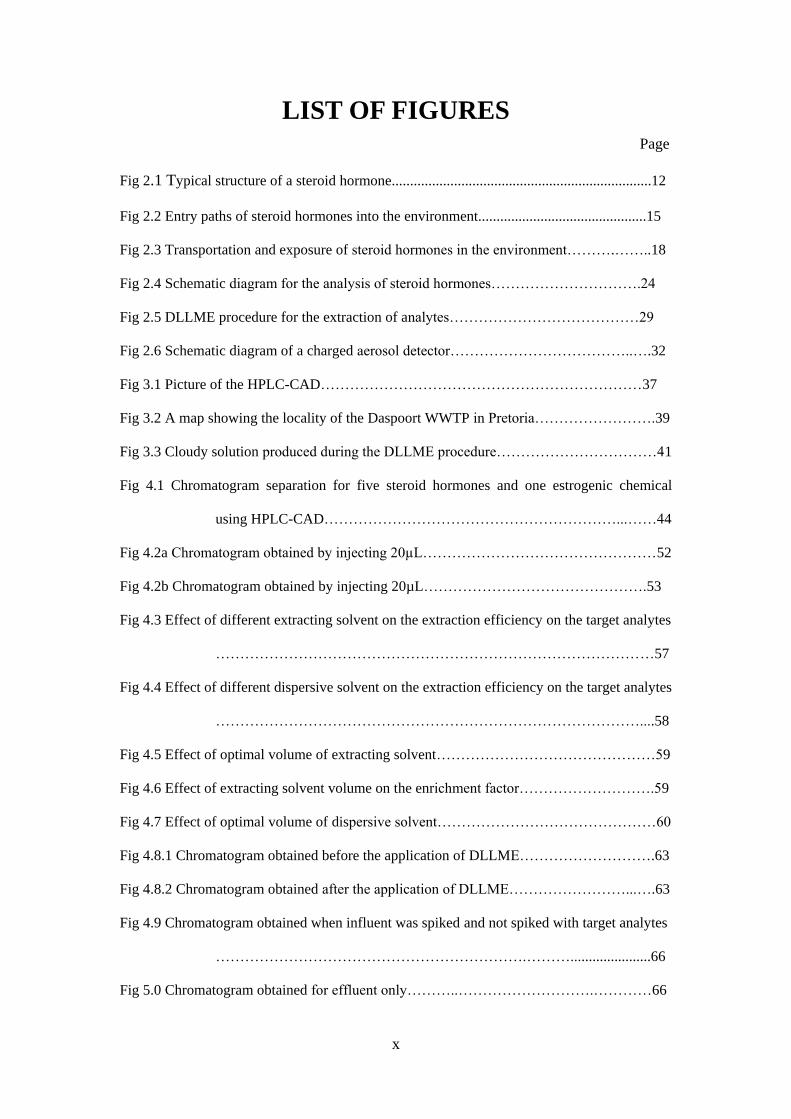
LIST OF FIGURES
Page
Fig 2.1 Typical structure of a steroid hormone.......................................................................12
Fig 2.2 Entry paths of steroid hormones into the environment..............................................15
Fig 2.3 Transportation and exposure of steroid hormones in the environment……….……..18
Fig 2.4 Schematic diagram for the analysis of steroid hormones………………………….24
Fig 2.5 DLLME procedure for the extraction of analytes…………………………………29
Fig 2.6 Schematic diagram of a charged aerosol detector………………………………..….32
Fig 3.1 Picture of the HPLC-CAD…………………………………………………………37
Fig 3.2 A map showing the locality of the Daspoort WWTP in Pretoria…………………….39
Fig 3.3 Cloudy solution produced during the DLLME procedure……………………………41
Fig 4.1 Chromatogram separation for five steroid hormones and one estrogenic chemical
using HPLC-CAD……………………………………………………...……44
Fig 4.2a Chromatogram obtained by injecting 20µL…………………………………………52
Fig 4.2b Chromatogram obtained by injecting 20µL……………………………………….53
Fig 4.3 Effect of different extracting solvent on the extraction efficiency on the target analytes
………………………………………………………………………………57
Fig 4.4 Effect of different dispersive solvent on the extraction efficiency on the target analytes
……………………………………………………………………………....58
Fig 4.5 Effect of optimal volume of extracting solvent………………………………………59
Fig 4.6 Effect of extracting solvent volume on the enrichment factor……………………….59
Fig 4.7 Effect of optimal volume of dispersive solvent………………………………………60
Fig 4.8.1 Chromatogram obtained before the application of DLLME……………………….63
Fig 4.8.2 Chromatogram obtained after the application of DLLME……………………...….63
Fig 4.9 Chromatogram obtained when influent was spiked and not spiked with target analytes
……………………………………………………….………......................66
Fig 5.0 Chromatogram obtained for effluent only………..……………………….…………66
xi
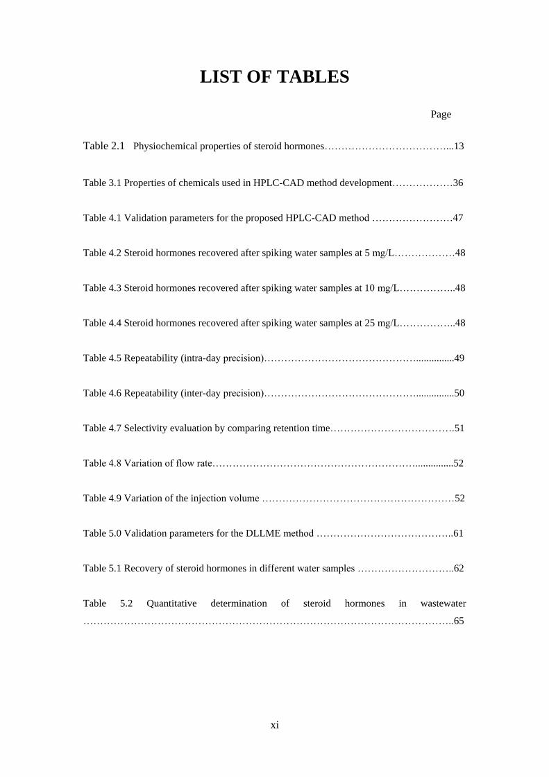
LIST OF TABLES
Page
Table 2.1 Physiochemical properties of steroid hormones………………………………...13
Table 3.1 Properties of chemicals used in HPLC-CAD method development………………36
Table 4.1 Validation parameters for the proposed HPLC-CAD method ……………………47
Table 4.2 Steroid hormones recovered after spiking water samples at 5 mg/L………………48
Table 4.3 Steroid hormones recovered after spiking water samples at 10 mg/L……………..48
Table 4.4 Steroid hormones recovered after spiking water samples at 25 mg/L……………..48
Table 4.5 Repeatability (intra-day precision)………………………………………...............49
Table 4.6 Repeatability (inter-day precision)………………………………………...............50
Table 4.7 Selectivity evaluation by comparing retention time……………………………….51
Table 4.8 Variation of flow rate……………………………………………………...............52
Table 4.9 Variation of the injection volume …………………………………………………52
Table 5.0 Validation parameters for the DLLME method …………………………………..61
Table 5.1 Recovery of steroid hormones in different water samples ………………………..62
Table 5.2 Quantitative determination of steroid hormones in wastewater
………………………………………………………………………………………………..65
xii
LIST OF ABBREVIATIONS
BPA = Bisphenol A
CAD = Charged aerosol detector
CE = Capillary electrophoresis
CPE = Cloud point extraction
DNA = Deoxyribonucleic acid
DAD = Diode array detection
DDT = Dichlorodiphenyltrichloroethane
DLLME = Dispersive liquid-liquid microextraction
FLD = Fluorescence detection
E1 = Estrone
E3 = Estriol
EDCs = Endocrine disruption chemicals
EF = Enrichment factor
ER = Estrogen receptors
EE = Extraction recovery /efficiency
ESI = Electrospray ionization
ESLD = Evaporating scattering light detection
FLD = Fluorescence detection
GC MS = Gas chromatography - mass spectrometry
GC MS/MS = Gas chromatography - tandem mass spectrometry
GLU = Glucuronide
HF-MMLE = Hollow fiber micro porous membrane liquid extraction
HF-SLME = Hollow fiber supported liquid microextraction
HPLC = High pressure liquid chromatography
IUPAC = International Union of Applied and Pure Chemistry
LLE = Liquid-liquid extraction
LC-MS = Liquid chromatography - mass spectrometry.
LC-MS/MS = Liquid chromatography-mass spectrometry/mass spectrometry
Log Kow = Octanol water partition coefficient
LOD = Limit of detection
LOQ = Limit of quantification
OP = Organic pollutants
PAH = Polycyclic aromatic hydrocarbon
P = Progesterone
xiii
pKa
= Acid Dissociation Constant
PPCPs = Pharmaceuticals and personal care products
PR = Progesterone receptors,
RI = Refractive index
RSD = Relative Standard Deviation
SPE = Solid-phase extraction
SPME = Solid phase microextraction
SPE-MIP = Solid phase microextraction-molecular imprinted polymer
STP = Sewage treatment plants
SUL = Sulfates
T = Testosterone
UV = Ultraviolet
WWTP = Waste water treatment plant
WHO = World health organization
β-E2 = Beta estradiol
α-E2 = Alpha estradiol
μg/L = Micro gram per litre
ng/L = Nano gram per litre
mg/L = Milli gram per litre
1
CHAPTER 1
INTRODUCTION
1.1 Background
Environmentalist and analytical chemists are concerned about the presence of emerging
organic pollutants (OP) which include pharmaceutical personal care products (PPCPs),
endocrine disrupting chemicals (EDCs) and some chemicals that are deliberately added into
our aquatic environment. Most of these compounds display estrogenic activities causing
undesirable adverse effects in humans, wildlife and the environment after long exposure
periods. Conventional wastewater treatment plants (WWTPs) are not designed to remove
steroid hormones or their metabolites present in wastewater [1-4], thus affecting the consumer
confidence about the quality of the water. Studies have shown that some hormones are
responsible for feminization of fish in aquatic environment [5, 8]. The EDCs causing these
effects are the natural, synthetic and other compounds that have the ability to mimic the
estrogenic activity. This increasingly publicized presence of these compounds has led
authorities to seek water treatment solutions and sensitive analytical methods proactively,
despite the fact that many OPs are not regulated.
Over the last decade, the presence and activity of EDCs in the environment has been a major
concern. These compounds alter the production and activities of endogenous hormones by
interacting with the endocrine system, presenting a potential threat to aquatic life and human
health [6]. According to Damstral and World Health Organization (WHO) [9], “any substance
or mixture that has the ability to alter the function(s), or to disrupt the synthesis, secretion,
transport, binding, reproduction, development or the behavior of an organism or its progeny
in the endocrine systems and consequently causes adverse health effects in an intact organism
2
has been defined as an EDCs”. EDCs have been categorized from various literatures mainly
into the following categories [6, 10];
• Pesticides and its metabolites (DDT, deldrin)
• Industrial and household chemicals (paints, detergents, UV sunscreen)
• PPCPs (clofibrates, sulfamethoxazole, ibuprofen)
• Polycyclic aromatic hydrocarbon (PAHs)
• Heavy metals (lead, cadmium)
• Steroid hormones (estrogens)
In this study, steroid hormones and bisphenol A will be our main focus as these compounds
have been reported to have ability to interfere with the hormonal systems of humans and
animals for over 80 years [11]. Some of the reasons why this class of compounds has received
more attention are their abundance in the human body and their estrogenic potency, as well as
the extent of their use in contraceptive pills and as growth promoters in animals.
Steroid hormones are mainly excreted from the body as estrogen conjugates, which comprise
sulphate (SUL) and glucoronide (GLU) in the urine [12-14]. Likewise, Hoffmann [15]
reported that female cattle also excrete mainly SUL and GLU estrogen conjugates.
Conjugated estrogens can also be found in some prescribed drugs as SUL estrogen salts,
which can be used in the treatment of hormonal imbalances, post-menopausal symptoms and
osteoporosis [12]. Steroid hormones have a strong endocrine disrupting effect, which
interferes with the reproductive functions of aquatic organisms [5, 10, 13, 16]. They have
been reported to cause testicular, prostate, and breast cancer [17-19]. Their presence has also
been reported to decrease sperm counts leading to reproductive disorders in men [18]. In fish,
their presence has been reported to be responsible for the reduction of fertility (decreased
sperm number and quality, or egg number), feminization in fish and frog abnormalities [15],
effects on the development of the gonads in male and juveniles yolk synthesis [20-22], and
3
reproductive alterations [15, 17]. Morphological changes have also been reported in teleost
fish and vertebrate’s thyroid gland as a result of exposure to organic pollutants [23-25].
The abilities of WWTPs in removal of steroid hormones have drawn a great deal of attention.
The removal rates of steroid hormones can vary from one WWTP to the other as the removal
of these compounds depends on the type of the treatment process and the physicochemical
properties. Recently, relative success has been achieved in the use of advanced technologies
such as granular activated carbon (GAC), membrane technology, ozonation, and ultraviolet
radiation in the removal of some PPCPs from wastewater [24, 26-27]. The removal of EDCs
falls into three categories; physical removal, biodegradation and chemical advanced oxidation
(CAO) [28]. The removal efficiency for estrone (E1), estradiol (E2) and ethylinestradiol
(EE2) in Germany, Canada and Brazil was studied using the activated sludge and was
reported to be 83%, 99.9% and 78%, respectively [14, 28]. In Korea, conventional drinking
water treatment methods were relatively inefficient for contaminant removal. The efficient
removal for (E1) was 99% and was achieved by GAC [26]. In some other studies, activated
sludge treatment steps removed hormones efficiently up to 77-79% for estriol (E3),
progesterone (P) 95%, estradiol (E2) 59%, and 17 alpha estradiol (17α-E2) 98% [14, 31- 32].
As much as these studies show the ability of the steroids to be removed from the treated
water, it is clear that the WWTPs have no capacity for 100 % removal efficiency. Thus the
WWTPs need improved treatments to completely remove these compounds from water
systems. The water industry is thus faced with the challenge to better understand the
mechanisms and methods of removal of EDCs to be able to offer cost effective solutions for
their removal in wastewater treatment.
1.2 Problem statement
There is much interest on the effects of steroid hormones on animals and human endocrine
system. Various types of steroid hormones and metabolites are introduced into the
environment through the discharges of WWTPs directly or indirectly, agricultural practices,

4
household and hospitals discharges. These activities and processes have increased the release
of steroids into the aquatic environment. These have affected the water body and water cycle
and hence a global concern. The whole world is struggling to keep up with the high demand
for water and its scarcity, with no alternative source of water, treatment plants tend to recycle
wastewater for drinking purposes [32]. In the process of recycling, some of the steroid
hormones might survive water treatment and then move to the underground water which
might pose some health effects. Determination of natural hormones such as estriol,
progesterone and estradiol has become of great interest due to the frequent detection in treated
and untreated WWTPs at low concentrations range of ng/L to μg/L. The natural and synthetic
steroid hormones have great higher estrogenic potentials. This is due to the fact that many of
their constituents are excreted unchanged as well as metabolites [12-14]. Inactive hormones
could be converted back in the environment to their active forms; this is due to the fact that
steroid hormones are not completely degraded biologically. This has led to increase in
development of preconcentration and analytical methods that can detect these compounds at
such concentration levels.
Numerous analytical methods have been used in the determination of these steroid hormones
in the environment [1, 11, 16, 25, 33] but some come with limitations. Currently, the
analytical determination of steroid hormones is dominated by gas chromatography- mass
spectrometry (GC-MS), and gas chromatography-tandem mass spectrometry (GC-MS/MS)
[6, 12, 13, 34- 37]. Due to the poor volatility of these compounds, derivatization steps are
prerequisite. This approach is unfortunately time consuming and labor intensive.
The use of liquid chromatography-tandem mass spectrometry (LC-MS/MS) has gained more
popularity for the analysis of steroid hormone due its ruggedness and sensitivity [6, 12, 34,
and 37]. An advantage of using LC-MS/MS is that it has the ability to analyze steroid
hormones in the environment without derivatization, or the need to hydrolyse the conjugated
form. However, using LC/MS/MS as an analytical tool is not without difficulties such as
5
matrix interference [37]. However, this approach is expensive for upcoming research and
development laboratories in emerging economies.
High pressure liquid chromatography (HPLC) with diode array (DAD) and other detectors is
also a preferred method of analysis of steroid hormones (12, 42). It is a fast, easy and simple
to operate. The concentration of analytes reported with DAD and the analytes were 0.2-1.6
µg/L [12, 42-44]. HPLC ultraviolet (UV) was able to detect at concentration levels of 0.3-1
µg/L [12, 18, 45-46]. HPLC has also been combined with mass spectrometry.
In this study, we propose an alternative technique for the determination of steroid hormones
in wastewater with the use of high pressure liquid chromatography coupled to a charged
aerosol detector (HPLC-CAD). CAD can be used in the determination of semi-volatile, basic,
neutral or acidic compounds without need for derivatization of the target analytes before
analysis. The mode of operation is by producing a signal that is proportional to the mass of
the sample and the response is independent of chemical structure [47- 49]. It detects
compounds electrically by ionizing them with charged nitrogen gas, an advantage CAD has
over the other detectors. When compared to other HPLC detectors, such as ultraviolet (UV),
diode array (DAD), fluorescence detector (FLD), there is a need for a derivatization step prior
to analysis. [47-51]. Evaporative light scattering detection (ESLD), is another type of detector
which is similar to the CAD in the mode of operation, it is able to detect steroid hormones
without a derivatization step prior to analysis.
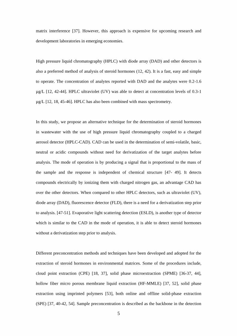
Different preconcentration methods and techniques have been developed and adopted for the
extraction of steroid hormones in environmental matrices. Some of the procedures include,
cloud point extraction (CPE) [18, 37], solid phase microextraction (SPME) [36-37, 44],
hollow fiber micro porous membrane liquid extraction (HF-MMLE) [37, 52], solid phase
extraction using imprinted polymers [53], both online and offline solid-phase extraction
(SPE) [37, 40-42, 54]. Sample preconcentration is described as the backbone in the detection
6
and quantification of steroid hormones in the environment. It will also be addressed by using
a more environmentally friendly approach, a preconcentration technique known as dispersive
liquid-liquid microextraction (DLLME). It is less time-consuming, inexpensive and simple
procedure which uses microliter volume of solvent and easy to operate [45-46, 55-56]
In this study, we intend to develop a new and alternative method for the determination of
steroid hormones in wastewater samples using HPLC-CAD. The main choice of this
technique is that there is no need for the derivatization of the compounds before analysis and
a preconcentration technique which uses less sample volume, fast and lower solvent
consumption with higher extraction efficiency will be used to extract the target analytes.
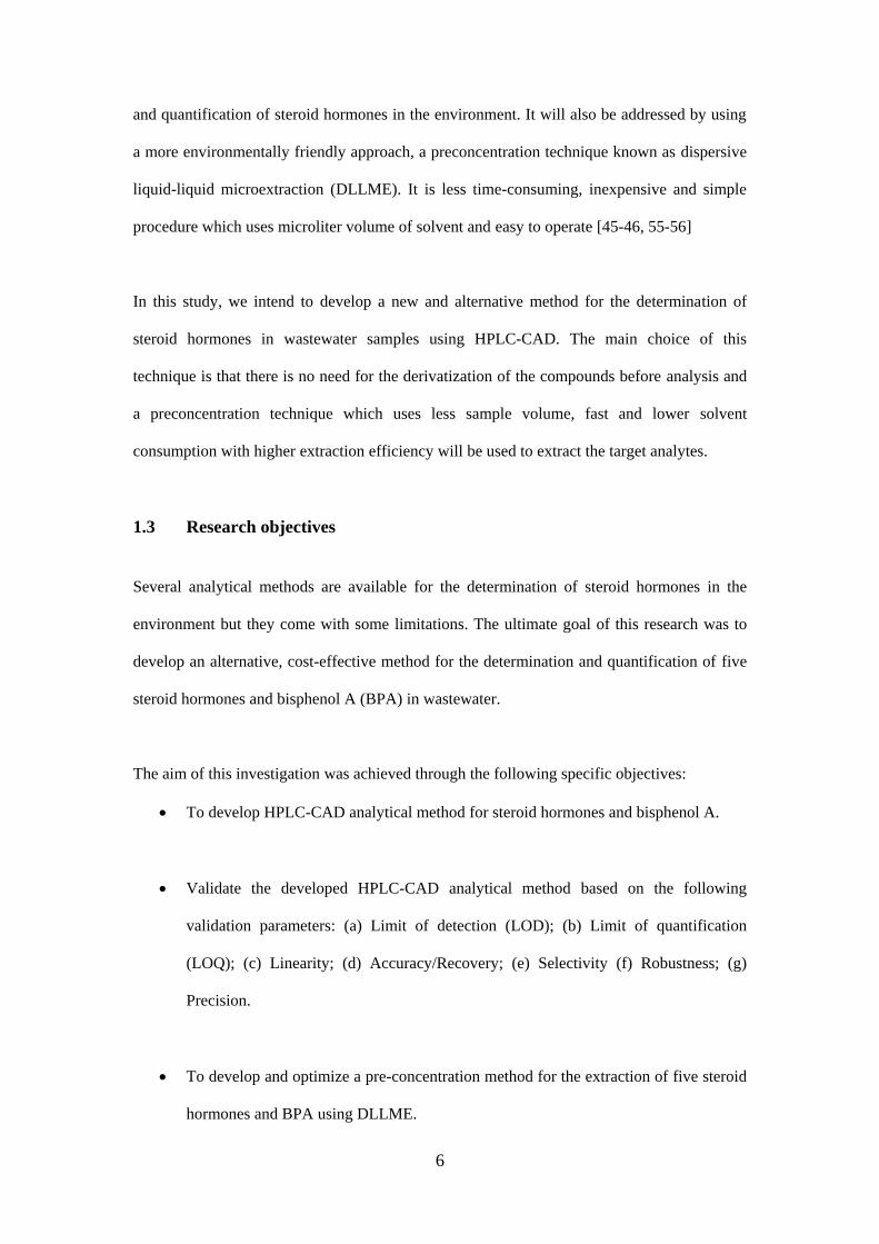
1.3 Research objectives
Several analytical methods are available for the determination of steroid hormones in the
environment but they come with some limitations. The ultimate goal of this research was to
develop an alternative, cost-effective method for the determination and quantification of five
steroid hormones and bisphenol A (BPA) in wastewater.
The aim of this investigation was achieved through the following specific objectives:
To develop HPLC-CAD analytical method for steroid hormones and bisphenol A.
Validate the developed HPLC-CAD analytical method based on the following
validation parameters: (a) Limit of detection (LOD); (b) Limit of quantification
(LOQ); (c) Linearity; (d) Accuracy/Recovery; (e) Selectivity (f) Robustness; (g)
Precision.
To develop and optimize a pre-concentration method for the extraction of five steroid
hormones and BPA using DLLME.
7
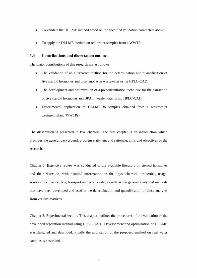
To validate the DLLME method based on the specified validation parameters above.
To apply the DLLME method on real water samples from a WWTP
1.4 Contributions and dissertation outline
The major contributions of this research are as follows:
The validation of an alternative method for the determination and quantification of
five steroid hormones and bisphenol A in wastewater using HPLC-CAD.
The development and optimization of a preconcentration technique for the extraction
of five steroid hormones and BPA in waste water using HPLC-CAD.
Experimental application of DLLME to samples obtained from a wastewater
treatment plant (WWTPs).
The dissertation is presented in five chapters. The first chapter is an introduction which
provides the general background, problem statement and rationale, aims and objectives of the
research.
Chapter 2: Extensive review was conducted of the available literature on steroid hormones
and their detection, with detailed information on the physiochemical properties, usage,
sources, occurrence, fate, transport and ecotoxicity, as well as the general analytical methods
that have been developed and used in the determination and quantification of these analytes
from various matrices.
Chapter 3: Experimental section. This chapter outlines the procedures of the validation of the
developed separation method using HPLC-CAD. Development and optimization of DLLME
was designed and described. Finally the application of the proposed method on real water
samples is described.
8
Chapter 4: This chapter presents the main findings and gives a detailed overview of the results
and graphs of the research
Chapter 5: The chapter provides the conclusions of this research, followed by
recommendations on future work.
1.5 Declaration of recent publications
The following recent articles have been presented and sent for review under this research
work.
Refereed journal publication
Osunmakinde, C.O., Dube, S and Nindi, M.M. Development of Dispersive Liquid-
Liquid Microextraction for the determination of six endocrine disruptors in
wastewater using High Pressure Liquid Chromatography-Charged Aerosol Detector.
Journal of analytical methods in chemistry. (Accepted ISI journal:171739)
Refereed conference publications
Osunmakinde, C.O., Nindi, M.M., and Dube, S. Method development for the
determination of steroid hormones in waste water using HPLC with charged aerosol
detector. Book of abstract 12th ICCA conference, Pretoria, South Africa 2013.
Osunmakinde, C.O., Nindi, M.M., and Dube, S. Investigation of steroids and
hormones in wastewater using HPLC with a charged aerosol detector. Book of
abstract of the 4th SEANAC International Conference, Maputo, pg 73, 2012.
Osunmakinde, C.O., Nindi, M.M., and Dube, S. Screening for steroids and hormones
in wastewater using HPLC with charged aerosol detector (CAD). Book of abstracts
9
at SACI YCS Symposium, 2012.
Research Reports presented, submitted and accepted by the Water Research
Commission South Africa K5/2094//3 (July, 2013).
10
CHAPTER 2
STEROID HORMONES AND THEIR
DETERMINATION
Steroid hormones belong to a class or group of compounds known as the EDCs. They are
biologically active organic compounds whose structure consists of a phenolic and hydroxyl
group. They are non-volatile, with moderate to high hydrophobicity [1]. Steroid hormones are
classified mainly into three main groups; natural, synthetic and xenoestrogens [6].
Natural steroid hormones are the bases of all sex hormones such as estradiol, estrone, estriol
and progesterone. They are produced in human beings, animals as well as in some plants.
Natural hormones are secreted by the adrenal cortex, testis, ovary, placenta and the endocrine
gland in human beings and animals [7] and they travel through the bloodstream. They can
bind to specific receptor sites in various organs and tissues and regulate a variety of biological
functions in mammals, such as controlling metabolism and reproductive function, as well as
maintaining blood pressure, glucose and ion levels, muscle and nervous system functions [8].
Synthetic hormones such as 17α-ethyinylestradiol, menstranol and its metabolites/conjugates
are chemically synthesized. They are the main active agents in hormone therapy, birth and
oral contraceptive pills [6, 17].
The term xenoestrogens is used to describe compounds that have shown to act like estrogens
in terms of their interference with the endocrine system. They comprise a multitude of
chemicals, which affect the endocrine (hormone) system of animals and humans. Some of
these compounds are the main active ingredient in pharmaceuticals and are used in
manufacturing industries, agriculture and households [6, 10, 12, 14]. They can mimic the
physiological processes such as growth, sexual differentiation or reproduction by binding to
11
the estrogen receptor. An example of such an estrogenic chemical compound is Bisphenol A
(BPA) [6, 10, 12, 20], Which is used in the production of plastics used for food product
packages, bottles for water, bottles for infant food and kitchen utensils [6, 45, 57-58].
Because of its polarity, persistence and solubility, BPA is often able to pass through treatment
plant operations and enter the aquatic environment [57]. BPA has also been demonstrated to
exhibit estrogenic activity and it has been classified as an endocrine disruptor [6, 8, 10, 57].
2.1 Mechanisms of steroid hormones
Steroid hormones are built from cholesterol, produced and secreted naturally or synthetically
by endocrine glands in the body. The endocrine system is an integrative system that controls
the cell function and activities of mammals, amphibians, birds, fish and various invertebrates
by communicating through chemical messengers called hormones [60]. The endocrine or
hormone system plays a critical role in development and growth, metabolism, reproduction,
natural defenses to stress, stimuli and nutritional balance of the blood. This system consists of
several glands (e.g. adrenal, pituitary, gonads, and ovaries) that synthesize and secrete
hormones. Steroid hormones are transported throughout the body via the circulatory system to
the target cells. On reaching a target cell, the hormone binds to a specific hormone receptor,
and then the receptor/hormone complex attaches to a specific segment of deoxyribonucleic
acid (DNA) called the response element to activate or inhibit gene expression, ultimately
leading to protein synthesis [60].
2.1.1 Molecular structure and physicochemical properties of steroid hormones
Steroid hormones have a tetracyclic molecular structure, which is derived from cholesterol. It
comprises four rings: a phenol group, two hexamethylene groups and a cyclopentane group.
Numbering and labeling of the four rings (A, B, C, D) as shown in Figure 2.1 are presented in
accordance with the International Union of Applied and Pure Chemistry’s (IUPAC)
recommendations for the nomenclature of steroids.
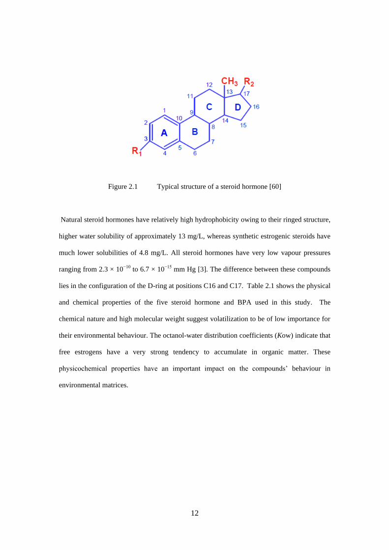
12
Figure 2.1 Typical structure of a steroid hormone [60]
Natural steroid hormones have relatively high hydrophobicity owing to their ringed structure,
higher water solubility of approximately 13 mg/L, whereas synthetic estrogenic steroids have
much lower solubilities of 4.8 mg/L. All steroid hormones have very low vapour pressures
ranging from 2.3 × 10⁻¹⁰ to 6.7 × 10⁻¹⁵ mm Hg [3]. The difference between these compounds
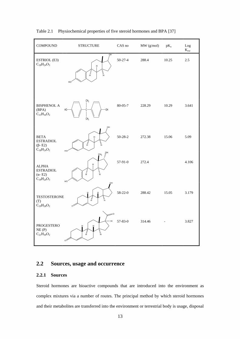
lies in the configuration of the D-ring at positions C16 and C17. Table 2.1 shows the physical
and chemical properties of the five steroid hormone and BPA used in this study. The
chemical nature and high molecular weight suggest volatilization to be of low importance for
their environmental behaviour. The octanol-water distribution coefficients (Kow) indicate that
free estrogens have a very strong tendency to accumulate in organic matter. These
physicochemical properties have an important impact on the compounds’ behaviour in
environmental matrices.
13
Table 2.1 Physiochemical properties of five steroid hormones and BPA [37]
COMPOUND
STRUCTURE
CAS no
MW (g/mol)
pKa
Log
Kow
ESTRIOL (E3)
C18H24O3
HO
H H
OH
H
50-27-4
288.4
10.25
2.5
BISPHENOL A
(BPA)
C15H16O2
C
CH3
CH3
OHHO
80-05-7
228.29
10.29
3.641
BETA
ESTRADIOL
(β- E2)
C18H24O2 HO
H H
OH
H
50-28-2
272.38
15.06
5.09
ALPHA
ESTRADIOL
(α- E2)
C18H24O3 HO
H H
CH3
H
OH
57-91-0
272.4
4.106
TESTOSTERONE
(T)
C19H28O2
H H
O
H
OH
H
58-22-0
288.42
15.05
3.179
PROGESTERO
NE (P)
C21H30O2 H H
O
H H
O
H
57-83-0
314.46
-
3.827
2.2 Sources, usage and occurrence
2.2.1 Sources
Steroid hormones are bioactive compounds that are introduced into the environment as
complex mixtures via a number of routes. The principal method by which steroid hormones
and their metabolites are transferred into the environment or terrestrial body is usage, disposal
14
and excretion by humans and farm animals (cattle and pigs). A combination of metabolized
and unmetabolized steroid hormones is excreted by humans. These are discharged from
private households, industrial waste waters, farm effluents and disposal systems of hospitals
and eventually enter municipal WWTPs [14]. Some steroid hormones can also enter the
wastewater via regular use during showering or bathing, and applications on the body [12, 14,
60].
Steroid hormones are also introduced into the environment either after extensive treatment in
WWTPs, mainly the urban domestic wastewater, or after minimal treatment from agricultural
wastes, e.g. irrigation water effluents from ponds, direct waste excretion by grazing livestock
without treatment. Thus, effluent from WWTPs becomes the most important source of
pollution. Conventional WWTPs have historically been used to improve dissolved oxygen
levels in water bodies, reduce nutrient loads and remove some biodegradable organic
compounds. Therefore, by design, they are not equipped to remove emerging contaminants
such as steroid hormones or any other PPCPs [2-4]. Hence, steroid hormones and their by-
products are discharged directly into surface waters, again without prior treatment at WWTPs
[57- 60].
Another significant source of steroid hormones in the environment that is common and
unregulated is the discharges from manufacturing, health facilities, and household disposal of
expired and unused medicine via toilet flush or domestic trash [2, 58, 61]. The release of
untreated sewage from WWTPs directly into the surface waters and the surroundings is also
another point. This is a major challenge particularly for plants or treatment facilities that are
designed to deal with large amounts of storm water, or erosion and other natural disasters that
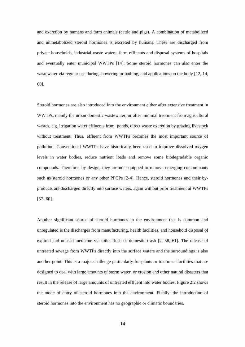
result in the release of large amounts of untreated effluent into water bodies. Figure 2.2 shows
the mode of entry of steroid hormones into the environment. Finally, the introduction of
steroid hormones into the environment has no geographic or climatic boundaries.
15
STORAGE
MANUFACTURING PLANTS
HOSPITALS AND DISPENSARY
FARM LAND ACTIVITIES
EXCRETIONDISPOSAL
AIRWATER BODIES SEWAGE
WASTE AND INCINERATION
AIRLAND FILL SITES
SEWAGE TREATMENT PLANTS
SOIL AND EDIBLES
SURFACE WATER
GROUND WATER
DRINKING WATER
ACTR
ING PLANTS
Figure 2.2 Sources of Steroid hormones in the environment adapted from Kummer [62]
2.2.2 Usage of steroids
A pharmaceutical (also frequently referred to as a drug or a medicinal product) can broadly be
defined as a compound of known chemical structure, which when administered into a host
organism produces biological effects. The US Food and Drug Administration classifies a
substance as a “drug” if it is intended to diagnose, treat, cure, mitigate or prevent diseases [9-
10]. Steroids are known to be endogenous and they comprise a wide group of substances that
mediate various types of biological responses and have been synthesized by chemists over the
years [63]. Cholesterol is the most widely spread steroid hormone in the body, which is
derived from dietary intake, but can also be synthesized in the body [12].
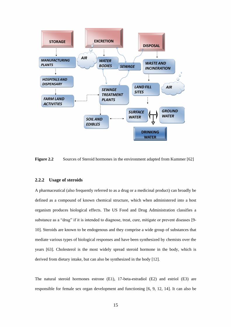
The natural steroid hormones estrone (E1), 17-beta-estradiol (E2) and estriol (E3) are
responsible for female sex organ development and functioning [6, 9, 12, 14]. It can also be
16
used in the treatment of various diseases such as allergic reactions, arthritis, some
malignancies and diseases resulting from hormone deficiencies or abnormal production [10,
12]. The synthetic steroid hormone such as 17-alpha ethinylestradiol (EE2) is used in female
contraceptives and in hormone replacement therapy [9, 12, 16-18]. In addition, synthetic
steroid hormones such as EE2 are used for therapy in treating menstrual syndrome, as a
hormone supplement, or as the main component of contraceptives [10, 12, 16, 20]. Other
synthetic steroids that mimic the action of progesterone are widely used as oral contraceptive
supplements and are also designed to stimulate the synthesis and muscle-building action of
protein in the body [63- 67].
Steroid hormones are used legally in veterinary medicine as growth promoters and for animal
fattening purposes. They have the capacity to increase weight gain and reduce the feed
conversion ratio [68]. Steroids are used in the agricultural sector for food production, mainly
for boosting the mass and quality of animal carcasses in food production for economic
reasons [68]. Anabolic steroids may also be used in sport or entertainment in order to improve
performance in competitive humans and animals [69]. These steroids may enhance
performance through a number of mechanisms, including increased muscle mass, enhanced
recovery from training, raised red blood cell count and heightened aggression [69-70]. Large
amounts of steroid hormones and synthetic hormones are illegally used, because of their
potential anabolic effects [69-70]. Natural and synthetic have been reported to be the most
potent of the EDCs [5].
2.2.3 Occurrence
The occurrence of steroid hormones and their metabolites has been studied in various
matrices over the years, owing to their possibly adverse effect on humans and wildlife [62].
Steroid hormones have been detected in various samples and their occurrence has been
documented in Europe, United Kingdom and China [14, 17, 19]. Their presence has been
reported in drinking water and bottled water at low concentration level of 0.01- 0. 9 ng/L [10,
17
73, 76], concentration levels of 0.2-114 ng/L have also been confirmed at WWTPs [7, 14, 65,
77-79]. In surface water, 0.3 – 1.7 ng/L [14, 17, 79-81], 2.6- 9.5 ng/L in sea water [10, 76],
and 6- 66 ng/L were detected in ground water [7, 76, 78, 82]. In the river water, 0.3 – 32.9
ng/L has been observed [60, 75, 83-84], 0.08 - 0.89 ng/g in the soil [7, 85-87], 0.9- 49 ng/g in
sediment and sludge [33, 76, 85, 87], and 0.6- 26 ng/g in some fish samples [88]. Finally the
presence of steroid hormones has also been confirmed in hospitals and industrial units, as well
as intensive animal breeding farms [7, 78].
2.3 Transportation of steroid hormones
The transport of steroid hormones is determined with respect to their partitioning into
different environmental compartments. They are non-volatile organic compounds and their
disposal or mode of entry into the environment is mainly by aqueous transport. The
partitioning into aqueous or solid phase depends on the compound-specific distribution
coefficients. Compounds that tend to stay in the dissolved phase are transported into surface
waters through the effluent discharge from WWTPs, while those with high sorption
capabilities end up in terrestrial environments, particularly if the treated sludge is used for
land applications. Pharmaceutical products residing in soil environments may reach ground
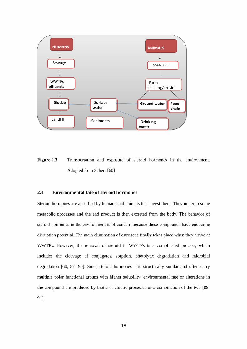
water upon leaching [88] or enter surface water again after run-off events. Figure 2.3 shows
the fate and transport pathways in which steroid hormones undergoes.
18
HUMANS
S Sludge
S Sewage
S WWTPs effluents
S Landfill
S Surface water
S Sediments
ANIMALS
S MANURE
S Farm leaching/erosion
Ground water Food chain
S Drinking water
Figure 2.3 Transportation and exposure of steroid hormones in the environment.
Adopted from Scherr [60]
2.4 Environmental fate of steroid hormones
Steroid hormones are absorbed by humans and animals that ingest them. They undergo some
metabolic processes and the end product is then excreted from the body. The behavior of
steroid hormones in the environment is of concern because these compounds have endocrine
disruption potential. The main elimination of estrogens finally takes place when they arrive at
WWTPs. However, the removal of steroid in WWTPs is a complicated process, which
includes the cleavage of conjugates, sorption, photolytic degradation and microbial
degradation [60, 87- 90]. Since steroid hormones are structurally similar and often carry
multiple polar functional groups with higher solubility, environmental fate or alterations in
the compound are produced by biotic or abiotic processes or a combination of the two [88-
91].
19
2.4.1 Abiotic transformations
An abiotic transformation is any process in which a substance is converted into simpler
products by a physical or chemical mechanism. Abiotic transformation of PPCPs products in
surface or wastewater may occur by hydrolysis and photolysis. Steroid hormones are usually
resistant to hydrolysis, whereas direct and indirect photolysis is a primary pathway for their
transformation. Direct photolysis of a compound is caused by the direct absorption of solar
light and indirect photolysis involves the use of natural photosensitizers, e.g. nitrate and
humic acid [90-92]. Steroid hormones and their metabolites undergo two major kinds of
abiotic transformations, these include photodegradation and sorption.
Photo degradation is a major transformation process for steroid hormones in the environment,
both in WWTPs and natural waters. In natural surface waters, photochemical reactivity is
strictly limited to the photic zone, i.e. the uppermost region of the water column, which is
affected by depth and attenuation [91-92]. Surface water with a high algal content or sediment
loading will have a very shallow photic zone because of light absorption and scattering. In
addition, humic substances can absorb or attenuate sunlight, which also decreases the depth of
light penetration, while colored dissolved organic matter is the main UV-absorbing
constituent in surface water and controls UV light penetration [89, 93].
Solar degradation of organic compounds can occur either directly or indirectly. In direct
photolysis, a compound absorbs appropriate radiation depending on the absorption spectrum
of the compound directly from the sunlight in which it becomes excited. After the excited
state, it will then undergo a chemical transformation, which enables it to generate a different
product based on the structure of the compound [88-93]. Indirect photolysis occurs when the
dissolved photosensitizers (nitrates, humic acids etc.) generate free radicals in surface water,
which then absorb solar radiation to get to an excited state [89-91]. Photo degradation

20
depends on various factors, such as the intensity of radiation, dissolved organic matter,
nutrient loads and water depth [93-94].
Adsorption or sorption occurs when suspended solids, particulates, sediments, colloids or
natural organic matter is a major elimination pathway of steroid hormones and metabolites in
WWTPs as well as in surface waters [91]. This results in the distribution of organic
contaminants between the aqueous and solid phases, which is governed by equilibrium
properties and non-hydrophobic mechanisms such as electrostatic interactions [90-95]. The
distribution of steroid hormones in the environment is based mainly on its Log Kow values
(solubility), which are the critical chemical parameters. Large Log Kow values (> 4) suggest
large hydrophobic molecules and will tend to dissociate from solid organic matter, i.e. high
sorption potential, while smaller Log Kow values (< 2.5) suggest low sorption potential [66].
Steroid hormones are hydrophobic substances and are readily absorbed onto the surface of
solid materials. They pose some risks to the environment, which includes soil, sediment,
aquatic and non-aquatic species [91]. Different studies have shown that sorption of steroid
hormones in the soil and sediment of aquatic environments is moderate to high [87, 89, 91-
92].
2.4.2 Biotic transformation (microbial degradation)
Biological transformation of pharmaceuticals occurs in two different ways in living
organisms: through the action of enzymes, e.g. cytochromes, and by the microorganisms
present in the digestive tract [67]. These transformations are aided by different species of
bacteria in the environment. Metabolism of drugs in the body is supported by different phases
of reactions such as hydrolysis, oxidation, hydroxylation and reduction [76]. The first phase
of the products generated is less toxic than the parent drug, but there are instances where the
metabolic product could be more toxic [89]. Hydrophilicity of the first phase products
generated is further enhanced by second phase conjugation reactions with the help of
21
glucuronic acid, SUL, acetyl, glutathione and amino acids. The highly polar conjugated
products are then excreted through urine and feces. Therefore, the extent to which a parent
compound or its metabolites are excreted depends on the compound-specific metabolism [94-
95]. High proportions of administered drugs are sometimes excreted unchanged [96-97]. The
excreted compounds in the second phase, which are the conjugated products, are sometimes
converted back to the parent compound with the aid of various treatment processes that occur
in WWTPs [97-100].
Microbial degradation constitutes an important process for PPCPs products. This degradation
is often part of the secondary treatment in WWTPs’ activities and involves the use of aerobic
activated sludge treatments and anaerobic sewage sludge digestion [28]. It is often desirable
that biodegradation results in complete mineralization. However, in addition to the differences
in the chemical characteristics of pharmaceutical compounds, the extent of biodegradation
(low, moderate or high) and hence the removal efficiency, depends on a variety of factors
including sludge age, hydraulic retention times, season, differences in WWTPs’ construction
and treatment technologies, amount of biomass, temperature, pH and oxygen saturation levels
[14, 64, 77, 89, 96, 98-99]. Conjugated estrogens are hydrolyzed into free estrogens and GLU
and sulfuric acid in the presence of fecal bacteria - Eschericia coli [13, 92, 101]. Very limited
information is available on environmental parameters such as nutrient levels, pH and other
recognized variables affecting microbial activity that influence or inhibit the degradation of
steroid hormones in the environment [13, 101-102].
2.5 Environmental significance and effects
2.5.1 Environmental significance
The presence of steroid hormones in the aquatic and terrestrial environments has increased
based on the effects of estrogens on target and non-target organisms. They interfere with
normal endocrine function by mimicking and blocking hormonal function [6, 10, 12, 24, 103].
22
The endocrine and reproductive effects of environmental contaminants are believed to be due
to:
(1) Mimicking endogenous hormones such as estrogens and androgens,
(2) Antagonizing normal, endogenous hormones,
(3) Altering the pattern of synthesis and metabolism of natural hormones, and
(4) Modifying hormone receptor levels.
The ecological relevance of endocrine disruption in wildlife is, however, difficult to quantify,
as there is limited understanding and few reports on how physiological changes affect the
individual animal and how individual responses affect the population and community [10,
103]. Furthermore, a major challenge faced by environmental biologists is the need to place
endocrine disruption into context with other environmental pressures faced by wildlife
populations, for example global warming.
There has been increasing evidence of endocrine disruption in fish and frogs due to estrogens
in the environment and several field and laboratory studies have reported inter-sex conditions
in fish exposed to estrogen compounds [16, 23]. Estradiol and 17α-ethinylestradiol (EE2) is
the active ingredient in many contraceptive pills and birth control methods. It induces the
synthesis of the yolk protein precursor vitellogenin, a specific and sensitive indicator of
exposure to xenoestrogens in fish at concentrations 1 600 000-fold lower than the
concentration necessary to cause the death of 50% of the test fish population [10, 105].
2.5.2 Environmental effects
Steroid hormones have interactions with the endocrine system mechanisms, however little has
been studied about the environmental effects. They are a potential risk for wildlife and
humans through the consumption of contaminated food or water. Some of the alterations in
the endocrine system can lead to disturbances of homeostasis, miscarriage, failures in

23
development and reproduction and even death [10, 12, 16, 102-106]. Steroid hormones may
affect not only the exposed individual but also the children and subsequent generations. These
effects may be transmitted not because of mutation of the DNA sequence, but rather through
modifications to factors that regulate gene expression, such as DNA methylation and histone
acetylation [103-104]. A detailed overview of the effects that estrogens may cause in the
environment is not the purpose of the current report. However, from an analytical perspective,
the concentration levels of the compounds in the environment are of interest since such data
determines the sensitivity requirements for analytical methods.
In humans, some steroid hormones have been linked to declining male reproductive health, by
both decreasing sperm counts and affecting quality leading to reproductive disorders in
human males [5, 10, 21, 103-106]. Women are normally exposed to estrogen, but the effects
on females are more difficult to track because of the estrous cycle and the resulting huge
differences in circulating hormone concentrations at different stages of the cycle [105-106].
The presence of estrogen-mimicking compounds in adult women can impair reproductive
capacity by interfering with natural hormone cycles, potentially rendering women unable to
conceive or to maintain pregnancy, and may promote abnormal cell growth [105- 109].
In aquatic environments, fish are considered the most sensitive of all species. Studies have
shown that steroid hormones EDCs have influenced the sexual development of fish in the
waters of the United Kingdom as far back as 15 years ago [107-109]. The blocking and
development of frog egg maturation [12, 16, 23, 108 -114], decline in alligator populations [8,
23, 110] and an increase in male fish possessing male and female characteristics [111-112].
Altered levels of circulating thyroid hormones and morphological changes in the thyroid
gland have been demonstrated in sea bass exposed to organic pollutants (OPs) [110-112]. A
number of studies have shown that male fish exposed to estrogens at ng/L level will exhibit
estrogenic responses, such as vitellogenin (VTG; precursor to yolk, a female-specific protein)
production [25, 109- 114].
24
2.6 Analytical determination of steroid hormones
The occurrence of steroid hormones in the environment is gaining wide attention owing to
their effects fate, transport and behavior. Modern and sensitive techniques are being
developed worldwide for their determination. Since steroid hormones and their metabolites
are found in the environment at lower concentrations, there is a need for sensitive analytical
methods that are reliable and reproducible. The most common advanced analytical techniques
used in the determination and quantification of steroid hormones are GC-MS, GC-MS/MS,
LC-MS, LC-MS/MS, HPLC and immunoassays [36-37,71. However, steroid hormones
cannot be analyzed directly without a sample preparation technique which is predominately
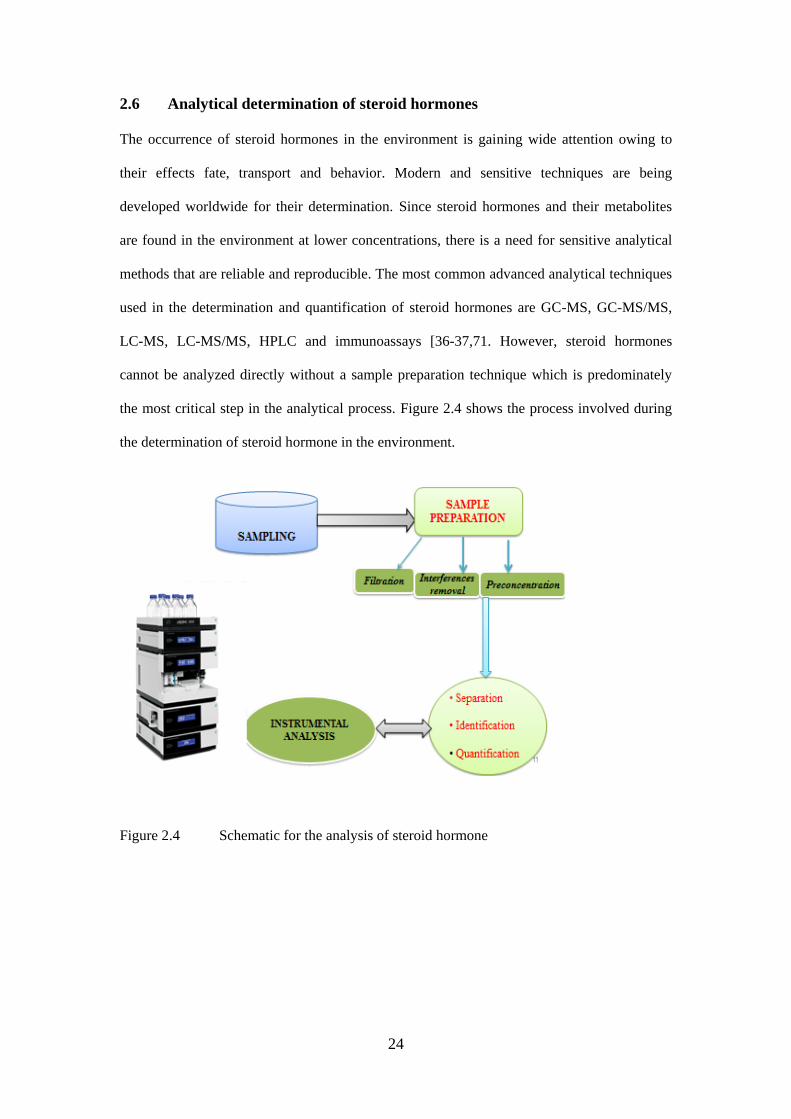
the most critical step in the analytical process. Figure 2.4 shows the process involved during
the determination of steroid hormone in the environment.
Figure 2.4 Schematic for the analysis of steroid hormone
25
2.6.1 Sample pre-treatment and clean-up methods
Sample pre-treatment and clean-up is usually a difficult step and time consuming segment in
the analysis of steroid hormones or other OPs in the environment. Due to the low level
concentrations in which they exists, as well as the complexity of the matrices in which they
are detected. However, sample preparation is necessary to convert the sample into a form that
is suitable for the analysis to be performed without the loss of the secondary sample [37]. The
process of analyzing a sample is based on the type of matrix and concentration level at which
the analytes are detected. It plays a major role in the data generated, as it can influence the
results in various ways.
Adequate optimization procedures and conditions are necessary to enhance sensitivity and
interference from the matrix. The application of sample preparation with high enrichment
achieves a low limit of detection (LOD) [37]. Sample preparation steps, such as filtration,
extraction, purification, and evaporation are employed before the final determination is
performed by bioassays or GC or HPLC.
2.6.1.1 Sampling and storage
The value of any data generated in the laboratory depends solely on the integrity of how the
sample was taken. Sample must be collected in such a way that nothing is added or lost
during the process. To examine the quality of wastewater discharged into the environment,
the characteristics of the water in question must be defined chemically or biologically. There
are different approaches in sampling organic pollutants in wastewater, such as grab sampling,
composite sampling and also the polar organic chemical integrative sampler (POCIS).
Samples are kept in ice-packs containers during transportation to the laboratories to avoid
further degradation of the target analytes.
26
2.6.1.2 Filtration
Because wastewater usually contains a high load of organic material and suspended particles,
filtration is usually the first step in sample preparation as the suspended solids easily clog the
absorbent bed. Similarly, when the analysis is performed by immunochemical assay, filtration
helps to avoid undesired adsorption onto antibodies. The filtration step is performed in
numerous ways, but most of the studies reviewed employed glass filters [90].
2.6.1.3 Isolation
Sample extraction from heavily contaminated samples (manure, soil and to some extent waste
water) often requires further clean-up before analysis [60, 90]. This has been achieved using
different techniques such as liquid-liquid extraction and solid-phase purification on C18
columns. Some of the purification procedures were developed to isolate the estrogenic active
fractions from the wastewater extract for further identification of the compounds responsible
for such activity, rather than for simple clean-up of the extract [90]. Good detection limits
without purification have only been reported by studies using biological techniques for
analysis or graphitized carbon black adsorbents as SPE [90].
2.6.1.4 Extraction
Extraction of natural and synthetic steroid hormones from wastewater is usually performed
using numerous techniques. Cloud point extraction (CPE) has been used as an alternative to
the liquid-liquid extraction (LLE) method in the determination of steroid hormones with LOD
values of 0.23-5.0 ng/L, and recoveries between 81-99.5% [18, 37]. In CPE technique
surfactants such as triton are normally used as the extraction solvent. The disadvantage of this
method however lies in the compatibility of the surfactant during analysis of the analytes with
instruments such as GC and HPLC. Also CPE is a time-consuming procedure [18, 37, 56,
113-115]. Hollow fiber micro porous membrane liquid extraction (HF-MMLE) has been
developed for determination of estrogens in sewage-water samples. The HF-MMLE is been
27
referred to as a green technique [37, 52]. This technique showed LOD as low as 1.6–10 ng/L
with high enrichment factors and extraction efficiency, but long processing time of 180 min
[37, 52]. Solid phase micro extraction (SPME) is a solvent free, fast and simple procedure
which was introduced by Pawliszyn for the extraction of environmental samples [36-37, 44].
It is a very fast technique and could be automated. The technique can also be coupled to
HPLC or GC [37, 8, 47, 53, 57, 62]. Using SPME as a preconcentration technique, the
instrumental detection of steroid hormones in environmental was as low as 6-100 ng/L with
recoveries of 88-116%. The limitation that comes with this technique is that it is expensive
and some of the fibers are fragile with limited life time. The desorption temperature and
sample carry over is also a major challenge during the procedure [37, 56, 115].
Solid phase extraction (SPE) could either be online or offline [37-38, 40-41, 54]. In most
cases discs and often specific cartridges have been employed for the use of extraction of
steroid hormones using SPE from wastewater. Both discs and cartridges have been used with
advantages and disadvantages. Cartridges have the advantage of easy system automation,
because devices are available for automated washing, conditioning, sample loading, drying
and elution of a large number of samples [37, 90]. It is time consuming and an expensive
technique which requires special instrumentation [37, 115]. Another disadvantage is that it
requires specific cartridges for target analytes, and these cartridges are not reusable. Analytes
were detected in water samples with LOD of 0.02-40 ng/L and recoveries of 79- 95%
recovery [38, 54]. Due to the various drawbacks of the existing methods, ongoing research
has led to the development of more preconcentration procedures to overcome these
limitations.
2.7 Dispersive liquid-liquid microextraction
The use of small amount or the elimination of solvent has led to the development of
dispersive liquid-liquid micro extraction (DLLME). DLLME was first reported by Assadi and
28
his co-workers in 2006 [55]. DLLME has also been used for the extraction of natural,
synthetic hormones as well as some EDCs [45-46, 56]. It is a liquid-phase microextraction
technique based on a ternary component solvent system in which appropriate mixture of
dispersing and extracting solvent are rapidly injected into an aqueous sample containing the
analytes of interest. The mixture of (water/disperser solvent/extraction solvent) is shaken in
order to produce a cloudy solution in the conical test tube. Centrifugation is done for a few
minutes, the fine particles of extracting solvent are sedimented at the bottom of the conical
test tube, and this is referred to as the sedimented phase. DLLME is a technique that can be
used for analytes with high or moderate lipophilic properties [47, 56-57,114-115].
DLLME uses an extracting solvent such as the chlorinated solvents that has a high density
than water such as carbon disulfide, carbon tetrachloride, tetrachloroethylene, chlorobenzene,
chloroform and dichloromethane [55-56]. The disperser should be miscible in water and
extracting solvent to help increase the surface area between the extracting solvent and
aqueous phase [114-115] thus ensuring that equilibrium is achieved quickly. The applicability
of the DLLME is based mainly on the distribution coefficient (k) which is defined as the ratio
of the analyte concentration in extraction solvent and sample solution [115]. The value of the
distribution coefficient should be greater than 500 to achieve a suitable application of
DLLME. However, for the acidic or alkaline analytes, distribution coefficient could be
increased by controlling the pH value of sample solution; making the analytes existing in
nonionic state [115-116]. Steroid hormones are largely neutral analytes, they have a great
potential to be extracted from the aqueous phase into the organic phase, and this makes them
a good candidate for this type of preconcentration procedure.
In 2011, DLLME was used in the determination of 17β-estradiol and diethylstilbestrol in
aqueous water samples using HPLC. The enrichment factors and extraction recoveries were
71.0 – 78.5 and 85.2 – 94.2 respectively. The limit of detection for 17β-estradiol was
0.010µg/L and diethylstilbestrol was 0.008 µg/L [56]. Bisphenol A has also been extracted in
29
water sample using DLLME and determined using HPLC-UV. Under optimum condition the
limit of detection was 0.07µg/L [45].
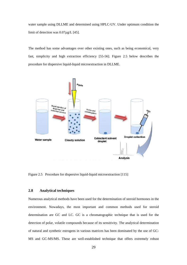
The method has some advantages over other existing ones, such as being economical, very
fast, simplicity and high extraction efficiency [55-56]. Figure 2.5 below describes the
procedure for dispersive liquid-liquid microextraction in DLLME.
Figure 2.5 Procedure for dispersive liquid-liquid microextraction [115]
2.8 Analytical techniques
Numerous analytical methods have been used for the determination of steroid hormones in the
environment. Nowadays, the most important and common methods used for steroid
determination are GC and LC. GC is a chromatographic technique that is used for the
detection of polar, volatile compounds because of its sensitivity. The analytical determination
of natural and synthetic estrogens in various matrices has been dominated by the use of GC-
MS and GC-MS/MS. These are well-established technique that offers extremely robust
30
instrumentation that is widely available in most laboratory settings [21]. Various
derivatization agents can be used for this process, but each of these steps can influence the
accuracy of the method. However, for the determination of concentration levels of conjugated
steroid hormones or metabolites a further step of enzymatic hydrolysis is needed and loss of
analytes can occur, which makes the derivatization reaction incomplete [6, 12-13, 76]. GC-
MS methods have recorded LODs ranging from 0.1- 1.5 ng/L [36], GC-MS/MS recorded
LOD values of 0.1-2.4 ng/L [17, 34-37, 58].The main drawback of GC-MS and GC-MS2
analysis is the need for derivatization in the analysis of compounds. It is labor-intensive and
requires long analysis time and suitable for steroid hormones derivatives.
Currently, LC-MS and LC-MS/MS have become widely used techniques for the
determination of steroid hormones in environmental samples because of their sensitivity and
specificity. LC-MS uses the direct method during the analysis of steroid hormones unlike the
GC-MS. Liquid chromatography coupled to mass spectrometry enables the determination of
both conjugated and non-conjugated estrogens without a derivatization step or hydrolysis [10,
34-41]. However, LC-MS as an analytical technique comes with some limitations such as
interferences [6, 12, 20, 37-41]. Matrix effects occurring in LC-MS can result in ion
suppression or enhancement of the signal of target analytes. Reported LODs on different
types of water samples vary between 1-20 ng/L [36-37], 0.05-3 ng/L [13, 17, 34, 37].
High performance liquid chromatography (HPLC), coupled to a wide variety of detection
systems has gained in popularity for analysis of steroid hormones. However, many LC–MS
methods have been developed to measure steroid hormones and hormone-like substances in
environmental samples [70] due to its high selectivity, specificity and sensitivity [12, 43].
However, many LC-MS methods have been developed to detect steroid hormones and
hormone like substances in environmental samples [37, 68]. HPLC has been combined with
detectors such as diode array (DAD), the concentration of analytes were 0.2-1.6µg/L [12, 42-
43]. HPLC ultraviolet (UV) was able to detect as low as 0.3-1µg/L [12, 18, 45-46].

31
Other methods that have also been used in the determination of Steroid hormones are
capillary electrophoresis (CE) and immunoassay. CE has been explored in the analysis of
polymers, monitoring of endocrine disruptors and for complex mixtures [12, 117-118].
Different types of detection modes can be coupled with the CE. The CE can be coupled to a
UV, FLD, MS and immunoassay. The use of MS detection with CE is quite sensitive, but it is
expensive and tedious process. CE has been used in the determination of steroid hormones in
different water samples, and it was able to detect estriol, estrone and 17β-estradiol with
detection limits of 8.9 × 10⁻⁸, 6.7 × 10⁻⁸, and 1.1 × 10⁻⁷ mol/L respectively [118]. The
limitations of CE application in the determination of EDCs involve sensitivity, sample matrix
interferences, high detection limit and sample ionic strength [12, 117].
In the field of environmental analysis, immunoassay techniques are getting more and more
attention because of their high sensitivity, ease of use, short analysis time cost-effectiveness
(they are cheaper than the LC-MS/MS) simplicity and the possibility of analyzing large sets
of samples even in the field [13, 119-120]. Positive results obtained by immunochemical
methods usually lead to more comprehensive analyses using sophisticated methods, such as
GC-MS, GC-MS/MS, LC-MS, and LC-MS/MS [68]. Immunochemical measurements can be
carried out directly, but sample pretreatment is sometimes necessary, as natural organic
matter may interfere with the analysis [120-122]. Immunoassays rely on the recognition
reaction between a specific antibody and the determinant in antigen [123]. Immunoassays are
fast and therefore suitable for screening a large number of samples. Disadvantages are the
cross-reactivity and sometimes scarceness of a specific antiserum. The detection limits vary
between 3 and 37 pg/ml [119-123].
32
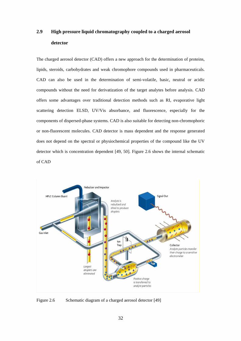
2.9 High pressure liquid chromatography coupled to a charged aerosol
detector
The charged aerosol detector (CAD) offers a new approach for the determination of proteins,
lipids, steroids, carbohydrates and weak chromophore compounds used in pharmaceuticals.
CAD can also be used in the determination of semi-volatile, basic, neutral or acidic
compounds without the need for derivatization of the target analytes before analysis. CAD
offers some advantages over traditional detection methods such as RI, evaporative light
scattering detection ELSD, UV/Vis absorbance, and fluorescence, especially for the
components of dispersed-phase systems. CAD is also suitable for detecting non-chromophoric
or non-fluorescent molecules. CAD detector is mass dependent and the response generated
does not depend on the spectral or physiochemical properties of the compound like the UV
detector which is concentration dependent [49, 50]. Figure 2.6 shows the internal schematic
of CAD
Figure 2.6 Schematic diagram of a charged aerosol detector [49]
33
Charged aerosol detection was introduced by Dixon and Peterson and this is based on aerosol
detection mode. The detection mechanism involves a three main stage namely: nebulization,
evaporation and detection [50]. Detection of analytes starts after the analytes moves through
the separation column, the HPLC eluent is transferred to the CAD where it is nebulized by
means of a small pressure effect produced by nitrogen (a carrier gas). In this situation,
nitrogen flows in a two or three dimensional way to the mobile phase eluting from the
chromatographic column.
The nebulized eluent is transforms the liquid phase into small droplets which are produced
into a heated drift tube. In the drift tube, a secondary stream of nitrogen becomes positively
charged as it passes through a high-voltage, platinum corona wire. This charge transfers to the
opposing stream of analyte particles and is then transferred to a collector where it is measured
by a highly sensitive electrometer as shown in figure 2.6. The detection of the resultant
analyte takes place through the electrical charging of the aerosol and the charged particles
using an electrical aerosol analyser The signal intensity generated by a charged aerosol
detector (CAD) is said to be directly proportional to the analyte concentration [49, 50]. CAD
does not depend on certain functional groups or moieties within the molecules when
compared with other detection method like UV, and FLD. Therefore a similar detector
response can be expected for different compounds [50], a considerable advantage when
screening for impurities where no structural information is available or where the detector
response is unknown.
CAD appears to be more sensitive, with a greater linear dynamic range, more consistent mass
response factor, and easier operation enabling quantification across a range that exceeds four
orders of magnitude. It has a more consistent analyte response; it has a compatible gradient
mode. Unlike UV/Vis, CAD detects analytes with or without chromophores and no
derivatization step is need before analysis of target compounds. CAD also has the same
limitation as other types of detector such as the response is affected by density and viscosity
34
of the mobile-phase composition. Higher organic content in the mobile phase leads to greater
transport efficiency of the nebulizer, which results in a larger number of particles reaching the
detector chamber and hence affecting the signal [50]. This problem has been resolved by
introducing secondary stream nitrogen to the mobile phase of exactly reverse composition,
this is provided by a second pump, this ensures a constant mobile-phase composition at the
detector inlet. Due to this procedure, a constant response that is independent of the mobile-
phase composition is usually obtained [50]. This procedure known as inverse gradient
compensation is done in high pressure liquid chromatography and supercritical fluid
chromatography. However, more studies are needed to fully investigate the effects of these
solvents.
According to Andruch in 2013 [116], various analytical techniques of HPLC, GC and LC
have been used in the determination of steroid hormones in environmental samples. To the
best of our knowledge, this is the first time that HPLC-CAD has been used in the
determination of steroid hormones in wastewater samples.
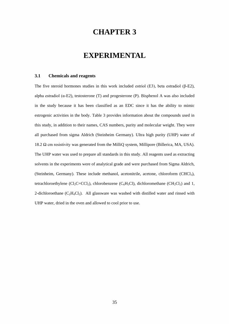
35
CHAPTER 3
EXPERIMENTAL
3.1 Chemicals and reagents
The five steroid hormones studies in this work included estriol (E3), beta estradiol (β-E2),
alpha estradiol (α-E2), testosterone (T) and progesterone (P). Bisphenol A was also included
in the study because it has been classified as an EDC since it has the ability to mimic
estrogenic activities in the body. Table 3 provides information about the compounds used in
this study, in addition to their names, CAS numbers, purity and molecular weight. They were
all purchased from sigma Aldrich (Steinheim Germany). Ultra high purity (UHP) water of
18.2 Ω cm resistivity was generated from the MilliQ system, Millipore (Billerica, MA, USA).
The UHP water was used to prepare all standards in this study. All reagents used as extracting
solvents in the experiments were of analytical grade and were purchased from Sigma Aldrich,
(Steinheim, Germany). These include methanol, acetonitrile, acetone, chloroform (CHCl3),
tetrachloroethylene (Cl2C=CCl2), chlorobenzene (C6H5Cl), dichloromethane (CH2Cl2) and 1,
2-dichloroethane (C2H4Cl2). All glassware was washed with distilled water and rinsed with
UHP water, dried in the oven and allowed to cool prior to use.
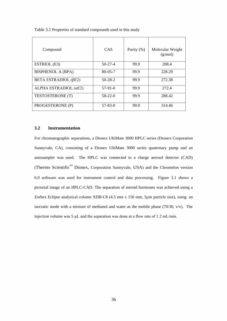
36
Table 3.1 Properties of standard compounds used in this study
Compound
CAS
Purity (%)
Molecular Weight
(g/mol)
ESTRIOL (E3) 50-27-4 99.9 288.4
BISPHENOL A (BPA) 80-05-7 99.9 228.29
BETA ESTRADIOL (βE2) 50-28-2 99.9 272.38
ALPHA ESTRADIOL (αE2) 57-91-0 99.9 272.4
TESTOSTERONE (T) 58-22-0 99.9 288.42
PROGESTERONE (P) 57-83-0 99.9 314.46
3.2 Instrumentation
For chromatographic separations, a Dionex UltiMate 3000 HPLC series (Dionex Corporation
Sunnyvale, CA), consisting of a Dionex UltiMate 3000 series quaternary pump and an
autosampler was used. The HPLC was connected to a charge aerosol detector (CAD)
(Thermo Scientific™
Dionex, Corporation Sunnyvale, USA) and the Chromelon version
6.0 software was used for instrument control and data processing. Figure 3.1 shows a
pictorial image of an HPLC-CAD. The separation of steroid hormones was achieved using a
Zorbex Eclipse analytical column XDB-C8 (4.5 mm x 150 mm, 5µm particle size), using an
isocratic mode with a mixture of methanol and water as the mobile phase (70/30, v/v). The
injection volume was 5 μL and the separation was done at a flow rate of 1.2 mL/min.
37
Figure 3.1 Picture of the HPLC-CAD
For sample preparation and drying purposes, a Thermo Electron centrifuge (Thermo Electron
Corporation, Massachusetts, USA) and a Thermo Electron SpeedVac concentrator (Model
SPD131DDA) coupled to a refrigerated vapor trap (Model RVT4104), and vacuum pump
(Model OFP400) (Thermo Scientific, CA, USA) were used.
3.3 Preparation of standard solutions
A stock standard solution of a concentrate on of 1000 mg/L was prepared by mixing 1 mg of
accurately weighed five steroid hormone standards and BPA (on a micro balance-Mettle
Telodo XP6U). The accurately weighed standards were dissolved and diluted to volume with
a mixture of methanol and water in the ratio (50/50 v/v). It was sonicated for 10 minutes to
dissolve the analytes. Working standard solutions were then prepared by appropriate serial
38
dilutions of the stock solution and adjusted to volume with methanol and water mixture.
These were all stored at 40C until ready for use.
3.4 Sample collection
In analytical chemistry, sampling is an important experimental procedure in the determination
of organic contaminants in wastewater. Wastewater samples (influent and effluent) were
collected from Daspoort, a WWTP located in Pretoria, one of the major cities in Gauteng
Province, South Africa. Figure 3.2 shows the map of the sampling site of the WWTP. The
Daspoort WWTP discharges into a neighbouring Apies river in its surroundings. Grab
sampling method was used and the samples were collected into amber glass bottles, which
had previously been washed and rinsed with UHP water and then flushed at least thrice with
wastewater before collection. Each sample (influent and effluent) was collected in duplicate
into 2.5 L bottles. All water samples were ice-packed during transportation to the laboratory.
The samples were vacuum filtered immediately through the Whatman (MN 615. 125 mm)
filter paper and extracted immediately to avoid degradation. The samples were then stored at
< 4 oC in the dark until ready for analysis.

39
Figure 3.2 A map showing the locality of the Daspoort WWTP in Pretoria
3.5 Calibration standard solutions
Standard solutions for the calibration curve were prepared in a mixture of methanol and water
(50/50 v/v %) at a concentration range of 0.1 to 100 mg/L. The calibration standards were
made by serial dilution of the stock solution and the calibration curves were calculated using
the linear regression model.
3.6 Validation of the HPLC-CAD method
The developed separation method for the five steroid hormone and BPA was achieved using a
Zorbex Eclipse analytical column XDB-C8 (4.5 mm x 150 mm, 5µm particle size), using
isocratic mode with a mixture of methanol and water as the mobile phase (70/30, v/v). The
40
developed method was evaluated by using the following parameters: linear range, correlation
of determination (R2), limit of detection (LOD), limit of quantification (LOQ) and intra-day
precision. For recovery experiments, MilliQ, tap water and wastewater (effluent) were spiked
with a mixture of all the analytes at three different concentration levels (5, 10 and 25 mg/L).
In this work the limit of detection (LOD) is defined as the lowest concentration of an analyte
in a sample that can be detected but not necessarily quantified. LOD was calculated using the
signal to noise ratio (S/N =3), which was then multiplied by the standard error (S) obtained
from the regression equation of the calibration curve. The value obtained was divided by the
slope (b) of the line of regression of y = bx + c in the calibration curve. It is represented
mathematically as;
LOD = 3 * S/b (1)
Limit of quantification (LOQ) is the minimum concentration that can be quantified at a
specific concentration level. LOQ in this study was calculated using the signal to noise ratio
(S/N =10), and then multiplied by the standard error (S) obtained from the calibration. The
value obtained was divided by the slope (b) of the analytes in the calibration curve. It is
represented mathematically as;
LOQ = 10 * S/ b (2)
An eight point calibration line was prepared at different concentration level. The recovery
experiments were carried out by spiking steroid hormone standards into MilliQ, tap water and
wastewater at different concentrations levels in triplicate.
3.7 Developing a DLLME method for the extraction of steroid hormones
A 5.0 mL water sample was placed in a 10 mL capped tube with a conical bottom and spiked
with 0.01 mL of a mixture of five steroid hormone standards plus BPA. Extracting solvents
such as; 1, 2 dichloroethane, chlorobenzene, chloroform, dichloromethane and
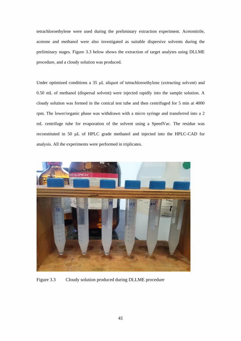
41
tetrachloroethylene were used during the preliminary extraction experiment. Acetonitrile,
acetone and methanol were also investigated as suitable dispersive solvents during the
preliminary stages. Figure 3.3 below shows the extraction of target analytes using DLLME
procedure, and a cloudy solution was produced.
Under optimized conditions a 35 μL aliquot of tetrachloroethylene (extracting solvent) and
0.50 mL of methanol (dispersal solvent) were injected rapidly into the sample solution. A
cloudy solution was formed in the conical test tube and then centrifuged for 5 min at 4000
rpm. The lower/organic phase was withdrawn with a micro syringe and transferred into a 2
mL centrifuge tube for evaporation of the solvent using a SpeedVac. The residue was
reconstituted in 50 µL of HPLC grade methanol and injected into the HPLC-CAD for
analysis. All the experiments were performed in triplicates.
Figure 3.3 Cloudy solution produced during DLLME procedure

42
3.7.1 Optimization of DLLME Method
The extraction efficiency of DLLME is influenced by several factors such as type and volume
of extraction and disperser solvent, extraction time, sample amount, pH, and salt addition [55-
56]. In this study the parameters optimized were the type and volume of extracting and
dispersing solvent. A series of preliminary experiments were performed using five extraction
solvents and three dispersive solvents to determine which best system was favorable for the
target analytes. The extracting solvents must have a density greater than water; the disperser
must also be soluble in the extracting solvent and miscible with water. The optimal volume of
extractants (35 µL) and disperser (500 µL) were also investigated to ensure high extraction
efficiency.
3.7.2 Validation of the DLLME method
The proposed DLLME method was validated under optimized conditions for the following
analytical characteristics: the LOD, LOQ, and coefficient of determination (R2) and linearity.
Relative standard deviation was used to express repeatability. The extraction recoveries and
enrichment factors were also determined. The validation data is presented in Table 4.
Linearity was established and the LODs and LOQs were calculated as described in Section
3.6.
3.8 Analysis of real wastewater samples
The developed and validated DLLME and HPLC-CAD methods were applied to influent and
effluent wastewater samples collected from Daspoort WWTP, in Gauteng South Africa.
Sampling was done during the winter season, and samples were collected in duplicates.
Wastewater samples were first vacuum filtered using the Whatman filter paper. 5 mL of the
water sample was placed in a 10 mL capped tube with a conical bottom. The target analytes
were extracted under optimized DLLME conditions. 35 μL aliquot of tetrachloroethylene
(extracting solvent) and 0.50 mL of methanol (dispersal solvent) were injected rapidly into
43
the sample solution. A cloudy solution was formed in the conical test tube and then
centrifuged for 5 min at 4000 rpm. The lower/organic phase was withdrawn with a micro
syringe and transferred into a 2 mL centrifuge tube for evaporation of the solvent using a
SpeedVac. The residue was reconstituted in 50 µL of HPLC grade methanol and injected into
the HPLC-CAD for analysis.
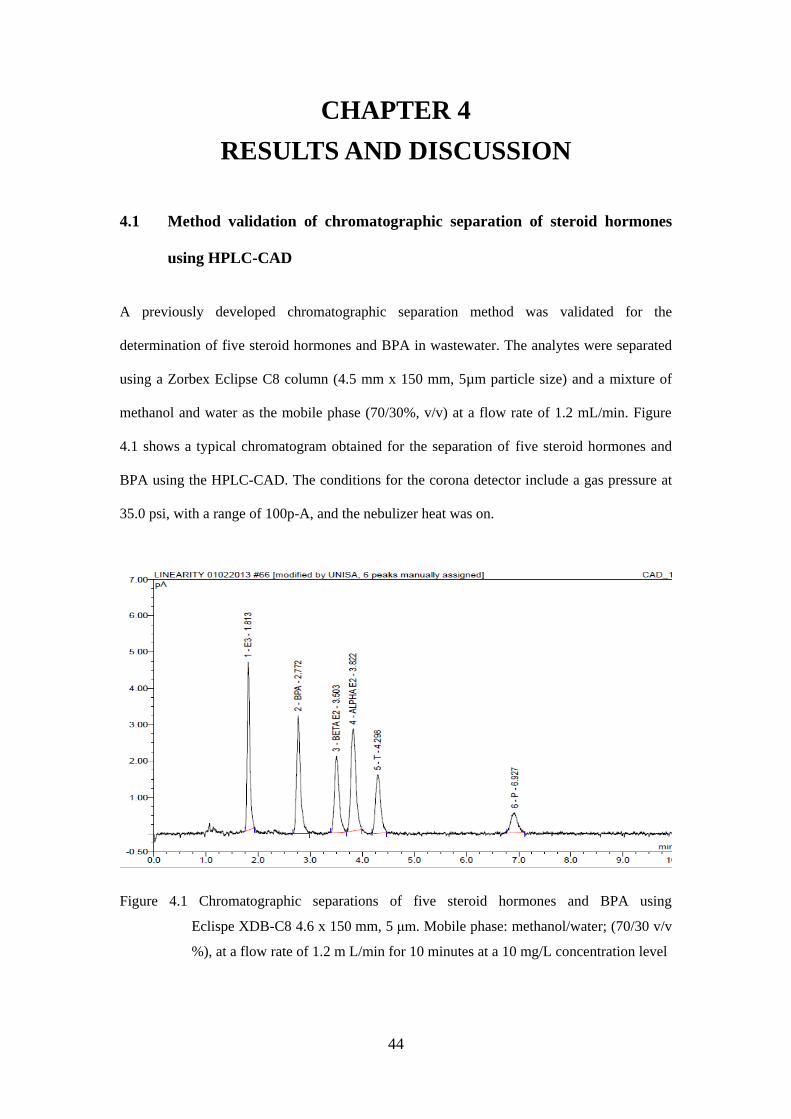
44
CHAPTER 4
RESULTS AND DISCUSSION
4.1 Method validation of chromatographic separation of steroid hormones
using HPLC-CAD
A previously developed chromatographic separation method was validated for the
determination of five steroid hormones and BPA in wastewater. The analytes were separated
using a Zorbex Eclipse C8 column (4.5 mm x 150 mm, 5µm particle size) and a mixture of
methanol and water as the mobile phase (70/30%, v/v) at a flow rate of 1.2 mL/min. Figure
4.1 shows a typical chromatogram obtained for the separation of five steroid hormones and
BPA using the HPLC-CAD. The conditions for the corona detector include a gas pressure at
35.0 psi, with a range of 100p-A, and the nebulizer heat was on.
Figure 4.1 Chromatographic separations of five steroid hormones and BPA using
Eclispe XDB-C8 4.6 x 150 mm, 5 μm. Mobile phase: methanol/water; (70/30 v/v
%), at a flow rate of 1.2 m L/min for 10 minutes at a 10 mg/L concentration level
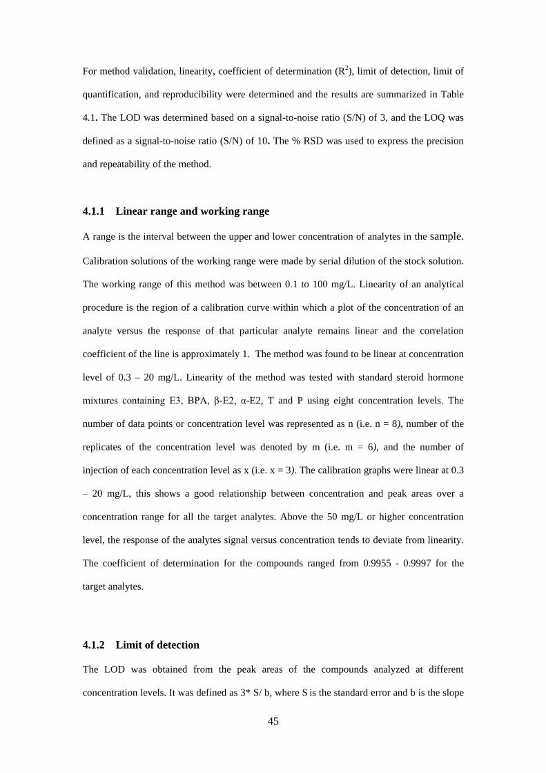
45
For method validation, linearity, coefficient of determination (R2), limit of detection, limit of
quantification, and reproducibility were determined and the results are summarized in Table
4.1. The LOD was determined based on a signal-to-noise ratio (S/N) of 3, and the LOQ was
defined as a signal-to-noise ratio (S/N) of 10. The % RSD was used to express the precision
and repeatability of the method.
4.1.1 Linear range and working range
A range is the interval between the upper and lower concentration of analytes in the sample.
Calibration solutions of the working range were made by serial dilution of the stock solution.
The working range of this method was between 0.1 to 100 mg/L. Linearity of an analytical
procedure is the region of a calibration curve within which a plot of the concentration of an
analyte versus the response of that particular analyte remains linear and the correlation
coefficient of the line is approximately 1. The method was found to be linear at concentration
level of 0.3 – 20 mg/L. Linearity of the method was tested with standard steroid hormone
mixtures containing E3, BPA, β-E2, α-E2, T and P using eight concentration levels. The
number of data points or concentration level was represented as n (i.e. n = 8), number of the
replicates of the concentration level was denoted by m (i.e. m = 6), and the number of
injection of each concentration level as x (i.e. x = 3). The calibration graphs were linear at 0.3
– 20 mg/L, this shows a good relationship between concentration and peak areas over a
concentration range for all the target analytes. Above the 50 mg/L or higher concentration
level, the response of the analytes signal versus concentration tends to deviate from linearity.
The coefficient of determination for the compounds ranged from 0.9955 - 0.9997 for the
target analytes.
4.1.2 Limit of detection
The LOD was obtained from the peak areas of the compounds analyzed at different
concentration levels. It was defined as 3* S/ b, where S is the standard error and b is the slope

46
of the calibration curve. Calibration was performed on a mixture of six standard solutions of
the compounds of interest. The calibration curves were constructed using the eight
concentration levels (i.e. n = 8). A sample solution at each concentration level was prepared 6
times (m= 6) and it was injected in triplicate (mx3). The calibration plots were approximated
by the linear regression equation, y = bx + c, where y is the peak area and x is the
concentration of determination compounds in mg/L. The results for the limit of detection are
summarized in Table 4.1. The LOD for E3 and P were 0.1 mg/L, while BPA, β-E2, α-E2 and
T was 0.2 mg/L. These values obtained are very high and at this concentration levels steroid
hormones will not be detected in the environmental samples. When these values were
compared to the detection limits obtained from a GC-MS or LC-MS, it is deduced that there is
a need for a cost friendly sample preconcentration technique, since the target analytes are only
found at lower concentration levels of µg/L or ng/L in the environment.
4.1.3 Limit of quantification
LOQ is defined as the minimum concentration that can be quantified at a specific level. It was
defined as 10 * S/b, where S is the standard error and b is the slope of the calibration curve.
Calibration was performed on a mixture of six standard solutions of the compounds of
interest. The calibration curves were constructed using the eight concentration levels (i.e. n =
8). A sample solution at each concentration level was prepared eight times (m= 8) and it was
injected in triplicate (mx3). The calibration plots were approximated by the linear regression
equation, y = bx + c, where y is the peak area and x is the concentration of determination
compounds in mg/L. The results for the limit of quantification are summarized in Table 4.1.
The LOQ for E3 was 0.3 mg/L, 0.7 mg/L for BPA, β-E2 was 0.8 mg/L, α-E2 was 1.1 mg/L, T
was 0.9 mg/L and P was found at 0.8 mg/L. With these high values obtained, the target
analytes cannot be quantified in the environment because they are found at lower
quantification levels. This suggests that there is a need for a preconcentration procedure that
will help to increase the sensitivity of the method on the HPLC-CAD.
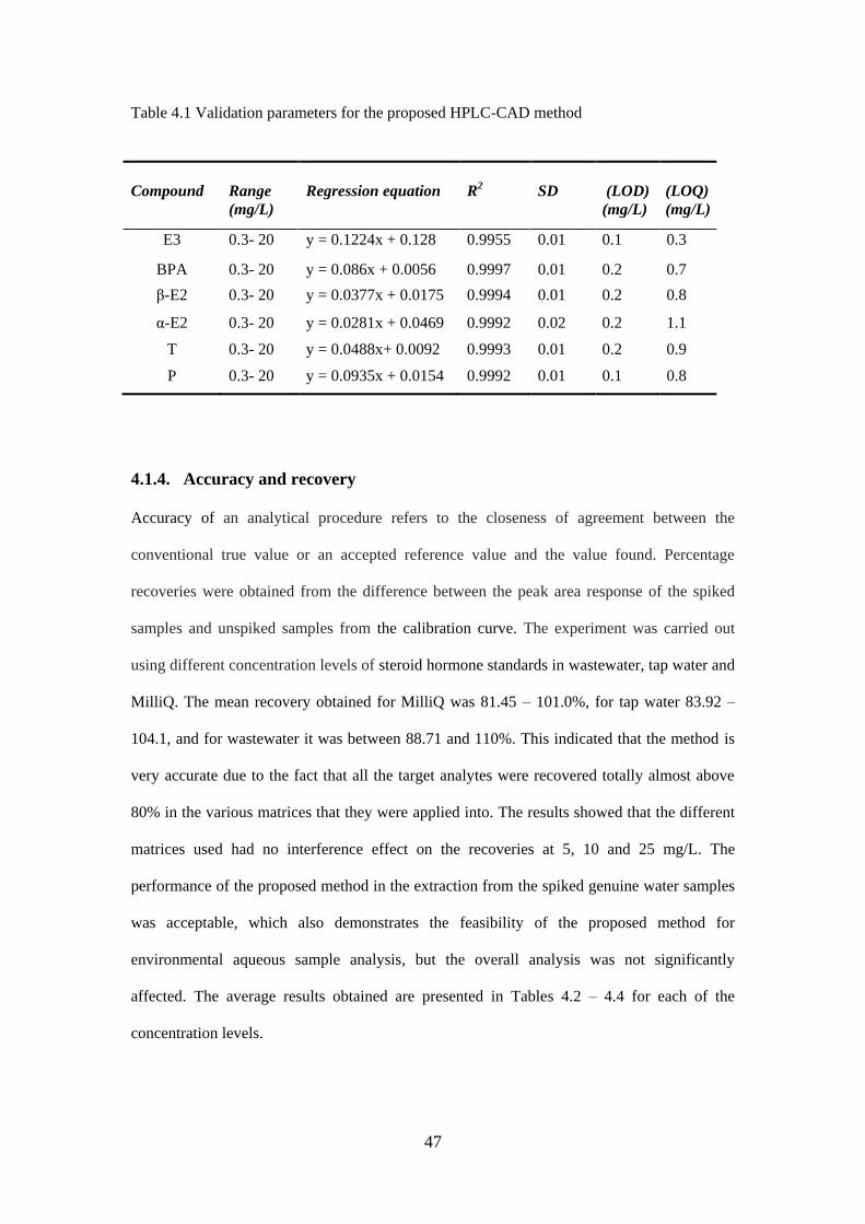
47
Table 4.1 Validation parameters for the proposed HPLC-CAD method
Compound
Range
(mg/L)
Regression equation
R2
SD
(LOD)
(mg/L)
(LOQ)
(mg/L)
E3 0.3- 20 y = 0.1224x + 0.128 0.9955 0.01 0.1 0.3
BPA 0.3- 20 y = 0.086x + 0.0056 0.9997 0.01 0.2 0.7
β-E2 0.3- 20 y = 0.0377x + 0.0175 0.9994 0.01 0.2 0.8
α-E2 0.3- 20 y = 0.0281x + 0.0469 0.9992 0.02 0.2 1.1
T 0.3- 20 y = 0.0488x+ 0.0092 0.9993 0.01 0.2 0.9
P 0.3- 20 y = 0.0935x + 0.0154 0.9992 0.01 0.1 0.8
4.1.4. Accuracy and recovery
Accuracy of an analytical procedure refers to the closeness of agreement between the
conventional true value or an accepted reference value and the value found. Percentage
recoveries were obtained from the difference between the peak area response of the spiked
samples and unspiked samples from the calibration curve. The experiment was carried out
using different concentration levels of steroid hormone standards in wastewater, tap water and
MilliQ. The mean recovery obtained for MilliQ was 81.45 – 101.0%, for tap water 83.92 –
104.1, and for wastewater it was between 88.71 and 110%. This indicated that the method is
very accurate due to the fact that all the target analytes were recovered totally almost above
80% in the various matrices that they were applied into. The results showed that the different
matrices used had no interference effect on the recoveries at 5, 10 and 25 mg/L. The
performance of the proposed method in the extraction from the spiked genuine water samples
was acceptable, which also demonstrates the feasibility of the proposed method for
environmental aqueous sample analysis, but the overall analysis was not significantly
affected. The average results obtained are presented in Tables 4.2 – 4.4 for each of the
concentration levels.
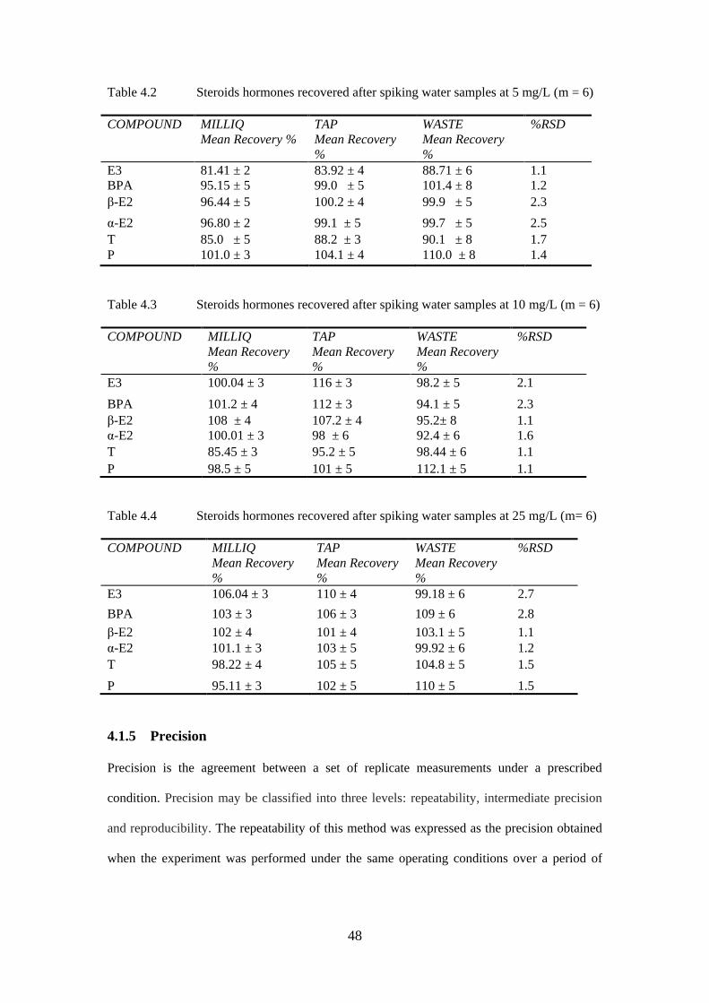
48
Table 4.2 Steroids hormones recovered after spiking water samples at 5 mg/L (m = 6)
COMPOUND MILLIQ
Mean Recovery %
TAP
Mean Recovery
%
WASTE
Mean Recovery
%
%RSD
E3 81.41 ± 2 83.92 ± 4 88.71 ± 6 1.1
BPA 95.15 ± 5 99.0 ± 5 101.4 ± 8 1.2
β-E2 96.44 ± 5 100.2 ± 4 99.9 ± 5 2.3
α-E2 96.80 ± 2 99.1 ± 5 99.7 ± 5 2.5
T 85.0 ± 5 88.2 ± 3 90.1 ± 8 1.7
P 101.0 ± 3 104.1 ± 4 110.0 ± 8 1.4
Table 4.3 Steroids hormones recovered after spiking water samples at 10 mg/L (m = 6)
COMPOUND MILLIQ
Mean Recovery
%
TAP
Mean Recovery
%
WASTE
Mean Recovery
%
%RSD
E3 100.04 ± 3 116 ± 3 98.2 ± 5 2.1
BPA 101.2 ± 4 112 ± 3 94.1 ± 5 2.3
β-E2 108 ± 4 107.2 ± 4 95.2± 8 1.1
α-E2 100.01 ± 3 98 ± 6 92.4 ± 6 1.6
T 85.45 ± 3 95.2 ± 5 98.44 ± 6 1.1
P 98.5 ± 5 101 ± 5 112.1 ± 5 1.1
Table 4.4 Steroids hormones recovered after spiking water samples at 25 mg/L (m= 6)
COMPOUND MILLIQ
Mean Recovery
%
TAP
Mean Recovery
%
WASTE
Mean Recovery
%
%RSD
E3 106.04 ± 3 110 ± 4 99.18 ± 6 2.7
BPA 103 ± 3 106 ± 3 109 ± 6 2.8
β-E2 102 ± 4 101 ± 4 103.1 ± 5 1.1
α-E2 101.1 ± 3 103 ± 5 99.92 ± 6 1.2
T 98.22 ± 4 105 ± 5 104.8 ± 5 1.5
P 95.11 ± 3 102 ± 5 110 ± 5 1.5
4.1.5 Precision
Precision is the agreement between a set of replicate measurements under a prescribed
condition. Precision may be classified into three levels: repeatability, intermediate precision
and reproducibility. The repeatability of this method was expressed as the precision obtained
when the experiment was performed under the same operating conditions over a period of
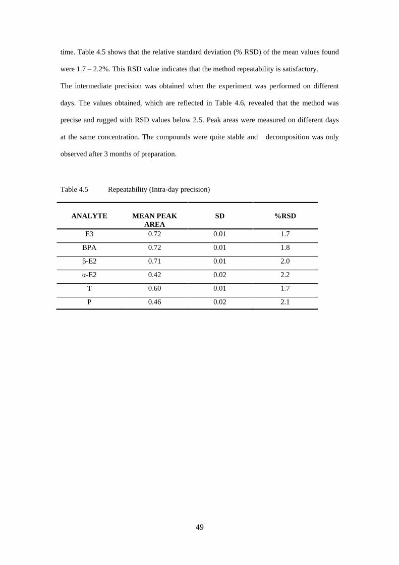
49
time. Table 4.5 shows that the relative standard deviation (% RSD) of the mean values found
were 1.7 – 2.2%. This RSD value indicates that the method repeatability is satisfactory.
The intermediate precision was obtained when the experiment was performed on different
days. The values obtained, which are reflected in Table 4.6, revealed that the method was
precise and rugged with RSD values below 2.5. Peak areas were measured on different days
at the same concentration. The compounds were quite stable and decomposition was only
observed after 3 months of preparation.
Table 4.5 Repeatability (Intra-day precision)
ANALYTE
MEAN PEAK
AREA
SD
%RSD
E3 0.72 0.01 1.7
BPA 0.72 0.01 1.8
β-E2 0.71 0.01 2.0
α-E2 0.42 0.02 2.2
T 0.60 0.01 1.7
P 0.46 0.02 2.1
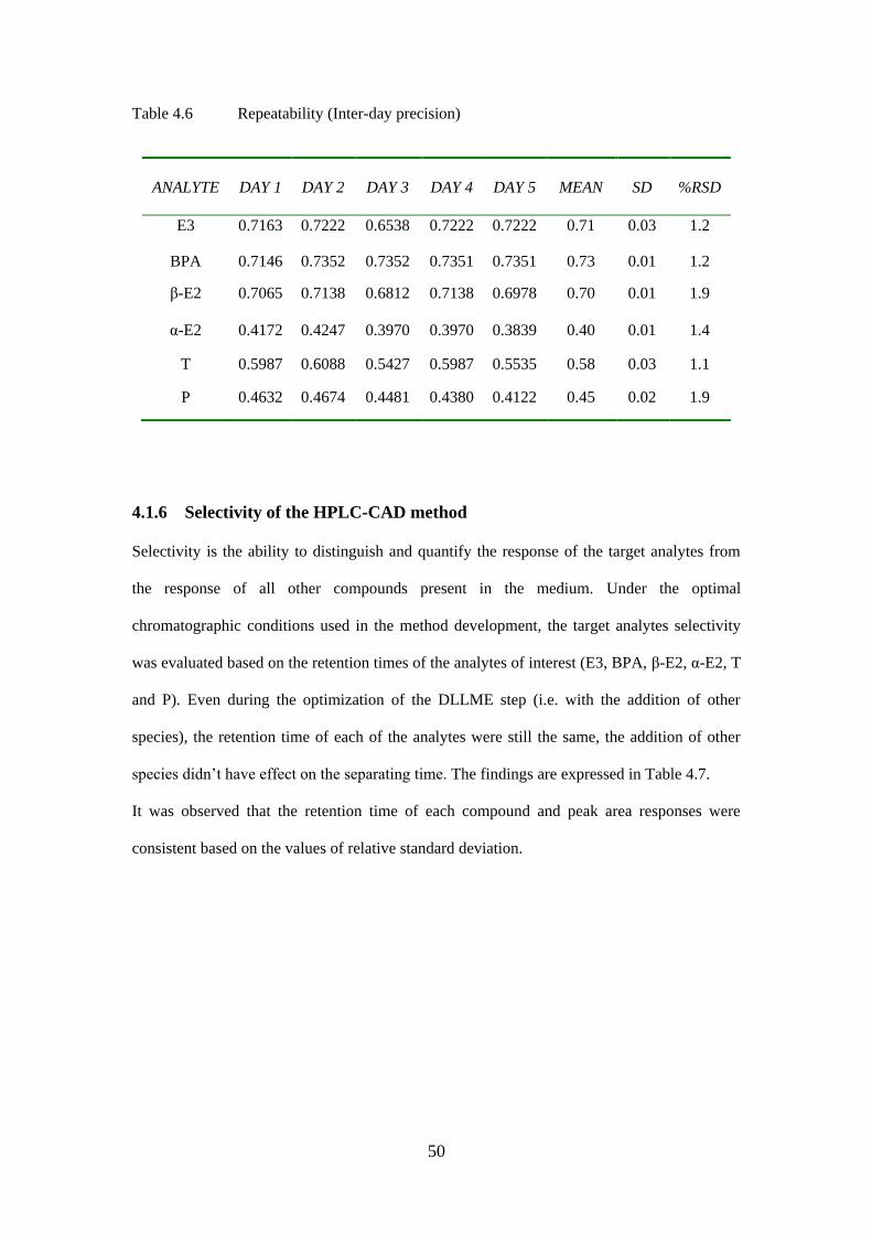
50
Table 4.6 Repeatability (Inter-day precision)
4.1.6 Selectivity of the HPLC-CAD method
Selectivity is the ability to distinguish and quantify the response of the target analytes from
the response of all other compounds present in the medium. Under the optimal
chromatographic conditions used in the method development, the target analytes selectivity
was evaluated based on the retention times of the analytes of interest (E3, BPA, β-E2, α-E2, T
and P). Even during the optimization of the DLLME step (i.e. with the addition of other
species), the retention time of each of the analytes were still the same, the addition of other
species didn’t have effect on the separating time. The findings are expressed in Table 4.7.
It was observed that the retention time of each compound and peak area responses were
consistent based on the values of relative standard deviation.
ANALYTE
DAY 1
DAY 2
DAY 3
DAY 4
DAY 5
MEAN
SD
%RSD
E3 0.7163 0.7222 0.6538 0.7222 0.7222 0.71 0.03 1.2
BPA 0.7146 0.7352 0.7352 0.7351 0.7351 0.73 0.01 1.2
β-E2
0.7065 0.7138 0.6812 0.7138 0.6978 0.70 0.01 1.9
α-E2 0.4172 0.4247 0.3970 0.3970 0.3839 0.40 0.01 1.4
T 0.5987 0.6088 0.5427 0.5987 0.5535 0.58 0.03 1.1
P 0.4632 0.4674 0.4481 0.4380 0.4122 0.45 0.02 1.9
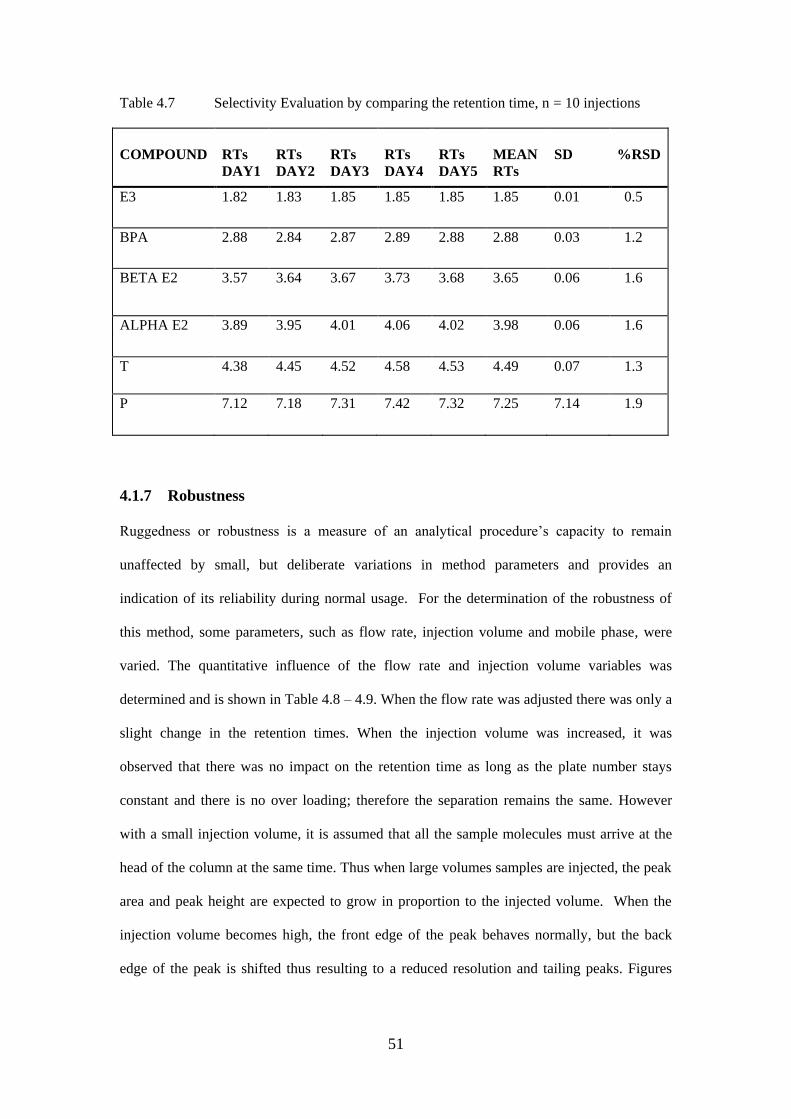
51
Table 4.7 Selectivity Evaluation by comparing the retention time, n = 10 injections
COMPOUND
RTs
DAY1
RTs
DAY2
RTs
DAY3
RTs
DAY4
RTs
DAY5
MEAN
RTs
SD
%RSD
E3 1.82 1.83 1.85 1.85 1.85 1.85 0.01 0.5
BPA 2.88 2.84 2.87 2.89 2.88 2.88 0.03 1.2
BETA E2 3.57 3.64 3.67 3.73 3.68 3.65 0.06 1.6
ALPHA E2 3.89 3.95 4.01 4.06 4.02 3.98 0.06 1.6
T 4.38 4.45 4.52 4.58 4.53 4.49 0.07 1.3
P 7.12 7.18 7.31 7.42 7.32 7.25 7.14 1.9
4.1.7 Robustness
Ruggedness or robustness is a measure of an analytical procedure’s capacity to remain
unaffected by small, but deliberate variations in method parameters and provides an
indication of its reliability during normal usage. For the determination of the robustness of
this method, some parameters, such as flow rate, injection volume and mobile phase, were
varied. The quantitative influence of the flow rate and injection volume variables was
determined and is shown in Table 4.8 – 4.9. When the flow rate was adjusted there was only a
slight change in the retention times. When the injection volume was increased, it was
observed that there was no impact on the retention time as long as the plate number stays
constant and there is no over loading; therefore the separation remains the same. However
with a small injection volume, it is assumed that all the sample molecules must arrive at the
head of the column at the same time. Thus when large volumes samples are injected, the peak
area and peak height are expected to grow in proportion to the injected volume. When the
injection volume becomes high, the front edge of the peak behaves normally, but the back
edge of the peak is shifted thus resulting to a reduced resolution and tailing peaks. Figures

52
4.2a and 4.2b shows the chromatograms obtained during this experiment. The % RSD
obtained showed the robustness of the method, which was satisfactory.
Table 4.8 Variation of the flow rate from 0.7 mL to 1.0 mL (m = 6)
Compound
0.7mL
(Av. peak
.area )
RTs
0.7mL
% RSD
RTs
1.0 mL
1.0 mL
(Av. peak
area)
% RSD
E3 0.16 2.31 0.7 2.26 0.15 0.4
BPA 0.21 3.22 1.0 3.66 0.36 1.0
β-E2 0.44 5.20 1.3 4.77 0.39 1.1
α-E2 0.55 6.78 1.5 5.22 0.45 1.4
T 0.63 7.43 1.2 5.86 0.38 1.1
P 0.54 8.36 1.0 9.51 0.19 1.3
Table 4.9 Variation of the injection volume between10 µL and 20 µL (m = 6)
Compound
10µL
(Av. peak)
RTs
(10 µL)
% RSD
20µL
(Av.
peak)
RTs
(20 µL)
% RSD
E3 0.2766 1.90 0.3 0.4651 1.89 0.5
BPA 0.2881 3.06 0.9 0.4051 2.93 0.2
β-E2 0.1792 3.97 1.1 0.8042 3.86 0.7
α-E2 0.2001 4.34 1.2 0.900 4.23 0.6
T 0.2133 4.88 0.9 1.289 4.74 1.2
P 0.0954 7.96 1.1 0.314 7.74 0.9
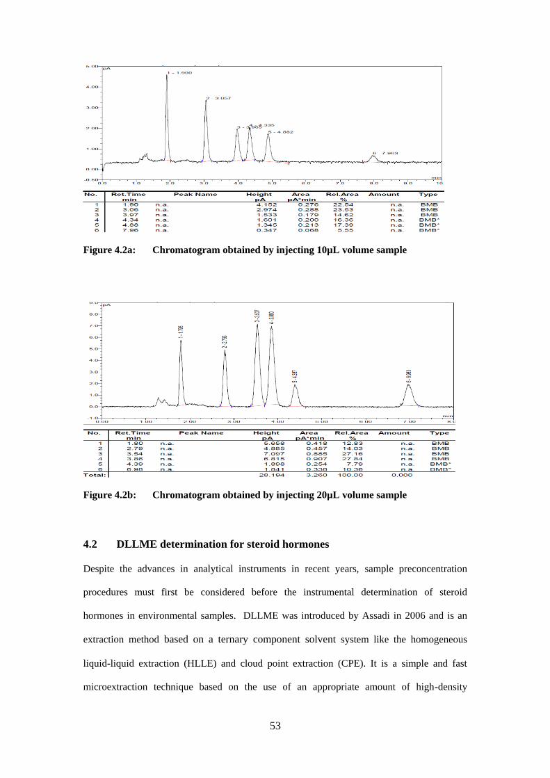
53
Figure 4.2a: Chromatogram obtained by injecting 10µL volume sample
Figure 4.2b: Chromatogram obtained by injecting 20µL volume sample
4.2 DLLME determination for steroid hormones
Despite the advances in analytical instruments in recent years, sample preconcentration
procedures must first be considered before the instrumental determination of steroid
hormones in environmental samples. DLLME was introduced by Assadi in 2006 and is an
extraction method based on a ternary component solvent system like the homogeneous
liquid-liquid extraction (HLLE) and cloud point extraction (CPE). It is a simple and fast
microextraction technique based on the use of an appropriate amount of high-density
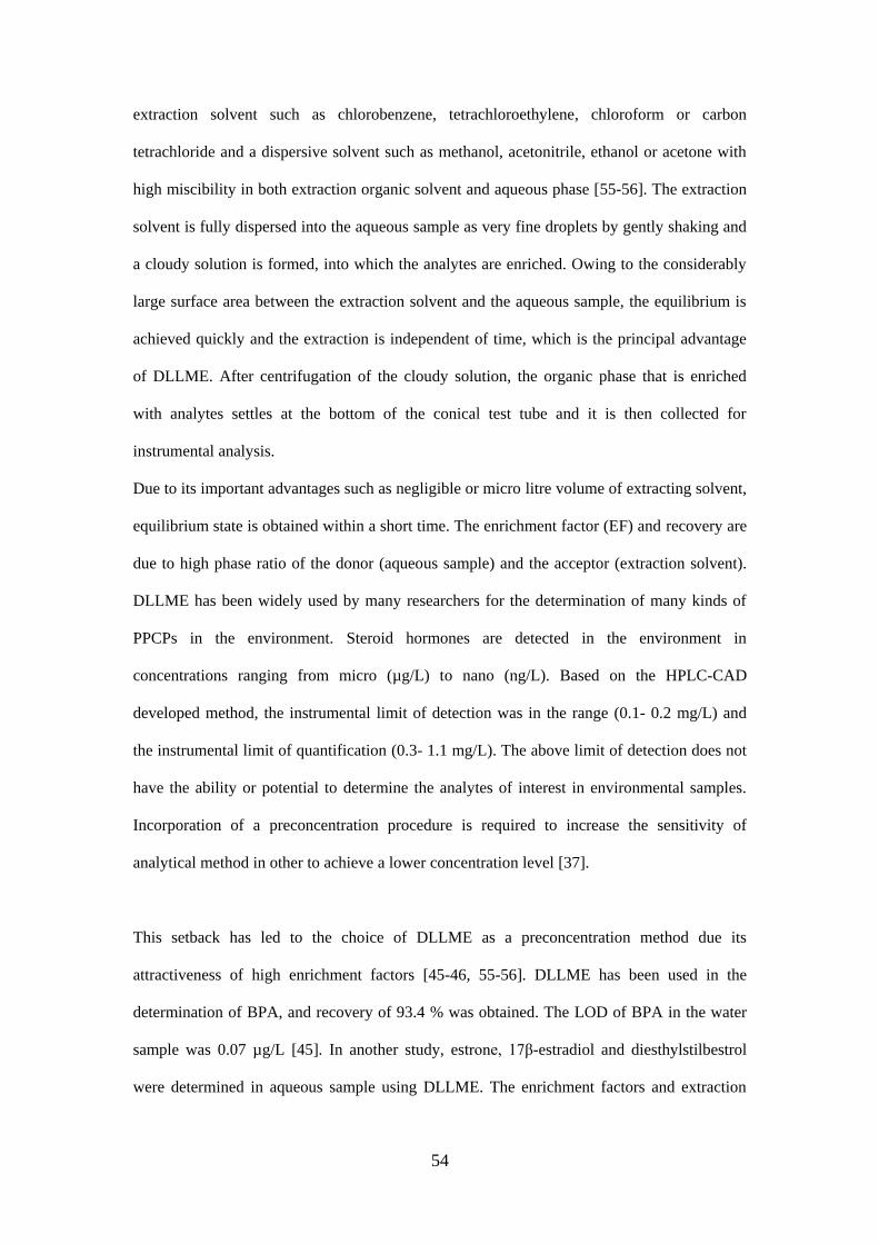
54
extraction solvent such as chlorobenzene, tetrachloroethylene, chloroform or carbon
tetrachloride and a dispersive solvent such as methanol, acetonitrile, ethanol or acetone with
high miscibility in both extraction organic solvent and aqueous phase [55-56]. The extraction
solvent is fully dispersed into the aqueous sample as very fine droplets by gently shaking and
a cloudy solution is formed, into which the analytes are enriched. Owing to the considerably
large surface area between the extraction solvent and the aqueous sample, the equilibrium is
achieved quickly and the extraction is independent of time, which is the principal advantage
of DLLME. After centrifugation of the cloudy solution, the organic phase that is enriched
with analytes settles at the bottom of the conical test tube and it is then collected for
instrumental analysis.
Due to its important advantages such as negligible or micro litre volume of extracting solvent,
equilibrium state is obtained within a short time. The enrichment factor (EF) and recovery are
due to high phase ratio of the donor (aqueous sample) and the acceptor (extraction solvent).
DLLME has been widely used by many researchers for the determination of many kinds of
PPCPs in the environment. Steroid hormones are detected in the environment in
concentrations ranging from micro (µg/L) to nano (ng/L). Based on the HPLC-CAD
developed method, the instrumental limit of detection was in the range (0.1- 0.2 mg/L) and
the instrumental limit of quantification (0.3- 1.1 mg/L). The above limit of detection does not
have the ability or potential to determine the analytes of interest in environmental samples.
Incorporation of a preconcentration procedure is required to increase the sensitivity of
analytical method in other to achieve a lower concentration level [37].
This setback has led to the choice of DLLME as a preconcentration method due its
attractiveness of high enrichment factors [45-46, 55-56]. DLLME has been used in the
determination of BPA, and recovery of 93.4 % was obtained. The LOD of BPA in the water
sample was 0.07 µg/L [45]. In another study, estrone, 17β-estradiol and diesthylstilbestrol
were determined in aqueous sample using DLLME. The enrichment factors and extraction
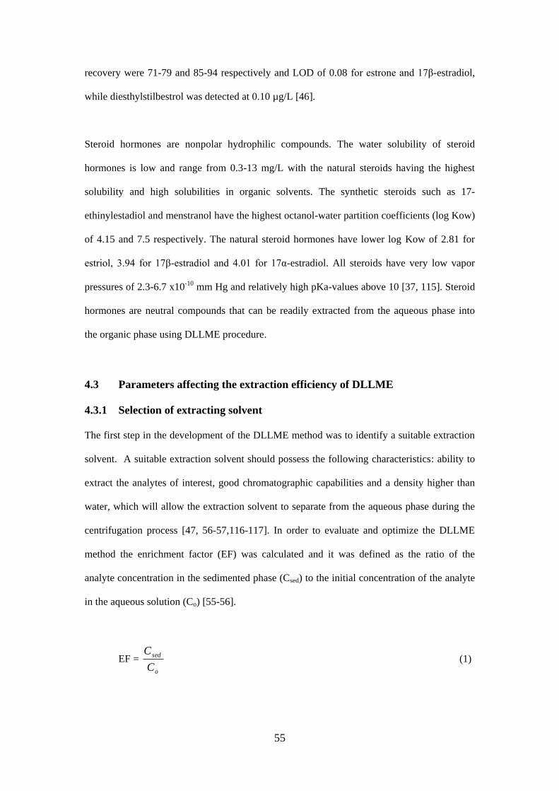
55
recovery were 71-79 and 85-94 respectively and LOD of 0.08 for estrone and 17β-estradiol,
while diesthylstilbestrol was detected at 0.10 µg/L [46].
Steroid hormones are nonpolar hydrophilic compounds. The water solubility of steroid
hormones is low and range from 0.3-13 mg/L with the natural steroids having the highest
solubility and high solubilities in organic solvents. The synthetic steroids such as 17-
ethinylestadiol and menstranol have the highest octanol-water partition coefficients (log Kow)
of 4.15 and 7.5 respectively. The natural steroid hormones have lower log Kow of 2.81 for
estriol, 3.94 for 17β-estradiol and 4.01 for 17α-estradiol. All steroids have very low vapor
pressures of 2.3-6.7 x10⁻¹⁰ mm Hg and relatively high pKa-values above 10 [37, 115]. Steroid
hormones are neutral compounds that can be readily extracted from the aqueous phase into
the organic phase using DLLME procedure.
4.3 Parameters affecting the extraction efficiency of DLLME
4.3.1 Selection of extracting solvent
The first step in the development of the DLLME method was to identify a suitable extraction
solvent. A suitable extraction solvent should possess the following characteristics: ability to
extract the analytes of interest, good chromatographic capabilities and a density higher than
water, which will allow the extraction solvent to separate from the aqueous phase during the
centrifugation process [47, 56-57,116-117]. In order to evaluate and optimize the DLLME
method the enrichment factor (EF) was calculated and it was defined as the ratio of the
analyte concentration in the sedimented phase (Csed) to the initial concentration of the analyte
in the aqueous solution (Co) [55-56].
EF = o
sed
C
C (1)
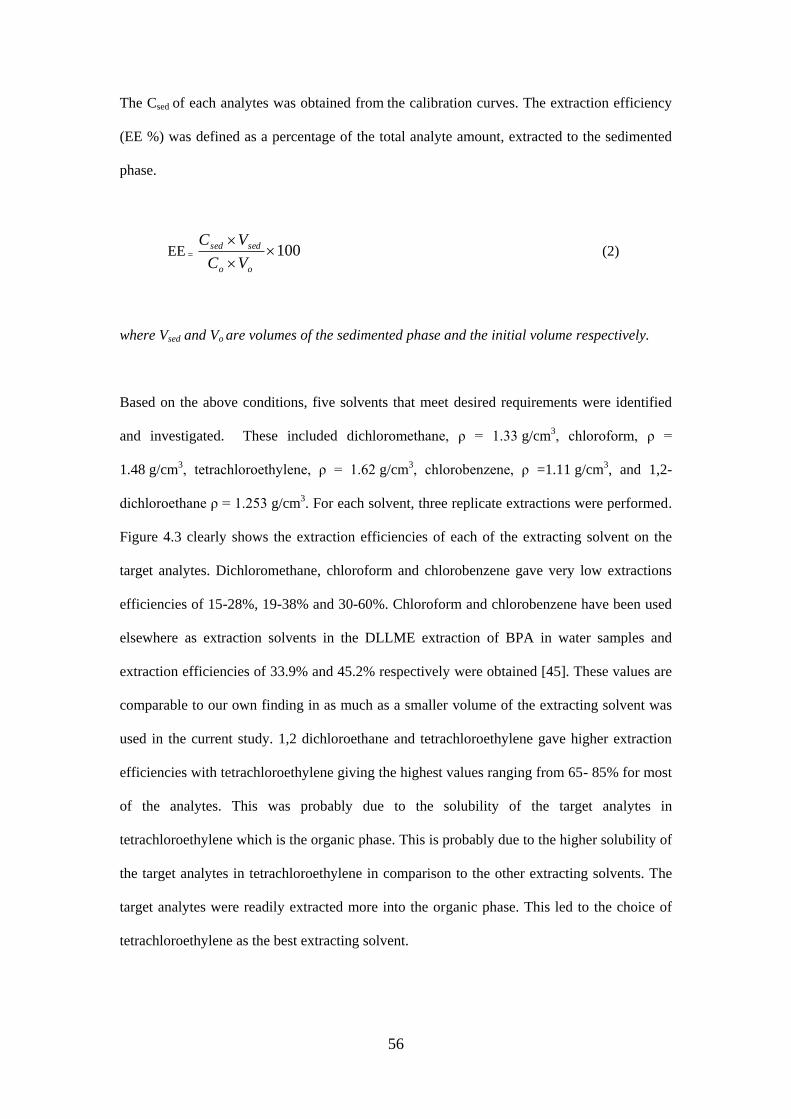
56
The Csed of each analytes was obtained from the calibration curves. The extraction efficiency
(EE %) was defined as a percentage of the total analyte amount, extracted to the sedimented
phase.
EE = 100
oo
sedsed
VC
VC (2)
where Vsed and Vo are volumes of the sedimented phase and the initial volume respectively.
Based on the above conditions, five solvents that meet desired requirements were identified
and investigated. These included dichloromethane, ρ = 1.33 g/cm3, chloroform, ρ =
1.48 g/cm3, tetrachloroethylene, ρ = 1.62 g/cm3, chlorobenzene, ρ =1.11 g/cm3, and 1,2-
dichloroethane ρ = 1.253 g/cm3. For each solvent, three replicate extractions were performed.
Figure 4.3 clearly shows the extraction efficiencies of each of the extracting solvent on the
target analytes. Dichloromethane, chloroform and chlorobenzene gave very low extractions
efficiencies of 15-28%, 19-38% and 30-60%. Chloroform and chlorobenzene have been used
elsewhere as extraction solvents in the DLLME extraction of BPA in water samples and
extraction efficiencies of 33.9% and 45.2% respectively were obtained [45]. These values are
comparable to our own finding in as much as a smaller volume of the extracting solvent was
used in the current study. 1,2 dichloroethane and tetrachloroethylene gave higher extraction
efficiencies with tetrachloroethylene giving the highest values ranging from 65- 85% for most
of the analytes. This was probably due to the solubility of the target analytes in
tetrachloroethylene which is the organic phase. This is probably due to the higher solubility of
the target analytes in tetrachloroethylene in comparison to the other extracting solvents. The
target analytes were readily extracted more into the organic phase. This led to the choice of
tetrachloroethylene as the best extracting solvent.
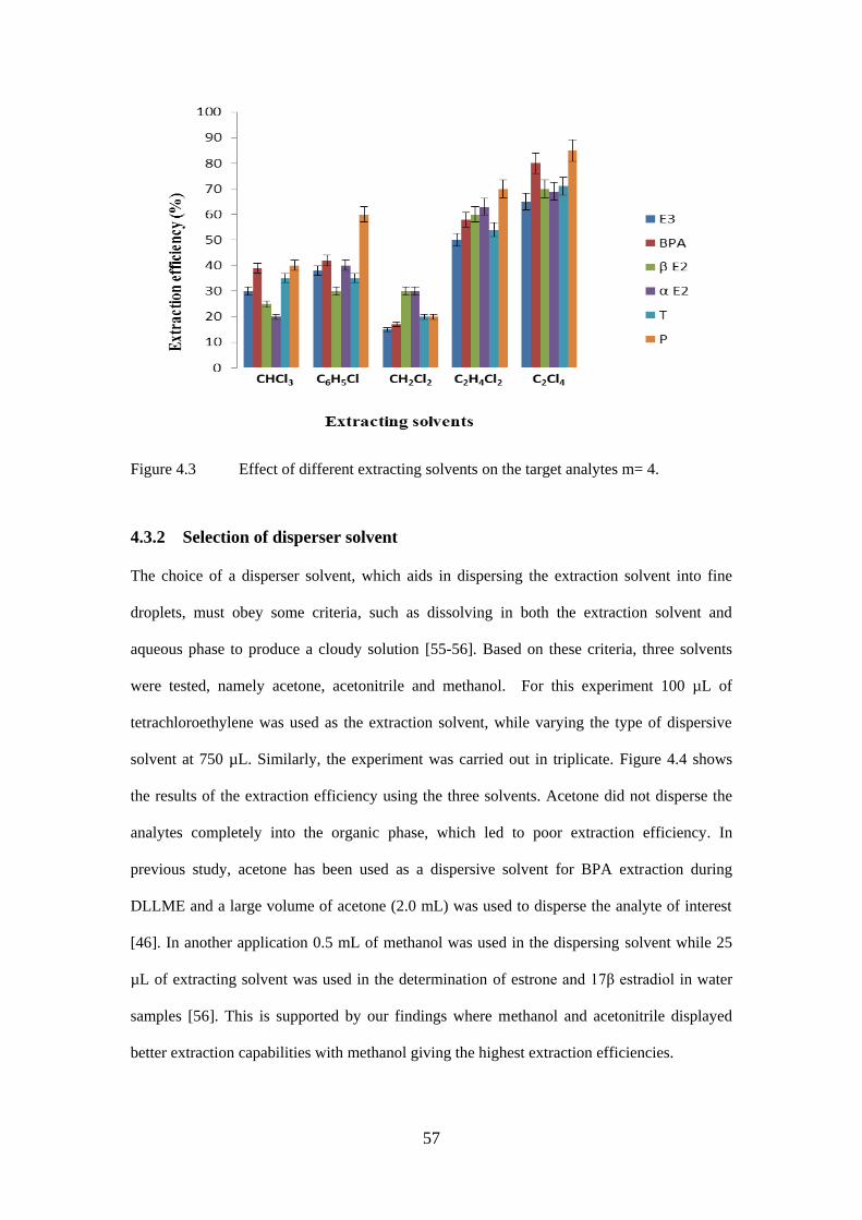
57
Figure 4.3 Effect of different extracting solvents on the target analytes m= 4.
4.3.2 Selection of disperser solvent
The choice of a disperser solvent, which aids in dispersing the extraction solvent into fine
droplets, must obey some criteria, such as dissolving in both the extraction solvent and
aqueous phase to produce a cloudy solution [55-56]. Based on these criteria, three solvents
were tested, namely acetone, acetonitrile and methanol. For this experiment 100 µL of
tetrachloroethylene was used as the extraction solvent, while varying the type of dispersive
solvent at 750 µL. Similarly, the experiment was carried out in triplicate. Figure 4.4 shows
the results of the extraction efficiency using the three solvents. Acetone did not disperse the
analytes completely into the organic phase, which led to poor extraction efficiency. In
previous study, acetone has been used as a dispersive solvent for BPA extraction during
DLLME and a large volume of acetone (2.0 mL) was used to disperse the analyte of interest
[46]. In another application 0.5 mL of methanol was used in the dispersing solvent while 25
µL of extracting solvent was used in the determination of estrone and 17β estradiol in water
samples [56]. This is supported by our findings where methanol and acetonitrile displayed
better extraction capabilities with methanol giving the highest extraction efficiencies.
58
Figure 4.4 Effect of different dispersive solvents on the target analytes m= 4
4.3.3. Determination of optimal volume of the extracting solvent
The extracting solvent volume also has great effects on the enrichment factor of the analytes
and it directly determines the volume of the lower/organic phase. The extraction solvent
volume is expected to be minimal, while at the same time allowing the target analytes to be
dispersed into the aqueous solution. To investigate this, different volumes of
tetrachloroethylene (between 10 and 50 μL) were mixed with a fixed volume of methanol
(750 μL) and rapidly injected into the sample. Increasing the volume of tetrachloroethylene
from 10 to 50 μL led to an increase in the volume of the sedimented phase from 30 - 105 μL.
The EF initially increases as the volume of the extracting solvent increases and gradually
decreases at volumes above 35 μL. Figures 4.5 show the extraction efficiency of each analyte
in the organic phase. At optimum volume of 35 μL, Progesterone showed the highest
extraction efficiency of 102%. E3 and testosterone had extraction efficiencies of 84%, β-E2
was 87% while BPA and α-E2 was 89%. Figure 4.6 shows the enrichment factor obtained for
the target analytes at optimal volume to be between 148 -258. In literature, DLLME has been
used in the determination of BPA, and recovery of 93.4 % was obtained. The LOD of BPA in
the water sample was 0.07 µg/L [45]. In another study, estrone, 17β-estradiol and
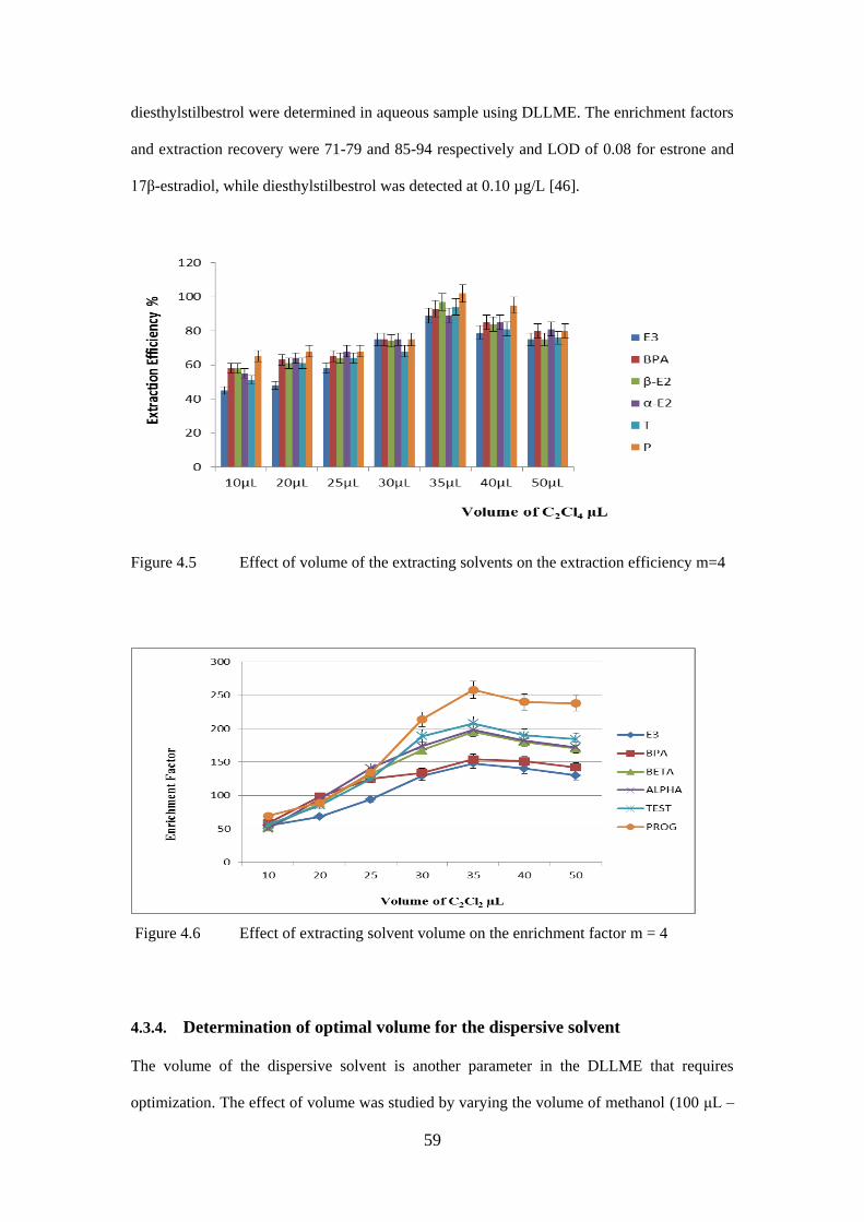
59
diesthylstilbestrol were determined in aqueous sample using DLLME. The enrichment factors
and extraction recovery were 71-79 and 85-94 respectively and LOD of 0.08 for estrone and
17β-estradiol, while diesthylstilbestrol was detected at 0.10 µg/L [46].
Figure 4.5 Effect of volume of the extracting solvents on the extraction efficiency m=4
Figure 4.6 Effect of extracting solvent volume on the enrichment factor m = 4
4.3.4. Determination of optimal volume for the dispersive solvent
The volume of the dispersive solvent is another parameter in the DLLME that requires
optimization. The effect of volume was studied by varying the volume of methanol (100 μL –
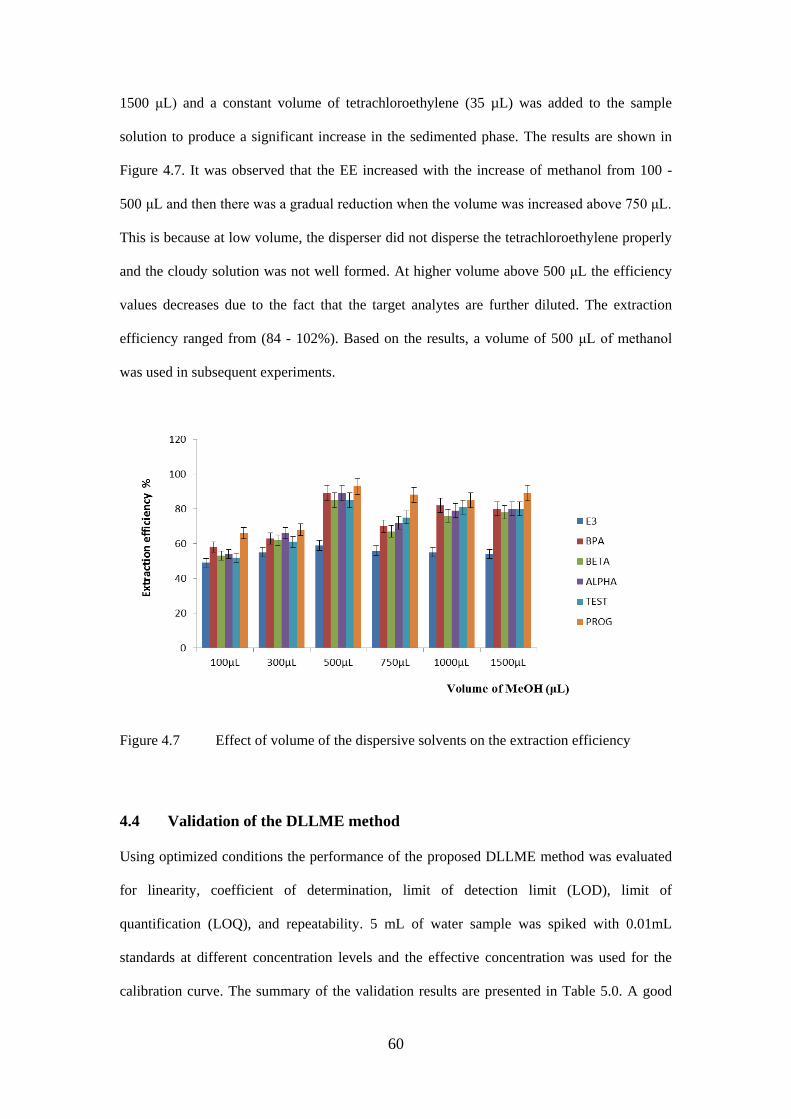
60
1500 μL) and a constant volume of tetrachloroethylene (35 µL) was added to the sample
solution to produce a significant increase in the sedimented phase. The results are shown in
Figure 4.7. It was observed that the EE increased with the increase of methanol from 100 -
500 μL and then there was a gradual reduction when the volume was increased above 750 μL.
This is because at low volume, the disperser did not disperse the tetrachloroethylene properly
and the cloudy solution was not well formed. At higher volume above 500 μL the efficiency
values decreases due to the fact that the target analytes are further diluted. The extraction
efficiency ranged from (84 - 102%). Based on the results, a volume of 500 μL of methanol
was used in subsequent experiments.
Figure 4.7 Effect of volume of the dispersive solvents on the extraction efficiency
4.4 Validation of the DLLME method
Using optimized conditions the performance of the proposed DLLME method was evaluated
for linearity, coefficient of determination, limit of detection limit (LOD), limit of
quantification (LOQ), and repeatability. 5 mL of water sample was spiked with 0.01mL
standards at different concentration levels and the effective concentration was used for the
calibration curve. The summary of the validation results are presented in Table 5.0. A good
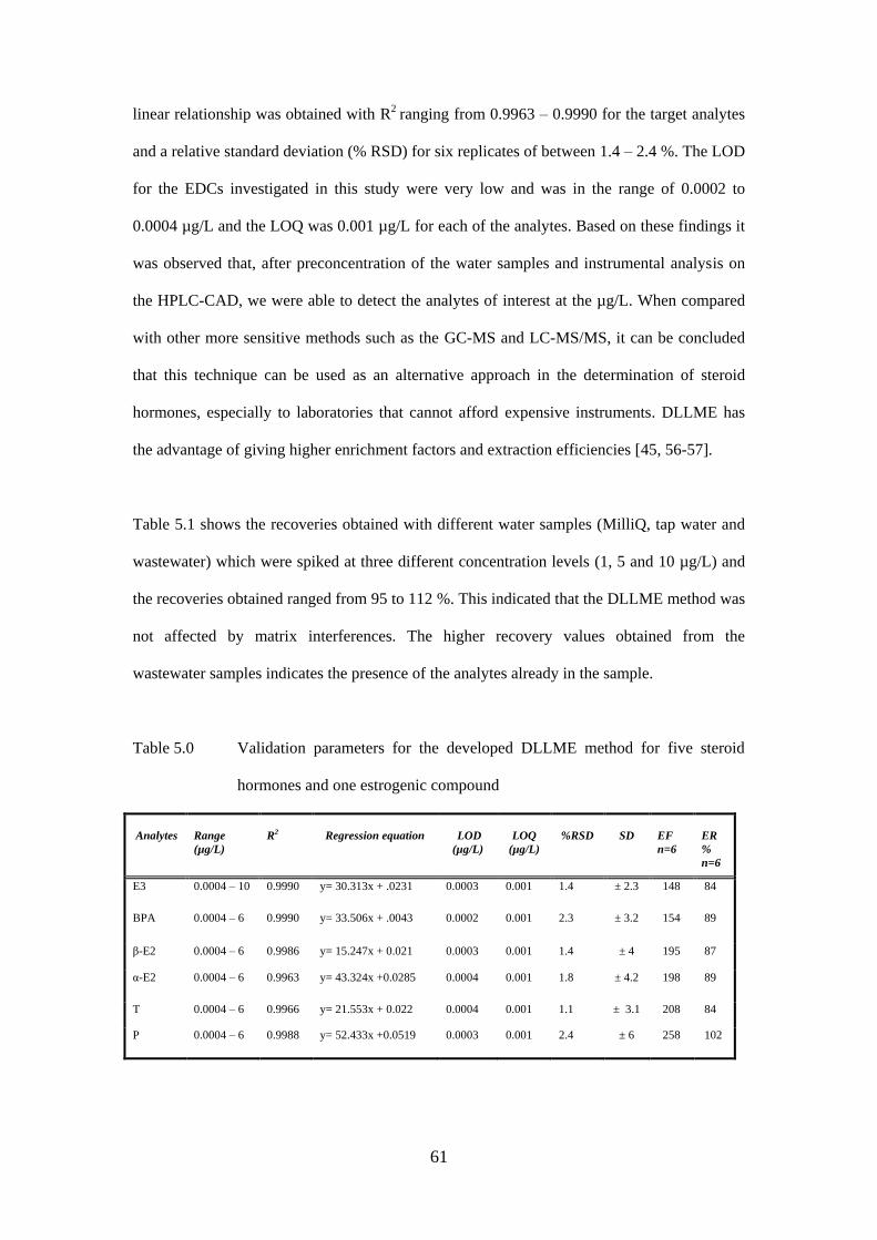
61
linear relationship was obtained with R2 ranging from 0.9963 – 0.9990 for the target analytes
and a relative standard deviation (% RSD) for six replicates of between 1.4 – 2.4 %. The LOD
for the EDCs investigated in this study were very low and was in the range of 0.0002 to
0.0004 µg/L and the LOQ was 0.001 µg/L for each of the analytes. Based on these findings it
was observed that, after preconcentration of the water samples and instrumental analysis on
the HPLC-CAD, we were able to detect the analytes of interest at the µg/L. When compared
with other more sensitive methods such as the GC-MS and LC-MS/MS, it can be concluded
that this technique can be used as an alternative approach in the determination of steroid
hormones, especially to laboratories that cannot afford expensive instruments. DLLME has
the advantage of giving higher enrichment factors and extraction efficiencies [45, 56-57].
Table 5.1 shows the recoveries obtained with different water samples (MilliQ, tap water and
wastewater) which were spiked at three different concentration levels (1, 5 and 10 µg/L) and
the recoveries obtained ranged from 95 to 112 %. This indicated that the DLLME method was
not affected by matrix interferences. The higher recovery values obtained from the
wastewater samples indicates the presence of the analytes already in the sample.
Table 5.0 Validation parameters for the developed DLLME method for five steroid
hormones and one estrogenic compound
Analytes
Range
(µg/L)
R2
Regression equation
LOD
(µg/L)
LOQ
(µg/L)
%RSD
SD
EF
n=6
ER
%
n=6
E3 0.0004 – 10 0.9990 y= 30.313x + .0231 0.0003 0.001 1.4 ± 2.3 148 84
BPA 0.0004 – 6 0.9990 y= 33.506x + .0043 0.0002 0.001 2.3 ± 3.2 154 89
β-E2 0.0004 – 6 0.9986 y= 15.247x + 0.021 0.0003 0.001 1.4 ± 4 195 87
α-E2 0.0004 – 6 0.9963 y= 43.324x +0.0285 0.0004 0.001 1.8 ± 4.2 198 89
T 0.0004 – 6 0.9966 y= 21.553x + 0.022 0.0004 0.001 1.1 ± 3.1 208 84
P 0.0004 – 6 0.9988 y= 52.433x +0.0519 0.0003 0.001 2.4 ± 6 258 102
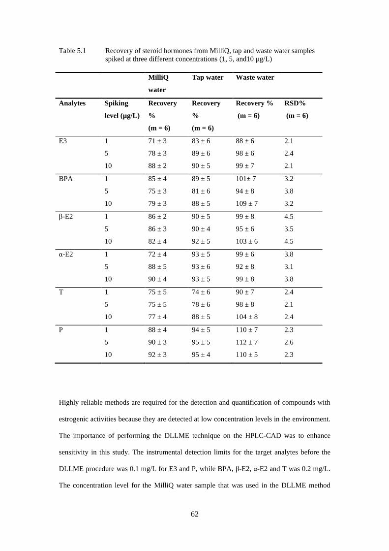
62
Table 5.1 Recovery of steroid hormones from MilliQ, tap and waste water samples
spiked at three different concentrations (1, 5, and10 µg/L)
MilliQ
water
Tap water Waste water
Analytes Spiking
level (µg/L)
Recovery
%
(m = 6)
Recovery
%
(m = 6)
Recovery %
(m = 6)
RSD%
(m = 6)
E3 1
5
10
71 ± 3
78 ± 3
88 ± 2
83 ± 6
89 ± 6
90 ± 5
88 ± 6
98 ± 6
99 ± 7
2.1
2.4
2.1
BPA 1
5
10
85 ± 4
75 ± 3
79 ± 3
89 ± 5
81 ± 6
88 ± 5
101± 7
94 ± 8
109 ± 7
3.2
3.8
3.2
β-E2 1
5
10
86 ± 2
86 ± 3
82 ± 4
90 ± 5
90 ± 4
92 ± 5
99 ± 8
95 ± 6
103 ± 6
4.5
3.5
4.5
α-E2 1
5
10
72 ± 4
88 ± 5
90 ± 4
93 ± 5
93 ± 6
93 ± 5
99 ± 6
92 ± 8
99 ± 8
3.8
3.1
3.8
T 1
5
10
75 ± 5
75 ± 5
77 ± 4
74 ± 6
78 ± 6
88 ± 5
90 ± 7
98 ± 8
104 ± 8
2.4
2.1
2.4
P 1
5
10
88 ± 4
90 ± 3
92 ± 3
94 ± 5
95 ± 5
95 ± 4
110 ± 7
112 ± 7
110 ± 5
2.3
2.6
2.3
Highly reliable methods are required for the detection and quantification of compounds with
estrogenic activities because they are detected at low concentration levels in the environment.
The importance of performing the DLLME technique on the HPLC-CAD was to enhance
sensitivity in this study. The instrumental detection limits for the target analytes before the
DLLME procedure was 0.1 mg/L for E3 and P, while BPA, β-E2, α-E2 and T was 0.2 mg/L.
The concentration level for the MilliQ water sample that was used in the DLLME method
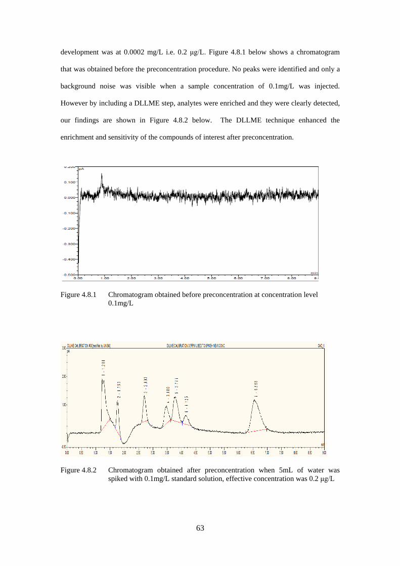
63
development was at 0.0002 mg/L i.e. 0.2 μg/L. Figure 4.8.1 below shows a chromatogram
that was obtained before the preconcentration procedure. No peaks were identified and only a
background noise was visible when a sample concentration of 0.1mg/L was injected.
However by including a DLLME step, analytes were enriched and they were clearly detected,
our findings are shown in Figure 4.8.2 below. The DLLME technique enhanced the
enrichment and sensitivity of the compounds of interest after preconcentration.
Figure 4.8.1 Chromatogram obtained before preconcentration at concentration level
0.1mg/L
Figure 4.8.2 Chromatogram obtained after preconcentration when 5mL of water was
spiked with 0.1mg/L standard solution, effective concentration was 0.2 μg/L
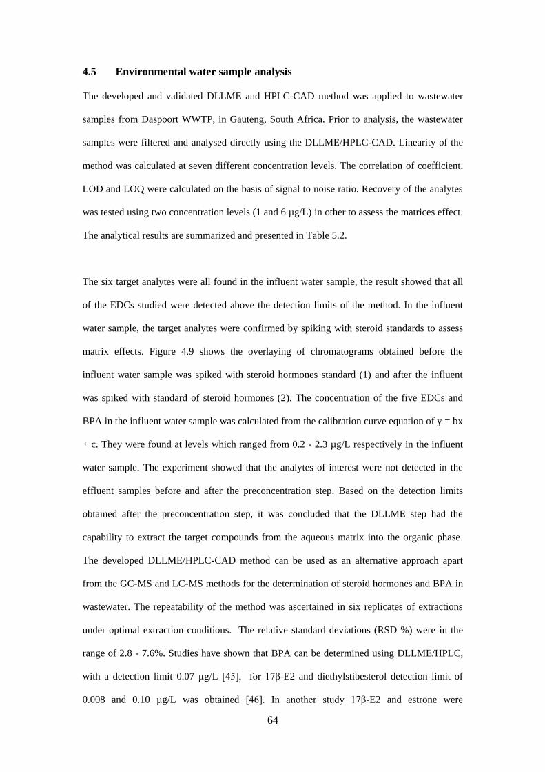
64
4.5 Environmental water sample analysis
The developed and validated DLLME and HPLC-CAD method was applied to wastewater
samples from Daspoort WWTP, in Gauteng, South Africa. Prior to analysis, the wastewater
samples were filtered and analysed directly using the DLLME/HPLC-CAD. Linearity of the
method was calculated at seven different concentration levels. The correlation of coefficient,
LOD and LOQ were calculated on the basis of signal to noise ratio. Recovery of the analytes
was tested using two concentration levels (1 and 6 µg/L) in other to assess the matrices effect.
The analytical results are summarized and presented in Table 5.2.
The six target analytes were all found in the influent water sample, the result showed that all
of the EDCs studied were detected above the detection limits of the method. In the influent
water sample, the target analytes were confirmed by spiking with steroid standards to assess
matrix effects. Figure 4.9 shows the overlaying of chromatograms obtained before the
influent water sample was spiked with steroid hormones standard (1) and after the influent
was spiked with standard of steroid hormones (2). The concentration of the five EDCs and
BPA in the influent water sample was calculated from the calibration curve equation of y = bx
+ c. They were found at levels which ranged from 0.2 - 2.3 µg/L respectively in the influent
water sample. The experiment showed that the analytes of interest were not detected in the
effluent samples before and after the preconcentration step. Based on the detection limits
obtained after the preconcentration step, it was concluded that the DLLME step had the
capability to extract the target compounds from the aqueous matrix into the organic phase.
The developed DLLME/HPLC-CAD method can be used as an alternative approach apart
from the GC-MS and LC-MS methods for the determination of steroid hormones and BPA in
wastewater. The repeatability of the method was ascertained in six replicates of extractions
under optimal extraction conditions. The relative standard deviations (RSD %) were in the
range of 2.8 - 7.6%. Studies have shown that BPA can be determined using DLLME/HPLC,
with a detection limit 0.07 µg/L [45], for 17β-E2 and diethylstibesterol detection limit of
0.008 and 0.10 µg/L was obtained [46]. In another study 17β-E2 and estrone were
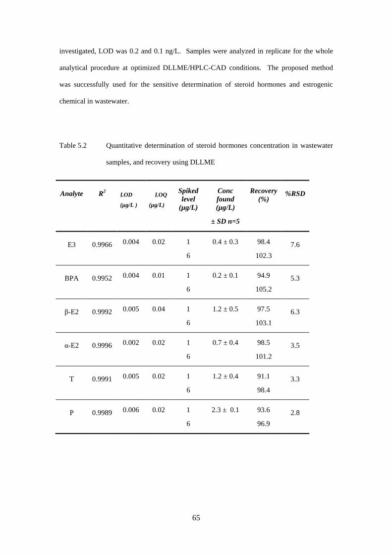
65
investigated, LOD was 0.2 and 0.1 ng/L. Samples were analyzed in replicate for the whole
analytical procedure at optimized DLLME/HPLC-CAD conditions. The proposed method
was successfully used for the sensitive determination of steroid hormones and estrogenic
chemical in wastewater.
Table 5.2 Quantitative determination of steroid hormones concentration in wastewater
samples, and recovery using DLLME
Analyte
R2
LOD
(µg/L )
LOQ
(µg/L)
Spiked
level
(µg/L)
Conc
found
(µg/L)
± SD n=5
Recovery
(%)
%RSD
E3
0.9966 0.004 0.02 1
6
0.4 ± 0.3 98.4
102.3
7.6
BPA
0.9952 0.004 0.01 1
6
0.2 ± 0.1 94.9
105.2
5.3
β-E2
0.9992 0.005 0.04 1
6
1.2 ± 0.5 97.5
103.1
6.3
α-E2
0.9996 0.002 0.02 1
6
0.7 ± 0.4 98.5
101.2
3.5
T
0.9991 0.005 0.02 1
6
1.2 ± 0.4 91.1
98.4
3.3
P
0.9989 0.006 0.02 1
6
2.3 ± 0.1 93.6
96.9
2.8
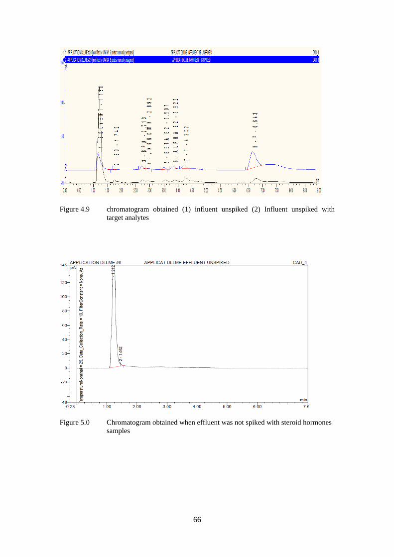
66
Figure 4.9 chromatogram obtained (1) influent unspiked (2) Influent unspiked with
target analytes
Figure 5.0 Chromatogram obtained when effluent was not spiked with steroid hormones
samples
67
CHAPTER 5
Conclusions and Future Work
5.1 Introduction
In this final chapter, a summary of the research study is provided by revisiting the research
objectives and goals that inspired the study. In addition, the main contributions arising from
the proposed DLLME/HPLC-CAD analysis are highlighted. Furthermore the research
limitations and challenges identified in the research study are also highlighted. Finally future
research work that could address some of the identified challenges are also discussed. In this
study, an alternative analytical method was developed for the determination five steroid
hormones and BPA.
5.2 Research Objectives
This section describes the approach in which the objectives presented in Chapter 1 section 1.3
were addressed and accomplished in the study. The already developed analytical separation
method was validated in order to confirm that the method or procedure was suitable for the
detection of five steroid hormones and BPA wastewater samples. Based on the validation
parameters, the results obtained showed that the method was accurate, had good repeatability
and was very robust. The LODs obtained using HPLC-CAD for the target analytes were as
follows: E3 and P were 0.1 mg/L, while BPA, β-E2, α-E2 and T was 0.2 mg/L. The
recoveries obtained for the analytes in wastewater was between 88.71 - 110%. From our
validation results, it was concluded that the method could separate the five steroid hormones
and BPA, but it would not be able to detect the target analytes in environmental without a
preconcentration step, since they are found in the environmental wastewater samples at low
concentration levels.
68
A preconcentration procedure known as DLLME was developed, optimized and also
validated. The optimization of the different conditions helped to enhance better sensitivity of
the analytes in environmental samples. Under optimum DLLME conditions, 35 µL of
tetrachloroethylene and 500 µL of methanol were used as extracting and dispersing solvent,
this led to higher extraction efficiencies of about 84- 102% for the analytes. Enrichment of the
analytes into the organic phase was also achieved at optimal values. The optimal volumes
helped to reduce the further dilution of the analytes of interest. After preconcentration, the
LOD values for the six analytes were found to be as low as 0.0002 µg/L for BPA. E3 and β-
E2 were detected at 0.0003 µg/L, while α-E2 and T were detected at 0.0004 µg/L
respectively. Based on the detection limits obtained after the preconcentration step, it was
concluded that the DLLME step had the capability to extract the target compounds from the
aqueous matrix into the organic phase and produce lower detection limits. Hence, it was
concluded that the DLLME/HPLC-CAD could be used as an alternative method for the
quantification of steroid hormones in the environment.
Finally the developed and optimized DLLME method was used as a preconcentration step in
the determination of five steroid hormones and BPA in a wastewater sample obtained from a
WWTP. The real wastewater sample (influent and effluent) were analysed using the
DLLME/HPLC-CAD technique. The concentration levels at which these analytes are found
in our own environment in South Africa, Pretoria using the HPLC-CAD were also
reported.The concentration of each of the target analytes in the influent sample was 0.4µg/L
for E3, α-E2 was 0.7 µg/L, BPA was 0.2 µg/L. Others found at higher concentration level
include β-E2 and T and was detected at 1.2 µg/L. P was found to be with the highest in the
environment with concentration level of 2.3 µg/L.
The target analytes were not detected in the wastewater effluents. Satisfactory recoveries for
the spiked tap water (95%), MilliQ water (92%) and wastewater (112%) were obtained.
Precision was quantified by relative standard deviation (% RSD) which was less than 4%.
69
Studies have shown that BPA can be determined using DLLME/HPLC, with a detection limit
0.07µg/L [45], for 17β-E2 and diethylstibesterol detection limit of 0.008 and 0.10 µg/L was
obtained [46]. In another study 17β-E2 and estrone were investigated, LOD was 0.2 and 0.1
ng/L. Based on our findings, determination of five steroid hormones and BPA in wastewater
sample was performed using DLLME/HPLC-CAD, the method was simple, fast, easy, and
more cost friendly for the extraction and preconcentration of the target analytes. The analysis
of the target analytes was achieved without any derivatization step prior to determination on
the HPLC. When DLLME/HPLC-CAD was compared with other methods for the extraction
and determination of EDCs on HPLC, the presented method has low limit of detection, high
recoveries and a short extraction time. This is the first time that five steroid hormones and one
estrogenic chemical BPA will be determined and quantified on an HPLC-CAD.
5.3 Limitation and Recommendation
During the process of this research project, the determination and quantification of steroid
hormone has been increasing globally and will continue to increase, since the intake of
steroids hormones is still taking place. The continuous release of steroid hormones and their
metabolites into the environment needs to be understood better and more scientific
documentation is necessary. The overall objectives of the study set out in Chapter 1 of the
dissertation were accomplished. However, the proposed DLLME/HPLC-CAD has the
potential to be improved as more analysis is carried out on the instrument. Despite recent
progress, there are still many gaps in scientific knowledge about the fate and behavior of
steroid hormones in the environment and the present dissertation provides some
recommendations for future research:
More preconcentration methods that are suitable for the determination of steroid
hormones and its metabolites need to be developed for the determination of steroid
hormone and its metabolites in the environment.
70
Improvement in analytical methods and instrumentation should be reviewed, as some
may not reveal the presence of steroid hormones, but they currently remain
undetected in the environment.
Monitoring of steroid hormones at different WWTPs should be done at regular
intervals to determine the extent of the release of these compounds in the
environment.
However, surrogate standard should be included in the future work of this method in
order to give a clear indication of the possibility of analyte loss during sampling of
water samples.
Finally, the exact amount of steroid hormones that would be tolerated by human
beings should also be documented for future purposes.

71
REFERENCES 1. Comerton, A.M., Andrews, R.C., Bagley, D.M. (2009). Practical overview of analytical
methods for endocrine-disrupting compounds, pharmaceuticals and personal care
products in water and waste water, Philosophical Transaction of the Royal Society A
(367): 3923-3939
2. Daughton, C., Ternes, T. (1999). Pharmaceuticals and personal care products in the
environment: Agents of subtle change? Environmental Health Perspectives (107): 907-
938.
3. Zhang Y., Zhou J.L (2008). Occurrence and removal of endocrine disrupting chemicals in
wastewater. Chemosphere (73) 848- 853
4. Briciu R.D., Wasik A.K., Namiesnik J (2009). Analytical Challenges and Recent
Advances in the Determination of Estrogens in Water Environments. Journal of
Chromatography Science, Vol. 47, (2):127-39
5. Routledge E. J., Sheahan D., Desbrow C., Brighty G.C., Waldock M., J.P. Sumpter
(1998). Identification of estrogenic chemicals in STW effluents. II. in vivo responses in
tout and roach. Environmental Science and Technology (32) 1559-1565.
6. Limpiyakorn, T., Homklin S., Ong, S.K. (2011). Critical reviews in Environmental
Science and Technology (41): 1231-11270
7. Ying G.-C., Kookana, R.S., Ru, Y.J.. (2002). Occurrence and fate of hormone steroids in
the environment. Environment International (28): 545-551.
8. Crisp, T.M., Clegg, E.D., Cooper, R.L, Wood, W.P., Anderson, D.G., Baetcke, K.P.,
Hoffman, J.L., Morrow, M.S., Rodier, D.J., Schaeffer, J.E., Touart, L.W., Zeeman M.G.,
Patel, Y.M. (1998). Environmental endocrine disruption: An effects assessment and
analysis. Environmental Health Perspective, (106): 11-56
9. Damstra T., Barlow S., Bergman A., Kavlock R., Kraak G (2002). Global assessment of
the state of the science of endocrine disruptors. An assessment prepared by an expert
group on behalf of the World Health Organization (WHO), the International Labor
72
Organization, and the United Nations Environment Programme Website publication
www.who.int/ipcs/publications/new issues/endocrine disruptors/en/index.html
10. Burkhardt-Holm, P. (2010) Endocrine disruptors and water quality: A state-of-the-art
review. International Journal of water Resources Development, Vol. 26, H. 3. S. 477-493.
11. Cook, J.W., Dodds, E.C., Hewett, C.L., Lawson, W. (1934). Estrogenic activity of some
condensed ring compounds in relation to their other biological activates. Proactive Royal
Society (114) 272-286.
12. LaFleur, A.D., Schug, K.A. (2011). A review of separation methods for the determination
of estrogens and plastics-derived estrogen mimics from aqueous systems. Analytica
Chimica Acta (696): 6–26.
13. Ascenzo, G.D., Di Corcia, A., Gentili, A., Mancini, R., Mastropasqua, R., Nazzari, M.,
Samperi, R. (2003). Fate of natural estrogen conjugates in municipal sewage transport
and treatment facilities. Science of the Total Environment (302): 199-209.
14. Ternes, T., Stumpf, M., Mueller, J., Haberer, K., Wilken, R., Servos, M. (1999). Behavior
and occurrence of estrogens in municipal sewage treatment plants-I. Investigations in
Germany, Canada and Brazil. Science of the Total Environ (225): 81-90
15. Hoffmann, B., Goes de Pinho, T., Schuler, G. (1997). Determination of free and
conjugated oestrogens in peripheral blood plasma, faeces and urine of cattle throughout
pregnancy. Experimental and Clinical Endocrinology & Diabetes 105(5), 296–303
16. Jobling, S., Nolan, M., Tyler, C. R., Brighty, G., Sumpter, J.P. (1998).Widespread sexual
disruption in wild fish. Environmental Science and Technology. (32): 2498-2506.
17. Lopez de Alda, M.J., Barcelo, D. (2001b). Determination of steroid sex hormones and
related synthetic compounds considered as endocrine disrupters in water by fully
automated on-line solid-phase extraction–liquid chromatography–diode array detection.
Journal of Chromatography A. (911): 203–11
18. Wang L., Cai Y.Q., He B., Yuan C.G., Shen D.Z, Shao J., Jiang G.B (2006).
Determination of estrogens in water by HPLC-UV using cloud point extraction. Talanta
(70) 47-51
73
19. Bergman A., Heindel J.J., Jobling S., Ki K A (2012). State of the science of endocrine
disrupting chemicals (2012). Summary for decision-makers. An assessment of the state of
the science of endocrine disruptors for the united states nations environmental programme
and world health organization
20. Chang, C.M., Chou, C.C., Lee, M.R. (2005). Determining leaching of bisphenol A from
plastic containers by solid-phase microextraction and gas chromatography-mass
spectrometry. Analytica Chimica Acta (539): 41-47.
21. Liu R., Zhou J.L., Wilding A (2004). Simultaneous determination of endocrine disrupting
phenolic compounds and steroids in water by solid-phase extraction–gas
chromatography–mass spectrometry. Journal of Chromatography A (1022): 179–189
22. Andersen L., Holbech H., Gessbo A., Norrgren L., Petersen G.I (2003). Effect of
exposure to 17α-ethinylestradiol during early development on sexual differentiation and
induction of vitellogenin in zebrafish (Danio rerio). Comparative Biochemistry and
Physiology Part C (134), 365–374.
23. Kidd K.A., Blanchfield P.J., Mills K.H., Palac V.P., Evans R.E., Lazorchak J.M., Flick
R.W (2007). Collapse of a fish population after exposure to a synthetic estrogen.
Environmental and Science vol 104, no 21, 8901.
24. Bolong N., Ismail A.F., Salim M.R., Matsuura T. (2009). A review of the effects of
emerging contaminants in wastewater and options for their removal. Desalination (239):
229-246
25. Schnitzler J.G., Koutrakis E., Siebert U., Thome J.P., Das K (2008). Effects of persistent
organic pollutants on the thyroid function of the European sea bass (Dicentrarchus
labrax) from the Aegean Sea, is it an endocrine disruption? Marine Pollution and
Bulletin. 56(10): 1755-1764
26. Behera S.K., Kim H. W., Oh J. E., Park H. S., (2011). Occurrence and removal of
antibiotics, hormones and several other pharmaceuticals in wastewater treatment plants of
the largest industrial city of Korea. Science of the total environ (409) 4351-4360
74
27. Xu N., Xu Y F., Xu S., Li J., Tao H. C., (2012). Removal of estrogens in municipal
wastewater treatment plants: A Chinese perspective. Environmental Pollution (165) 215-
224
28. Liu, Z.H., Kanjo, Y., Mizutani, S., (2009a). Removal mechanisms for endocrine
disrupting compounds (EDCs) in wastewater treatment-physical means, biodegradation,
and chemical advanced oxidation: a review. Science of the Total environment (407) 731-
74
29. Kim S.D, Cho J, Kim I. S., Vanderford B.J., Snyder S.A. (2007).Occurrence and removal
of pharmaceuticals and endocrine disruptors in South Korean surface, drinking, and waste
waters. Water Research ( 41) 1013 – 1021
30. Joss A., Andersen H., Ternes T., Richie P.R., Siegrist H., (2004). Removal of estrogens in
municipal wastewater treatment under aerobic and anaerobic conditions: Consequences
for plant Optimization. Environmental and Science Technology. (38), 3047- 3055
31. Baronti C, Curini R, D'Ascenzo G, Corcia AD, Gentili A, Samperi R.(2000). Monitoring
natural and synthetic estrogens at activated sludge sewage treatment plants and in
receiving river water. Environmental and Science Technology (34):5059–66
32. Chang H, Wu SM, Hu JY, Asami M, Kunikane S. (2008). Trace analysis of androgens
and progestogens in environmental waters by ultra-performance liquid chromatography–
electrospray tandem mass spectrometry. Journal of Chromatography A (1195):44–51.
33. Liu S., Ying G.-G., Zhao J.L., Chen F., Yang B., Zhou L.J., Lai H.J. (2011). Trace
analysis of 28 steroids in surface water, wastewater and sludge samples by rapid
resolution liquid chromatography-electrospray ionization tandem mass spectrometry.
Journal of Chromatography A (1218):1367–78
34. Grover D.P., Z.L. Zhanga Z.L., Readmanb J.W, Zhoua J.L., (2009). A comparison of
three analytical techniques for the measurement of steroidal estrogens in environmental
water samples. Talanta (78) 1204–1210
75
35. Petrovic, M., Eljarrat, E., Lopez de Alda, M.J., Barcelo, D. (2002). Recent advances in
the mass spectrometric analysis related to endocrine disrupting compounds in aquatic
environmental samples. Journal of Chromatography A (974): 23–51
36. Quintana J.B., Carpinteiro J., Rodrıguez I., Lorenzo R.A., Carro A.M., Cela R (2004).
Determination of natural and synthetic estrogens in water by gas chromatography with
mass spectrometric detection. Journal of Chromatography A (1024) 177–185.
37. Tomsikova H., Aufartova J., Solich P., Ferrera J., Rodrı´guez J.J.S., Novakova L. (2012).
High-sensitivity analysis of female-steroid hormones in environmental samples Trends in
Analytical Chemistry (34) 35-55 doi:10.1016/j.trac.2011.11.008
38. Pailler J. Y, Krein A., Pfister L., Hoffmann L., Guignard C., (2009). Solid phase
extraction coupled to liquid chromatography-tandem mass spectrometry analysis of
sulfonamides, tetracyclines, analgesics and hormones in surface water and wastewater in
Luxembourg. Science of the Total Environ. (407) 4736-4743.
39. Wang H X., Zhou Y., Jiang Q W (2012). Simultaneous screening of estrogens,
progestogens, and phenols and their metabolites in potable water and river water by ultra-
performance liquid chromatography coupled with quadrupole time-of-flight mass
spectrometry. Microchemical Journal (100) 83–94
40. Viglino L., Aboulfadl K., Prevost M., Sauve S. (2008) Analysis of natural and synthetic
estrogenic endocrine disruptors in environmental waters using online preconcentration
coupled with LC-APPI-MS/MS. Talanta (76) 1088–1096
41. Sanchez B.A., Capote F. P, Jimenez J R, Luque de Castro M.D (2008). Automated solid-
phase extraction for concentration and clean-up of female steroid hormones prior to liquid
chromatography–electrospray ionization–tandem mass spectrometry: An approach to
lipidomics. Journal of Chromatography A (1207) 46–54
42. Maria J. Lopez de Alda., Damia Barcelo (2001). Use of solid-phase extraction in various
of its modalities for sample preparation in the determination of estrogens and
progestogens in sediment and water. Journal of Chromatography A (938) 145–153

76
43. Wu C Q., Chen D Y., Feng Y S., Deng H M., Liu Y H., Zhou A J. (2012) Determination
of estrogens in water samples by ionic liquid-based dispersive liquid-liquid
microextraction combined with high performance liquid chromatography. Analytical
Letters, (45): 1995–2005
44. Aufartova J., Maria Esther Torres-Padron., Sosa-Ferrera Z., Novakova L., Solich P.,
Santana-Rodríguez J.J. (2014). Development of a novel in-tube solid phase
microextraction based on micellar desorption followed by LC-DAD FD for the
determination of some endocrine disruptor compounds in environmental liquid samples.
Journal of Liquid Chromatography & Related Technology (37):12, 1654-1672
45. Rezaeea M., Yaminia. Y., Shariati. S., Esrafili A., Shamsipur M. (2009). Dispersive
liquid–liquid microextraction combined with high-performance liquid chromatography-
UV detection as a very simple, rapid and sensitive method for the determination of
bisphenol A in water samples. Journal of Chromatography A (1216) 1511–1514
46. Hadjmohammadi M.R., Ghoreishi S.S (2011). Determination of Estrogens in Water
Samples Using Dispersive Liquid Liquid Microextraction and High Performance Liquid
Chromatography. Acta Chimica Slova. (58) 765–771
47. Vehoveca, T., Obrezab, A. (2010). Review of operating principle and applications of the
charged aerosol detector. Journal of Chromatography A, 1217: 1549–1556.
48. Gorecki T, Lynen F, Szucs R., Sandra P., (2006). Universal response in liquid
chromatography using charged aerosol detection, Analytical Chemistry (78) 3186–3193
49. Thermoscientific. Dionex Corporation 1228 Titan Way P.O. Box 3603 Sunnyvale, CA.
Corona ultra TM. ESA Biosciences, Inc., Chelmsford, MA
50. Fox C.B., Sivananthan S.J., Mikasa T.J.T., Lin S., Parker S.C (2013). Charged aerosol
detection to characterize components of dispersed-phase formulations. Advances in
Colloid and Interface Science (199–200) 59–65
51. Almeling S., Ilko D., and Holzrabe U., (2012). Review of charged aerosol detection in
pharmaceutical analysis. Journal of Pharmaceutical and Biomedical Analysis (69) 50– 63
77
52. Zorita S., Hallgren P., Mathiasson L. (2008). Steroid hormone determination in water
using an environmentally friendly membrane based extraction technique. Journal of
Chromatography A (1192), 1-8
53. Sadowski R., and Kopciuch R.G., (2013). Research Article Isolation and determination of
estrogens in water samples by solid-phase extraction using molecularly imprinted
polymer. Journal of Separation Science. (36), 2299–2305
54. Salvador A., Moretton C., Piram A., Faure R., (2007). On-line solid-phase extraction with
on-support derivatization for high-sensitivity liquid chromatography tandem mass
spectrometry of estrogens in influent/effluent of wastewater treatment plants. Journal of
Chromatography A, (1145) 102–109
55. Rezaee, M., Assadi, Y., Milani Hosseini, M.-R., Aghaee, E., Ahmadi, F., Berijani, S.
(2006). Determination of organic compounds in water using dispersive liquid–liquid
microextraction. Journal of Chromatography A 1116 (1–2): 1–9.
56. Xiwei Du, Wang, X. Li, Y. Ye, F. Dong1, Q. and Huang, C. (2010). Determination of
estrone and 17b-estradiol in water samples using dispersive liquid–liquid microextraction
followed by LC. Chromatographia (71) 405-410
57. Li X, Ying G.G., Su H.C., Yang X.B., Wang L (2010). Simultaneous determination and
assessment of 4-nonylphenol, bisphenol A and triclosan in tap water, bottled water and
baby bottles. Environment International (36) 557–562
58. Vandenberg L.N., Hauser R., Marcus Michele., Olea N., Welshons W.V. (2007). Human
exposure to bisphenol A (BPA). Reproductive toxicology (24) 139-177
59. Markey C.M., Rubin B.S., Soto A.M., Sonnenschein C. (2003). Endocrine disruptors:
from wingspread to environmental developmental biology. Journal of steroid
biochemistry & molecular biology (83) 235-244
60. Scherr, F.F., Sarmah, A.K., Di, H.J., Cameron, K.C. (2008). Modelling degradation and
metabolite formation kinetics of estrone-3-sulphate in agricultural soils. Environmental
Science and Technology 42(22): 8388–8394. doi:10.1021/es801850a.
78
61. Geyer, H.J., Rimkus, G., Scheunert, I.m Kaune, A., Schramm K.W., Kettrup A., [41]
Zeeman, M., Muir, D.C.G., Hansen, L.G. and Mackay, D. (2000) Bioaccumulation and
occurrence of endocrine disrupting chemicals, persistent organic pollutants, and other
organic compounds in fish and other organisms including humans. In Beek B. (ed.): The
handbook of environmental chemistry Vol. 2, Part Bioaccumulation. Berlin, Heidelberg:
Springer Verlag
62. Kummerer, K., 2009. The presence of pharmaceuticals in the environment due to human
use-present knowledge and future challenges. Journal of Environmental Management
(90), 2354-2366
63. Lalumera, G., Calamari, D., Galli, P., Castiglioni, S., Crosa, G. Fanelli, R. (2004).
Preliminary investigation on the environmental occurrence and effects of antibiotics used
in aquaculture in Italy. Chemosphere (54): 661-668.
64. Czajka, C.P. Londry, K.L. (2006). Anaerobic biotransformation of estrogens. Science of
the Total Environment (367): 932- 941.
65. Williams, R.T., 2005. Human health pharmaceuticals in the environment–an introduction.
In: Williams, R.T. (Ed.). Human pharmaceuticals: assessing the impact on aquatic
ecosystems. SETAC press, Pensacola, FL.
66. Bound, J., Kitsou, K., Voulvoulis, N., (2006). Household disposal of pharmaceuticals and
perception of risk to the environment. Environmental Toxicology and Pharmacology (21),
301-307.
67. Bhawanu. S.A, Sulaiman, O., Hashim, R. Mohamad, I.M.N. (2010). Thin-layer
chromatographic analysis of steroids: A review. Tropical Journal of Pharmaceutical
Research. 9 (3): 301-313.
68. Noppe H., Le Bizec B., Verheyden K., De Brabander H. F (2008). Novel analytical
methods for the determination of steroid hormones in edible matrices. Analytica Chimica
Acta (611) 1-16
69. Kicman A.T (2008). Pharmacology of anabolic steroids. British journal of pharmacology,
154 (3), 502-521
79
70. Hartqens F., Kuipers H (2004). Effects of androgen-anabolic steroids in athletes. Sports
and Medicine 34 (8): 513-54
71. Gomes R.L., Scrimshaw M.D., Lester J.N (2003). Determination of endocrine disrupters
in sewage treatment and receiving waters. Trends in Analytical Chemistry, Vol. 22, No.
10
72. Packov V., Loukotkov L., Boskov Z., Stulk K.(2009). Review: Analysis for estrogens as
environmental pollutants Journal of Seperation Science (32), 867 – 882
73. Wagner, M. Oehlmann, J. (2009) Endocrine disruptors in bottled mineral water: Total
estrogenic burden and migration from plastic bottles. Environmental Science and
Pollution Research International 16(3): 278-286
74. Adler, P., Steger-Hartmann, T., Kalbfus, W. (2001). Distribution of natural and synthetic
estrogenic steroid hormone in water samples from southern and middle Germany. Acta
Hydrochimica Hydrobiology. 29(4): 227-241.
75. Wang, H.-X., Zhou, Y. Jiang, Q.-W. (2012). Simultanueous screening of estrogens,
progestogens, and phenols and their metabolites in portable water and river water by
ultra-performance liquid chromatography coupled with quardupole time-of-flight
spectrometry. Microchemical Journal (100): 83-94.
76. Maria Kostopoulou and Anastasia Nikolaou (2008). Analytical problems and the need for
sample preparation in the determination of pharmaceuticals and their metabolites in
aqueous environmental matrices. Trend in analytical chemistry, vol 27, No. 11
77. Carballa, M., Omil, F., Lema, J., Llompart, M., Garcia, C., Rodriguez, I., Gomez, M.,
Ternes, T., (2005). Behaviour of pharmaceuticals and personal care products in a sewage
treatment plant of northwest Spain. Water Science and Technology (52): 29-35.
78. Snow, Bartelt-Hunt, S.L., Saunders, S.E., Devivo, S.L., Cassada, D.A. (2008). Detection,
occurrence and fate of emerging contaminants in agricultural environments.
doi:10.2175/106143008X328518. Water Environment Research, 80(10).
80
79. Desbrow C, Routledge EJ, Brighty GC, Sumpter JP, Waldock M. (1998). Identification of
estrogenic chemicals in STW effluent. 1. Chemical fractionation and in vitro biological
screening. Environment Science and Technology 32(11):1549–1558.
80. Vulliet E., Wiest L., Baudot R., Grenier-Loustalot M.F (2008). Multi-residue analysis of
steroids at sub-ng/L levels in surface and ground-waters using liquid chromatography
coupled to tandem mass spectrometry. Journal of Chromatography A, (1210) 84–91
81. Koplin D.W., Furlong E.T., Meyer M.T., Thurman E.M., Zaugg S.D., Barber L.B Buxton
H.T. (2002). Pharmaceutical, hormones and other organic wastewater contaminants in US
streams, 1999-2000, A national reconnaissance. Environmental Science and Technology
(36): 1202-1211.
82. Kjaer, J., Olsen, P., Bach, K., Barlebo, H. C., Ingerslev, F., Hansen, M. Sorensen, B.H.
(2007). Leaching of estrogenic hormones from manure-treated structured soils.
Environmental science and technology (41) 3911-3917
83. Sun, L., Yong, W., Chu, X., Lin, J. (2009). Simultaneous determination of 15 steroidal
oral contraceptives in water using solid-phase disk extraction followed by high
performance liquid chromatography-tandem mass spectrometry. Journal of
Chromatography A (1216): 5416–23.
84. Al-Odaini N.A., Zakaria M.P., Yaziz M.I. and Surif, S. (2010). Multi-residue analytical
method for human pharmaceuticals and synthetic hormones in river water and sewage
effluent by solid-phase extraction and liquid chromatography-tandem mass spectrometry.
Journal of Chromatography A (1217): 6791–806.
85. Perez-Carrera E, Hansen M, Leon V, Bjorklund E, Krogh K, Halling- Sorensen B et al
(2010) Multiresidue method for the determination of 32 human and veterinary
pharmaceuticals in soil and sediment by pressurized-liquid extraction and LC-MS/MS.
Analytical and Bioanalytical Chemistry (398):1173–1184
86. Gineys N., Giroud B., Vulliet E. (2010) Analytical method for the determination of trace
levels of steroid hormones and corticosteroids in soil, based on PLE/SPE/LC-MS/MS.
Analytical and Biolanalytical Chemistry; (397): 2295–302
81
87. Ting C. Chen , Te S. Chen , Kuei J. Yeh , Yu C. Lin , How R. Chao & Yi L. Yeh (2012)
Sorption of estrogens estrone, 17β-estradiol, estriol, 17α-ethinylestradiol, and
diethylstilbestrol on sediment affected by different origins, Journal of Environmental
Science and Health, Part A: Toxic/Hazardous Substances and Environmental
Engineering, (47):12, 1768-1775,
88. Xu Y.J., Tian X.H., Zhang X.Z., Gong X.H., Zhang S.J., Liu H.H., Zhang L.M. (2010).
Simultaneous determination of hormone residues in aquatic products by UPLC-ESI-MS-
MS. Am laboratory (42):11–6
89. Heberer, T. (2002). Occurrence, fate, and removal of pharmaceutical residues in the
aquatic environment: A review of recent research data. Toxicology Letters (131): 5-17.
90. Gopinath C.N. (2010). Determination of bioconcentration potential of selected
pharmaceuticals in fathead minnow, pimephales promelas, and channel catfish, ictalurus
punctatus. PhD thesis. University of North Texas.
91. Scherr Frank (2009). Sorption, Degradation and Transport of Estrogens and Estrogen
Sulphates in Agricultural Soil. Thesis for PhD at Lincoln University
92. Nikolaou, A. Meric, S. Fatta, D. (2007). Occurrence patterns of pharmaceuticals in water
and wastewater environments. Analytical and Bioanalytical Chemistry (387): 1225-1234
93. Zepp, R.G. Cline, D.M. (1977). Rate of direct photolysis in the aquatic environment.
Environmental Science and Technology. (11): 359-366
94. Petrovic, M., Barceló, D., (2007). LC-MS for identifying photodegradation products of
pharmaceuticals in the environment. Trends in Analytical Chemistry (26): 486-493
95. Ternes, T., Hermann, N., Bonerz, M., Knacker, T., Siegrist, H., Joss, A. (2004a). A rapid
method to measure the solid-water distribution coefficient (Kd) for pharmaceuticals and
musk fragrances in sewage sludge. Water Research (38): 4075-4084.
96. Boxall, A., Fogg, L., Blackwell, P., Blackwell, P., Kay, P., Pemberton, E., Croxford A.
(2004). Veterinary medicines in the environment. Reviews of Environmental
Contamination and Toxicology, (180): 1-91.
82
97. Ternes, T., (1998). Occurrence of drugs in German sewage treatment plants and rivers.
Water Research (32): 3245-3260
98. Hirsch, R., Ternes, T., Haberer, K. Kratz, K., (1999). Occurrence of antibiotics in the
aquatic environment. The Science of the Total Environment (225): 109-118.
99. Halling-Sorensen, B., Nors Nielsen, S., Lanzky, P., Ingerslev, F., Holten Lützhøft, H.,
Jorgensen, S. (1998). Occurrence, fate and effects of pharmaceutical substances in the
environment - A review. Chemosphere (36): 357-393.
100. Fent, K., Weston, A., Caminada, D. (2006). Ecotoxicology of human pharmaceuticals.
Aquatic Toxicology (76): 122-159.
101. Daughton, C., Ternes, T. (1999). Pharmaceuticals and personal care products in the
environment: Agents of subtle change? Environmental and Health Perspectives (107):
907-938.
102. Samirkumarkhanal B., Thompson M., Keeong S S, Vanleeuwen A H (2006). Fate,
Transport and Biodegradation of Natural Estrogens in the Environment and Engineered
Systems. Vol. 40, no. 21, Environmental Science and Technology (9), 6537
103. Sonnenschein C., Soto A.M.J. (1998). Steroid Biochem. Molec. Biol. vol. 65 No. 1±6,
pp.143±150. An Updated Review of Environmental Estrogen and Androgen Mimics and
Antagonists
104. Jobling S., Tyler, R. (2006) Introduction: The ecological relevance of chemically induced
endocrine disruption in wildlife. Environmental and Health Perspective (114): 7–8.
105. Sumpter, J. P., Johnson, A. C. (2005) Lessons from endocrine disruption and their
application to other issues concerning trace organics in the aquatic environment,
Environmental Science and Technology, 39(12), pp. 4321–4332.
106. Toppari, J., Larsen, J.C., Christiansen, P., Giwercman, A., Grandjean, P., Guillette, L.J.
Jr., Jegou, B., Jensen, T. K., Jouannet, P., Keiding, N., Leffers, H., McLachlan, J. A.,
Meyer, O., (1996). Male reproductive health and environmental xenostrogens.
Environmental and Health Perspectives - Vol 104, Supplement 4
83
107. Sharpe, R.M., Skakkebaek, N.E. (1993) Are estrogens involved in falling sperm counts
and disorders of the male reproductive-tract. Lancet 341(8857): 1392-1395.
108. Carlsen, E., Giwercman, A., Keiding, N., Skakkebaek, N.E. (1992). Evidence for
deacreasing quality of semen during past 50 years. British Medical Journal 305(6854):
609-613
109. Purdom C.E., Hardiman, P.A., Bye, V.V.J., Eno, N.C., Tyler, C.R., Sumpter, J.P. (1994)
Estrogenic effects of effluents from sewage treatment works. Chemistry and Ecology
8(4), 275–285
110. Pickford, D.B., Morris, I.D. (1999). Effects of endocrine disrupting contaminants on
amphibian oogenesis: Methoxychlor inhibits progesterone-induced maturation of
Xenopus laevis oocytes in vitro. Environmental and Health Perspective. (107): 285-292.
111. Crane, D.A. Guillette Jr. L.J., Pickford, D.B., Percival H.F., Woodward, A.R. (1998).
Sex-steroid and thyroid hormone concentrations in juvenile alligators (Alligator
mississippiensis) from contaminated and reference lakes in Florida, USA. Environmental
and Toxicology Chemistry (17): 446-452
112. Jobling, J. S., Sumpter, J. S. (1993). Detergent components in sewage effluent are weakly
oestrogenic to fish: an in vitro study using rainbow trout (Oncorynchus mykiss)
hepatocytes. Aquatic Toxicology (27), 361-372
113. Zhou, J., Zhu, X.-S., Cai, Zhong-Hua (2009). Endocrine disruptors: An overview and
discussion on issues surrounding their impact on marine animals. Journal of Marine
Animals and their Ecology, 2(2).
114. Zang X.H., Wu Q. H., Zhang M.Y., XI G.H., Wang Z. (2009). Developments of
dispersive liquid liquid microextraction technique Chinese Journal of Analytical
Chemistry Vol 37 (2) 161-168
115. Wei Y. and Ming L.J (2010). Progress in sample pretreatment for the analysis of
estrogens with liquid chromatography-mass spectrometry. Chinese Journal of Analytical
Chemistry 38 (4), 598-606
84
116. Andruch V., Balogh I.S., Kocurova L., Sandrejova J (2013). Five Years of Dispersive
Liquid–Liquid Microextraction 48:3, 161-259, DOI: 10.1080/05704928.2012.697087
117. Tsukagoshi, K, Kameda, T., Yamamoto, M., Nakajima, R. (2002). Separation and
determination of phenolic compounds by capillary electrophoresis with
chemiluminescence detection. Journal of Chromatography A (978): 213–330.
118. Li P., Zhao W., Wang X., Shi G., Zhou T. (2013). Research Article Stacking and
simultaneous determination of estrogens in water samples by CE with electrochemical
detection Journal of Separation Science (36), 334–340
119. Sylvain D.G., Daniel M., Pierre B., Michel F. Possible (1995). Mechanisms of action of
environmental contaminants on St. Lawrence beluga whales (Delphinapterus leucas).
Environmental Health and. Perspect.103 (suppl 4): 73-77.
120. Wudy, S.A., Hartmann, M.F. (2004). Gas chromatography-mass spectrometry profiling
of steroids in times of molecular biology. Hormone Metabolic Research (36): 415-422.
121. Snyder S.A., Keith T.L., Verbrugge D.A., Snyder E.M., Gross T.S., Kannan K., Giesy J.P
(1999). Analytical methods for Detection of Selected Estrogenic Compounds in Aqueous
Mixtures. Environmental Science and Technology (33):2814-2820.
122. Sherry J. (1997). Environmental immunoassays and other bioanalytical methods:
Overview and update. Chemosphere (34):1011-1025.
123. Emon J.M.V., Gerlach C.L. (1998). Environmental monitoring and human exposure
assessment using immunochemical techniques. Journal of Microbiological Methods
(32):121-131