disease symptoms and their frequency of occurrence …
TRANSCRIPT

ACTA AGROBOTANICAVol. 60 (1): 123 133
2007
DISEASE SYMPTOMS AND THEIR FREQUENCY OF OCCURRENCEIN SYCAMORES (ACER PSEUDOPLATANUS L.) IN THE RYMANÓW
FOREST UNIT STANDS
Tadeusz Kowalski, Paulina Materniak
Department of Forest Pathology, Agricultural University, Al. 29 Listopada 46, 31 425 Krakówe mail: rltkowal@cyf kr.edu.pl
Received: 25.10.2006
S u m m a r y
Field studies were conducted in the years 2003 2005 in the Rymanów Forest Unit in 13 stands aged between 40 to 100 years, which had 10% 60% of the sycamore in their species composition. They grew on a mountain forest site (12 stands) and mountain riparian forest (1 stand). In each of them 100 trees were examined, growing next to each other in the central part of the stands. The disease symptoms, on trunks and in the crown area of each tree, and their intensity were determined according to the predefined symptomatic developmental code.
More than 80 fragments of wood and bark were collected from trunks of living and dead trees with local cankers and bark peeling off exposing wood. From the samples, 798 isolations were made on 2% malt agar medium.
The examined sycamores in the Rymanów Forest Unit showed a large variation in the disease symptoms and their occurrence frequency. Among 1300 analyzed trees, only 13.7% did not show external, macroscopic disease symptoms. There was a relatively large share of dead trees (15.0%), which in individual stands ranged 4.0 32.0%. The most frequent symptoms in crowns were as follows: top dying (6.3% trees), entire branch dying (16.2%) or only their tops (9.6%), crown thinning (19.4%), leaf atrophy (10.8%) and leaf discoloration (11.6%). On sycamores trunks, the following symptoms were found: plate like and strip like necrosis of bark that was breaking, falling off and exposing wood (8.6% trees), local bark cankers (14.7%), among which healed ones dominated (10.3%), bark cracks (14.3%) and tree cancer symptoms (3.8%). Bark necrosis and wood exposure formed 1.5 times more frequently on the northern and western side than on the southern and eastern side, bark cracks appeared most frequently on the southern trunk side. On the cross sections of sycamore trunks, the following symptoms were found predominantly: T shaped discolorations which appeared in the place of local healed cankers, dead wood regions in the places of local unhealed cankers and widespread bark cankers, sometimes taking the form of a sector reaching the part near the pith, and greyish green or greenish brown wood discolorations in the form of
numerous stains, especially in the trunk periphery part. On the trunks of 184 (14.2%) sycamores, perithecia of
Nectria coccinea were present. They formed in the area of can
kers on bark and exposed wood alike. Fruiting bodies of Nectria cinnabarina, Eutypa acharii, Melanomma pulvis-pyrius, conidiomata of Cytospora ambiens, Aposphaeria cf. pulviscula and conidiomata of Stegonsporium pyriforme occurred sporadically. From wood, the following were isolated predominantly: Basi-diomycetes sp. 1, Chalara sp. 1, Cadophora fastigiata, Nectria cinnabarina and Cytospora ambiens. Chalara sp.1, with its morphological features, best matched the anamorph of Ceratocystis coerulescens sensu lato.
Key words: Acer pseudoplatanus, disease symptoms, fungi
INTRODUCTIONAmong native maple species, only sycamore
(Acer pseudoplatanus L.), occurring mainly in moun-tain and piedmont regions in the south of Poland, is of significance in terms of forest-forming processes (Boratyński, 1999). It forms its own stands or, more frequently, occurs in a single or group form, performing the role of the improvement admixtures (Ja w o r s k i , 1994). For several years, in many such stands a rela-tively high frequency of occurrence of disease symp-toms and dieback of sycamores has been observed, in particular in middle and older age classes. The reasons for these disturbing processes are not known yet.
Periodic intensifications of the disease processes of sycamore have already been observed in other coun-tries, in particular Austria, Switzerland and the United Kingdom (Pe a c e , 1962; R a w l i n g , 1972; B e v e r -c o m b e and R a y n e r , 1978; Mu r r a y , 1978; G r e -g o r y , 1982; Jansen et al., 1992; C e c h , 1995). Atten-tion should be paid to the great role of extreme climatic conditions in this process, in particular drought and low temperatures. The following are most often included in fungi contributing to the development of cankers on syc-amore branches and trunks: Nectria cinnabarina (Tode: Fr.) Fr., Nectria coccinea (Pers.:Fr.) Fr., Dichomera

Tadeusz Kowalski, Paulina Materniak124
saubinetii (Mont.) Cooke, Valsa ambiens (Pers.: Fr.) Fr., Phomopsis pustulata Died. i Diplodina acerina (Pass.) Sutton (B e v e r c o m b e i R a y n e r , 1978; G r e g o r y , 1982; G r z y w a c z , 1999). A systemic disease of syca-mores, the so-called verticiliosis, is caused by Verticil-lium albo-atrum Reinke et Berth. and Verticilium dahlie Kleb. (Smith, 1979). In North America and Western Europe, sycamore trunks are also affected by Crypto-stroma corticale (Ell. et Ev.) Gregory et Waller (Peace, 1962; Cech, 2004). There is also a possibility of infec-tion of sycamores by fungi causing dangerous diseases of other maple species, the diversity of which is particu-larly great in North America (Hepting, 1971).
The aim of the studies in the Rymanów Forest Unit was to determine disease symptoms in sycamores, the frequency of their occurrence in different-aged trees and the frequency of sycamore in the species composi-tion, as well as to identify fungi most frequently occur-ring in the area of bark cankers on trunks and accompa-nying internal symptoms in trunk wood.
The study was conducted under a Ministry of Science and Higher Education research project No. 2 P06L 036 26.
MATERIAL AND METHODSField studies were conducted in the years 2003
– 2005 in 13 stands of the Rymanów Forest Unit aged between 40 to 100 years, which had 10% – 60% of syca-more in their species composition (Tab. 1). They grew on a mountain forest site, except for one stand (comp. 55b) which occurred in a mountain riparian forest. In each of them 100 trees were examined, growing next to each oth-er in the central part of the stands, also making a meas-urement of their diameter at breast height. The disease symptoms, on trunks and in the crown area of each tree, and their intensity were determined according to the pre-defined symptomatic – developmental code.
During the field studies, more than 80 fragments were collected from trunks of living and dead trees showing symptoms of local cankers and bark peeling off exposing wood. In the laboratory, fungi were identi-fied based on fruiting bodies present on them, a descrip-tion and photo documentation of the disease symptoms were made, as well as they were prepared for the iso-lation of fungi. After disinfection of the samples with 96% ethanol, their more deeply situated tissues were exposed, fragments with the dimensions of about 5 x 2 x 2 mm were sampled with a sterile scalpel and placed on Petri dishes on 2% malt – agar solidified medium (Difco, Sparks, USA). In order to isolate fungi, a total of 798 wood fragments were plated on the medium. The incubation took place at a temperature of 20oC, with no access of light. The growing fungi colonies were isolat-ed and, along with the occurrence of spores, they were identified for species.
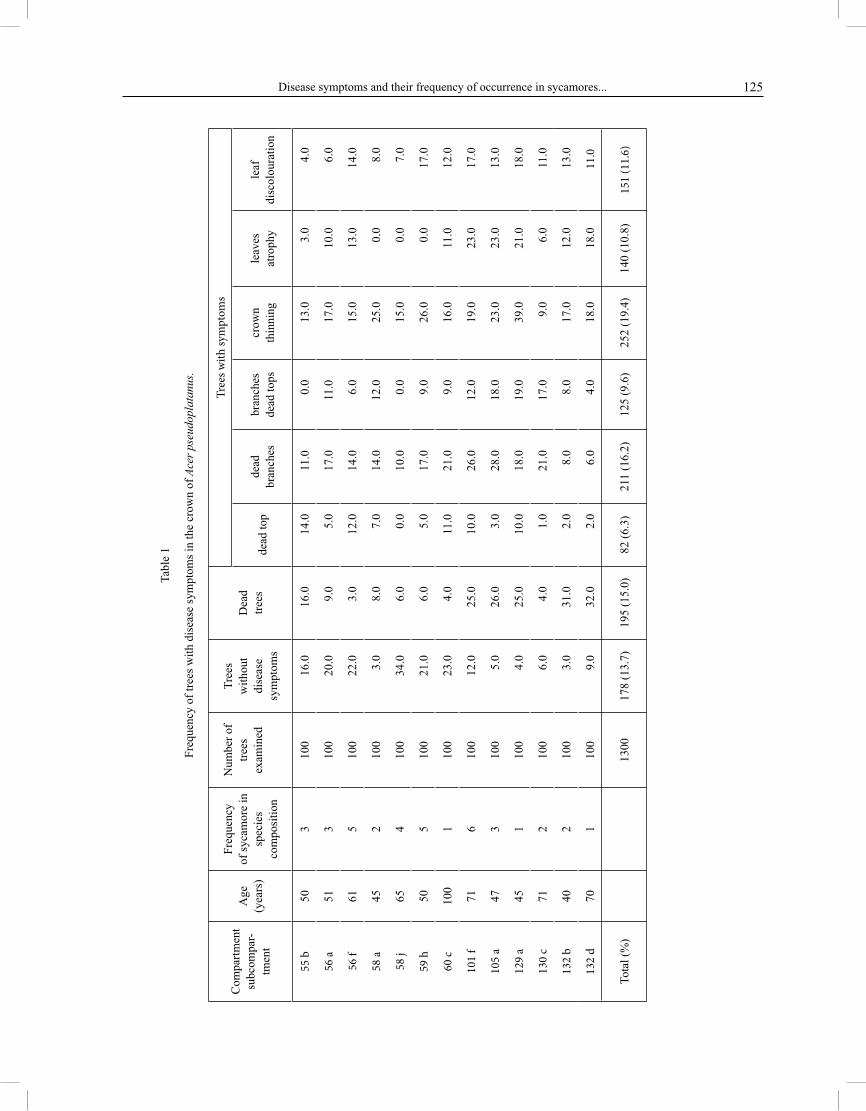
RESULTSThe examined sycamores in the Rymanów Forest
Unit showed a large variation in the disease symptoms and their occurrence frequency. Among 1300 analysed trees, only 13.7% did not show external, macroscopic disease symptoms (Tab. 1). There was a relatively large share of dead trees (15.0%), which in individual stands ranged 4.0 – 32.0 % (Tab. 1). The diameter at height breast in dead trees most often ranged from 18 to 29 cm (Tab. 3). Top dying, found in 6.3% of trees, was a symp-tom occurring in tree crowns which evidenced the ad-vanced disease process. In some stands, this symptom was proportionate to the number of dead trees, in oth-er stands these relations were reverse (Tab. 1). Other symptoms in tree crowns included: entire branch dy-ing or top dying, crown thinning, leaf atrophy and leaf discoloration (Tab. 1). Dead branches were present in 16.2% of trees, but in most of them less than 10% of branches in the crown had died (Fig. 1). Among trees with thinning crowns, the largest number of them had the loss of leaves ranging between 10% and 30% (Fig. 2). Trees with the diameter at breast height of 33 – 50 cm showed crown thinning more than twice more often than trees with the diameter at breast height below 17 cm (Tab. 3). Leaves on thicker trees also had yellow-brown discoloration (Tab. 3). In 20.6% of the examined sycamores, symptoms of tar-spot disease were found, caused by Rhytisma acerinum (Pers.) Fr.
The disease symptoms occurring on syca-more trunks were also characterised by a large vari-ation. The further proper growth of trees was most threatened by plate-like or strip-like necrosis of bark which then cracked and fell off, exposing wood. Such a symptom was observed in 8.6% of trees with the di-ameter at breast height ranging between 18 and 40 cm (Tab. 2, 3). Bark necrosis and wood exposure formed 1.5 times more frequently on the northern and west-ern side than on the southern and eastern side (Fig. 3). In 14.7% of trees, local bark cankers occurred on trunks (Tab. 2), among which healed cankers were predominant (10.3%). On trunks of 3.8% of trees, symptoms of tree cancer occurred, with different degrees of development. On trunks of 14.3% of the examined sycamores, lon-gitudinal bark cracks occurred, sometimes exceed-ing even 30 cm in length. In 8.1% of trees, bark was cracked along the section from the rootstock up to 2 metres high, and only in 0.6% at a height of more than 4 meters. Bark cracks which did not cover more than ¼ of trunk girth were predominant (7.2%). They were present most frequently on the southern side of trunks (Fig. 3). Frost ribs occurred only sporadically, they were found on the trunks of 5 trees (Tab. 2). Epi-cormic shoots also formed relatively rarely on trunks. They occurred more numerously only on 47-year-old sycamores in comp. 105a (Tab. 2).
Disease symptoms and their frequency of occurrence in sycamores... 125
Tabl
e 1
Freq
uenc
y of
tree
s with
dis
ease
sym
ptom
s in
the
crow
n of
Ace
r pse
udop
lata
nus.
Com
partm
ent
subc
ompa
r-tm
ent
Age
(yea
rs)
Freq
uenc
yof
syca
mor
e in
spec
ies
com
posi
tion
Num
ber o
ftre
esex
amin
ed
Tree
sw
ithou
tdi
seas
esy
mpt
oms
Dea
dtre
es
Tree
s with
sym
ptom
s
dead
top
dead
bran
ches
bran
ches
dead
tops
crow
nth
inni
ngle
aves
atro
phy
leaf
disc
olou
ratio
n
55 b
503
100
16.
0
16.
0
1
4.0
1
1.0
0
.0
1
3.0
3.
0
4.0
56 a
513
100
20.
0
9.
0
5.0
1
7.0
11
.0
1
7.0
10.
0
6.0
56 f
615
100
22.
0
3.
0
1
2.0
1
4.0
6
.0
1
5.0
13.
0
1
4.0
58 a
452
100
3.
0
8.
0
7.0
1
4.0
12
.0
2
5.0
0.
0
8.0
58 j
654
100
34.
0
6.
0
0.0
1
0.0
0
.0
1
5.0
0.
0
7.0
59 h
505
100
21.
0
6.
0
5.0
1
7.0
9
.0
2
6.0
0.
0
1
7.0
60 c
100
110
0
2
3.0
4.0
11.
0
21.
0
9.0
16.
0
1
1.0
12.
0
101
f71
610
0
1
2.0
2
5.0
10.
0
26.
0
12.0
19.
0
2
3.0
17.
0
105
a47
310
0
5.0
2
6.0
3.
0
28.
0
18.0
23.
0
2
3.0
13.
0
129
a45
110
0
4.0
2
5.0
10.
0
18.
0
19.0
39.
0
2
1.0
18.
0
130
c71
210
0
6.0
4.0
1.
0
21.
0
17.0
9.
0
6.0
11.
0
132
b40
210
0
3.0
3
1.0
2.
0
8.
0
8.0
17.
0
1
2.0
13.
0
132
d70
110
0
9.0
3
2.0
2.
0
6.
0
4.0
18.
0
1
8.0
11.
0
Tota
l (%
)13
0017
8 (1
3.7)
195
(15.
0)82
(6.3
)21
1 (1
6.2)
125
(9.6
)25
2 (1
9.4)
140
(10.
8)15
1 (1
1.6)

Tadeusz Kowalski, Paulina Materniak126
Fig. 1. Branch dying intensity in crowns of A. pseudoplatanus, frequency of dead branches: a to 10%, b 11 30%, c 31 50%,d over 50%.
Fig. 2. Frequency of trees with crown thinning, with leaves loss: a to 10%, b 11 30%, c 31 50%, d over 50%.
8,5
5,3
1,11,3
0
1
2
3
4
5
6
7
8
9
10
a b c d
%
7,4
6,4
4,1
1,5
0
1
2
3
4
5
6
7
8
9
10
a b c d
%


Tadeusz Kowalski, Paulina Materniak128
Table 3Disease symptoms frequency appearance on Acer pseudoplatanus according to its diameter.
* DBH diameter at breast hight
Table 4Fungi isolated from living A. pseudoplatanus trunks with symptoms of local cankers and bark falling off exposing wood.
Disease symptoms typeFrequency of trees in diameter sections (DBH*, cm)
3 8 9 17 18 29 30 40 41 55 Total (%)
dead trees 22 (13.1) 63 (21.4) 76 (15.6) 28 (11.0) 6 (6.2) 195 (15.0)
leaf atrophy 0 (0.0) 18 (6.1) 74 (15.2) 41 (16.1) 7 (7.2) 140 (10.8)
leaf discolouration in crown 4 (2.4) 27 (9.2) 53 (10.9) 39 (15.4) 28 (28.9) 151 (11.6)
crown thinning 22 (13.1) 37 (12.5) 85 (17.5) 78 (30.7) 30 (30.9) 252 (19.4)
dead branches in a crown 10 (6.0) 21 (7.1) 85 (17.5) 56 (22.0) 39 (40.2) 211 (16.2)
bark cracks on a trunk 3 (1.8) 23 (7.8) 94 (19.3) 57 (22.4) 9 (9.3) 186 (14.3)necrosis and bark falling offon a trunk 2 (1.2) 11 (3.7) 66 (13.6) 28 (11.0) 5 (5.2) 112 (8.6)
Number (%) of trees indiameter sections 168 (12.9) 295 (22.7) 486 (37.4) 254 (19.5) 97 (7.5) 1300 (100.0)
Fungi
Number of inhabited wood
Total (%)fragments in trunk part
periphery inner
Alternaria alternata (Fr.) Keissler 2 2 (0.3)Aposphaeria cf. pulviscula (Sacc.) Sacc. 11 11 (1.4)Basidiomycetes sp.1 148 148 (18.5)Basidiomycetes sp. 2 6 6 (0.8)Cadophora fastigiata Lagerb. & Melin 21 21 (2.6)Chalara sp. 1 29 28 57 (7.1)Chalara sp.2 4 4 (0.5)Cytospora ambiens Sacc. 22 22 (2.8)Diplodina acerina (Pass.) Sutton 1 1 (0.1)Eutypa acharii Tul. 9 9 (1.1)Fusarium solani (Mart.) Sacc. 12 12 (1.5)Mollisia sp. 47 47 (5.9)Nectria cinnabarina (Tode: Fr.) Fr. 28 28 (3.5)Nectria coccinea (Pers.: Fr.) Fr. 64 271 335 (42.0)Stegonosporium pyriforme (Hoffm.: Fr.) Corda 6 6 (0.8)Trichoderma harzianum Rifai 2 11 13 (1.6)Non sporulating fungi (3 species) 6 6 (0.8)Number of „sterile” fragments 32 6 38 (4.8)Number of examined wood fragments 186 612 798

Disease symptoms and their frequency of occurrence in sycamores... 129
Fig. 4. Changes in sycamore wood in local healed canker place.
Fig. 5. Free space forming between bark and wood in place of the canker on sycamore trunk.
Fig. 6. Sector of dead wood as a result of stripe like bark canker on sycamore trunk.
Fig. 7. Numerous local discolorations on sycamore trunk cross section.
Fig. 8. Perithecia of N. coccinea on sycamore trunk in places of canker and bark falling off.
Fig. 9. Conidia of N. coccinea produced in culture on malt agar medium.

Tadeusz Kowalski, Paulina Materniak130
On the cross sections of sycamore trunks, the following internal disease symptoms occurred pre-dominantly: T-shaped discolorations occurring in the place of local healed cankers (Fig. 4.), free spaces formed between bark and wood in the place of un-healed bark cankers (Fig. 5), dead wood regions in the place of local unhealed cankers and more wide-spread bark cankers, sometimes taking the form of a wedge-like sector reaching the part near the pith, in the area of which wood showed grey-brown dis-coloration or rot (Fig. 6), as well as grey-green or grey-brown wood discolorations in the form of spots arranged in a characteristic pattern, in particular in the part near the pith (Fig. 7).
On trunks of 184 (14.2%) sycamores, perithe-cia of Nectria coccinea were present. They formed in the area of cankers on bark and exposed wood alike (Fig. 8). Fruiting bodies of Nectria cinnabarina, Eutypa acharii, Melanomma pulvis-pyrius and conidiomata of Cytospora ambiens, Aposphaeria cf. pulviscula and condiomata of Stegonsporium pyriforme occurred sporadically. As a result of isolations onto malt – agar medium, 19 fungi species were obtained from 798 wood fragments. The following were isolated most frequently: Nectria coccinea, Basidiomycetes sp.1, Chalara sp.1, Mollisia sp., Cadophora fastigiata, Nectria cinnabarina and Cytospora ambiens (Tab. 4). N. coccinea developed in vitro the anamorphic stage, known under the name of Cylindrocarpon candidum (Link) Wollenw. (Fig. 9). Species from the genus Chalara differed primarily in their size and the shape of phialoconidia (Figs. 10, 11). Chalara sp.1, with its morphological features, best matched the anamorph of Ceratocystis coerulescens Muench s.l.
DISCUSSIONThe analysis conducted in the stands of the Ry-
manów Forest Unit shows the advanced progression of the disease process in sycamores, leading relatively often to their dying. This process was observed in all the stands, irrespective of the age or the frequency of sycamore in the species composition; differences only related to its intensity. The disease symptoms found on trunks and in crowns of sycamores were characterised by a large variation, what makes them similar to the condition observed in other deciduous trees, in particu-lar beech, oak and ash (K o w a l s k i , 1991; K o w a l s k i and Ł u k o m s k a , 2005; R o j e k , 2005). Some disease symptoms in sycamore crowns were a result of the di-rect infection by fungi of organs in this part of the tree. A typical example may be the symptoms of tar-spot disease of leaves caused by Rhytisma acerinum. The symptoms of leaf atrophy and discoloration, as well as Fig. 11. Phialoconidia of Chalara sp. 2.
Fig. 10. Phialoconidia of Chalara sp. 1.

Disease symptoms and their frequency of occurrence in sycamores... 131
the dieback of branches not affected by pathogenic fun-gi, observed in crowns, could be secondary symptoms resulting from disease-related changes on sycamore trunks, which included bark cankers of different type, shape and size. Local cankers which were healed, visible on the trunk cross section with the shape of the letter T, can be considered to be the least dangerous for the fur-ther growth of the tree. Plate-like or strip-like cankers, combined with bark cracking and falling off, should be included among the most dangerous ones. A high formation frequency of of such bark cankers type was observed in certain periods of time also in other countries. Their reasons are attributable both to abiot-ic and biotic factors. It has been proved in the United Kingdom that the mass occurrence of bark cankers in sycamores was related to the previous occurrence of a drought period (B e v e r c o m b e and R a y n e r , 1978; Mu r r a y , 1978; G r e g o r y , 1982). Bark dieback was always connected with anatomical changes in vessels and cells of the pith rays in the area of late wood of the youngest annual growth ring (Mu r r a y , 1978). However, it could not be shown whether the cam-bium in the regions of cankers was dying as a result of drought or infection by fungi of trees being under the stress conditions. In the area of such cankers, the pres-ence of Nectria coccinea, Verticillium tenereum (Nees ex Pers.) Link, Diplodina acerina, Phomopsis pustulata and Dichomera saubinetii was found most frequently (B e v e r c o m b e and R a y n e r , 1978; Mu r r a y , 1978; G r e g o r y , 1982). Nectria coccinea was also found nu-merously in the area of cankers on sycamore trunks in Upper Austria, where not climatic factors were recog-nised as the factor making sycamores more susceptible, but changes in the substrate associated with mining of lignite. In these sycamores, an additional symptom was several-metre-long bark cracks in the above-ground por-tion of trunks (C e c h , 1994). Nectria coccinea was also the species which was present most often on dead bark and in wood of trunks of the examined sycamores in the Rymanów Forest Unit. This species was similarly often found on beeches inhibited by the beech scale Cryp-tococcus fagisuga Lind. (K o w a l s k i and K l e b a n , 2000). The ability to cause bark cankers by N. coccinea in sycamore and beech has been shown experimental-ly in the event that the inoculum was introduced in the place of injury on trunks of living trees, however, if these trees were in a good condition, cankers were of limited size and were healed (P a r k e r , 1975; Gregory, 1982;K o w a l s k i and K l e b a n , 2000). Fusarium solani, cur-rently isolated from wood in the area of T-shaped healed cankers, is a fungus species found in association with such symptoms also in other species of deciduous trees (Wo o d and S k e l l y , 1964; P r z y b y ł , 1985; K o w a l -s k i , 1991). Wo o d and S k e l l y (1964) have shown that it is able to cause, by itself, cankers of sugar maple only
in the case of inoculation in the period of vegetation dor-mancy. P r z y b y ł (1985) has confirmed the pathogenic-ity of F. solani, inoculating four-month injured shoots of poplar. The most probable factor affecting the de-velopment of injuries on sycamore trunks, allowing the abovementioned infections and other fungus spe-cies to occur (Tab. 4), could be weather anomalies pri-marily related to the occurrence of low temperatures. They are particularly dangerous when they occur at the end stage of winter, and their characteristic feature is that temperature drops follow over a dozen day-long warmings, what has been shown experimentally with re-spect to common oak (K o w a l s k i , 1981; Th o m a s and H a r t m a n n , 1992).
Bark cankers and dieback of sycamores in the Ry-manów Forest Unit were accompanied by characteristic internal disease symptoms, visible on trunk cross sec-tions. The results of isolations do not show that only one fungus species is connected with a particular type of discoloration. A lot of evidence shows that the ob-served discolorations are an effect of an accumulation of phenolic substances in the so-called reaction regions as a result of infection of sapwood by various fungi and bacteria. In maples, they often have greenish ororange discoloration (Z i m m e r m a n n , 1974; Pe a r c e , 1996). Such numerous isolations of Nectria coccinea from discolored sapwood of the examined sycamores show that this species, after causing bark cankers, is able to grow across the deep layers of wood. As its mycelium may grow already at a temperature of several degrees above zero, most probably in nature the wood of sycamore trunks is infested also during the period of vegetation dormancy (Mu r r a y , 1978). In the explana-tion context of the of sycamore diseases causes, special attention should be paid to the establishment of the of internal symptoms presence (Fig. 7), very similar to the a disease symptoms of maples, mainly sugar maple, in North America termed “sapstreak disease of maple” (H e p t i n g , 1971), and the isolation, from sycamores with such symptoms, of the fungus Chalara sp.1 with its features similar to the anamorph of Ceratocystis coerulescens s.l. However, some taxonomic problems appear in this respect. In North America, Ceratocystis virescens (Davidson) Moreau is currently reported as the perpetrator of “sapstreak disease”. In Europe, a morphologically similar species Ceratocystis coer-ulescens was treated as the perpetrator of wood blue stain of coniferous trees (B u t i n , 1996). Hu n t (1956) recognised these species as synonyms and they were treated in this way for several dozen years in most my-cological monographs and studies (Z a j o n c and Wu l f , 1997). In many studies, however, differences are indi-cated in the vegetation stages of these species belonging to the genus Chalara (N a g R a j and K e n d r i c k , 1975; K i l e and Wa l k e r , 1987). Ultimately, based on molec-

Tadeusz Kowalski, Paulina Materniak132
ular studies, it was proved that Ceratocy stis virescens is a separate species, and within Ceratocystis coerules-cens five morphological types were first distinguished(Wi t t h u h n et al. 1998), and then they were described as new species (H a r r i n g t o n and Wi n g f i e l d , 1998). The explanation of affiliation to species of the cultures isolated from diseased sycamores and currently identi-fied as fungi from the genus Chalara will require further methodologically oriented taxonomic studies.
REFERENCESBevercombe G. P., Rayner A.D.M., 1978. Dichomera saubi-
netii and bark diamond canker formation in sycamore. Trans. Brit. Mycol. Soc. 71: 505 507.
Boratyński A., 1999. Systematyka i geograficzne rozmieszczenie. W: Klony / Systematics and geographic distribution. In: Maples (ed. Bugała W.) Polska Akademia Nauk, Bogucki Wyd. Naukowe, Poznań, s. 15 73.
Butin H., 1996. Krankheiten der Wald und Parkbaeume. Georg Thieme Verlag, Stuttgart.
Cech T. L., 1995. Absterben von Bergahorn (Acer pseudoplata-nus L.) in Oberoesterreich. Forstschutz Aktuell, 16: 2 3.
Cech T.L., 2004. Bemerkenswerte Krankheiten in 2004. Forstschutz Aktuell, 32: 31 34.
Gregory S. C., 1982. Bark necrosis of Acer pseudoplatanus L. in northern Britain. Eur. J. For. Path. 12: 157 167.
Grzywacz A., 1999. Ważniejsze choroby infekcyjne. W: Klony / More important infection diseases. In: Maples (ed. Bugała W.) Polska Akademia Nauk, Bogucki Wyd. Naukowe, Poznań, s. 427 469.
Harrington C., Wingfield M. J., 1998. The Ceratocystis species on conifers. Can. J. Bot. 76, 8: 1446 1457.
Hepting G. H., 1971. Diseases of forest and shade trees of the United States. USDA Forest Service, Handbook No. 386.
Hunt J., 1956. Taxonomy of the genus Ceratocystis. Lloydia, 19: 1 58.
Jansen E., Forster B., Engesser R., Odermatt O., Meier F., 1992. Die Forstschutzsituation in der Schweiz 1991. Allg. Forstzeitung, 7: 359.
Jaworski A., 1994. Charakterystyka hodowlana drzew leśnych / Growing characteristics of forest trees. Gutenberg, Kraków.
Kile G.A., Walker J., 1987. Chalara australis, a vascular pathogen of Nothofagus cunninghamii in Australia and its relationship to other Chalara species. Australian Journ. Bot. 35: 1 32.
Kowalski T., 1981. Grzyby powodujące zamieranie drzew Acer platanoides L. ’Globosum’ w zadrzewieniach przyulicznych w Tarnowie / Fungi causing the dieback of trees Acer platanoides L. ’Globosum’ in street tree plantings in Tarnów. Zesz Nauk. Akad. Roln. w Krakowie Nr 164, ser. Leśnictwo, z. 13: 83 92.
Kowalski T., 1991. Oak decline: I. Fungi associated with various disease symptoms on overground portions of mid
dleaged and old oak (Quercus robur L.). Eur. J. For. Path. 21: 136 151.
Kowalski T., Kleban J., 2000. Badania nad występowaniem i przyczynami lokalnych nekroz na pniach Fagus sylva-tica L. w wybranych drzewostanach Nadleśnictwa Dukla / Studies on the occurrence and causes of local cankers on trunks of Fagus sylvatica L. in selected stands of the Dukla Forest Unit. Zesz. Naukowe AR w Krakowie nr 362, ser. Leśnictwo, 28: 27 38.
Kowalski T., Łukomska A., 2005. Badania nad zamieraniem jesionu (Fraxinus excelsior L.) w drzewostanach Nadleśnictwa Włoszczowa / Studies on the dieback of ash (Fraxinus excelsior L.) in stands of the Włoszczowa Forest Unit. Acta Agrobot. 59, 2: 429 440.
Murray J. S., 1978. Death of bark in Acer pseudoplatanus associated with drought. Eur. J. For. Path. 8: 65 75.
Nag Raj T. R., Kendrick B., 1975. A Monograph of Chalara and Allied Genera. Wilfrid Laurier Univ. Press, Waterloo, Ontario, Canada.
Parker E. J., 1975. Some investigation with beech bark disease Nectria in southern England. Eur. J. For. Path. 5: 118 124.
Peace T. R., 1962. Pathology of trees and shrubs. Clarendon Press, Oxford.
Pearce R.B., 1996. Antimicrobial defences in the wood of living trees. New Phytol. 132: 203 233.
Przybył K., 1985. Mikroflora grzybowa z pni topoli z objawami brunatnej plamistości parchatej i raka typu tarczy / Fungal microflora in poplar trunks with symptoms of brown spot and target canker diseases. Arboretum Kórnickie, 30: 269 283.
Rawling K. L., 1972. Death of sycamore trees at Otley, Yorks. Quarterly J. Forestry, 66: 201 207.
Rojek P., 2005. Występowanie symptomów chorobowych i wybranych czynników biotycznych związanych ze zjawiskiem zamierania buka ( Fagus sylvatica L.) w drzewostanach Magurskiego Parku Narodowego / The occurrence of disease symptoms and selected biotic factors associated with the dieback of beech (Fagus sylvatica L.) in the Magurski National Park. Acta Agrobot. 59, 2: 441 452.
Smith L. D., 1979. Verticillium wilt of landscape trees. Journ. Arboriculture, 5(9): 193 197.
Thomas F. M., Hartmann G., 1992. Frosthaerte des Bastes aelterer Traubeneichen auf besonnten und absonnigen Stammseiten. Forst u. Holz, 47: 462 464.
Witthuhn R. C., Wingfield B.D., Wingfield M.J., Wolfaardt M., 1998. Monophyly of the conifer species in the Ceratocystis coerulescens complex based on DNA sequence data. Mycologia, 90(1): 96 101.
Wood F. A., Skelly J. M., 1964. The etiology of an annual canker on maple. Phytopathology, 54: 269 272.
Zajonc J., Wulf A., 1997. Gefaerdet die durch Ceratocystis virescens verursachte Splintstreifenkrankheit die europaeischen Ahornarten. Nachrichtenbl. Deut. Pflanzenschutzd., 49(12): 297 300.
Zimmermann G., 1974. Untersuchungen ueber Art und Ursachen von Verfaerbungen an Bergahorn Stammholz (Acer pseudoplatanus L.). Forstw. Cbl. 93: 247 261.

Disease symptoms and their frequency of occurrence in sycamores... 133
Symptomy chorobowe i nasilenieich występowania u jaworów
(Acer pseudoplatanus L.) w drzewostanachNadleśnictwa Rymanów
S t r e s z c z e n i eBadania terenowe prowadzono w latach 2003
– 2005 w Nadl. Rymanów w 13 drzewostanach w wieku 40 do 100 lat, w których udział jaworu w składzie gatun-kowym wynosił od 10 do 60%. Rosły one na siedlisku lasu górskiego (12 drzewostanów) i na lesie łęgowym górskim (1 drzewostan). W każdym z nich poddano ana-lizie 100 rosnących obok siebie drzew w środkowej czę-ści drzewostanu. Określono objawy chorobowe na pniu i w obrębie korony każdego drzewa oraz ich nasilenie według uprzednio przygotowanego kodu symptomolo-giczno-rozwojowego.
Pobrano ponad 80 fragmentów kory i drewna z pni martwych drzew oraz drzew żywych wykazują-cych objawy lokalnych nekroz oraz złuszczania kory odsłaniającego drewno, z których wykonano 798 izola-cji na 2% pożywkę agarowo-maltozową.
Badane jawory w Nadleśnictwie Rymanów wy-kazywały duże zróżnicowanie symptomów chorobo-wych i nasilenia ich występowania. Spośród poddanych analizie 1300 drzew tylko 13,7% nie wykazywało ze-wnętrznych makroskopowych objawów chorobowych. Stosunkowo duży był udział drzew obumarłych (15,0%), który wynosił w poszczególnych drzewostanach od 4,0 do 32,0%. Do najczęstszych symptomów w koronach drzew należały: zamieranie wierzchołka (6,3% drzew), zamieranie całych gałęzi (16,2%) lub ich szczytów (9,6%), przerzedzenie korony (19,4%), atrofia liści (10,8%) i przebarwienie liści (11,6%).
Na pniach jaworów stwierdzano: placowatą lub pasmową martwicę kory, która pękała, wykruszała się i odsłaniała drewno (8,6% drzew), lokalne nekro-zy kory (14,7%), wśród których dominowały nekrozy zabliźnione (10,3%), podłużne spękania kory (14,3%) oraz objawy raka drzewnego (3,8%). Do martwicy kory i odsłaniania drewna ok. 1,5 razy częściej docho-dziło od strony północnej i zachodniej niż południowej i wschodniej, spękania kory występowały najczęściej od strony południowej pni.
Na przekrojach poprzecznych pni jaworów naj-częściej występowały: przebarwienia w kształcie litery „T”, występujące w miejscu lokalnych zabliźnionych nekroz, obumarłe strefy drewna w miejscu lokalnych niezabliźnionych nekroz i rozległych martwic kory, przyjmujące niekiedy formę sektora sięgającego czę-ści przyrdzeniowej, i szarozielone lub zielonobrunatne przebarwienia drewna w postaci licznych plam, zwłasz-cza w części przyobwodowej pnia.
Na pniach 184 (14,2%) jaworów obecne były peritecja Nectria coccinea. Wykształcały się one w ob-rębie nekroz, zarówno na korze jak i na odsłoniętym drewnie. Sporadycznie występowały owocniki Nectria cinnabarina, Eutypa acharii, Melanoma pulvis-pyrius oraz conidiomata Cytospora ambiens, Aposphaeria cf. pulviscula i Stegonsporium pyriforme. Z drewna naj-liczniej izolowano: Nectria coccinea, Basidiomycetes sp.1, Chalara sp.1, Mollisia sp., Cadophora fastigiata, Nectria cinnabarina i Cytospora ambiens. Chalara sp.1 swymi cechami morfologicznymi najbardziej odpowia-dała anamorfie Ceratocystis coerulescens sensu lato.
