direct retino-raphe projection alters serotonergic tone ... · open direct retino-raphe projection...
TRANSCRIPT
OPEN
Direct Retino-Raphe Projection Alters Serotonergic Toneand Affective Behavior
Chaoran Ren1,2,3,10, Liju Luan1,2,3,10, Benson Wui-Man Lau4,5,6, Xin Huang1,2,3, Jian Yang4,5,6, Yuan Zhou1,2,3,Xihong Wu3,7, Jie Gao1,2,3, Gary E Pickard8,9, Kwok-Fai So*,4,5,6 and Mingliang Pu*,1,2,3
1Department of Anatomy, School of Basic Medical Sciences, Peking University, Beijing, China; 2Key Laboratory on Machine Perception (Ministry of
Education), Peking University, Beijing, China; 3Key Laboratory for Visual Impairment and Restoration (Ministry of Education), Peking University,
Beijing, China; 4Department of Anatomy and Research Center of Heart, Brain, Hormone and Healthy Aging, LKS Faculty of Medicine, Pokfulam,
Hong Kong, China; 5The State Key Laboratory of Brain and Cognitive Sciences, The University of Hong Kong, Pokfulam, Hong Kong, China; 6GHM
Institute of CNS Regeneration, Jinan University, Guangzhou, China; 7Department of Machine Intelligence, Peking University, Beijing, China; 8School
of Veterinary Medicine and Biomedical Sciences, University of Nebraska-Lincoln Lincoln, NE, USA; 9Department of Ophthalmology and Visual
Sciences, University of Nebraska Medical Center, Omaha, NE, USA
Light is a powerful modulator of higher-order cognitive processes such as mood but it remains unclear which neural circuits mediate the
impact of light on affective behavior. We found that light deprivation produces a depressive-like behavioral state that is reversed by
activation of direct retinal signals to the serotonergic dorsal raphe nucleus (DRN) in a manner equivalent to treatment with the selective
serotonin reuptake inhibitor fluoxetine. Surprisingly, the DRN-projecting retinal ganglion cells (RGCs) are indistinguishable from the
classic alpha/Y-like RGC type that contributes to image-forming visual pathways. Silencing RGC firing or specific immunotoxin ablation of
DRN-projecting RGCs increased depressive-like behavior and reduced serotonin levels in the DRN. Serotonin has a key role in the
pathophysiology of depression, and these results demonstrate that retino-raphe signals modulate DRN serotonergic tone and affective
behavior.
Neuropsychopharmacology advance online publication, 27 February 2013; doi:10.1038/npp.2013.35
Keywords: dorsal raphe nucleus; retinal ganglion cell; depression; affective visual information; SSRI
��������������������������������������������������
INTRODUCTION
Photon capture by retinal photoreceptors is the initial stepin visual perception (Yau and Hardie, 2009). Additionally,light stimulation of the retina is a powerful modulator ofnon-visual functions such as cognition, conditioned fearresponses, circadian rhythms, and affective behavior(Vandewalle et al, 2009; Wirz-Justice et al, 2009; Warthenet al, 2011). Light therapy can be an effective treatment forboth seasonal and non-seasonal mood disorders (Lewy et al,1987; Golden et al, 2005) while depressive-like behavior canbe induced in mammals across species by light deprivation(Wilson, 2002; Gonzalez and Aston-Jones, 2008; Monje et al,
2011; Lau et al, 2011). Our past study showed thatalpha/Y-like retinal ganglion cells (RGCs) innervate themidbrain dorsal raphe nucleus (DRN; Luan et al, 2011) andthe DRN has a crucial role in regulating affective behavior.Recent evidence shows that a group of intrinsicallyphotosensitive RGCs (ipRGCs) is responsible for processingnonimage-forming visual functions (Guler et al, 2008).However, it remains to be defined whether ipRGCs orconventional RGCs contribute to affective behavior. Herewe show that alpha/Y-like RGCs with no apparent intrinsicphotosensitivity send signals to serotonergic neurons of themidbrain DRN mediating the effects of light both ondepressive-like behavior and DRN serotonin (5-HT) levels.Moreover, we demonstrate that the effect of RGC input tothe DRN is similar to that of selective serotonin reuptakeinhibitor (SSRI) treatment on depressive-like behavior.
MATERIALS AND METHODS
All experiments were performed in accordance withPeking University guidelines for animal research and theAssociation for Research in Vision and Ophthalmology(ARVO) Statement for the Use of Animals in Ophthalmicand Vision Research. The experimental animal protocol was
*Correspondence: Professor K-F So, LKS Faculty of Medicine, 21Sassoon Road, Pokfulam, Hong Kong, China, Tel: +852 2819 9216,Fax: +852 2817 6821, E-mail: [email protected] M Pu, Anatomy/Embryology, School of Basic MedicalSciences; Key Laboratory on Machine Perception; Key Laboratory forVisual Impairment and Restore, Peking University, 38 Xueyuan Road,Beijing 100191, China, Tel: +8610 8280 2972, Fax +8610 8280 2877,E-mail: [email protected] authors contributed equally to this work.Received 8 August 2012; revised 24 December 2012; accepted28 January 2013; accepted article preview online 31 January 2013
Neuropsychopharmacology (2013), 1–13
& 2013 American College of Neuropsychopharmacology. All rights reserved 0893-133X/13
www.neuropsychopharmacology.org
approved by Peking University Institutional Animal Careand Use Committee (IACUC).
Animals
Male adult Mongolian gerbils (2–3-months old, 65–77 g)were used in these experiments. Upon arrival all animalswere maintained in 12L : 12D conditions (LD) with food andwater provided ad libitum. For light-deprivationexperiments, all conditions were the same except animalswere kept in complete darkness (DD) for 14 days.
Stereotaxic Surgeries
Cholera toxin B subunit (CTB)-conjugated Alexa Fluor 488or 594 (C34777 or C22842, Invitrogen Inc., Grand Island,NY) was deposited into the DRN or the superior colliculus(SC) as described previously (Luan et al, 2011). Briefly,animals were anesthetized with 60 mg/kg sodium pento-barbital by intraperitoneal injection (IP) and placed in astereotaxic device (Stoelting, Wood Dale, IL). After acraniotomy was performed, a Hamilton syringe was insertedinto the DRN or SC and 1.5 ml of CTB-conjugated AlexaFluor was injected, the wound was sutured, and 3 days wereallowed for retrograde transport of CTB to the retina.
Immunocytochemistry and Image Processing
All animals were anesthetized (60 mg/kg sodium pentobar-bital, IP) and perfused intracardially with 0.9% salinefollowed by 4% paraformaldehyde in phosphate-bufferedsaline (PBS). Brains and eyes were removed.
Detection of melanopsin-expressing RGCs was performedusing immunocytochemical procedures on retinal wholemounts. Retinas were placed in 0.1 M PBS containing 10%normal goat serum (Vector Laboratories, Burlingame, CA)with 0.3% Trition-X-100 (T8787, Sigma-Aldrich, St Louis,MO) for 1 h before incubation for 3 days at 4 1C in a rabbitanti-melanopsin antibody diluted 1 : 600; antibody wasgenerated against an N-terminal amino-acid sequenceof melanopsin protein (PA1-780, Fisher Scientific Inc.,Barrington, IL). This was followed by incubation with asecondary antibody (Dylight488 or Dylight549) goat-anti-rabbit IgG (1 : 200, Vector Laboratories).
Immunocytochemical staining of serotonergic neuronswas performed on free-floating brain sections. Briefly,40-mm thick cryostat sections collected through the DRNwere placed in 0.1 M PBS containing 10% normal goatserum with 0.3% Trition-X-100 for 1 h before incubation inrabbit anti-serotonin antibody (1 : 500, Sigma-Aldrich) for72 h at 4 1C. Brain sections were then placed in secondaryantibody Dylight488 goat-anti-rabbit IgG (1 : 200, VectorLaboratories) for 6 h at room temperature.
The method for converting intracellularly injectedLucifer Yellow (Sigma-Aldrich) into a permanent dye hasbeen described previously (Pu, 1999). Briefly, after thecompletion of electrophysiological recording, retinascontaining DRN-projecting RGCs intracellularly injectedwith Lucifer Yellow were incubated in 4% paraformalde-hyde in 0.1 M PBS for 2 h. Retinas were rinsed in 0.1 M PBSfor 10 min three times and placed in 10% normal goatserum containing 2% Trition-X-100 and 0.5% dimethyl
sulfoxide (Sigma-Aldrich) for 48 h at 4 1C. Retinas were thenincubated in rabbit-anti-Lucifer Yellow antibody (1 : 1000,Sigma-Aldrich) for 48 h at 4 1C followed by secondaryantibody Dylight 549 goat-anti-rabbit IgG (1 : 200, VectorLaboratories) for 6 h at room temperature. All sectionsand retinas were rinsed in 0.1 M PBS and cover-slipped inanti-fading aqueous mounting medium (EMS, Hatfield, PA).
Confocal Microscopy and Three-DimensionalReconstruction
RGCs injected with Lucifer Yellow and immunocytochemi-cally stained were scanned with a confocal microscope (TCSSP5 II, Leica Microsystems, Wetzlar, Germany). The Z axisinterval was 0.2 mm. Each stack of optical sections covered aretinal area of 325.75� 325.75 mm2 (1024� 1024 pixels).Using Image J and Photoshop CS5 (Adobe Corp., San Jose,California), each stack of optical sections was arranged as amontage and projected to a 01 X–Y plane and a 901 Y–Zplane to obtain a three-dimensional reconstruction of thecell. Details of three-dimensional reconstruction andconfocal calibration procedures are described elsewhere(Pu, 1999).
Selective Elimination of Retinal Photoreceptors
To selectively eliminate retinal rod and cone photorecep-tors, animals received a single IP injection of N-methyl-N-nitrosourea (MNU; N1517, Sigma-Aldrich). MNU is adirect-acting alkylating agent that targets photoreceptorscausing rapid apoptosis while sparing RGCs (Herrold, 1967;Wenzel et al, 2000; Yoshizawa and Tsubura, 2005; Boudardet al, 2009). MNU was dissolved in saline with the finalconcentration of 10 mg/ml and the injected dosage was80 mg/kg body weight, a dose in the range of MNU used inother species (Yoshizawa and Tsubura, 2005). This dosagewas chosen specifically based on a pilot study in which adose was determined that could effectively eliminatephotoreceptors without affecting the health status ofanimals. MNU-treated animals were returned to their homecages and maintained under LD conditions for at least7 days before they were assigned to an experimental group.
Selective Targeting of ipRGCs, SC-projecting RGCs,DRN-projecting RGCs and Blocking Retinal SpikingActivity
We adopted the immunotoxin method described by Gozet al (2008) to selectively destroy melanopsin-expressingipRGCs. Briefly, under deep anesthesia, animals (n¼ 6/group) received intravitreal injections of anti-melanopsin–saporin conjugate (2 mg/eye, #IT-44, Advanced TargetingSystems, San Diego, CA) using a 5-ml Hamilton syringefitted with a 30-G needle. The syringe was held in place for2 min after immunotoxin delivery and gradually withdrawnover the next 60 s. Animals were returned to their homecage for the next 14 days.
To selectively remove SC-projecting RGCs, animalsthat had received CTB-conjugated Alexa Fluor 488(1.5 ml/animal) deposited into the SC 3 days earlier, receivedintravitreal injection of a custom conjugate made betweensaporin (#IT-27-100, Advanced Targeting Systems) and an
Retinal ganglion cells regulate affective behaviorC Ren et al
2
Neuropsychopharmacology
affinity-purified anti-CTB antibody (GCHL-55A-Z, Immu-nology Consultants Laboratory, Portland, OR). Animals(n¼ 6/group) received bilateral intravitreal injectionsof different quantities (0 mg, 0.5 mg, 2 mg, 4 mg) of theimmunotoxin and were returned to their home cage for14 days.
To selectively remove DRN-projecting RGCs, animalsthat had received CTB-conjugated Alexa Fluor 594(1.5 ml/animal) deposited in the DRN 3 days earlier, receivedintravitreal injections (2 mg/eye n¼ 6/group) of the sameimmunotoxin used for the ablation of SC-projecting RGCsdescribed above. This dose was chosen based on the resultsof a pilot study in which a dose was determined that couldeffectively eliminate the CTB-labeled SC-projecting RGCs.After intravitreal injection, animals were returned to theircages for at least 14 days before being assigned to anexperimental group.
To block the action potentials produced by RGCsin MNU-treated animals, we adopted a tetrodotoxin(TTX) intravitreal injection protocol described by othergroups (Edwards and Cline, 1999; Viswanathan et al, 2000;Witkovsky et al, 2004). TTX is a potent voltage-gatedsodium channel blocker that blocks sodium-dependentaction potentials in RGCs and prevents transmission ofvisual signals from retina. TTX (1069, Tocris Bioscience,Ellisville, MO) was prepared as 3 mM in distilled water;6 mM TTX in a volume of 5ml (5 ml saline serves as vehicle,n¼ 6/group) was injected into the vitreous chamber of eacheye using a 30-G needle attached to a 5-ml Hamilton syringe.After injections, animals were returned to their home cagefor 90 min before they were assigned to behavioral tests.
Chronic Treatment with Anti-depressants
The light-deprived (DD) animals received daily IP injectionsof fluoxetine (10 mg/kg, n¼ 6), imipramine (10 mg/kg,n¼ 6), or vehicle (saline, 400 ml, n¼ 6) for 21 days.The dosage for chronic treatment with anti-depressantswas determined based on previous publications (Detke et al,1996; Bijak and Papp, 1995; Page et al, 1999; Svenningssonet al, 2002; Rodrıguez-Gaztelumendi et al, 2009; Machadoet al, 2012). Fluoxetine hydrochloride (F132, Sigma-Aldrich)and imipramine hydrochloride (I7379, Sigma-Aldrich) weredissolved in saline with a final concentration of 2 mg/ml.Daily injections were delivered at 1700 hours under dim redlight (5–9 lux), except on testing days. To minimize possibleperitoneal irritation, IP injection sites were alternatedbetween left and right lower abdominal quadrants on adaily basis. After injection, animals were returned to theirhome cages and kept in complete darkness.
Physiological Recording and Data Analysis
DRN-projecting RGCs were recorded as previouslydescribed (Luan et al, 2011). Briefly, the eyes wereenucleated under dim red light; lens and vitreous werecarefully removed with a pair of fine forceps. The eyecupwas flat-mounted, sclera side down, directly on the bottomof a recording chamber, and was superfused by oxygenated(95% O2/5% CO2) Ames medium (Sigma-Aldrich) at a fixedrate (5 ml/min) at room temperature between 22–24 1C.Visual stimuli were generated by programming the
Psychophysics Toolbox in Matlab displayed on a Samsungmini LED projector (Samsung SP-P310ME, SamsungElectronics Co Ltd, Suwon City, Korea) and imaged with afirst-surface mirror and lens (Edmond Scientific, Barring-ton, NJ) on the film plane of the microscope’s camera port.The luminance level of the projector was measured with adigital radiometer (S370 Radiometer, UDT Instruments,San Diego, CA) using a � 40 water-immersion objective(Carl Zeiss, Thornwood, NY). The irradiance was furtherreduced using neutral density filters (Oriel Corp., Stratford,CT). Visual responses and spontaneous activity of DRN-projecting RGCs were recorded using a glass microelectrodeand amplified with a patch clamp amplifier (Multiclamp700B) and digitized (Digidata 1430; Axon Instrument, Inc.,Forest City, CA). RGCs retrogradely labeled by CTBwere targeted for recording. To test the blocking effect ofTTX on the action potentials of DRN-projecting RGCs,20 ml oxygenated (95% O2/5% CO2) Ames medium contain-ing 0.1 mM TTX was superfused into the chamber 5 minbefore cell recording. The concentration of TTX wasdetermined based on the work of Margolis and Detwiler(2007). Once the physiological profiling was completed,recorded RGCs were intracellularly filled with LuciferYellow for morphological evaluation. The acquired physio-logical data were further analyzed off-line (pCLAMP9; AxonCorp., CA).
Modified Forced Swimming Test (FST)
Modified forced swimming tests (FST) were conducted aspreviously described (Lau et al, 2011). Briefly, animals ineach experimental group were tested in two trials. In thefirst trial, animals were forced to swim in a glass cylinder(silicon dioxide, diameter: 30 cm, height: 50 cm, waterdepth: 30 cm, water temperature: 28 1C) for 15 min.Then animals were dried and returned to their cages for24 h. The second trial lasted for 5 min. The entire procedurewas recorded with an infrared camera placed above theglass cylinder. Three featured behaviors during the secondsession were measured: swimming (active movement thatcrossed at least one quadrant of the cylinder); climbing(pulling movements of forepaws that scratched at the wall ofcylinder); and immobility (no active movement except thatneeded to keep the animal from drowning). A behavior-sampling technique (Cryan et al, 2005a) was used to scoremovements as defined above, which measure the frequencyof behaviors during 5 s. To observe any time-of-dayinfluence on animal’s affective behavior, animals in theLD, DD, MNU-treated (MNU-LD and MNU-DD) groups(n¼ 6/group at each time point) were tested at fourdifferent Zeitgeber Time (ZT) points (ZT0, ZT6, ZT12,ZT18) where ZT0¼ light onset and ZT12¼ light offsetin LD. Animals in the other groups were tested at ZT6.To minimize the effect of light on the affective behaviorof animals, each trial was conducted under dim red light(25 lux).
Tail Suspension Test (TST)
Depression-like behavior of animals was assayed using thetail suspension test (TST), which is widely used fortesting behavioral despair of rodents, including the gerbil
Retinal ganglion cells regulate affective behaviorC Ren et al
3
Neuropsychopharmacology
(Steru et al, 1985; Varty et al, 2003). Briefly, the animal, heldby the tail, was suspended 50 cm from the floor and thebehavior of the animal was recorded by a video camerafor 6 min. The immobile time during the last 4 min wascounted. Each test was performed at ZT6 under dim redlight (25 lux).
Open Field Test (OFT)
Motor activity was measured in an open field test box(74� 74� 40 cm). A 4� 4 grid was superimposed onto thefloor to score the locomotor activity. This experimentincluded two sessions. In the first, each animal was allowedto explore in the box for 3 min to reduce gerbil’s ‘defensiveconvulsions in response to novel environment’ (Pickleset al, 2012). Then each animal was returned to its homecage. The second session was performed 24 h later. Animalsin each group were placed in the center of the open field andleft to explore for 60 min. All animal activity was recordedwith an infrared camera placed above the box. The distancetraveled by each animal during the second test sessionwas measured. Each test started at ZT6 under dim redlight (25 lux). The box was wiped clean with a paper towelsoaked in 50% ethanol and dried thoroughly between eachtest session.
Sucrose Preference Test (SPT)
Animals were tested for preference of a 2% sucrose solution(Sucrose, Sigma-Aldrich), using a two-bottle choice proce-dure as described by Monteggia et al (2007) with slightmodifications. Each animal was housed individually duringthe 2-day test period. Animals were given two bottles,
one of sucrose (2%) and one of tap water. Every 24 h theamount of sucrose and water consumed was recorded. Toprevent potential location preference of drinking, theposition of the bottles was changed every 24 h. Food andwater were available ad libitum prior to the SPT. Thepreference for the sucrose solution was determined asthe percentage of sucrose solution ingested relative to thetotal intake.
Statistical Analysis
Assignment of subjects to various experimental groups wasrandom. Immobile scores at four different time points(Figure 1a) were analyzed by a two-way ANOVA test(with the two factors being type and time points) followedby Tukey’s post hoc test if necessary. Other behavioral dataand cell/spikes counting data were analyzed through theone-way ANOVA test followed by Tukey’s post hoc test ifnecessary. To determine whether the level of serotonin inDRN and the forced swimming test are related, a possiblecorrelation between the number of 5-HTþ neurons in DRNand the score of three different movements in forcedswimming test was explored (Pearson’s test). Data areexpressed as the mean±SEM. Statistical significance was setas po0.05.
Experimental Groups:
LD: Animals were housed in 12L : 12D (LE 300 lux; D¼ 0lux) for 2 weeks.
DD: Animals were transferred from LD into completedarkness for 2 weeks.
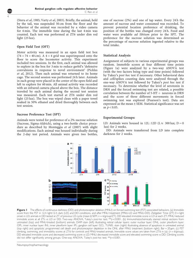
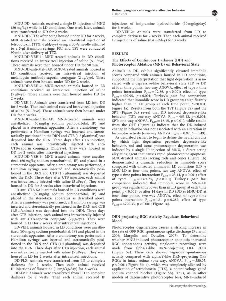
Figure 1 The effects of continuous darkness (DD) and photoreceptor ablation (MNU) on forced swimming test (FST)-associated behaviors. (a) Immobilescore from the FST in 12-h light:12-h dark (LD) and DD conditions, and after MNU treatment (MNU-LD and MNU-DD). Zeitgeber Time (ZT) 0¼ lightonset in LD; animals in DD tested at ZT of previous LD cycle (mean±SEM, n¼ 6/group/ZT). DD elevated immobile score vs LD at each ZT; MNU reducedimmobile score at all ZTs vs LD or DD. Two-way ANOVA, Tukey’s post-hoc test; **po0.001. (b) Immunohistochemically stained retinal sections fromuntreated (top) and MNU-treated (bottom) animals. DAPI stain (left) illustrating retinal cellular layers: outer nuclear layer: ONL; outer plexiform layer:OPL; inner nuclear layer: INL; inner plexiform layer: IPL; ganglion cell layer: GCL. TUNEL stain (right) illustrating absence of apoptosis in untreated animal(top right) and apoptotic programmed cell death and photoreceptor depletion in the ONL after MNU treatment (bottom right). Bar¼ 20 mm. (c) FSTclimbing, swimming, and immobility scores at ZT6 for controls and MNU-treated animals. Immobile score values are taken from ZT6 in (a); (n¼ 6/group).DD elevated immobile score and decreased swimming score vs LD; MNU decreased immobile score and elevated swimming score vs DD. Climbing scoresdid not differ significantly among groups. One-way ANOVA, Tukey’s post-hoc test; **po0.001.
Retinal ganglion cells regulate affective behaviorC Ren et al
4
Neuropsychopharmacology
MNU-DD: Animals received a single IP injection of MNU(80 mg/kg) while in LD conditions. One week later, animalswere transferred to DD for 2 weeks.
MNU-DD-TTX: After being housed under DD for 2 weeks,MNU-treated animals received an intravitreal injection oftetrodotoxin (TTX; 6 mM/eye) using a 30-G needle attachedto a 5-ml Hamilton syringe. FST and TST were conducted90 min after delivery of TTX.
MNU-DD-VEH-1: MNU-treated animals in DD condi-tions received an intravitreal injection of saline (5 ml/eye).These animals were then housed under DD for 90 min.
MNU-DD-anti-Mel-SAP: MNU-treated animals housed inLD conditions received an intravitreal injection ofmelanopsin antibody-saporin conjugate (2 mg/eye). Theseanimals were then housed under DD for 2 weeks.
MNU-DD-VEH-2: MNU-treated animals housed in LDconditions received an intravitreal injection of saline(5 ml/eye). These animals were then housed under DD for2 weeks.
DD-VEH-1: Animals were transferred from LD into DDfor 2 weeks. Then each animal received intravitreal injectionof saline (5 ml/eye). These animals were then housed underDD for 2 weeks.
MNU-DD-anti-CTB-SAP: MNU-treated animals wereanesthetized (60 mg/kg sodium pentobarbital, IP) andplaced in a stereotaxic apparatus. After a craniotomy wasperformed, a Hamilton syringe was inserted and stereo-taxically positioned in the DRN and CTB (1.5 ml/animal) wasdeposited into the DRN. Three days after CTB injection,each animal was intravitreally injected with anti-CTB-saporin conjugate (2 mg/eye). They were housed inDD for 2 weeks after intravitreal injections.
MNU-DD-VEH-3: MNU-treated animals were anesthe-tized (60 mg/kg sodium pentobarbital, IP) and placed in astereotaxic apparatus. After a craniotomy was performed, aHamilton syringe was inserted and stereotaxically posi-tioned in the DRN and CTB (1.5 ml/animal) was depositedinto the DRN. Three days after CTB injection, each animalwas intravitreally injected with saline (5 ml/eye). They werehoused in DD for 2 weeks after intravitreal injections.
LD-anti-CTB-SAP: animals housed in LD conditions wereanesthetized (60 mg/kg sodium pentobarbital, IP) andplaced in the stereotaxic apparatus as described above.After a craniotomy was performed, a Hamilton syringe wasinserted and stereotaxically positioned in the DRN and CTB(1.5 ml/animal) was deposited into the DRN. Three daysafter CTB injection, each animal was intravitreally injectedwith anti-CTB-saporin conjugate (2 mg/eye). They werehoused in LD for 2 weeks after intravitreal injections.
LD-VEH: animals housed in LD conditions were anesthe-tized (60 mg/kg sodium pentobarbital, IP) and placed in thestereotaxic apparatus. After a craniotomy was performed, aHamilton syringe was inserted and stereotaxically posi-tioned in the DRN and CTB (1.5 ml/animal) was depositedinto the DRN. Three days after CTB injection, each animalwas intravitreally injected with saline (5 ml/eye). They werehoused in LD for 2 weeks after intravitreal injections.
DD-FLX: Animals were transferred from LD to completedarkness for 2 weeks. Then each animal receivedIP injections of fluoxetine (10 mg/kg/day) for 3 weeks.
DD-IMI: Animals were transferred from LD to completedarkness for 2 weeks. Then each animal received IP
injections of imipramine hydrochloride (10 mg/kg/day)for 3 weeks.
DD-VEH-2: Animals were transferred from LD tocomplete darkness for 2 weeks. Then each animal receivedIP injections of saline (0.4 ml/day) for 3 weeks.
RESULTS
The Effects of Continuous Darkness (DD) andPhotoreceptor Ablation (MNU) on Behavioral State
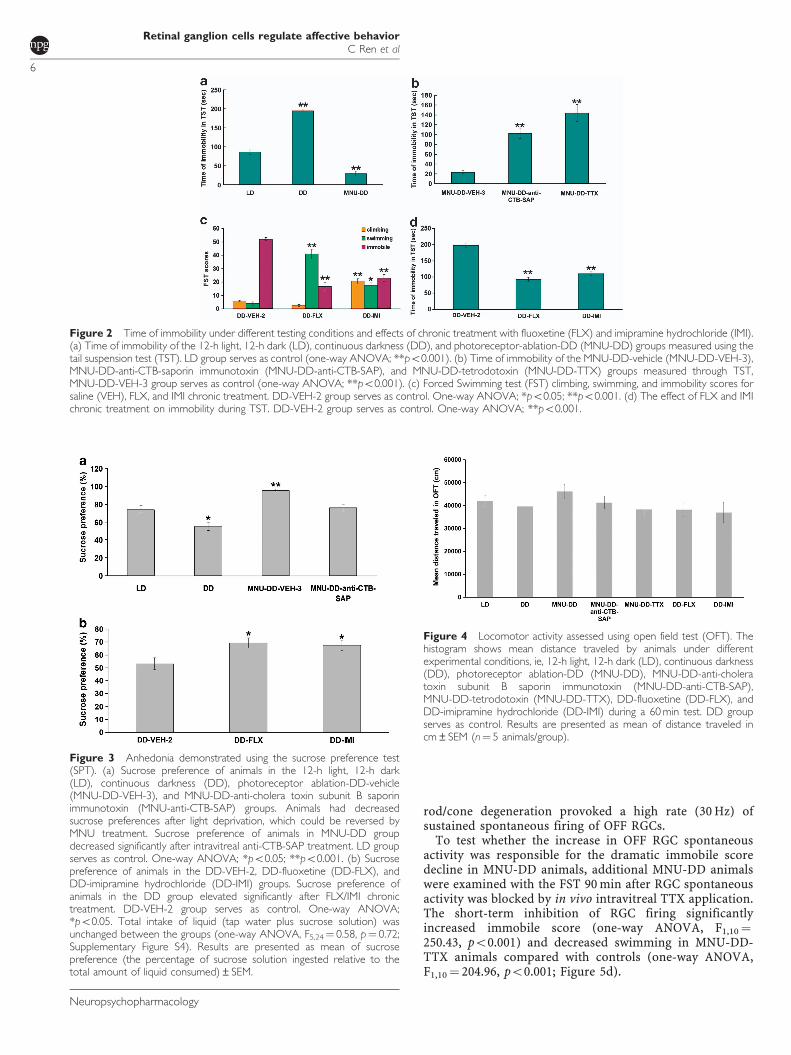
Animals in DD exhibit significantly elevated immobilescores compared with animals housed in LD conditions,supporting the interpretation that light deprivation is asso-ciated with a depressive-like behavioral state (LD vs DDat four time points, two-way ANOVA, effect of type� timepoints interaction: F3,40¼ 12.86, po0.001; effect of type:F1,40¼ 407.95, po0.001; Turkey’s post hoc comparisonindicated that immobile score in DD group was significantlyhigher than in LD group at each time point, po0.001;Figure 1a). Results from both the TST (Figure 2a) and theSPT (Figure 3a) reveal that DD induced depressive-likebehavior (TST: one-way ANOVA, F1,10¼ 463.12, po0.001;SPT: one-way ANOVA, F1,10¼ 16.15, po0.01), while resultsfrom the OFT (Figure 4) indicate that the DD-inducedchange in behavior was not associated with an alteration inlocomotor activity (one-way ANOVA, F6,28¼ 0.92, p¼ 0.49).
As described earlier, to begin to define the neural circuitsby which light deprivation provokes depressive-likebehavior, rod and cone photoreceptor degeneration wasinduced by a single IP injection of MNU, a direct-actingalkylating agent that causes rapid photoreceptor apoptosis.MNU-treated animals lacking rods and cones (Figure 1b)demonstrated a dramatic reduction in immobile scorecompared with untreated animals in LD conditions (LD vsMNU-LD at four time points, two-way ANOVA, effect oftype� time points interaction: F3,40¼ 23.44, po0.001; effectof type: F1,40¼ 1374.35, po0.001; Turkey’s post hoccomparison indicated that immobile score in MNU-LDgroup was significantly lower than in LD group at each timepoint, po0.001) or after 14 days in DD (DD vs MNU-DD atfour time points, two-way ANOVA, effect of type� timepoints interaction: F3,40¼ 1.3, p¼ 0.287; effect of type:F1,40¼ 4790.33, po0.001; Figure 1a).
DRN-projecting RGC Activity Regulates BehavioralMood
Photoreceptor degeneration causes a striking increase inthe rate of OFF RGC spontaneous spike discharge (Pu et al,2006; Margolis and Detwiler, 2007). To determinewhether MNU-induced photoreceptor apoptosis increasedRGC spontaneous activity, single-unit recordings weremade from alpha/Y-like DRN-projecting OFF RGCs(Figure 5a). These cells showed vigorous spontaneousactivity compared with alpha/Y-like DRN-projecting OFFRGCs in intact retinas (one-way, ANOVA, F1,18¼ 380.05,po0.001; Figure 5b–c), which was completely silenced byapplication of tetrodotoxin (TTX), a potent voltage-gatedsodium channel blocker (Figure 5b). Thus, as in othermodels of degenerative photoreceptor loss, MNU-induced
Retinal ganglion cells regulate affective behaviorC Ren et al
5
Neuropsychopharmacology
rod/cone degeneration provoked a high rate (30 Hz) ofsustained spontaneous firing of OFF RGCs.
To test whether the increase in OFF RGC spontaneousactivity was responsible for the dramatic immobile scoredecline in MNU-DD animals, additional MNU-DD animalswere examined with the FST 90 min after RGC spontaneousactivity was blocked by in vivo intravitreal TTX application.The short-term inhibition of RGC firing significantlyincreased immobile score (one-way ANOVA, F1,10¼250.43, po0.001) and decreased swimming in MNU-DD-TTX animals compared with controls (one-way ANOVA,F1,10¼ 204.96, po0.001; Figure 5d).
Figure 3 Anhedonia demonstrated using the sucrose preference test(SPT). (a) Sucrose preference of animals in the 12-h light, 12-h dark(LD), continuous darkness (DD), photoreceptor ablation-DD-vehicle(MNU-DD-VEH-3), and MNU-DD-anti-cholera toxin subunit B saporinimmunotoxin (MNU-anti-CTB-SAP) groups. Animals had decreasedsucrose preferences after light deprivation, which could be reversed byMNU treatment. Sucrose preference of animals in MNU-DD groupdecreased significantly after intravitreal anti-CTB-SAP treatment. LD groupserves as control. One-way ANOVA; *po0.05; **po0.001. (b) Sucrosepreference of animals in the DD-VEH-2, DD-fluoxetine (DD-FLX), andDD-imipramine hydrochloride (DD-IMI) groups. Sucrose preference ofanimals in the DD group elevated significantly after FLX/IMI chronictreatment. DD-VEH-2 group serves as control. One-way ANOVA;*po0.05. Total intake of liquid (tap water plus sucrose solution) wasunchanged between the groups (one-way ANOVA, F5,24¼ 0.58, p¼ 0.72;Supplementary Figure S4). Results are presented as mean of sucrosepreference (the percentage of sucrose solution ingested relative to thetotal amount of liquid consumed)±SEM.
Figure 4 Locomotor activity assessed using open field test (OFT). Thehistogram shows mean distance traveled by animals under differentexperimental conditions, ie, 12-h light, 12-h dark (LD), continuous darkness(DD), photoreceptor ablation-DD (MNU-DD), MNU-DD-anti-choleratoxin subunit B saporin immunotoxin (MNU-DD-anti-CTB-SAP),MNU-DD-tetrodotoxin (MNU-DD-TTX), DD-fluoxetine (DD-FLX), andDD-imipramine hydrochloride (DD-IMI) during a 60 min test. DD groupserves as control. Results are presented as mean of distance traveled incm±SEM (n¼ 5 animals/group).
Figure 2 Time of immobility under different testing conditions and effects of chronic treatment with fluoxetine (FLX) and imipramine hydrochloride (IMI).(a) Time of immobility of the 12-h light, 12-h dark (LD), continuous darkness (DD), and photoreceptor-ablation-DD (MNU-DD) groups measured using thetail suspension test (TST). LD group serves as control (one-way ANOVA; **po0.001). (b) Time of immobility of the MNU-DD-vehicle (MNU-DD-VEH-3),MNU-DD-anti-CTB-saporin immunotoxin (MNU-DD-anti-CTB-SAP), and MNU-DD-tetrodotoxin (MNU-DD-TTX) groups measured through TST,MNU-DD-VEH-3 group serves as control (one-way ANOVA; **po0.001). (c) Forced Swimming test (FST) climbing, swimming, and immobility scores forsaline (VEH), FLX, and IMI chronic treatment. DD-VEH-2 group serves as control. One-way ANOVA; *po0.05; **po0.001. (d) The effect of FLX and IMIchronic treatment on immobility during TST. DD-VEH-2 group serves as control. One-way ANOVA; **po0.001.
Retinal ganglion cells regulate affective behaviorC Ren et al
6
Neuropsychopharmacology
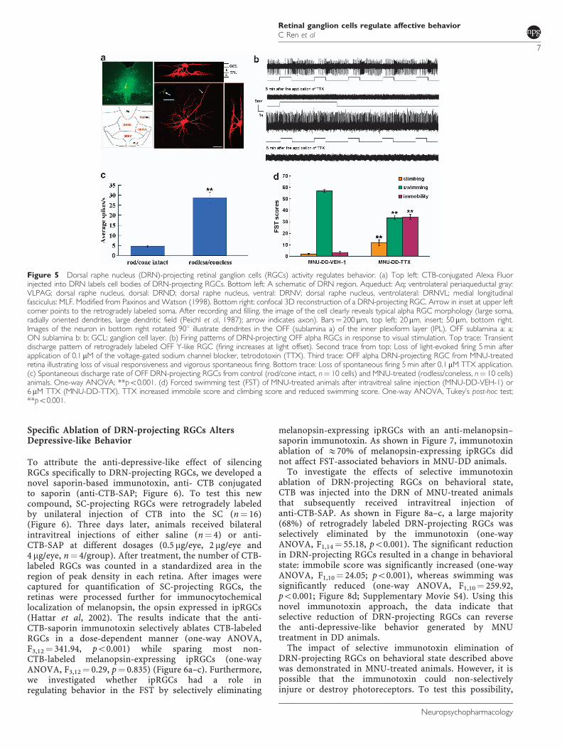
Specific Ablation of DRN-projecting RGCs AltersDepressive-like Behavior
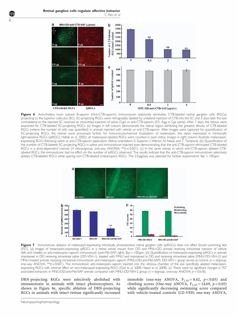
To attribute the anti-depressive-like effect of silencingRGCs specifically to DRN-projecting RGCs, we developed anovel saporin-based immunotoxin, anti- CTB conjugatedto saporin (anti-CTB-SAP; Figure 6). To test this newcompound, SC-projecting RGCs were retrogradely labeledby unilateral injection of CTB into the SC (n¼ 16)(Figure 6). Three days later, animals received bilateralintravitreal injections of either saline (n¼ 4) or anti-CTB-SAP at different dosages (0.5 mg/eye, 2mg/eye and4 mg/eye, n¼ 4/group). After treatment, the number of CTB-labeled RGCs was counted in a standardized area in theregion of peak density in each retina. After images werecaptured for quantification of SC-projecting RGCs, theretinas were processed further for immunocytochemicallocalization of melanopsin, the opsin expressed in ipRGCs(Hattar et al, 2002). The results indicate that the anti-CTB-saporin immunotoxin selectively ablates CTB-labeledRGCs in a dose-dependent manner (one-way ANOVA,F3,12¼ 341.94, po0.001) while sparing most non-CTB-labeled melanopsin-expressing ipRGCs (one-wayANOVA, F3,12¼ 0.29, p¼ 0.835) (Figure 6a–c). Furthermore,we investigated whether ipRGCs had a role inregulating behavior in the FST by selectively eliminating
melanopsin-expressing ipRGCs with an anti-melanopsin–saporin immunotoxin. As shown in Figure 7, immunotoxinablation of E70% of melanopsin-expressing ipRGCs didnot affect FST-associated behaviors in MNU-DD animals.
To investigate the effects of selective immunotoxinablation of DRN-projecting RGCs on behavioral state,CTB was injected into the DRN of MNU-treated animalsthat subsequently received intravitreal injection ofanti-CTB-SAP. As shown in Figure 8a–c, a large majority(68%) of retrogradely labeled DRN-projecting RGCs wasselectively eliminated by the immunotoxin (one-wayANOVA, F1,14¼ 55.18, po0.001). The significant reductionin DRN-projecting RGCs resulted in a change in behavioralstate: immobile score was significantly increased (one-wayANOVA, F1,10¼ 24.05; po0.001), whereas swimming wassignificantly reduced (one-way ANOVA, F1,10¼ 259.92,po0.001; Figure 8d; Supplementary Movie S4). Using thisnovel immunotoxin approach, the data indicate thatselective reduction of DRN-projecting RGCs can reversethe anti-depressive-like behavior generated by MNUtreatment in DD animals.
The impact of selective immunotoxin elimination ofDRN-projecting RGCs on behavioral state described abovewas demonstrated in MNU-treated animals. However, it ispossible that the immunotoxin could non-selectivelyinjure or destroy photoreceptors. To test this possibility,
Figure 5 Dorsal raphe nucleus (DRN)-projecting retinal ganglion cells (RGCs) activity regulates behavior. (a) Top left: CTB-conjugated Alexa Fluorinjected into DRN labels cell bodies of DRN-projecting RGCs. Bottom left: A schematic of DRN region. Aqueduct: Aq; ventrolateral periaqueductal gray:VLPAG; dorsal raphe nucleus, dorsal: DRND; dorsal raphe nucleus, ventral: DRNV; dorsal raphe nucleus, ventrolateral: DRNVL; medial longitudinalfasciculus: MLF. Modified from Paxinos and Watson (1998). Bottom right: confocal 3D reconstruction of a DRN-projecting RGC. Arrow in inset at upper leftcorner points to the retrogradely labeled soma. After recording and filling, the image of the cell clearly reveals typical alpha RGC morphology (large soma,radially oriented dendrites, large dendritic field (Peichl et al, 1987); arrow indicates axon). Bars¼ 200 mm, top left; 20mm, insert; 50 mm, bottom right.Images of the neuron in bottom right rotated 901 illustrate dendrites in the OFF (sublamina a) of the inner plexiform layer (IPL). OFF sublamina a: a;ON sublamina b: b; GCL: ganglion cell layer. (b) Firing patterns of DRN-projecting OFF alpha RGCs in response to visual stimulation. Top trace: Transientdischarge pattern of retrogradely labeled OFF Y-like RGC (firing increases at light offset). Second trace from top: Loss of light-evoked firing 5 min afterapplication of 0.1mM of the voltage-gated sodium channel blocker, tetrodotoxin (TTX). Third trace: OFF alpha DRN-projecting RGC from MNU-treatedretina illustrating loss of visual responsiveness and vigorous spontaneous firing. Bottom trace: Loss of spontaneous firing 5 min after 0.1 mM TTX application.(c) Spontaneous discharge rate of OFF DRN-projecting RGCs from control (rod/cone intact, n¼ 10 cells) and MNU-treated (rodless/coneless, n¼ 10 cells)animals. One-way ANOVA; **po0.001. (d) Forced swimming test (FST) of MNU-treated animals after intravitreal saline injection (MNU-DD-VEH-1) or6 mM TTX (MNU-DD-TTX). TTX increased immobile score and climbing score and reduced swimming score. One-way ANOVA, Tukey’s post-hoc test;**po0.001.
Retinal ganglion cells regulate affective behaviorC Ren et al
7
Neuropsychopharmacology
DRN-projecting RGCs were selectively abolished withimmunotoxin in animals with intact photoreceptors. Asshown in Figure 8e, specific ablation of DRN-projectingRGCs in animals with intact retinas significantly increased
immobile (one-way ANOVA, F1,10¼ 8.62, po0.05) andclimbing scores (One-way ANOVA, F1,10¼ 14.69, po0.05)while significantly decreasing swimming score comparedwith vehicle-treated controls (LD-VEH; one-way ANOVA,
Figure 6 Anti-cholera toxin subunit B-saporin (Anti-CTB-saporin) immunotoxin selectively eliminates CTB-labeled retinal ganglion cells (RGCs)projecting to the superior colliculus (SC). SC-projecting RGCs were retrogradely labeled by unilateral injection of CTB into the SC and 3 days later the eyecontralateral to the injected SC received an intravitreal injection of saline (5ml) or anti-CTB-saporin (0.5–4 mg in 5ml saline). After 7 days, the retinas wereexamined for CTB-labeled SC-projecting RGCs. (a) Images in left column demonstrate the retinal region exhibiting the greatest density of CTB-labeledRGCs (where the number of cells was quantified) in animals injected with vehicle or anti-CTB-saporin. After images were captured for quantification ofSC-projecting RGCs, the retinas were processed further for immunocytochemical localization of melanopsin, the opsin expressed in intrinsicallylight-sensitive RGCs (ipRGCs; Hattar et al, 2002); all melanopsin-labeled RGCs were counted in each retina. Images in right column illustrate melanopsin-expressing RGCs following saline or anti-CTB-saporin application. Retina orientation: S: Superior, I: Inferior, N: Nasal, and T: Temporal. (b) Quantification ofthe number of CTB-labeled SC-projecting RGCs in saline and immunotoxin-injected eyes demonstrating that the anti-CTB-saporin eliminated CTB-labeledRGCs in a dose-dependent manner (4 retinas/group, one-way ANONA; **po0.001). (c) In the same retinas in which anti-CTB-saporin ablated CTB-labeled RGCs, the immunotoxin had no effect on the number of ipRGCs observed. The results indicate that the anti-CTB-saporin immunotoxin selectivelyablates CTB-labeled RGCs while sparing non-CTB-labeled (melanopsin) RGCs. The 2.0 mg/eye was selected for further experiments. Bar¼ 100 mm.
Figure 7 Immunotoxin ablation of melanopsin-expressing intrinsically photosensitive retinal ganglion cells (ipRGCs) does not affect forced swimming test(FST). (a) Images of melanopsin-expressing ipRGCs in a retinal whole mount from DD and MNU-DD animals receiving intravitreal injection of vehicle(left and middle) or anti-melanopsin–saporin immunotoxin (anti-Mel-SAP; right). Bar¼ 100mm. (b) Quantification of melanopsin-expressing ipRGCs in animalsmaintained in DD receiving intravitreal saline (DD-VEH-1), treated with MNU and maintained in DD and receiving intravitreal saline (MNU-DD-VEH-2) andMNU-treated animals receiving intravitreal immunotoxin anti-melanopsin–saporin (MNU-DD-anti-Mel-SAP), DD-VEH-1 group serves as control. (n¼ 6/group,one-way ANOVA; **po0.001). The immunotoxin anti-melanopsin–saporin injected into the vitreous chamber of the eye specifically ablated melanopsin-expressing RGCs with minimal effect on non-melanopsin-expressing RGCs (Goz et al, 2008; Hatori et al, 2008). (c) There were no significant changes in FSTassociated behaviors in MNU-DD-anti-Mel-SAP animals compared with MNU-DD-VEH-2 group (n¼ 6/group, one-way ANOVA; p¼ 0.618).
Retinal ganglion cells regulate affective behaviorC Ren et al
8
Neuropsychopharmacology

F1,10¼ 50.44, po0.001). These results are consistent withthe interpretation that DRN-projecting RGCs modulatebehavioral state.
The results from the TST paralleled the robust effectsobserved with the FST. DD animals had signifi-cantly increased immobile time compared with LD animals(one-way ANOVA, F1,10¼ 463.12, po0.001) andMNU treatment significantly reduced immobile time inMNU-DD compared with DD animals (one-way ANOVA,F1,10¼ 889.22, po0.001; Figure 2a). Conversely, MNU-DDanimals receiving intravitreal injection of anti-CTB-SAP(one-way ANOVA, F1,10¼ 79.72, po0.001) or TTX (one-wayANOVA, F1,10¼ 78.56, po0.001) showed greatly increasedimmobile times (Figure 2b). These findings are similarto the results obtained in the FST.
As shown in Figure 3a, maintain animals in DD wasassociated with a significant decrease in sucrose preferencecompared with animals maintained in LD conditions(one-way ANOVA, F1,10¼ 9.26, po0.05). Consistent withthe results from the other behavioral tests, MNU treatmentwas anti-depressive, significantly increasing sucrose pre-ference compared with DD animals (one-way ANOVA,F1,10¼ 71.23, po0.001), whereas selective removal of DRN-projecting RGCs in MNU-DD animals by immunotoxinsignificantly decreased sucrose preference compared withMNU-DD-VEH-3 animals (one-way ANOVA, F1,10¼ 21.44,po0.01). Taken together, the findings demonstrate thatDRN-projecting RGCs have an important role in regulatingbehavioral state.
Relationship between DRN 5-HT-positive Neurons andFST Behaviors
Next, we investigated DRN serotonergic tone usingimmunocytochemical staining for 5-HT in a subset of
animals (n¼ 4) from each group. The number of 5-HT-positive (þ ) neurons in the dorsal and lateral divisionsof the DRN varied significantly among experimental groups(one-way ANOVA, dorsal division: F5,18¼ 123.92, po0.001;lateral division: F5,18¼ 101.63, po0.001); conditions thatincreased RGC activity (MNU-DD) increased the number ofDRN 5-HT neurons and conversely, conditions thatdecreased RGC activity (DD, MNU-DD-TTX; MNU-DD-SAP) decreased the number of DRN 5-HTþ neuronsrelative to levels in animals housed in LD conditions(one-way ANOVA, MNU-DD vs LD: F1,6¼ 156.31, po0.001;DD vs LD: F1,6¼ 77.25, po0.001; MNU-DD-TTX vs LD:F1,6¼ 18.98, po0.01; MNU-DD-SAP vs LD: F1,6¼ 18.21,po0.01). These effects were more prominent in the lateralDRN (MNU-DD increased lateral 5-HTþ neurons 12-foldover DD but only sevenfold over DD in the dorsal DRN;Figure 9a–b). Chronic treatment of DD animals with theSSRI anti-depressant fluoxetine (DD-FLX) significantlyreduced immobile scores in the FST (one-way ANOVA,F1,10¼ 92.80, po0.001) and TST (one-way ANOVA,F1,10¼ 112.97, po0.001; Figure 2c–d and SupplementaryMovie S5) and significantly elevated 5-HT levels over DDconditions (Figure 9b). DD animals treated for 21 days withimipramine, a non-SSRI tricyclic anti-depressant (DD-IMI),also reversed the depressive-like behavior evoked by DD(FST: one-way ANOVA, F1,10¼ 84.24, po0.001; TST:one-way ANOVA, F1,10¼ 84.31, po0.001), as illustrated inFigure 2c–d. Fluoxetine and imipramine also reversedthe reduced sucrose preference behavior evoked by DD(one- way ANOVA, F2,12¼ 5.75, po0.05; Turkey’s post hoccomparison indicated that sucrose preference ofDD-VEH-2 group is significantly lower than that in DD-FLX (po0.05) and DD-IMI group (po0.05)) (Figure 9b).As revealed in Figure 2c, the impacts of the twoanti-depressants are different on FST behavior; this could
Figure 8 Specific ablation of dorsal raphe nucleus-projecting retinal ganglion cells (DRN-projecting RGCs) alters depressive-like behavior. (a) Examples ofretrogradely labeled DRN-projecting RGCs in retinal whole mounts after cholera toxin subunit B (CTB) injection into DRN and then after intravitreal (left)saline injection or (right) anti-CTB-saporin (anti-CTB-SAP) immunotoxin (2mg/eye) injection. Bar¼ 20 mm. (b) Distribution of CTB-labeled DRN-projectingRGCs in retinal whole mounts in photoreceptor ablation-continuous darkness (MNU-DD)-treated animals 7 days after intravitreal vehicle (left) or anti-CTB-SAP immunotoxin (2 mg/eye; right). Retina orientation: S: Superior, I: Inferior, N: Nasal, and T: Temporal. (c) Quantification of DRN-projecting RGCs in saline(MNU-DD-VEH-3) and immunotoxin-treated eyes from MNU-DD animals illustrating significant reduction in the number of CTB-labeled RGCs (87.7±11.4 MNU-DD-VEH-3 vs 28.2±6.4 MNU-DD-anti-CTB-SAP; n¼ 8 retinas/group), one-way ANOVA; **po0.001. (d) Forced swimming test (FST)scores in MNU-DD animals after CTB injection into the DRN followed by intravitreal injection of vehicle or immunotoxin. Specific ablation of DRN-projecting RGCs (MNU-DD-anti-CTB-SAP) significantly increased immobile and climbing scores, whereas decreasing swimming score compared withcontrols. One-way ANOVA; **po0.001. (e) FST climbing, swimming, and immobility scores at Zeitgeber Time 6 for animals housed in LD conditionstreated with saline and anti-CTB-SAP. Specific ablation of DRN-projecting RGCs (LD-anti-CTB-SAP) significantly increased immobile and climbing scores,whereas decreasing swimming score. LD-VEH group serves as control. One-way ANOVA; *po0.05; **po0.001.
Retinal ganglion cells regulate affective behaviorC Ren et al
9
Neuropsychopharmacology
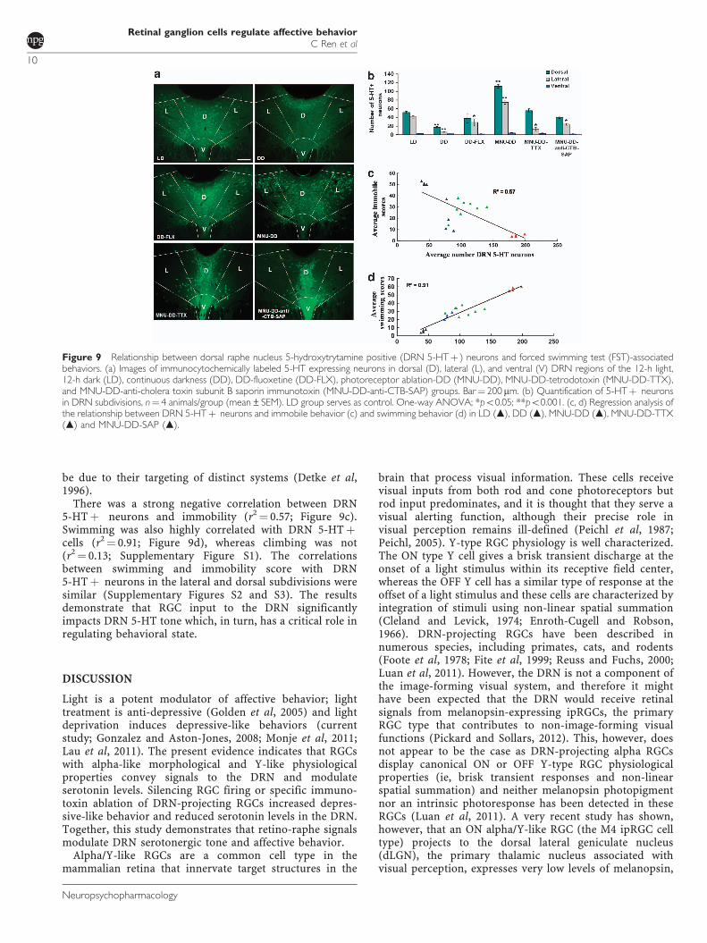
be due to their targeting of distinct systems (Detke et al,1996).
There was a strong negative correlation between DRN5-HTþ neurons and immobility (r2¼ 0.57; Figure 9c).Swimming was also highly correlated with DRN 5-HTþcells (r2¼ 0.91; Figure 9d), whereas climbing was not(r2¼ 0.13; Supplementary Figure S1). The correlationsbetween swimming and immobility score with DRN5-HTþ neurons in the lateral and dorsal subdivisions weresimilar (Supplementary Figures S2 and S3). The resultsdemonstrate that RGC input to the DRN significantlyimpacts DRN 5-HT tone which, in turn, has a critical role inregulating behavioral state.
DISCUSSION
Light is a potent modulator of affective behavior; lighttreatment is anti-depressive (Golden et al, 2005) and lightdeprivation induces depressive-like behaviors (currentstudy; Gonzalez and Aston-Jones, 2008; Monje et al, 2011;Lau et al, 2011). The present evidence indicates that RGCswith alpha-like morphological and Y-like physiologicalproperties convey signals to the DRN and modulateserotonin levels. Silencing RGC firing or specific immuno-toxin ablation of DRN-projecting RGCs increased depres-sive-like behavior and reduced serotonin levels in the DRN.Together, this study demonstrates that retino-raphe signalsmodulate DRN serotonergic tone and affective behavior.
Alpha/Y-like RGCs are a common cell type in themammalian retina that innervate target structures in the
brain that process visual information. These cells receivevisual inputs from both rod and cone photoreceptors butrod input predominates, and it is thought that they serve avisual alerting function, although their precise role invisual perception remains ill-defined (Peichl et al, 1987;Peichl, 2005). Y-type RGC physiology is well characterized.The ON type Y cell gives a brisk transient discharge at theonset of a light stimulus within its receptive field center,whereas the OFF Y cell has a similar type of response at theoffset of a light stimulus and these cells are characterized byintegration of stimuli using non-linear spatial summation(Cleland and Levick, 1974; Enroth-Cugell and Robson,1966). DRN-projecting RGCs have been described innumerous species, including primates, cats, and rodents(Foote et al, 1978; Fite et al, 1999; Reuss and Fuchs, 2000;Luan et al, 2011). However, the DRN is not a component ofthe image-forming visual system, and therefore it mighthave been expected that the DRN would receive retinalsignals from melanopsin-expressing ipRGCs, the primaryRGC type that contributes to non-image-forming visualfunctions (Pickard and Sollars, 2012). This, however, doesnot appear to be the case as DRN-projecting alpha RGCsdisplay canonical ON or OFF Y-type RGC physiologicalproperties (ie, brisk transient responses and non-linearspatial summation) and neither melanopsin photopigmentnor an intrinsic photoresponse has been detected in theseRGCs (Luan et al, 2011). A very recent study has shown,however, that an ON alpha/Y-like RGC (the M4 ipRGC celltype) projects to the dorsal lateral geniculate nucleus(dLGN), the primary thalamic nucleus associated withvisual perception, expresses very low levels of melanopsin,
Figure 9 Relationship between dorsal raphe nucleus 5-hydroxytrytamine positive (DRN 5-HTþ ) neurons and forced swimming test (FST)-associatedbehaviors. (a) Images of immunocytochemically labeled 5-HT expressing neurons in dorsal (D), lateral (L), and ventral (V) DRN regions of the 12-h light,12-h dark (LD), continuous darkness (DD), DD-fluoxetine (DD-FLX), photoreceptor ablation-DD (MNU-DD), MNU-DD-tetrodotoxin (MNU-DD-TTX),and MNU-DD-anti-cholera toxin subunit B saporin immunotoxin (MNU-DD-anti-CTB-SAP) groups. Bar¼ 200 mm. (b) Quantification of 5-HTþ neuronsin DRN subdivisions, n¼ 4 animals/group (mean±SEM). LD group serves as control. One-way ANOVA; *po0.05; **po0.001. (c, d) Regression analysis ofthe relationship between DRN 5-HTþ neurons and immobile behavior (c) and swimming behavior (d) in LD (m), DD (m), MNU-DD (m), MNU-DD-TTX(m) and MNU-DD-SAP (m).
Retinal ganglion cells regulate affective behaviorC Ren et al
10
Neuropsychopharmacology
and exhibits a weak intrinsic photoresponse (Estevez et al,2012). Specialized immunocytochemical amplificationmethods were necessary to detect melanopsin in these cellsand even then not all physiologically confirmed M4 ipRGCswere melanopsin-positive (Estevez et al, 2012). It is likelythat the standard immunocytochemical protocol used tolabel melanopsin RGCs (Luan et al, 2011) would have beeninsufficient to detect melanopsin in DRN-projecting RGCs ifthese cells are M4 ipRGCs. Although it cannot be said withcertainty that the ON alpha/Y-like RGCs that innervatethe DRN are different from the M4 ipRGCs described byEstevez et al (2012), their brisk transient response to photicstimuli differs from the tonic-sustained responses of M4cells and, more importantly, Estevez et al (2012) did notobserve any OFF alpha/Y-like ipRGCs expressing melanop-sin. In the DD-maintained MNU-treated animals used as amodel in the present study, OFF cells in particular had acrucial role in altering behavioral mood as it was thespontaneous activity of OFF RGCs only that increaseddramatically following MNU treatment.
Under normal conditions, the DRN receives input fromboth ON and OFF Y-type RGCs (Luan et al, 2011) and theirmaintained firing frequency is dependent on the luminancelevel (Barlow and Levick, 1969; Troy and Robson, 1992).Under DD conditions, both ON and OFF Y-type cells ceaselight-evoked responses but maintain a low spontaneousfiring rate (Figure 5c). After MNU-induced photoreceptordepletion, OFF Y-type cells subsequently showed a sixfoldincrease in spontaneous firing rate, which, in turn,improved affective behavior. Although a relatively smallnumber of alpha/Y-like RGCs innervates the DRN, becausethese RGCs have a broad dendritic field (Luan et al, 2011),only E700 are necessary to tile the entire retinal surface(Supplementary Table S1). All alpha/Y-like RGCs haverelatively large diameter axons; perhaps the high conduc-tion velocity of these RGCs (Peichl et al, 1987) is the salientcomponent of the visual signal being conveyed to the DRN(Chen and Regehr, 2003).
The findings in the present study showed that the level ofneural activity of DRN-projecting RGCs was directly relatedto behavioral state. Moreover, DRN-projecting RGC activitywas correlated with DRN 5-HT levels and affective behavior;MNU-DD animals contained the greatest number of DRN5-HTþ neurons, whereas DD animals had the fewest.Reducing RGC signals to the DRN in MNU-DD animals,either by acute TTX inhibition of firing or by immunotoxinablation of DRN-projecting RGCs, decreased DRN 5-HTlevels and significantly elevated behavioral indicesof depressive-like behavior. Chronic inhibition of DRN-projecting RGCs or more complete immunotoxin lesioningof DRN-projecting RGCs in MNU-DD animals would likelyhave produced even greater effects on DRN 5-HT tone anddepressive-like behavior.
We also examined whether the SSRI anti-depressantfluoxetine would improve the depressive-like behaviorevoked by DD treatment in a manner similar to the MNU-evoked increase in DRN-projecting RGC spontaneousactivity. Indeed, as shown in Figure 2c and SupplementaryMovie S5, chronic fluoxetine treatment significantlyreduced immobile score, although elevating swimmingwhile having no effect on climbing behavior in the FST.This result is consistent with previous reports that SSRIs are
both anti-depressive and elevate swimming without affect-ing climbing behavior in the FST (Lucki, 1997) and similarto the effects observed by MNU treatment of DD animals.Thus, the depressive-like behavior induced by lightdeprivation was alleviated both by fluoxetine and increasedactivity of DRN-projecting RGCs. Importantly as shownpreviously (Lucki, 1997), fluoxetine effects in this studywere specific, in that swimming time in the FST waselevated without an increase in general locomotor activity.Similarly, the anti-depressant effects of MNU treatment didnot generate an increase in locomotor activity, in agreementwith the lack of MNU-evoked increased locomotor activityreported by Boudard et al (2009). Moreover, DD animalstreated with imipramine, a non-SSRI tricyclic anti-depres-sant, also showed a reversal in depressive-like behavior inthe FST and TST without affecting the locomotor activity inthe OFT. DRN 5-HT levels were also highly correlatedwith FST swimming but not climbing behavior. Collectively,the data support the view that increased retinal input to theDRN is anti-depressive.
We describe a non-pharmacological anti-depressanttreatment, activation of direct retino-raphe input to theDRN that modulates serotonergic tone and depressive-likebehavior. This novel result sheds new light on our currentunderstanding of affective visual information processing.
ACKNOWLEDGEMENTS
This work was supported by National Basic ResearchProgram (2011CB510206 and 2009CB320901 to MP);NSFC grants (31170986 and 30831160516 to MP), ResearchScheme under the contract N_HKU750/08 (KFS), theFundamental Research Funds for the Central Universities(21609101, KFS); National Basic Research Program of China(973 Program) (2011CB707501, KFS), and the USA NationalInstitutes of Health grants EY 017809 and NS 077003 (GEP).
This work was supported by the government grants fromChina, Hong Kong, and the United States.
DISCLOSURE
The authors declare no conflict of interest.
REFERENCES
Barlow HB, Levick WR (1969). Changes in the maintaineddischarge with adaptation level in the cat retina. J Physiol 202:699–718.
Bijak M, Papp M (1995). The effect of chronic treatment withimipramine on the responsiveness of hippocampal CA1 neuronsto phenylephrine and serotonin in a chronic mild stress model ofdepression. Eur Neuropsychopharmacol 5: 43–48.
Boudard DL, Mendoza J, Hicks D (2009). Loss of photicentrainment at low illuminances in rats with acute photoreceptordegeneration. Eur J Neurosci 30: 1527–1536.
Chen C, Regehr WG (2003). Presynaptic modulation of theretinogeniculate synapse. J Neurosci 23: 3130–3135.
Cleland BG, Levick WR (1974). Brisk and sluggish concentricallyorganized ganglion cells in the cat’s retina. J Physiol 240:421–456.
Cryan JF, Valentino RJ, Lucki I (2005a). Assessing substratesunderlying the behavioral effects of antidepressants using the
Retinal ganglion cells regulate affective behaviorC Ren et al
11
Neuropsychopharmacology
modified rat forced swimming test. Neurosci Biobehav Rev 29:547–569.
Detke MJ, Rickels M, Lucki I (1996). Active behaviors in the ratforced swimming test differentially produced by serotonergicand noradrenergic antidepressants. Psychopharmacology (Berl)121: 66–72.
Edwards JA, Cline HT (1999). Light-induced calcium influxinto retinal axons is regulated by presynaptic. J Neurophysiol81: 895–907.
Enroth-Cugell C, Robson JG (1966). The contrast sensitivity ofretinal ganglion cells of the cat. J Physiol (Lond) 187: 517–552.
Estevez ME, Fogerson PM, Ilardi MC, Borghuis BG, Chan E, WengS et al (2012). Form and function of the m4 cell, an intrinsicallyphotosensitive retinal ganglion cell type contributing togeniculocortical vision. J Neurosci 32: 13608–13620.
Fite KV, Janusonis S, Foote W, Bengston L (1999). Retinal afferentsto the dorsal raphe nucleus in rats and Mongolian gerbils.J Comp Neurol 414: 469–484.
Foote WE, Taber-Pierce E, Edwards L (1978). Evidence for a retinalprojection to the midbrain raphe of the cat. Brain Res 156:135–140.
Golden RN, Gaynes BN, Ekstrom RD, Hamer RM, Jacobsen FM,Suppes T et al (2005). The efficacy of light therapy in thetreatment of mood disorders: a review and meta-analysis of theevidence. Am J Psychiatry 162: 656–662.
Gonzalez MM, Aston-Jones G (2008). Light deprivation damagesmonoamine neurons and produces a depressive behavioralphenotype in rats. Proc Natl Acad Sci USA 105: 4898–4903.
Goz D, Studholme K, Lappi DA, Rollag MD, Provencio I, Morin LP(2008). Targeted destruction of photosensitive retinal ganglioncells with a saporin conjugate alters the effects of light on mousecircadian rhythms. PLoS One 3: e3153.
Guler AD, Ecker JL, Lall GS, Haq S, Altimus CM, Liao HW et al(2008). Melanopsin cells are the principal conduits for rod-coneinput to non-image-forming vision. Nature 453: 102–105.
Hatori M, Le H, Vollmers C, Keding SR, Tanaka N, Buch T et al(2008). Inducible ablation of melanopsin-expressing retinalganglion cells reveals their central role in non-image formingvisual responses. PLoS One 3: e2451.
Hattar S, Liao HW, Takao M, Berson DM, Yau KW (2002).Melanopsin-containing retinal ganglion cells: architecture,projections, and intrinsic photosensitivity. Science 295:1065–1070.
Herrold KM (1967). Pigmentary degeneration of the retina inducedby N-methyl-N-nitrosourea. An experimental study in Syrianhamsters. Arch Ophthalmol 78: 650–653.
Lau BW, Ren C, Yang J, Yan SW, Chang RC, Pu M et al (2011).Light deprivation induces depression-like behavior andsuppresses neurogenesis in diurnal mongolian gerbil (Merionesunguiculatus). Cell Transplant 20: 871–881.
Lewy AJ, Sack RL, Miller LS, Hoban TM (1987). Antidepressantand circadian phase-shifting effects of light. Science 235:352–354.
Luan L, Ren C, Lau BW, Yang J, Pickard GE, So KF et al (2011).Y-like retinal ganglion cells innervate the dorsal raphe nucleus inthe Mongolian gerbil (Meriones unguiculatus). PLoS One 6:e18938.
Lucki I (1997). The forced swimming test as a model for coreand component behavioral effects of antidepressant drugs.Behav Pharmacol 8: 523–532.
Machado DG, Cunha MP, Neis VB, Balen GO, Colla A, Grando Jet al (2012). Fluoxetine reverses depressive-like behaviorsand increases hippocampal acetylcholinesterase activityinduced by olfactory bulbectomy. Pharmacol Biochem Behav103: 220–229.
Margolis DJ, Detwiler PB (2007). Different mechanisms generatemaintained activity in ON and OFF retinal ganglion cells.J Neurosci 27: 5994–6005.
Monje FJ, Cabatic M, Divisch I, Kim EJ, Herkner KR, Binder BRet al (2011). Constant darkness induces IL-6-dependent depres-sion-like behavior through the NF-kB signaling pathway.J Neurosci 31: 9075–9083.
Monteggia LM, Luikart B, Barrot M, Theobold D, Malkovska I,Nef S et al (2007). Brain-derived neurotrophic factor conditionalknockouts show gender differences in depression-relatedbehaviors. Biol Psychiatry 61: 187–197.
Page ME, Detke MJ, Dalvi A, Kirby LG, Lucki I (1999). Serotonergicmediation of the effects of fluoxetine, but not desipramine,in the rat forced swimming test. Psychopharmacology (Berl) 147:162–167.
Paxinos G, Watson C (1998). The Rat Brain in StereotaxicCoordinates. 4th edn.
Peichl L (2005). Diversity of mammalian photoreceptor properties:adaptations to habitat and lifestyle? Anat Rec A Discov Mol CellEvol Biol 287: 1001–1012.
Peichl L, Ott H, Boycott BB (1987). Alpha ganglion cells inmammalian retinae. Proc R Soc Lond B Biol Sci 231: 169–197.
Pickard GE, Sollars PJ (2012). Intrinsically photosensitiveretinal ganglion cells. Rev Physiol Biochem Pharmacol 162:59–90.
Pickles AR, Hagan JJ, Jones DN, Hendrie CA (2012). Short-termindividual housing induced social deficits in female Mongoliangerbils: attenuation by chronic but not acute imipramine.Neuropharmacology 62: 1993–1998.
Pu M (1999). Dendritic morphology of cat retinalganglion cellsprojecting to suprachiasmatic nucleus. J Comp Neurol 414:267–274.
Pu M, Xu L, Zhang H (2006). Visual response properties of retinalganglion cells in the royal college of surgeons dystrophic rat.Invest Ophthalmol Vis Sci 47: 3579–3585.
Reuss S, Fuchs E (2000). Anterograde tracing of retinal afferentsto the tree shrew hypothalamus and raphe. Brain Res 874:66–74.
Rodrıguez-Gaztelumendi A, Rojo ML, Pazos A, Dıaz A (2009).Altered CB receptor-signaling in prefrontal cortex from ananimal model of depression is reversed by chronic fluoxetine.J Neurochem 108: 1423–1433.
Steru L, Chermat R, Thierry B, Simon P (1985). The tail suspensiontest: a new method for screening antidepressants in mice.Psychopharmacol (Berl) 85: 367–370.
Svenningsson P, Tzavara ET, Witkin JM, Fienberg AA,Nomikos GG, Greengard P (2002). Involvement of striataland extrastriatal DARPP-32 in biochemical and behavioraleffects of fluoxetine (Prozac). Proc Natl Acad Sci USA 99:3182–3187.
Troy JB, Robson JG (1992). Steady discharges of X and Y retinalganglion cells of cat under photopic illuminance. Vis Neurosci 9:535–553.
Vandewalle G, Maquet P, Dijk DJ (2009). Light as a modulator ofcognitive brain function. Trends Cog Sci 13: 429–438.
Varty GB, Cohen-Williams ME, Hunter JC (2003). The antide-pressant-like effects of neurokinin NK1 receptor antagonists in agerbil tail suspension test. Behav Pharmacol 14: 87–95.
Viswanathan S, Frishman LJ, Robson JG (2000). The uniform fieldand pattern ERG in macaques with experimental glaucoma:removal of spiking activity. Invest Ophthalmol Vis Sci 41:2797–2810.
Warthen DM, Wiltgen BJ, Provencio I (2011). Light enhanceslearned fear. Proc Natl Acad Sci USA 108: 13788–13793.
Wenzel A, Grimm C, Marti A, Kueng-Hitz N, Hafezi F, Niemeyer Get al (2000). C-fos controls the ‘private pathway’ of light-inducedapoptosis of retinal photoreceptors. J Neurosci 20: 81–88.
Wilson N (2002). Depression and its relation to light deprivation.Psychoanal Rev 89: 557–567.
Wirz-Justice A, Benedetti F, Terman M (2009). Chronotherapeuticsfor Affective Disorders. Karger: New York.
Retinal ganglion cells regulate affective behaviorC Ren et al
12
Neuropsychopharmacology

Witkovsky P, Veisenberger E, Haycock JW, Akopian A,Garcia-Espana A, Meller E (2004). Activity-dependent phosphor-ylation of tyrosine hydroxylase in dopaminergic neurons of therat retina. J Neurosci 24: 4242–4249.
Yau KW, Hardie RC (2009). Phototransduction motifs andvariations. Cell 139: 246–264.
Yoshizawa K, Tsubura A (2005). Characteristics of N-methyl-N-nitrosourea-induced retinal degeneration in animals and
application for the therapy of human retinitis pigmentosa.Nihon Ganka Gakkai Zasshi 109: 327–337.
This work is licensed under a Creative CommonsAttribution-NonCommercial-NoDerivs 3.0 Un-
ported License. To view a copy of this license, visit http://creativecommons.org/licenses/by-nc-nd/3.0/
Supplementary Information accompanies the paper on the Neuropsychopharmacology website (http://www.nature.com/npp)
Retinal ganglion cells regulate affective behaviorC Ren et al
13
Neuropsychopharmacology