the serotonergic inhibitory postsynaptic potential in ... · the serotonergic inhibitory...
TRANSCRIPT

The Journal of Neuroscience, January 1995. 75(l): 223-229
The Serotonergic Inhibitory Postsynaptic Potential in Prepositus Hypoglossi Is Mediated by Two Potassium Currents
Daniel H. Bobkerl and John T. Williams2
‘Department of Neurology and zVollum Institute, Oregon Health Sciences University, Portland, Oregon 97201
Synaptic inhibition mediated by the activation of potassium channels has been reported from several types of neurons. In each case, despite mediation by different neurotransmit- ters, the.K+ conductance underlying the synaptic potential is activated by a G protein and inwardly rectifies. We report here a second K+ current that contributes to synaptic inhi- bition. Intracellular recordings were made from guinea pig nucleus prepositus hypoglossi in vitro, where we have de- scribed a 5-HT-mediated IPSP. Voltage-clamp analysis of the current induced by applied 5-HT revealed two separate conductances: an inwardly rectifying, rapidly activating K+ current (I,,) and an outwardly rectifying, slowly activating K+ current (I,,). /,, was blocked by extracellular Ba*+ (200 PM) and TEA+ (126 mnn). loR was insensitive to this concentration of Ba*+ and TEA+, but was inhibited by Cd2+ and intracellular BAPTA, indicating Ca dependence.
Single focal electrical stimuli evoked a 5-HT-mediated IPSP, or under voltage clamp, an inhibitory postsynaptic cur- rent (IPSC). Ba*+ blocked only a component of this IPSC, which corresponded to the current caused by /,,. When mul- tiple stimuli were applied (to prolong the release of trans- mitter), the time-dependent current loR was more fully acti- vated, resulting in an augmentation of the IPSC. We conclude that the IPSC is caused by both currents and that its ampli- tude can be modulated by the degree to which I,,, is acti- vated. This represents a mechanism by which synaptic re- sponses can be potentiated.
[Key words: 54-/T, inhibitory postsynaptic potential, po- tassium channels, Ca-dependent K current, inward rectifier, outward rectifier]
Potassium channels are important for neuronal functioning be- cause they maintain resting membrane potential, help regulate repetitive firing and contribute to some postsynaptic potentials (Hille, 1992). Among the latter, a frequently observed response is an IPSP mediated by a G protein-coupled K+ channel. Such an IPSP has been reported in the heart (muscarinic receptor mediated), hippocampus (GABA, receptor), locus coeruleus (o(~ adrenoceptor), dorsal raphe (5-HT,, receptor), submucous plex-
Received Apr. 18, 1994; revised June 13, 1994; accepted June 16, 1994. This work was supported by USPHS Grants NS 01574 (D.H.B.) and DA 04523
(J.T.W.), and a grant from the National Association for Research on Schizophrenia and Affective Disorders (D.H.B.). We thank Drs. N. Marrion, C. Jahr, and M. Simmons for comments on the experiments and manuscript.
Correspondence should be addressed to Daniel H. Bobker, Department of Neu- rology, L226, Oregon Health Sciences University, 3181 SW Sam Jackson Park Road, Portland, OR 97201. Copyright 0 1995 Society for Neuroscience 0270-6474/95/150223-07$05.00/O
us (somatostatin receptor), and other regions (Del Castillo and Katz, 1955; Egan et al., 1983; Mihara et al., 1987; Dutar and Nicoll, 1988; North, 1989; Pan et al., 1989). In each example, the inhibitory current is caused by a K+ conductance that passes inward current more readily than outward current (inward rec- tifier or I,,& develops rapidly during voltage-clamp steps and is sensitive to inhibition by extracellular barium. Recently, the K+ channel coupled to muscarinic receptors in atria1 myocytes has been cloned and determined to be a member of a new family of channels (Kubo et al., 1993).
We have observed an IPSP in the guinea pig nucleus prepos- itus hypoglossi (PH). Previous investigations have demonstrat- ed that the IPSP is caused by 5-HT,, receptor activation, leading to an increase in Ki conductance (Bobker and Williams, 1990). In other brain regions, the 5-HT,, receptor has been shown to both activate an inward rectifier K+ current (Andrade and Ni- ~011, 1987; Williams et al., 1988) and to reduce calcium currents (Pennington and Kelly, 1990). Our voltage-clamp studies in the PH have revealed that this receptor activates two separable K+ currents: an inward rectifier and a novel, outward rectifier. The purpose of this investigation was to characterize these currents and determine their respective roles in mediating the IPSP.
Materials and Methods Intracellular recordings. Slices of brainstem (thickness of 300 pm) from male guinea pigs (200-300 gm) containing the PH (media1 medulla) were cut in the horizontal plane in a vibratome, submerged in a 0.5 ml tissue bath and superfused with physiological saline (1.5 ml/min) at 35°C. The content of the superfusate was (mM) NaCl, 126; KCI, 2.5 (unless otherwise stated); NaH>PO,, 1.2; MgCl,, 1.2; CaCI,, 2.4; glucose, 11; and NaHCO,, 24, saturated with 95% O,, 5% CO,. Microelectrodes were filled with 2 M KC1 and were from 40-80 Mfi in resistance. Mea- surement of current and voltage were done with an Axoclamp-2A am- plifier and recorded onto CHART (ADInstruments) or ~CLAMP (Axon Instruments). Voltage-clamp experiments were done with a discontin- uous single-electrode voltage clamp (sample rate 2-6 kHz, filtered at 0.1 kHz) with continuous monitoring of the headstage for adequacy of the clamp. Intracellular filling of cells with BAPTA was done by either adding it to the microelectrode solution (50-100 mM with 2 M KCI, pH 7.2) or by pretreating the brain slice with BAPTA/AM (SO-100 PM with pluronic F- I27 1% for 2-6 hr). Drugs were applied in known concen- trations by bath superfusion. Drug ised were 5-HT creatinine sulfate, APV. BAPTA (Sigma Chemical Co.): BAPTA/AM. oluronic F- 127 (Calbiochem); i-C?, S-OH-DPAT, NAN- 190, (-)b&ulline, ketan- serin (Research Biochemicals Inc.); CNQX (Cambridge Research Bio- chemicals).
Synaptic potentials or currents were evoked using focal electrical stim- ulation (0.111 mA, 0.5 msec; single stimuli or trains as indicated at 40 Hz) with bipolar tungsten electrodes placed within 500 pm of the re- cording electrode. Note that trains ofstimuli only approximate sustained release-of 5-HT, because of feedback inhibition by 5-HT (Bobker and Williams, 1990). Therefore, while 20 stimuli at 40 Hz would trigger release for 500 msec, the concentration of 5-HT is likely declining during the train. For the measurement of the sAHP, cells were held at -70

224 Bobker and Williams * Two K+ currents in PH
A S-CT (30 nM) B - , I I
1 0.6 D 1 0.6
J-O.4
subtracted
i -0.2 0 0.4 0.2 2 6 ; E 5
IA
-0.4
Figure 1. 5-CT activated two separate K+ conductances. A, Bath application of 5-CT (30 nM, indicated by bar) caused a membrane hyperpolar- ization. The fast pulses were electrotonic potentials (evoked by 500 msec, 100 pA current pulses) that demonstrate the associated decrease in input resistance. Holding potential was -70 mV. B, Focal electrical stimulation (at arrow) caused a 5-HT-mediated IPSP with a duration of about 1 set (Bobker and Williams, 1990). C, Under voltage clamp, the steady state Z/V relationship for 5-CT (0.1 PM) in control (open circles) had inflection points at - 110 and -80 mV. Adding Ba*+ (0.2 mM; solid circles) to the Derfusine. solution blocked all 5-CT current negative to about -90 mV. leaving an outwardly rectifying current. Currents measured at the knd of a-2 set step, with a holding potential of -90 rnL? Baseline currents (those present before the addition of agonist) have been subtracted. D. The control S-CT current minus that in Ba2+ (from data in C) is shown (solid squares). This current rectified inwardly.
mV and action potentials were evoked with a 1.5 msec depolarizing current pulse; four consecutive spikes were digitally averaged on Scope (ADInstruments)
Data analysis. Agonist-induced currents were determined by sub- tracting the baseline currents from those obtained in the presence of agonist. An exceDtion was TEA+ (126 mM). which activated I,. itself. ,. -.. , most probably by releasing endogknous 5-HT. Therefore, the baseline currents were obtained by blocking the 5-HT current with NAN-190 (1 PM); the current remaining was subtracted from that obtained with 5-HT in the presence of TEA+. All subtractions were done directly with the digitized data. Exponential curve fitting was done using the Che- byshev algorithm with a double exponential and an added constant (AXOGRAPH, Axon Instruments, CA). The significance of differences be- tween means was determined using Student’s t test. All mean values are expressed as the SEM.
Results Selection of cells Three cell types can be distinguished in the PH. Briefly, type I PH neurons had a fast action potential (about 0.5 msec), were spontaneously active (firing frequencies from 1 to 20 Hz) and were found within 500 pm of the wall of the fourth ventricle. Application of 5-HT to these cells caused a hyperpolarization followed by a late depolarization. Focal electrical stimulation caused an IPSP (Fig. 1) and a slow EPSP, both mediated by 5-HT (Bobker and Williams, 1990; Bobker, 1994). Type II neu- rons had a broader action potential, were hyperpolarized by 5-HT and were located in the most rostra1 region of the nucleus. Type III neurons were also spontaneously active with a fast action potential and were located from 300 to 1000 wrn from the ventricular wall. 5-HT depolarizes these neurons by acti-
vating h-current (Bobker and Williams, 1989). This study in- cluded type I cells only, which could be selected on the basis of their location close to the ventricle and their response to 5-HT.
Idnic currents caused by applied agonists The ionic mechanism of the IPSP was characterized with cur- rent- and voltage-clamp studies using intracellular recordings from neurons in the slice preparation, as has been described previously (Bobker and Williams, 1990). Recordings were made from 65 type I PH neurons. Superfusion of 5-HT (10-100 PM)
caused a membrane hyperpolarization that ranged from 10 to 35 mV and was associated with a decrease in input resistance. Because 5-HT also produced a late depolarization via 5-HT, receptors (Bobker, 1994), it was applied in the presence of the 5-HT, antagonist ketanserin. In some experiments, the selective 5-HT, agonist 5-carboxamidotryptamine (5-CT) or the 5-HT,, agonist 8-hydroxy-2-(di-n-propylamino)-tetralin (8-OH-DPAT) was used because they caused a hyperpolarization only (Fig. IA).
Under voltage clamp, 5-HT (30 MM) in ketanserin (1 PM),
5-CT (0.1 PM), or 8-OH-DPAT (0.1 PM) caused currents with an identical current/voltage (Z/L’) relationship. The steady-state Z/Vfor 5-CT demonstrated an outward current at -60 mV that reversed polarity at - 103 f 1 mV (n = 9; in another four cells, the current did not reverse with steps to - 130 mV). The current was nonlinear; inflection points were observed at about - 110 and - 80 mV. To determine if this I/V relationship was due to the presence of two distinct currents, Ba*+ (0.2 mhi) was added
The Journal of Neuroscience, January 1995, 75(l) 225
control S-CT wash
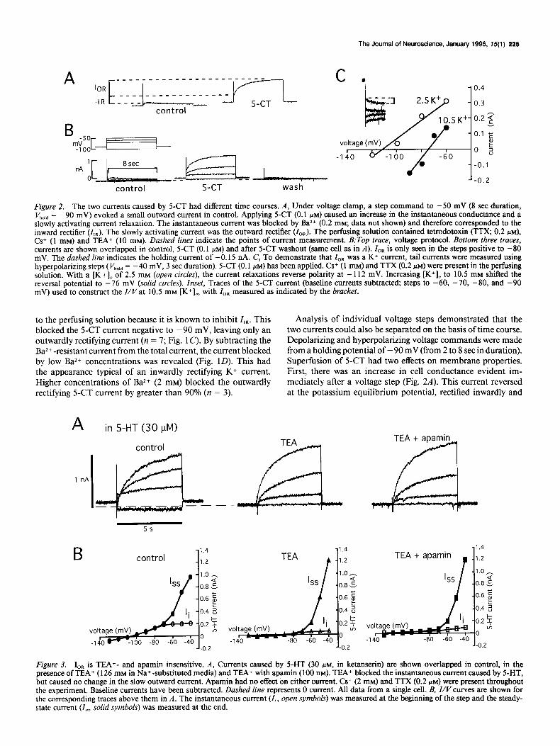
Figure 2. The two currents caused by 5-CT had different time courses. A, Under voltage clamp, a step command to -50 mV (8 set duration, V hold = -90 mV) evoked a small outward current in control. Applying 5-CT (0.1 PM) caused an increase in the instantaneous conductance and a slowly activating current relaxation. The instantaneous current was blocked by Ba2+ (0.2 mM; data not shown) and therefore corresponded to the inward rectifier (I,,). The slowly activating current was the outward rectifier (I,,,). The perfusing solution contained tetrodotoxin (TTX; 0.2 PM), Cs+ (1 mM) and TEA+ (10 mM). Dashed lines indicate the points of current measurement. B:rop truce, voltage protocol. Bottom three traces, currents are shown overlapped in control, 5-CT (0.1 PM) and after S-CT washout (same cell as in A). ZOR is only seen in the steps positive to -80 mV. The dashed line indicates the holding current of -0.15 nA. C, To demonstrate that ZOR was a K+ current, tail currents were measured using hyperpolarizing steps ( Vhold = -40 mV, 3 set duration). 5-CT (0.1 PM) has been applied. Cs+ (1 mM) and TTX (0.2 PM) were present in the perfusing solution. With a [K+], of 2.5 mM (open circles), the current relaxations reverse polarity at - 112 mV. Increasing [K+], to 10.5 mM shifted the reversal potential to -76 mV (solid circles). Inset, Traces of the 5-CT current (baseline currents subtracted, steps to -60, -70, -80, and -90 mV) used to construct the Z/V at 10.5 mM [K’],, with Z,, measured as indicated by the bracket.
to the perfusing solution because it is known to inhibit Z,n. This Analysis of individual voltage steps demonstrated that the blocked the 5-CT current negative to -90 mV, leaving only an two currents could also be separated on the basis of time course. outwardly rectifying current (n = 7; Fig. 1 C). By subtracting the Depolarizing and hyperpolarizing voltage commands were made Ba2+ -resistant current from the total current, the current blocked from a holding potential of - 90 mV (from 2 to 8 set in duration). by low Ba2+ concentrations was revealed (Fig. 1D). This had Superfusion of 5-CT had two effects on membrane properties. the appearance typical of an inwardly rectifying K+ current. First, there was an increase in cell conductance evident im- Higher concentrations of Ba2+ (2 mM) blocked the outwardly mediately after a voltage step (Fig. 2A). This current reversed rectifying S-CT current by greater than 90% (n = 3). at the potassium equilibrium potential, rectified inwardly and
A in 5-HT (30 pM)
control
1 nA
control
1.0 - Q
0.8 .!?
0.6 E
0.4 2
0.2 2 A
0
-0.2
-
TEA
TEA + apamin
a
TEA + apamin
r
-1.4
-1.2
-1.0 -
.0.8 2
.0.6 E
-0.4 2
- 0.2 2 3,
-0
-0.2
Figure 3. I,, is TEA+- and apamin insensitive. A, Currents caused by 5-HT (30 PM, in ketanserin) are shown overlapped in control, in the presence of TEA+ (126 mM in Na+-substituted media) and TEA+ with apamin (100 nM). TEA+ blocked the instantaneous current caused by 5-HT, but caused no change in the slow outward current. Apamin had no effect on either current. Cs+ (2 mM) and TTX (0.2 PM) were present throughout the experiment. Baseline currents have been subtracted. Dashed line represents 0 current. All data from a single cell. B, Z/V curves are shown for the corresponding traces above them in A. The instantaneous current (I,, open symbols) was measured at the beginning of the step and the steady- state current (I,,, so/id symbols) was measured at the end.

226 Bobker and Williams * Two K+ currents in PH
A, control
A2 BAPTA
B
4&--y.T 4L----Jlom” 1 0.6
I ss l
J -0.4
Figure 4. BAPTA inhibited I,,. A,, The effectiveness of intracellular BAPTA delivery was assessed by measurement of the sAHP. In a control cell, the sAHP is 16 mV. Dashed line represents holding potential of - 70 mV. The action potential has been truncated, and the trace is the average of four consecutive spikes. A,, In a cell impaled with a BAPTA- filled electrode (100 mM), the sAHP was nearly blocked after 15 min. B, I/Vcurve from another cell impaled for 15 min with a BAPTA-filled electrode (50 mM). 5-HT (30 PM, with ketanserin) has been applied. The instantaneous current (I,, open circles) reversed at - 110 mV and rec- tified inwardly. The steady-state current (I,, solid diamonds) still had a slow outward component, but it is markedly reduced as compared with control cells. Inset, Representative steps at -50, -70, - 100, and - 120 mV are shown (I’,,,, = -90 mV, 5 set duration). Baseline currents have been subtracted.
was blocked by Ba2+ (0.2 mM). Second, 5-CT caused a slowly developing outward current relaxation that was noninactivating. This time-dependent current caused the outward rectification seen with low Ba*+ (0.2 mM) and will be termed Z,,. ZOR was first apparent in the steps made positive to -80 mV (Fig. 2B); its amplitude was 63 ? 25 pA at -70 mV and 779 & 74 pA at -40 mV (n = 6).
The reversal potential of I,, was determined by using a tail- current protocol. In the presence and absence of 5-CT, hyper- polarizing steps ( Vhold = -40 mV) were made to measure the decay of Z,,. Current reversal occurred at - 100 -t 3 mV in 2.5 [K+],, (n = 6, with two cells not reversing). Increasing [K+], to 10.5 mM shifted the reversal to -69 + 4 mV (n = 3; Fig. 2C). This shift was close to what would be predicted by the Nemst equation for a K+ conductance.
Effects of K+ channel blockers
As indicated above, extracellular BaZ+ at low concentration (0.2 mM) blocked I,,, while high concentration (2 mM) blocked ZOR. Tetraethylammonium chloride (TEA+; 126 mM in Na+-substi- tuted media) was found to provide the best separation of the currents, as it had no effect on ZOR while causing a near complete block of I,, (n = 4; Fig. 3). TEA+ did cause the release of some endogenous 5-HT, however, as indicated by the appearance of
A -60
-65
0.1 I -80 -75 -70 -65 -60 -55
voltage (mV)
Figure 5. Deactivation kinetics of I,,. A, In 5-HT (30 PM, with ke- tanserin), tail currents are shown (only the time-dependent component) for steps to the indicated potential (I’,,,, = -50 mV, 15 set duration). The double exponential curve fit is superimposed (solid line) and was used to determine time constants r, and r1 (indicated next to each curve). All data from a single cell. Cs+ (1 mM) was present in the perfusant. B, Semilogarithmic plot oftime constant versus membrane potential. Data are averaged from three cells. Error bars are the SEM and are not shown when they fell within the data symbol.
ZOR before the application of 5-HT. Baseline currents for sub- traction were therefore obtained by applying the 5-HT,, antag- onist I -(2-methoxyphenyl)-4-[4-(2-phthalimmido)butyl]pi- perazine (NAN- 190; 1 KM; Glennon et al., 1988) which blocked both ZOR and I,, (n = 3). Other compounds that had no effect on ZOR were Cs+ (2 mM), 4-aminopyridine (5 mM), and apamin (100 nM; Fig. 3). Z,, was blocked by internal Cs+ (I M in the recording electrode as CsCl)
Calcium dependence
The slow activation and voltage dependence of I,,, suggested possible calcium dependence. Superfusion of CdCl, (100-500 PM in phosphate-free media) caused a 78 _t 11% reduction in ZOR at - 50 mV (n = 3). The effect of the calcium buffer BAPTA was then tested. Intracellular loading was achieved either by adding BAPTA to the microelectrode solution or by using the

The Journal of Neuroscience, January 1995, 75(l) 227
membrane-permeant BAPTA/AM. With either method, reduc- tion of the slow afterhyperpolarization (sAHP) was used as a measure of effective loading. Treated neurons had a reduced sAHP, with a peak amplitude of 8.4 f 1.0 mV (n = 11); this compares with a control sAHP of 15.8 + 0.5 mV (n = 5, P < 0.01; Fig. 4A). However, their input resistance (100-200 Ma), spontaneous firing rates (3-10 Hz), and location within the slice were typical of type I PH neurons. 5-HT (30 FM) applied to BAPTA-loaded neurons caused an instantaneous current that reversed at -98 f 8 mV and rectified inwardly (Fig. 4B). In contrast, IOR was completely absent in some cells and reduced in others; at - 50 mV, the amplitude of the slow current relax- ation was 508 + 65 pA in control cells (n = 6) and 55 + 32 pA in BAPTA treated cells (n = 4, P < 0.01).
Kinetics of I,,
ZOR activated slowly, reaching half-maximal amplitude in 1.2 ? 0.2 set at -50 mV (n = 5). Deactivation kinetics were deter- mined using the tail protocol and 15 set long steps to allow the current to reach steady state. Current relaxations were obtained between -60 and -75 mV (l/hold = -50 mV). The current deactivation had an early fast decay followed by a later slow decay. An exponential curve fitting algorithm closely approxi- mated these relaxations using the sum of two exponential func- tions (Fig. 5A), while it could not do so with a single exponential. At -60 mV, the resulting time constants for 7, (fast decay) and r2 (slow decay) were 1.3 +- 0.2 and 3.4 f 0.2 set, respectively (n = 3). Both time constants were voltage-dependent, with an e-fold change occurring in 10 mV for T, and in 20 mV for 72 (Fig, 5B).
Ionic currents underlying the IPSP Having determined that 5-CT (as well as 5-HT and 8-OH- DPAT) activated two K+ currents, we investigated which cur- rents were responsible for generating the IPSP. A single electrical stimulus near the impaled neuron caused a 5-HT-mediated IPSP with a duration of 1 set and a peak amplitude of lo-35 mV occurring at about 200 msec (Bobker and Williams, 1990). In voltage clamp, the inhibitory postsynaptic current (IPSC) amplitude ranged from 200 to 600 pA and peaked at 207 ? 7 msec (n = 5; at -55 mV). The IPSC reversed polarity at - 109 ? 3 mV in 2.5 mM [K+], (n = 3). The reversal of the IPSC suggested that at least a component of the current was due to I,,, as IOR does not pass inward current under these conditions. We hypothesized that if I,, was the only current contributing to the IPSC, then low concentrations of Ba*+ should completely block the IPSC. Instead, Ba*+ (0.2 mM) caused most of its in- hibition at more negative potentials, effectively blocking the reversal of the IPSC (Fig. 6A). At - 55 mV, the IPSC amplitude was 410 + 95 pA in control and 323 + 113 pA in Ba2+ (dif- ference not significant); at - 125 mV, amplitudes were - 2 13 + 55 pA in control and -3 k 3 pA in Ba*+ (P < 0.05, n = 3). A plot of IPSC amplitude versus voltage (IPSC/ k’) in the presence and absence of Ba*+ (Fig. 6B) therefore recapitulated the Z/I/ curve of 5-CT under those conditions (see Fig. 1 C).
A second prediction, based on the slow activation of ZoR, was that longer exposures to transmitter should activate more of ZOR and increase the amplitude of the IPSC. Trains of stimuli (3- 10 pulses) at 40 Hz were used to create a long “burst” of 5-HT release (see methods). Figure 7A shows IPSCs evoked at two potentials. After holding the cell at - 50 mV for at least 10 set, a single stimulus or a train of stimuli were delivered. The train
A coytrol in Ba2+(0.2 mM)
ro.4 I5
-1
L-0.2
Figure 6. Both I,, and I,, contribute to the IPSC. A, In voltage clamp, IPSCs were evoked with a single stimulus (at mww, shown in first trace only) from three different potentials as indicated. In control, the reversal of the IPSC occurred at about - 110 mV. Ba2+ (0.2 mM) reduced the amplitude of the IPSC primarily at more negative potentials and pre- vented its reversal. The brief downward deflections are stimulation artifacts. These experiments were done in the presence of 6-cyano-7- nitroquinoxaline-2,3-dione (CNQX; 10 PM), 2-amino-5-phosphonov- alerate (APV, 10 PM) and (-)bicuculline (10 WM), because Ba*+ caused some spontaneous glutamatergic and GABAergic synaptic potentials. B, In another cell, the same type of experiment was performed to con- struct an IPSC/Vcurve (control, open squares; in Ba’+ (0.2 mM), solid circles).
caused an augmentation of the IPSC that increased with the number of stimuli. At a holding potential of -90 mV, where I,, is inactive, no augmentation of the IPSC was seen with multiple stimuli. This resulted in a voltage-dependent augmen- tation that closely followed the voltage dependence of IOR (Fig. 7B). This experiment supports the idea that I,, contributes to the IPSC and can significantly augment it depending on the number of stimuli.
Discussion This study demonstrates that in type I PH neurons bath applied 5-HT or 5-CT activated two K+ currents, I,, and I,,. This is supported by the experiments with TEA+ and BAPTA, showing selective blockade of I,, and IOR, respectively. I,, has properties similar to those described for classical G protein<oupled in- wardly rectifying K+ currents: it activated rapidly and was sen- sitive to Ba2+ and TEA+. This finding is similar to what has been reported for the 5-HT,, receptor in other brain regions (Andrade and Nicoll, 1987; Williams et al., 1988). However, the observation of a second K+ conductance being activated by the 5-HT,, receptor has not been reported. ZOR had properties that were quite distinct from I,,: it activated slowly, rectified outwardly, and was much less sensitive to Ba*+. It is of interest to note that in rat CA1 hippocampal neurons, p-opioid receptors

228 Bobker and Williams l Two K+ currents in PH
B 0.6
-50 mV -90 mV
Figure 7. I,, potentiated the IPSC. A, In the upper three traces, at -50 mV IPSCs were evoked (at arrow, indicated infirst trace) using 1 (1 x), 3 (3 x), or 10 (10 x) stimuli (supramaximal stimulus, train at 40 Hz). Longer trains of stimuli increased the IPSC amplitude. Dashed line indicates the amplitude of the 1 x IPSC for comparison. Fast upward deflections are stimulation artifacts. The lower three traces show same experiment conducted at -90 mV. All traces from one cell. B, Averaged data from four cells showing the IPSC amplitude at - 50 and -90 mV. Hatched bars indicate the amplitude following a single stimulus; solid bars, for 10 stimuli. The IPSC amplitude was increased (82 f 14%, P <: 0.05, Student’s t test) by multiple stimuli at -50 mV, while not being significantly affected at -90 mV. Error bars indicate the SEM.
also ‘activate inwardly and outwardly rectifying K+ conduc- tances (Wimpey and Chavkin, 199 1). The latter current reported by these investigators was different from Z,, reported here, how- ever. They described a current that activated rapidly, appeared only with strong depolarizations (positive to -60 mV) and was insensitive to Cd*+. In addition, they did not observe inward and outward rectifiers occurring in the same cell. Therefore, Gi coupled receptors may have the ability to activate a variety of K+ conductances in different cell types.
Our experiments to characterize ZoR demonstrate that it has properties in common with both Ca-activated K+ currents and the muscarine-sensitive M-current. The inhibition of ZoR by Cd2+ and intracellular BAPTA suggests that it is Ca dependent and may therefore belong to the former group. Furthermore, Ca-dependent K+ channels often exhibit slow activation kinetics (Brown and Griffith, 1983; Lancaster and Adams, 1986; North and Tokimasa, 1987) and may be modulated by neurotrans- mitters (Trautmann and Marty, 1984; Lancaster and Adams, 1986). Arguing against this is its activation at relatively negative potentials and insensitivity to TEA+ and apamin, inhibitors of BK and SK channels, respectively. However, as apamin-insen- sitive SK channels have been described, we cannot eliminate the possibility that ZOR is such a conductance (Latorre et al., 1989). In comparing ZoR with M-current, they are found to share a similar voltage dependence, display insensitivity to most K+ channel blockers and are both blocked by high concentrations of Ba*+ (Brown and Adams, 1980; Adams et al., 1982). External Cd2+ has been reported to inhibit M-current by some investi- gators (Tokimasa and Akasu, 1990) while others have not seen an effect (Adams et al., 1982; Madison et al., 1987). There are two important differences between Z,, and M-current, however. First, ZOR has significantly slower kinetics of activation and de- activation (Marrion et al., 1992). Second, ZOR appears to be dependent on agonist activation; in contrast, while M-current can be modulated by various transmitters (Brown and Adams, 1980; Moore et al., 1988; Sims et al., 1988), depolarization alone is sufficient for activation. In summary, the properties of I,, are most consistent with a Ca-activated K+ current. The source of Ca*+ could be voltage-dependent entry through calcium chan- nels, as suggested by the Cd*+ inhibition of Z,,. However, CdZ+
is known to have effects unrelated to channel blockade. In ad- dition, the failure of ZoR to activate with depolarization alone argues against this possibility. A more likely source ofCa2+ then, would be receptor-mediated release from intracellular stores.
The pharmacological data indicates that the 5-HT,, receptor is responsible for activating both Z,R and ZoR. This is supported by the observation that 5-CT and 8-OH-DPAT activated both currents. Although 5-CT is an agonist at all 5-HT, receptors, 8-OH-DPAT is quite selective for the 5-HT,, subtype (Gozlan et al., 1983). In addition, NAN- 190 blocked both currents. With respect to second messengers, previous studies of the cloned human 5-HT,, receptor have demonstrated that it couples neg- atively to adenylate cyclase through a G, protein (Fargin et al., 1989), as well as stimulating phospholipase C and increasing cytosolic Ca2+ levels (Raymond et al., 1992). It also known that G proteins can directly activate some channels (Brown, 1990) including some inward rectifiers (Yatani et al., 1987; Kubo et al., 1993). Therefore, it is possible that 5-HT,, receptor acti-
vation leads to a single second messenger that triggers two con- ductances or that distinct transducers are involved. Our exper- iments suggest that the pathway leading to Ca2+ release may be critical for evoking ZOR.
A significant finding of this report was that both Z,R and ZoR contributed to generating the current that underlies the IPSP. Two lines of evidence support this contention. First, we dem- onstrated that superfusion of 5-CT and 5-HT activated the two conductances. There is no reason to anticipate that synaptically released 5-HT would not have a similar effect. Second, low concentrations of Ba2+ blocked Z,R during bath application of 5-HT, with the remaining current being ZoR. The same concen- tration of Ba2+ reduced the amplitude of the IPSC at more negative potentials and prevented its reversal. If the IPSC was mediated by Z,R alone, then Ba2+ should have caused a near complete inhibition of it. Instead, it revealed an IPSC/I/curve that was similar to the Z/I/ for the outward rectifier. This in- dicates that the IPSC has components of each current.
In addition to contributing to generation of the IPSP, ZOR may serve to modulate its amplitude. In our experiments, we used a high frequency train of stimuli to cause a prolonged release of transmitter. This resulted in a voltage-dependent augmen-

The Journal of Neuroscience, January 1995, 15(l) 229
tation of the IPSC that closely followed the voltage-dependence of I,,. The IPSP amplitude may therefore be determined by a relatively constant input from I,, and a variable input from ZoR. This variation might be observed physiologically if the presyn- aptic neuron undergoes burst-firing. The findings presented here are significant then for two reasons. First, they indicate that a K+ current other than Z,, can contribute to an IPSP. Second, this represents a manner by which neurotransmission may be potentiated at the postsynaptic membrane and suggests that one function of slowly activating currents may be regulation of the intensity of synaptic transmission.
References Adams P. Brown DA, Constanti A (1982) Pharmacological inhibition
of the M-current. J Physiol (Lond) 332:223-262. Andrade R. Nicoll RA (1987) Pharmacologically distinct actions of
serotonin on single pyramidal neurons of the rat hippocampus re- corded in vitro. J Physiol (Lond) 394:99-124.
Bobker DH (1994) A slow excitatory postsynaptic potential mediated bv 5-HT, receptors in nucleus prepositus hypoglossi. J Neurosci 14: 2428-2434. -
Bobker DH, Williams JT (1989) Serotonin augments the cationic cur- rent I, in central neurons. Neuron 2: 1535-l 540.
Bobker DH, Williams JT (1990) Serotonin-mediated inhibitory post- synaptic potential in guinea-pig prepositus hypoglossi and feedback inhibition bv serotonin. J Phvsiol (Lond) 422:447-462.
Brown AM (i990) Ionic channels and their regulation by G protein subunits. Annu Rev Physiol 52: 197-2 13.
Brown DA. Adams PR (1980) Muscarinic suppression of a novel voltage-sensitive K+ current ‘in a vertebrate neurone. Nature 283: 673-675.
Brown DA, Griffith WH (1983) Calcium-activated outward current in voltage-clamped neurones of the guinea-pig, J Physiol (Lond) 337: 287-301.
Del Castillo J, Katz B (1955) Production of membrane potential changes in the frog’s heart by inhibitory nerve impulses. Nature 175:1035.
Dutar P, Nicoll RA (1988) A physiological role for GABA, receptors in the central nervous system. Nature 332: 156-l 58.
Egan T, Henderson G, North RA, Williams JT (1983) Noradrenaline- mediated synaptic inhibition in rat locus coeruleus neurones. J Physiol (Lond) 345~477-488.
Fargin A, Raymond J, Regan J, Cotecchia S, Lefkowitz R, Caron M (1989) Effector coupling of the cloned 5-HT,, receptor. J Biol Chem 264:14848-14852.
Glennon RA, Naiman N, Pierson M, Titeler M, Lyon R, Weisberg E (1988) NAN- 190: an arylpiperazine analog that antagonizes the stim- ulus effects of the 5-HT,, agonist 8-hydroxy-2-(di-n-propylami- no)tetralin (8-OH-DPAT). Eur J Pharmacol 154:339-34 1.
Gozlan H, El Mestikawy S, Pichat L, Glowinski J, Hamon M (1983) Identification of presynaptic serotonin autoreceptors using a new li- gand: ‘H-PAT. Nature 305: 140-142.
Hille B (1992) Potassium channels and chloride channels. In: Ionic channels of excitable membranes, pp 115-l 39. Sunderland, MA: Sin- auer.
Kubo Y, Reuveny E, Slesinger PA, Jan YN, Jan LY (1993) Primary structure and functional expression of a rat G-protein-coupled mus- carinic potassium channel. Nature 364:802-806.
Lancaster B, Adams PR (1986) Calcium-dependent current generating the afterhyperpolarization of hippocampal neurons. J Neurophysiol 55:1268-1282.
Latorre R, Oberhauser A, Labarca P, Alvarez 0 (1989) Varieties of calcium-activated potassium channels. Annu Rev Physiol 5 1:385- 399.
Madison DV, Lancaster B, Nicoll RA (1987) Voltage-clamp analysis of cholinergic action in the hippocampus. J Neurosci 7:733-74 1.
Marrion NV, Adams PR, Gruner W (1992) Multiple kinetic states underlying macroscopic M-currents in bullfrog sympathetic neurons. Proc R Sot Lond [Biol] 248:207-214.
Mihara S, Nishi S, North RA, Surprenant A (1987) A nonadrenergic, noncholinergic slow inhibitory postsynaptic potential in neurones of the guinea-pig submucous plexus. J Physiol (Lond) 390:357-365.
Moore SD, Madamba SC, Joels M, Siggins CR (1988) Somatostatin augments the M-current in hippocampal neurons. Science 239:278- 280.
North RA (1989) Drug receptors and the inhibition of nerve cells. Br J Pharmacol 98: 13-28.
North RA, Tokimasa T (1987) Persistent calcium-sensitive potassium current and the resting properties of guinea-pig myenteric neurones. J Physiol (Lond) 386:333-353.
Pan ZZ, Colmers WF, Williams JT (1989) 5-HT-mediated synaptic potentials in the dorsal raphe nucleus: interactions with excitatory amino acid and GABA neurotransmission. J Neurophysiol 62:48 l- 486.
Pennington NJ, Kelly JS (1990) Serotonin receptor activation reduces calcium current in an acutely dissociated adult central neuron. Neuron 4~75 l-758.
Raymond JR, Albers FJ, Middlemiss JP (1992) Functional expression of human 5-HT,, receptors and differential coupling to second mes- sengers in CHO cells. Naunyn Schmiedebergs Arch Pharmacol 346: 127-137.
Sims SM, Singer JJ, Walsh JV (1988) Antagonistic adrenergic-mus- carinic regulation of M-current in smooth muscle cells. Science 239: 190-192.
Tokimasa T, Akasu T (1990) ATP regulates muscarine-sensitive po- tassium current in dissociated bull-frog primary afferent neurones. J Physiol (Lond) 4261241-264.
Trautmann A, Marty A (1984) Activation of Ca-dependent K channels by carbamoylcholine in rat lacrimal glands. Proc Nat1 Acad Sci USA 81:611-615.
Williams JT, Colmers WF, Pan ZZ (1988) Voltage- and ligand-acti- vated inwardly rectifying currents in dorsal raphe neurons in vitro. J Neurosci 8:3499-3506.
Wimpey TL, Chavkin C (199 1) Opioids activate both an inward rec- tifier and a novel voltage-gated potassium conductance in the hip- pocampal formation. Neuron 6:28 l-289.
Yatani A, Codina J, Brown AM, Bimbaumer L (1987) Direct acti- vation of mammalian atria1 muscarinic potassium channels by GTP regulatory protein G,. Science 235:207-2 11.