diplococcus rhesus monkey -...
TRANSCRIPT

Alterations in Thyroid Hormone Economy
during Acute Infection with
Diplococcus pneumoniae in the Rhesus Monkey
KENNETHA. WOEBERwith the assistance of WILLIAM A. HARRISON
From the U. S. Army Medical Research Institute of Infectious Diseases,Frederick, Maryland 21701
A B S T R A C T In order to study the alterations in thy-roid hormone economy that accompany an acute bacterialinfection, rhesus monkeys were inoculated i.v. with avirulent Diplococcus pneumoniae culture containing ap-proximately 108 organisms per dose. This was found toproduce a well-defined febrile illness followed in mostinstances by spontaneous recovery, thereby permittingsequential observations to be made during progressionfrom the healthy state through acute infection into con-valescence. During the acute febrile period of the in-fection, the clearance of both exogenously labeled L-thy-roxine (TV) and 3,3',5-triiodo-L-thyronine (Ts) fromtheir peripheral pools was accelerated. This alterationwas often evident by 8 hr after inoculation with thevirulent culture and could not be ascribed to a decreasein extracellular binding. Despite the accelerated hor-monal clearance, the concentrations of both endogenouslylabeled thyroid hormone and stable T4 in the sera of thesurviving monkeys remained essentially unchanged orincreased, indicating that hormonal secretion must haveincreased during this period. During the convalescentperiod, hormonal clearance was similar to preinfectioncontrol values. Leukocytes isolated from blood obtained6 hr after inoculation with the virulent culture dis-played enhanced T4-deiodinative activity.
INTRODUCTION
The effects of acute infection on thyroid hormone econ-omy are uncertain. Although acute infection has beenshown to influence thyroid function and the peripheralmetabolism of L-thyroxine (T4) and 3,3',5-triiodo-L-thy-
This work was presented in part at the Annual Meetingof the American Thyroid Association in Chicago, Illinois,November 1969.
Received for publication 9 July 1970 and in revised form7 November 1970.
ronine (TB) in several animal species, the results of suchstudies tend to be conflicting, appearing to vary with thenature of the species examined. In the rat, for example,acute streptococcal and pneumococcal infections appearto result in a depression of thyroid function (1, 2),whereas in man data obtained after treatment of acutepneumococcal pneumonia had been instituted suggestthat an increase in thyroid function might accompanythis type of stress (3).
We undertook the present study in order to assessin a sequential fashion various aspects of thyroid hor-mone economy during progression from the healthy statethrough an acute bacterial infection into convalescence.The rhesus monkey was employed as the animal modelbecause we felt that its response should be most closelyrepresentative of that of man. Diplococcus pneumoniaewas employed as the infecting microorganism.
METHODSMale rhesus monkeys (Macaca mulatta) weighing between2.1 and 3.7 kg were secured in primate chairs. In the ex-periment in which it was desired to label the intrathyroidaliodine pool, eight monkeys were fed a low iodine diet.'After a 3 wk period of adaptation had elapsed, each monkeywas given an i.v. injection of 25 ,uCi of carrier-free inor-ganic 125I 2 to label the intrathyroidal iodine pool. 1 ml bloodsamples were obtained by saphenous venipuncture dailythereafter. 10 days later, each monkey was given an i.v.injection of 10 /ACi (approximately 0.3 ,ug) of 'I-labeled L-thyroxine' (T4-'I) in 1 ml of 1% (w/v) human serumalbumin to label the peripheral hormonal pool. After a 5
'Modified SKF monkey diet containing vitamin fortifica-tion mix and U.S.P. salt mix (less iodine), obtained fromGeneral Biochemicals, Div., North American Mogul Prod-ucts Co., Chagrin Falls, Ohio.
2 Obtained from New England Nuclear Corp., Boston,Mass.
'Obtained from Abbott Laboratories, Chemical MarketingDiv., North Chicago, Ill.
378 The Journal of Clinical Investigation Volume 50 1971

day control period had elapsed, six monkeys were inocu-lated i.v. with a Diplococcus pneumoniae (Type 1-A) cul-ture containing approximately 108 virulent organisms perdose, and two were sham-inoculated with an equal volumeof normal saline. This was immediately followed by asecond i.v. injection of 10 AGCi of T4-'31I. 1 ml blood sampleswere obtained every 8 hr for the first 32 hr after inocula-tion, and collections were continued daily thereafter. 5 daysafter inoculation, the surviving monkeys were given a thirdi.v. injection of 10 ,uCi of T4-l"I, and collection of bloodsamples was continued for a further 5 days. Two additionalmonkeys were employed to assess the effects of a heat-killed D. pneumoniae culture on the peripheral metabolismof T4-'"I. Here, the culture was subjected to a temperatureof 57.5'C for 30 min, and plating out confirmed the absenceof viable organisms. Immediately after inoculation with theheat-killed culture, 10 ACi of T4-13I was injected i.v., andblood samples were obtained over the next 5 days.
In the experiment in which it was desired to assess con-currently the peripheral metabolism of both T4 and T3,four additional monkeys were maintained on a diet whichhad an iodine content of 1.6 ppm.4 Two monkeys wereinoculated i.v. with the virulent D. pnteumoniae culture, andthis was followed immediately by an i.v. injection of 10 ,Ciof T4-"'I and 5 GCi (approximately 0.1 /.g) of 'SI-labeledT35 (T3-'I). The remaining two monkeys served as con-trols. 1 ml blood samples were obtained by saphenous veni-puncture every 8 hr for the first 48 hr after inoculation andthen daily for an additional 3 days.
In all experiments, all injections were given between 1and 2 p.m., and days or fractions thereof are in relationto this time. Temperature was measured rectally with athermistor thermometer.
The concentrations in serum of protein-bound 125I (PB'Iand T3-'5I) and protein-bound I (T4-'31I) were measuredas follows. To 250 Al of serum were added 1 drop of 2 Mpotassium iodide and a few milligrams of thiouracil, andthe protein-bound radioactivity was precipitated with cold20% trichloroacetic acid. The precipitates were washed twicewith cold 5%o trichloroacetic acid and then dissolved with2 N NaOH to a standard volume for counting in a well-type scintillation counter. Corrections were made for thecontribution of the 'I to the '5I counts in those samplesthat contained both isotopes. Counting standards were pre-pared from the injection solutions immediately after ad-ministration and, in the case of T4V-I and T3-'I, werealso subjected to precipitation with trichloroacetic acid afterthe addition of a small amount of 25% (w/v) human serumalbumin. 94-96% of both the labeled T4 and T3 was re-covered in the precipitate.
After each injection of T4-'31I, the declining concentrationof radioactivity in serum during the subsequent 5 day periodwas plotted against time. By 16 hr after injection, thecurve appeared to conform to a single exponential function,indicating that distribution equilibrium of the residual T4-131I had been attained. The data obtained at 16 hr andthereafter were therefore used to calculate values for thefractional rate of T4 disappearance. In the infected monkeys,the rate of disappearance of T4-'31I from serum slowedabruptly between 2 and 3 days after inoculation with theculture. Accordingly, values for the fractional disappearancerate were calculated separately from the data obtainedduring the first 2 days and from the data obtained from
4Monkey chow obtained from Ralston Purina Co., St.Louis, Mo.
5 Obtained from Abbott Laboratories.
days 3 through 5. Values for the volume of T4 distributionwere calculated as the quotient of injected radioactivityand the concentration of radioactivity in serum at the timeof injection as obtained by backward extrapolation of thedisappearance curve. Correction was always made for theconcentration of radioactivity remaining from a previousinjection of T4-'311. In the case of the infected monkeys,values for the volume of distribution during the periodfrom days 3 through 5 after inoculation with the culturewere calculated as follows. Since it was not possible to ob-tain complete urine and stool collections, the residual T4-3"I at the end of the first 2 days was estimated as theproduct of the values for the calculated volume of distribu-tion during this period and the concentration of T4-'I inserum on day 2. The volume of distribution during theperiod from days 3 through 5 was then calculated as thequotient of this value and the value for the concentrationof T4-13I in serum on day 2 derived by backward extrapola-tion of the disappearance curve for days 3 through 5. Therate of T4 clearance was calculated as the product of volumeof distribution and fractional disappearance rate, and theabsolute rate of T4 disappearance as the product of clear-ance rate and the concentration of endogenous T4 in serum.
In the case of T3, the disappearance curve did not con-form to a single exponential function, but slowed progres-sively with time. Accordingly, the method of "peeling" wasemployed to derive an estimate of the rate of disappearanceof T3-'I. Owing to the small number of later points andhence the essentially arbitrary nature of the peeling, noattempt was made to calculate kinetic data for T3 disap-pearance.
The binding of T4 in serum was assessed by enrichingserum samples with the equivalent of approximately 70 Agof T4-'I per 100 ml and subjecting them to reverse-flowfilter paper electrophoresis in glycine (0.2 M) -acetic acid(0.13 M) buffer at pH 8.6, using a Durrum-type electro-phoresis cell (4). Serum samples from each monkey werealways subjected to electrophoresis concurrently in a singlecell. The distribution of T4-'"I among the binding proteinswas quantitated by cutting out the radioactive zones on thefilter paper strips with the aid of radioautographs andcounting them in a well-type scintillation counter.
The concentration of endogenous T4 in serum was mea-sured by the binding displacement method of Murphy andPattee (5).' Owing to the small volumes of serum, it wasnecessary to pool several samples for this determination.
The effects of acute infection in vivo on the deiodinationof TAXlI by leukocytes in vitro was assessed in two ex-periments. In each experiment, two fresh monkeys wereemployed of which one was inoculated i.v. with the D.pneumoniae culture containing approximately 108 virulentorganisms per dose and the other with the heat-killed cul-ture. In the one experiment, heparinized blood samples werecollected in chilled. tubes on the day before inoculation,immediately before inoculation, 6 hr after inoculation, dailythereafter for 3 days, and on day 6 after inoculation. In theother experiment, heparinized blood samples were obtainedimmediately before and again 6 hr after inoculation. Imme-diately after collection, the processing of the blood was be-gun, and the whole leukocyte isolation procedure was carriedout in chilled laboratory ware. The incubation with T4-AXIwas begun immediately after the leukocyte isolation had beencompleted. The samples from both monkeys in each ex-periment were handled concurrently. Plastic laboratory ware
'Performed by the Boston Medical Laboratory, Waltham,Mass.
Thyroid Hormone Economy in Pneumococcal Infection in the Rhesus Monkey 379

or siliconized glassware were used throughout. The leuko-cytes were isolated from the blood by the method of Bertinoet al. (6). Briefly, the blood was sedimented in a dextran-saline solution, and the supernatant layer containing theleukocytes was centrifuged. The erythrocytes remaining inthe leukocyte pellet were lysed with cold, distilled waterand removed by washing. Finally, the leukocytes were sus-pended in Krebs-Ringer phosphate buffer at pH 7.4 con-taining 2 mg of glucose per ml (KRPG). 1 ml of the leuko-cyte suspensions was added to Erlenmeyer flasks containing1 ml of KRPG and 50 jul (approximately 0.25 ,ug) ofT4-A'I. Additional flasks containing 2 ml of KRPG and50 jul of T4-]'I were prepared to serve as tissue-free con-trols. All flasks were prepared in duplicate. The flasks wereincubated at 370C in 100% oxygen in a metabolic shaker.Preliminary experiments had indicated that plateau valuesfor T4-AI deiodination by leukocytes are attained by ap-proximately 1 hr. Consequently, in the one experiment inwhich it was desired to obtain plateau measurements, incu-bation was allowed to proceed for 2 hr. After incubation,500 til of 25% (w/v) human serum albumin containingpotassium iodide and thiouracil was added to each flask to
stop the reaction. In the other experiment, the rate of T4-AX'Ideiodination was assessed by withdrawing samples from theincubation medium after 10, 15, and 30 min of incubationand transferring them to tubes containing serum albumin,potassium iodide, and thiouracil. 20 jul from each flask ortube were then subjected to ascending chromatography inNo. 1 Whatman filter paper strips in a butanol-acetic acid-water (120: 30: 50) solvent system (7). The percentageof the total radioactivity present as inorganic iodide andorigin material, representing the percentage of T4-11I de-iodinated, was quantitated by cutting out the radioactivezones on the filter paper strips with the aid of radioauto-graphs and counting them in a well-type scintillation counter.These values were corrected for spontaneous deiodination bysubtracting from them the corresponding tissue-free controlvalue.
RESULTS
Fig. 1 depicts the results of an experiment conducted ineight monkeys in which the intrathyroidal iodine poolwas labeled with inorganic 'I and the peripheral hor-
SHAM-INOCULATED
-!
INFECTED SURVIVORS
lI\.I !..
i -4 - 3 -2 -I O 1 2 3 4 5i~~~~~
DAYS DAYS DAYS
FIGURE 1 The effects of acute infection with Diplococcus pneumoniae on the concentrationin serum of endogenously synthesized protein-bound 'I (PB'1I) and on the disappearancefrom serum of injected '3I-labeled L-thyroxine (T4-AI). The time of inoculation with theculture or sham-inoculation with normal saline is designated as day 0.
380 K. A. Woeber
INFECTED NONSURVIVORS
w
W4
0.
w
I-
an
14D2.0
a .z
Een 0
2
In:
I

TABLE I
The Effects of Acute Infection with Diplococcus pneumoniae on the Kinetics of Peripheral31I-Labeled L-Thyroxine (T4-'311) Metabolism
Fractional T4 Absolute T4Monkey Volume of T4 disappearance T4 clearance disappearance
No. Weight Period* distribution rate rate Serum T4 rate
kg ml %/day mi/day Ag/100 ml jug/day
Sham-inoculated with normal saline1 2.28 Control
Postinoculation
2 2.26 ControlPostinoculation
Infected by inoculation with the virulent culture3 2.36 Control
Acute (104.40F at 24 hr)Convalescent IConvalescent II
4 2.48 ControlAcute (105.40F at 24 hr)Convalescent IConvalescent II
5 2.38 ControlAcute (105.10F at 32 hr)Convalescent IConvalescent II
6 2.44 ControlAcute (105.20F at 16 hr)Convalescent IConvalescent II
7 2.44 ControlAcute (105.30F at 16 hr)
8 2.13 ControlAcute (104.00F at 16 hr)
Inoculated with the heat-killed culture:9 3.67 Postinoculation
10 3.60 Postinoculation
336327
284305
270310340305
323554619421
260400480306
272253284333
278247
260449
341
372
38 128 5.542 137 5.5
7.07.5
53 15151 156
35 94 6.0 5.652 161 7.0 11.326 88 7.5 6.630 92 7.5 6.9
45 14553 29440 24844 185
52 13570 28026 12537 113
50 13698 24816 4537 123
41 114119 294
62 161105 471
39 133
38
6.0 8.76.0 17.67.0 17.4
4.5 6.15.8 14.48.5 3.86.5 8.0
4.56.0
5.117.6
141
* In this column, the term "acute" refers to the first 2 days after inoculation with the virulent culture, and the maximumtemperature and its time of occurrence are indicated in parentheses. Convalescent I refers to days 3 through 5 afterinoculation. The same injection of T4-1311I was used to assess the kinetics of T4 metabolism during both these periods.Convalescent II refers to the period following the fresh injection of T4-'31I on day 5 after inoculation.t Control observations prior to inoculation were not obtained in this group. Nevertheless, the values following inoculationare similar to the control values presented here.
monal pool with TA-XI. The time of inoculation is de-signated as zero time and is indicated by the dashedvertical lines. As depicted in the upper panels, fever wasusually present by 8 hr after inoculation with the cultureand lasted for 2-3 days. A moderate neutrophilia wasalso present during this period in the four monkeys thatsurvived the infection, whereas leukopenia occurred in
the two nonsurvivors.7 This initial period will be termedthe acute period, whereas the period from day 3 onwardswill be termed the convalescent period. Table I sum-marizes the data for the kinetics of THAI metabolism in
'A necropsy was performed on one of these (monkey No.7) and revealed pulmonary congestion and edema. Pure cul-tures of D. pneumoniae were obtained from heart blood andfrom lung and liver tissue.
Thyroid Hormone Economy in Pneumococcal Infection in the Rhesus Monkey 381

the individual monkeys and also includes the data ob-tained in the two monkeys inoculated with the heat-killed culture. The values for the concentration of en-dogenously synthesized PB"I in the sera of the indi-vidual monkeys are presented in Table II.
During the acute febrile period of the illness, the rateof disappearance of T4-'I from serum, depicted in thelower panels of Fig. 1, was increased in all six infectedmonkeys relative to their control values (Table I). Infour of the six infected monkeys, the calculated volume ofT4 distribution was increased during the acute febrileperiod (Table I). Consequently, the calculated rate of T4clearance was greatly increased, and for the group ofsix infected monkeys as a whole this increase was sig-nificant statistically as judged from the paired t test(P <0.01). These alterations were greater in the twononsurviving monkeys than in the four infected sur-vivors. Despite the increased rate of T4 clearance, serumT4 was increased in three infected monkeys relative totheir control values and unchanged in one so that theabsolute rate of T4 disappearance was increased duringthis period (Table I).
The values for endogenously synthesized PB'I in se-rum are depicted in the middle panels of Fig. 1 as a percent of the mean of the values obtained during the con-trol period from days - 5 through 0 for each monkeyand are also presented in Table II. During the acutefebrile period, serum PB'I changed little in two of theinfected survivors and increased in the other two despitethe increased rate of T4 clearance. In the two non-survivors, serum PB'I decreased in one and fluctuatedwidely in the other.
During the convalescent period from day 3 onwardswhen the temperature had returned toward control values,a decrease in the rate of disappearance of T4-'"'I fromserum was observed (Fig. 1), and the values were con-sistently less than the control values prior to infection(Table I, convalescent I). On the other hand, the in-crease in the calculated volume of T4 distribution per-sisted, with the result that the calculated rate of Toclearance was similar to the preinfection control values(Table I, convalescent I). As depicted in Fig. 2, T4-'I given as a fresh injection during the convalescentperiod on day 5 after inoculation with the culture be-
TABLE I IThe Effects of Acute Infection with Diplococcus pneumoniae on the Concentration in Serum of Endogenously
Synthesized Protein-Bound 1211 (PB1251) after the Administration of Inorganic 1251 on Day - 10*
Serum PB126I on dayMonkey
No. -5 -4 -3 -2 -1 0 0.33 0.67 1 1.33 2 3 4 5
%Admin. dose X 103/100 mlSham-inoculated with normal saline
1 60.2 61.7 68.5 48.4 44.4 55.3 35.4 43.8 41.0 44.6 48.6 43.8 36.8 31.1(63)t (78) (73) (79) (86) (78) (65) (55)
2 21.9 35.9 30.5 35.1 12.1 42.9 38.3 36.5 22.2 29.3 17.7 41.2 25.3 22.5(129) (123) (75) (99) (60) (139) (85) (76)
Infected by inoculation with the virulent culture3 67.3 55.7 70.9 55.8 59.6 64.9 45.4 71.5 33.0 61.0 56.3 54.6 66.3 88.2
(73) (115) (53) (98) (90) (88) (106) (141)
4 31.4 11.1 29.3 31.3 32.9 23.0 30.1 34.2 27.5 10.8 44.1 49.6 33.8 46.6(114) (129) (104) (41) (166) (187) (128) (176)
5 23.6 28.7 15.9 15.1 14.6 21.7 36.3 21.1 28.6 13.2 29.3 18.2 16.6 26.1(182) (106) (144) (66) (147) (91) (83) (131)
6 28.4 21.0 17.4 20.7 32.3 38.8 20.2 24.5 29.3 16.4 55.7 68.6 63.6 54.4(77) (93) (111) (62) (211) (260) (241) (206)
7 103.9 115.2 102.4 105.4 70.2 97.9 11.2 65.2 46.8 42.7 Died(11) (66) (47) (43)
8 32.0 29.6 17.7 28.0 33.9 35.4 8.1 58.1 11.3 Died(28) (198) (38)
* The time of inoculation is designated as day 0 and immediately followed the collection of blood for the measurements shown inthis column.t The values in parentheses are those depicted in Fig. 1 and are the per cent of the mean of the values during the control periodfrom days -5 through 0 for each monkey.
382 K. A. Woeber

- N'-00 6
la
2-
-5 -4 -3 -2 -I 0 2 3 4 5 6 7 8 9 10
DAYS
FIGURE 2 The disappearance from serum of "31I-labeled L-thyroxine (T4-'I) given as a fresh injection on day 5 after inoculation with the culture.The preceding disappearance curves are those depicted in Fig. 1 for theinfected survivors.
haved in a similar fashion. Its rate of disappearancefrom serum was less and its calculated volume of dis-tribution greater than the control values in all foursurvivors (Table I, convalescent II). Values for serumT4 in the three survivors in which this determinationwas obtained were greater than the values in the samemonkeys during the preceding acute and control periods(Table I). Distinct increases in serum PB'I were alsopresent during the convalescent period in two of thefour survivors (Table II), and the monkey who dis-played the greatest increase (monkey No. 6) also hadthe greatest increase in serum T4.
No alterations in the distribution of T4-'I amongT4-binding globulin (TBG), albumin, or Ti-bindingprealbumin (TBPA) were observed during the acuteperiod of the illness (Table III). However, on day 6during the convalescent period, a slight increase in theper cent of T4 bound by TBG and a corresponding de-crease in the per cent bound by TBPA were observed.
No alterations in the kinetics of T4-I metabolism,in serum PB'I, or in T.-binding in serum were ob-served in the two monkeys sham-inoculated with normalsaline. Likewise, in the two monkeys inoculated withthe heat-killed culture, the values for the kinetics of T4-"I metabolism were similar to the control values in theother eight monkeys presented in Table I.
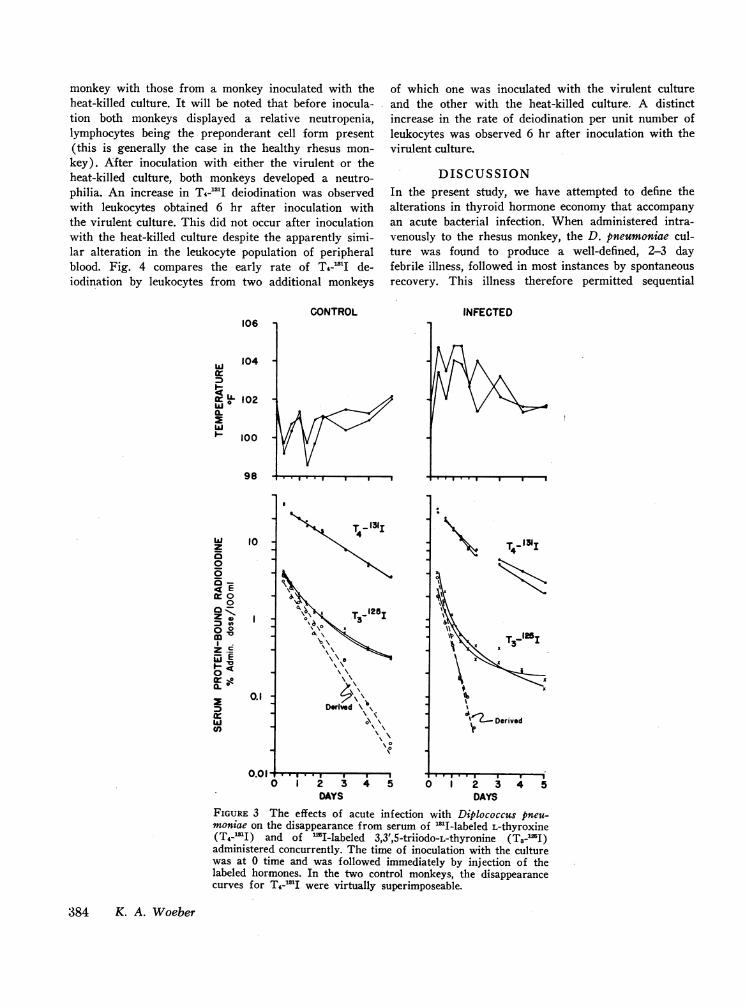
Fig. 3 compares the kinetics of T3-W2I and T4-'Idisappearance in two infected monkeys with those intwo control monkeys. In the infected monkeys, the dura-tion of the acute febrile period and the alterationsin the kinetics of T&-'I disappearance were the same asthose described earlier. In both the control and infectedmonkeys, the rate of disappearance of T3-'I from serumdecreased progressively with time. Nevertheless, the
slope of the straight line derived by the peeling tech-nique was greater in the infected monkeys during theacute period, indicating an increased fractional rate ofTs disappearance.
Table IV compares the plateau values for in vitrodeiodination of T4-13I by leukocytes from an infected
TABLE IIIThe Effects of Acute Infection with Diplococcus pneumoniae
on the Binding of '3ll-Labeled L-Thyroxine (T4-131I)by T4-Binding Globulin (TBG) and T4-Bind-
ing Prealbumin (TBPA) in Serum*
Day:Monkey
No. -5 -3 0.33 2 6
Sham-inoculated with normal saline1 %T4-TBG 30 32 28 32 31
%T4-TBPA 39 38 38 37 35
2 %T4-TBG 23 18 30 26 23%T4-TBPA 28 26 26 28 28
Infected by inoculation with the virulent culture3 %T4-TBG 27 22 23 26 29
%T4-TBPA 41 40 48 46 394 %T4-TBG 19 28 29 31 34
%T4-TBPA 45 36 37 37 245 %T4-TBG 22 21 21 28 34
%T4-TBPA 49 47 51 43 396 %T4-TBG 31 25 22 23 45
%T4-TBPA 41 45 49 37 31
* Serum samples were enriched with the equivalent of approxi-mately 70 .g of T4-1311 per 100 ml.t The time of inoculation is designated as day 0.
Thyroid Hormone Economy in Pneumococcal Infection in the Rhesus Monkey 383
monkey with those from a monkey inoculated with theheat-killed culture. It will be noted that before inocula-tion both monkeys displayed a relative neutropenia,lymphocytes being the preponderant cell form present(this is generally the case in the healthy rhesus mon-key). After inoculation with either the virulent or theheat-killed culture, both monkeys developed a neutro-philia. An increase in T4-131I deiodination was observedwith leukocytes obtained 6 hr after inoculation withthe virulent culture. This did not occur after inoculationwith the heat-killed culture despite the apparently simi-lar alteration in the leukocyte population of peripheralblood. Fig. 4 compares the early rate of T4-'I de-iodination by leukocytes from two additional monkeys
106 -
IrA 4! i
t I 102
LUa.- ~ j
98 -
CONTROL
of which one was inoculated with the virulent cultureand the other with the heat-killed culture. A distinctincrease in the rate of deiodination per unit number ofleukocytes was observed 6 hr after inoculation with thevirulent culture.
DISCUSSIONIn the present study, we have attempted to define thealterations in thyroid hormone economy that accompanyan acute bacterial infection. When administered intra-venously to the rhesus monkey, the D. pneumoniae cul-ture was found to produce a well-defined, 2-3 dayfebrile illness, followed in most instances by spontaneousrecovery. This illness therefore permitted sequential
INFECTED
A2 3DAYS
4 5I . I.. . I I
0 1 2 3DAYS
FIGURE 3 The effects of acute infection with Diplococcus pneu-moniae on the disappearance from serum of 'MI-labeled L-thyroxine(T4-'I) and of 'I-labeled 3,3',5-triiodo-L-thyronine (T3-l2I)administered concurrently. The time of inoculation with the culturewas at 0 time and was followed immediately by injection of thelabeled hormones. In the two control monkeys, the disappearancecurves for THA'I were virtually superimposeable.
384 K. A. Woeber
10
I
0.1I -~
za0
a -
4Eher 00
ogZ S0 °
0 0
coIDI
.',, E
la<
n
T4- 3i
T3- 2I3
Derived
4 5
X 1
7\ \
d \ )s\0\ \
\ 0
f

TABLE IVThe Effect of Acute Infection with Diplococcus pneumoniae In Vivo on the Deiodination of 1311-Labeled
L- Thyroxine (T4-131I) by Leukocytes In Vitro
Monkey inoculated with heat-killed culture Monkey inoculated with virulent culture
Leukocytes in whole blood Leukocytes in whole bloodLeukocytes T4-131I Leukocytes T4-131I
Day* Total PMN Bands in incubate deiodination Total PMN Bands in incubate deiodination
number/pl % % number/pl % number/ld % % number/pl %- 1 8100 17 3500 4.3 14,750 8 2700 1.6
(4.02, 4.56)t (1.59, 1.66)0 8250 8 2750 4.0 13,200 13 2700 4.5
(3.85, 4.23) (4.22, 4.70)0.25 14,000 41 2500 3.5 15,500 51 2050 11.8
(3.49, 3.57) (11.56, 12.01)1 15,200 72 1 3400 4.2 15,100 54 1 3000 3.7
(4.15, 4.26) (3.62, 3.79)2 10,100 26 2800 7.9 13,900 57 11 3050 3.0
(7.60, 8.24) (2.82, 3.26)3 9700 25 1 2400 5.0 11,600 48 8 2900 2.8
(4.90, 5.07) (2.71, 2.89)6 7900 33 1 2660 1.8 26,100 55 4 3140 3.0
(1.53, 2.15) (2.09, 3.99)
* The time of inoculation is designated as day 0 and immediatelyt The values in parentheses are the values in duplicate flasks.
observations to be made during progression from thehealthy state through acute infection into convalescence.
The earliest detectable alteration following inocula-tion with the virulent culture was accelerated clearanceof both To and T3 from their respective peripheral pools.A similar alteration has previously been observed inman during the acute phase of pneumococcal pneumonia(3). This alteration was often evident by 8 hr afterinoculation, as judged from the lower values for theconcentration of labeled hormone in serum at this timerelative to the control values. A decrease in extracellularbinding of hormone did not appear to be responsiblefor the accelerated hormonal clearance for two reasons.First, no alteration in the distribution of labeled T4among the binding proteins was observed in serum ob-tained during the acute febrile period. Second, the frac-tional rates of disappearance of both T4 and T3 fromserum were increased during this period. This is con-trary to what would be expected were a decrease inextracellular binding responsible, since alterations inextracellular binding induce alterations in the fractionalrates of To and Ti disappearance that are the converseof one another (8, 9). Rather, the data are more inkeeping with enhanced cellular uptake and metabolismof both hormones during the acute febrile period. Therole of fever in the pathogenesis of this alteration can-not be assessed in our study owing to the relatively
followed the collection of blood for the measurements shown.
small number of animals employed. However, in thestudy of acute pneumococcal pneumonia in man citedabove (3), no correlation appeared to exist betweenthe magnitude of the febrile response and the accelera-tion of hormonal disappearance.
During the convalescent period, the increased volumeof T4 distribution persisted, but a decreased fractionalrate of disappearance was observed. The latter phenom-enon has also previously been observed during recoveryfrom acute pneumococcal pneumonia in man (3). Thedecreased fractional rate of disappearance might havebeen ascribed either to more rapid thyroidal recyclingof labeled iodide liberated from more rapid peripheraldegradation of hormone or to more rapid formation ofthe iodoprotein product of T4 metabolism, or to both.Both these explanations would be consistent with anincreased flux of hormone to the cells during the pre-ceding acute febrile period. However, the T4-OI thatwas administered as a fresh injection during the con-valescent period behaved in a similar fashion. Not onlywas its volume of distribution greater and its rate ofdisappearance from serum less than the control values,but in no instance was a progressive slowing of thedisappearance curve observed. Consequently, neither in-creased thyroidal recycling of liberated iodide nor in-creased iodoprotein accumulation could be implicated asa major factor in the slowing that was observed.
Thyroid Hormone Economy in Pneumococcal Infection in the Rhesus Monkey 385

Another factor that might have been implicated inthe slowing of the fractional rate of hormonal disap-pearance was an increase in extracellular binding. Onday 6 during the convalescent period, a slightly greaterproportion of To was associated with TBG in serumenriched with approximately 70 sg of T4 per 100 ml.Although the degree of saturation of TBG was notassessed owing to the small supply of serum, this con-centration of added T4 should approach that requiredto measure binding capacity if the binding capacity ofTBG in rhesus serum is similar to that in man. Conse-quently, the increased proportion of T4 associated withTBG reflected in all likelihood an increased bindingcapacity of the protein. Nevertheless, it is unlikely thatthe increase in extracellular binding was the sole mecha-nism responsible for the alterations in the kinetics ofhormonal disappearance during the convalescent periodbecause an abrupt decrease in the volume of distributiondid not occur; in fact, the volume of distribution wasconsistently greater than that observed during the con-trol period. Rather, the data suggest that increasedcellular uptake of T4 persists into the convalescentperiod (and this would be in accord with the persistentincrease in the volume of To distribution that was ob-served), but that its access to sites of rapid metabolismis retarded by the T4 accumulated at an increased rateduring the preceding acute febrile period.
The behavior of the endogenously synthesized, labeledhormone is also of some interest. During the acutefebrile period, its concentration in the sera of the foursurviving monkeys either remained essentially un-
1.6
u IA0
t 1.2o -
x 0 1.00
= 1.0 6 hr postlnoculotionm 0.80
2 0.62Z 20.
0 .4-_ C~~~~~~ontrol
- 0.2-
changed or increased in the face of an increased rateof hormonal clearance, indicating that the rate of hor-monal secretion must have increased during this period.Although a direct comparison between the endoge-nously labeled hormone and the endogenous stable Tois not possible owing to the fact that the latter repre-sents the value for several samples of serum pooledduring each period, the failure of serum T4 to decline inthe face of accelerated hormonal clearance also supportsincreased hormonal secretion as occurring during theacute febrile period. The mechanism by which this in-crease was evoked is not known. However, it is tempt-ing to speculate that it might have been secondary tothe accelerated hormonal clearance. In one of the twononsurvivors, the concentration of endogenously labeledhormone was depressed, reflecting the greatly acceler-ated hormonal clearance, whereas in the other the con-centration in serum tended to fluctuate widely, suggest-ing possibly a transient burst in hormonal secretion.
Our data, as well as those obtained previously in manduring acute pneumococcal pneumonia (3), indicatethat during the acute febrile period the flux of hormoneto the cells is increased. Accordingly, the question towhich we next addressed ourselves concerned the rolesubserved by this increased cellular availability of hor-mone. Wetherefore directed our attention to the periph-eral blood leukocytes. This was prompted both by theirready accessibility and by the observation of Klebanoff(10) that the phagocytosis of bacteria is followed bytheir iodination and that this may represent a microbi-cidal mechanism in the leukocyte. Many peripheral
0 10 20 30 0 10 20 30MINUTES MINUTES
FIGURE 4 Comparison of the effects of inoculation with the heat-killed(left graph) and virulent (right graph) Diplococcus pneumoniae cultureson the rate of T4-1I deiodination by leukocytes in vitro.
386 K. A. Woeber

tissues possess a T4-dehalogenase, and one of the prod-ucts of this dehalogenation reaction is iodine in arelatively oxidized state (11) that should be capableof iodinating bacteria. In the present study, leukocytesobtained from monkeys 6 hr after inoculation with thevirulent culture displayed enhanced T4-deiodinative ac-tivity, whether assessed by early rate measurements orby plateau measurements. This did not appear to berelated to the alteration in leukocyte population becausea similar alteration followed inoculation with the heat-killed culture, and this was accompanied by little orno change in deiodinative activity. During this earlyperiod after inoculation, the bacteria are undergoingmultiplication and are being rapidly phagocytosed.Thus, our observation could be interpreted as providinga source of readily available iodine in a relatively oxi-dized state for bacterial iodination though we have noevidence that bears directly on this point. On the otherhand, since the T4-dehalogenase may be a peroxidase(12) and since phagocytosis is accompanied by in-creased peroxidatic activity and hydrogen peroxidegeneration (13-15), it is entirely possible that the en-hanced deiodinative activity of leukocytes was merelyan accompanying phenomenon unrelated to any microbi-cidal action.
The observed alterations in over-all hormonal econ-omy, however, cannot be ascribed to the enhanced de-iodinative activity of the peripheral blood leukocytesfor this was short-lived. Consequently, enhanced cellularuptake of hormone must have occurred in other sites.What these sites are and what role the hormone thereinsubserves during an acute bacterial infection remainsto be determined. Finally, the similarity between thepresent data and that obtained previously in man duringacute pneumococcal pneumonia (3) suggests that therhesus monkey is a suitable animal model for studyingthe influence of infection-related stress on thyroid hor-mone economy.
ACKNOWLEDGMENTSIn conducting the research reported herein, the "Guide forLaboratory Facilities and Care" established by the Com-mittee on the Guide for Laboratory Animal Resources, Na-tional Academy of Sciences-National Research Council, wasadhered to.
I am grateful to Specialist E-5 Robert D. Egbert forexcellent technical assistance and to Captain James B. Moe,VC, U. S. Army, for performing the necropsy and pro-viding the pathological data.
REFERENCES
1. Reichlin, S., and R. J. Glaser. 1958. Thyroid function inexperimental streptococcal pneumonia in the rat. J. Exp.Med. 107: 219.
2. Shambaugh, G. E., III, and W. R. Beisel. 1966. Altera-tions in thyroid physiology during pneumococcal septi-cemia in the rat. Endocrinology. 79: 511.
3. Gregerman, R. I., and N. Solomon. 1967. Accelerationof thyroxine and triiodothyronine turnover during bac-terial pulmonary infections and fever: implications forthe functional state of the thyroid during stress and insenescence. J. Clin. Endocrinol. Metab. 27: 93.
4. Elzinga, K. E., E. A. Carr, Jr., and W. H. Beierwaltes.1961. Adaptation of the standard Durrum-type cell for re-verse-flow paper electrophoresis. Amer. J. Clin. Pathol.36: 125.
5. Murphy, B. E. P., and C. J. Pattee. 1964. Determina-tion of thyroxine utilizing the property of protein-binding.J. Clin. Endocrinol. Metab. 24: 187.
6. Bertino, J. R., R. Silber, M. Freeman, A. Alenty, M.Albrecht, B. W. Gabrio, and F. M. Huennekens. 1963.Studies on normal and leukemic leukocytes. IV. Tetra-hydrofolate-dependent enzyme systems and dihydrofolicreductase. J. Clin. Invest. 42: 1899.
7. Wilkinson, J. H., and C. H. Bowden. 1960. Iodoamino-acids and related compounds. In Chromatographic andElectrophoretic Techniques. I. Smith, editor. WilliamHeinemann Ltd., London, 2nd edition. 1: 166.
8. Zaninovich, A. A., R. Volpe, and C. Ezrin. 1969. Effectsof variations of thyroxine-binding globulin capacity onthe disappearance of triiodothyronine from the plasma.J. Clin. Endocrinol. Metab. 29: 1601.
9. Woeber, K. A., E. Hecker, and S. H. Ingbar. 1970. Theeffects of an acute load of thyroxine on the transport andperipheral metabolism of triiodothyronine in man. J. Clin.Invest. 49: 650.
10. Klebanoff, S. J. 1967. Iodination of bacteria: a bacterici-dal mechanism. J. Exp. Med. 126: 1063.
11. Galton, V. A., and S. H. Ingbar. 1961. The mechanismof protein iodination during the metabolism of thyroidhormones by peripheral tissues. Endocrinology. 69: 30.
12. Galton, V. A., and S. H. Ingbar. 1963. Role of peroxi-dase and catalase in the physiological deiodination of thy-roxine. Endocrinology. 73: 596.
13. Evans, W. H., and M. Rechcigl, Jr. 1967. Factors in-fluencing myeloperoxidase and catalase activities in poly-morphonuclear leukocytes. Biochim. Biophys. Acta. 148:243.
14. Iyer, G. Y. N., D. M. F. Islam, and J. H. Quastel. 1961.Biochemical aspects of phagocytosis. Nature (London).192: 535.
15. Paul, B., and A. J. Sbarra. 1968. The role of the phago-cyte in host-parasite interactions. XIII. The direct quan-titative estimation of H202 in phagocytizing cells. Bio-chim. Biophys. Acta. 156: 168.
Thyroid Hormone Economy in Pneumococcal Infection in the Rhesus Monkey 387