differential contribution of the mitochondrial translation...
TRANSCRIPT
Differential contribution of the mitochondrialtranslation pathway to the survival of diffuse largeB-cell lymphoma subsets
Erik Norberg1,6,7, Ana Lako1,7, Pei-Hsuan Chen1, Illana A Stanley1, Feng Zhou1,2, Scott B Ficarro1,2, Bjoern Chapuy3, Linfeng Chen3,Scott Rodig4, Donghyuk Shin5, Dong Wook Choi1, Sangho Lee5, Margaret A Shipp3, Jarrod A Marto1,2,4 and Nika N Danial*,1
Diffuse large B-cell lymphomas (DLBCLs) are a highly heterogeneous group of tumors in which subsets share molecular featuresrevealed by gene expression profiles and metabolic fingerprints. While B-cell receptor (BCR)-dependent DLBCLs are glycolytic,OxPhos-DLBCLs rely on mitochondrial energy transduction and nutrient utilization pathways that provide pro-survival benefitsindependent of BCR signaling. Integral to these metabolic distinctions is elevated mitochondrial electron transport chain (ETC)activity in OxPhos-DLBCLs compared with BCR-DLBCLs, which is linked to greater protein abundance of ETC components. Togain insights into molecular determinants of the selective increase in ETC activity and dependence on mitochondrial energymetabolism in OxPhos-DLBCLs, we examined the mitochondrial translation pathway in charge of the synthesis of mitochondrialDNA encoded ETC subunits. Quantitative mass spectrometry identified increased expression of mitochondrial translation factorsin OxPhos-DLBCL as compared with the BCR subtype. Biochemical and functional assays indicate that the mitochondrialtranslation pathway is required for increased ETC activity and mitochondrial energy reserves in OxPhos-DLBCL. Importantly,molecular depletion of several mitochondrial translation proteins using RNA interference or pharmacological perturbation of themitochondrial translation pathway with the FDA-approved inhibitor tigecycline (Tigecyl) is selectively toxic to OxPhos-DLBCL celllines and primary tumors. These findings provide additional molecular insights into the metabolic characteristics of OxPhos-DLBCLs, and mark the mitochondrial translation pathway as a potential therapeutic target in these tumors.Cell Death and Differentiation (2017) 24, 251–262; doi:10.1038/cdd.2016.116; published online 21 October 2016
Cells adapt their metabolism to satisfy changing biosyntheticand bioenergetic needs.1,2 Investigation of metabolic repro-gramming in cancer has provided insights into the metaboliccontrol of proliferation and survival.3–5 Although the initialfocus of this field has been aerobic glycolysis (the Warburgeffect),6 increasing evidence points to a complex landscape oftumor metabolic circuitries beyond aerobic glycolysis, includ-ing varied contribution of mitochondria to tumor metabolism aswell as heterogeneity in fuel utilization pathways.7–12
Diffuse large B-cell lymphomas (DLBCLs) are a geneticallyheterogeneous group of tumors that can be classified intodistinct molecular subtypes based on gene expressionprofiles. The cell-of-origin (COO) classification defined DLBCLsubsets that shared certain components of their RNA profileswith normal germinal center B cells (GCBs) or in vitro-activated B cells (ABCs), and a third undefined subsetdesignated ‘type 3’.13 Using an independent approach, the
consensus cluster classification (CCC) framework comparedthe transcriptional profiles of DLBCL groups with each otherwithout referencing normal B cells.14 CCC identified tumor-intrinsic distinctions in three highly reproducible clusters; theB-cell receptor/proliferation cluster (BCR-DLBCL) showingincreased expression of BCR signaling components, theoxidative phosphorylation cluster (OxPhos-DLBCL) markedby increased expression of genes encoding for mitochondrialelectron transport chain (ETC) components, and the hostresponse cluster (HR-DLBCL) characterized by a T-cell-richinflammatory immune cell infiltrate.14,15 COO and CC classifi-cations capture different aspects of DLBCL biology. Forexample, CCC-defined BCR-DLBCLs include BCR-depen-dent tumors of both ABC and GCB COO subtypes,16,17
whereas CC-classified OxPhos-DLBCLs include BCR-independent tumors.7,16 Our previous functional analyses ofDLBCL subtypes also uncovered quantitative proteome- and
1Department of Cancer Biology, Dana-Farber Cancer Institute, Boston, MA 02115, USA; 2Blais Proteomics Center, Dana-Farber Cancer Institute, Boston, MA 02115, USA;3Department of Medical Oncology, Dana-Farber Cancer Institute, Boston, MA 02115, USA; 4Department of Pathology, Brigham and Women’s Hospital, Harvard MedicalSchool, Boston, MA 02115, USA and 5Department of Biological Sciences, Sungkyunkwan University, Suwon 16419, Korea*Corresponding author: NN Danial, Department of Cancer Biology, Dana-Farber Cancer Institute, Harvard Medical School, 450 Brookline Avenue, CLSB 11-143, Boston,MA 02115, USA. Tel: 617 6326 436; Fax: 617 6325 363; E-mail: [email protected] address: Department of Physiology and Pharmacology, Karolinska Institute, Solna 17177, Sweden.7These authors contributed equally to this work.
Received 24.6.16; revised 08.9.16; accepted 13.9.16; Edited by N Chandel; published online 21.10.16
Abbreviations: DLBCL, diffuse large B-cell lymphoma; BCR, B-cell receptor; OxPhos, oxidative phosphorylation; ETC, electron transport chain; mtDNA,mitochondrial DNA; COO, cell-of-origin; CCC, consensus cluster classification; FAO, fatty acid oxidation; iTRAQ, isobaric tags for relative and absolute quantification; DEEPSEQ, deep efficient peptide sequencing and quantification; TUFM, Tu translation elongation factor mitochondrial; GFM1, G elongation factor mitochondria 1; MRP,mitochondrial ribosomal protein; shRNA, short hairpin RNA; ROS, reactive oxygen species; NAC, N-acetyl cysteine; SRC, spare respiratory capacity; AML, acute myeloidleukemia
Cell Death and Differentiation (2017) 24, 251–262& 2017 Macmillan Publishers Limited, part of Springer Nature. All rights reserved 1350-9047/17
www.nature.com/cdd

metabolome-level signatures associated with differences innutrient and energy metabolism.7 Specifically, these studiesshowed that BCR-dependent DLBCLs have greater glycolyticflux typical of the Warburg phenotype. Unlike BCR-DLBCLs,OxPhos-DLBCLs channel the majority of glucose-derivedpyruvate into the mitochondria, display elevated ETC activity,ATP production and fatty acid oxidation (FAO). Importantly,these metabolic distinctions are associated with subtype-selective survival mechanisms. Moreover, acute inhibition ofBCR signaling in BCR-DLBCLs increased their FAO capacity,thus revealing a reciprocal relationship between BCR signal-ing and FAO.7 Overall, these findings underscore the utility ofcapturing and dissecting metabolic distinctions in DLBCLsubtypes.The ETC is comprised of a series of large multi-subunit
complexes housed within the mitochondrial inner membrane,which carry out multiple redox reactions that ultimately lead tothe reduction of molecular oxygen to water. The initial electrondonors for these reactions are supplied by the tricaboxylic acidcycle in the form of NADH and FADH2. Respiratory chaincomplexes I (NADH dehydrogenase also known as NADH-ubiquinone oxidoreductase), III (ubiquinol-cytochrome creductase), and IV (cytochrome c oxidase) extrude protonsacross the inner membrane while transferring electrons. Theresulting proton gradient is subsequently coupled with ATPsynthesis by the activity of the F0F1 ATP synthase(complex V), completing the process of oxidative phosphor-ylation (OXPHOS). Except for complex II (succinate dehy-drogenase), the protein constituents of the ETC complexesare encoded by two independently transcribed and translatedgenomes; nuclear and mitochondrial.18,19 The mitochondrialDNA (mtDNA) encodes 13 subunits contributing to complexesI, III, IV, and V, 22 transfer RNAs, and 2 ribosomal RNAs. Themechanism for decoding the mitochondrial genome requiresnuclear-encoded factors, including ribosomes, translationinitiation, and elongation factors, and tRNA synthetases thatare distinct from the cytoplasmic counterparts dedicated totranslation of nuclear transcripts.20 Mutations in mtDNA andmitochondrial translation factors are associated with ETCfailure in several human pathologies,20,21 highlighting thefunctional relevance of the mitochondrial genome. Functionalfidelity of the ETC not only requires the coordinate synthesis ofrespiratory chain subunits encoded by the nuclear andmitochondrial genomes but also proper assembly andorganization of ETC complexes into higher-order super-complexes in the inner membrane. The ETC structuralorganization is modulated by dedicated chaperones andassembly factors, mitochondrial membrane morphology, andmembrane lipid composition.22,23 The differences in ETCactivity and OXPHOS dependency among DLBCL subtypeshas prompted investigation of pathways in charge of synthesisand assembly of respiratory chain complexes and theircontribution to metabolic heterogeneity in DLBCL subsets.Here we interrogated the mitochondrial translation machineryand its functional contribution to energy metabolism andsurvival of OxPhos-DLBCLs versus non-OxPhos/BCR-dependent subtypes.
Results
Increased expression of mitochondrial translation pro-teins in OxPhos-DLBCLs. Our previous assessment of themitochondrial proteome in OxPhos- and non-OxPhos/BCR-dependent DLBCLs revealed the enrichment of several ETCsubunits and ETC assembly factors in OxPhos-DLBCLs thatis consistent with increased ETC activity in this subtype.7
These previous analyses, based on a high-performance,single-dimension liquid chromatography–tandem mass spec-trometry platform,24 quantified predominantly nuclear-encoded ETC subunits. Because OxPhos-DLBCLs displayincreased activity of several ETC complexes that areencoded by both the nuclear and mitochondrial genomes,7
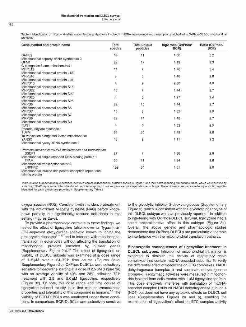
we predicted that the protein-level enrichment of ETCsubunits in this subtype would also include mtDNA-encoded subunits. To enable more extensive interrogationof the mitochondrial proteome, we utilized deep efficientpeptide sequencing and quantification (DEEP SEQ) massspectrometry25 in conjunction with isobaric tags for relativeand absolute quantification (iTRAQ) labeling. Mitochondriaisolated from three independent OxPhos- and three non-OxPhos/BCR-DLBCL cell lines were analyzed using thisplatform. The DLBCL subtype designation of these cells linesbased on the CCC and COO classifications has beenpreviously reported13,16,26,27 (Supplementary Information).The DEEP SEQ platform not only quantified the enrichmentof mtDNA-encoded ETC subunits in OxPhos-DLBCLs com-pared with BCR counterparts but also revealed significantlyhigher levels of numerous protein components of themitochondrial translation machinery (Figures 1a–e; Table 1;Supplementary Tables 1 and 2). These analyses indicatedhigher expression across several classes of mitochondrialtranslation proteins, including mitochondrial elongation fac-tors (TUFM (Tu translation elongation factor, mitochondrial)and GFM1 (G elongation factor, mitochondrial, 1)), mitochon-drial ribosomal proteins (mitochondrial ribosomal protein(MRP) S5, S7, S9, S16, S22, S25, L12, and L46), andproteins in charge of mitochondrial tRNA synthesis andfunction (DARS2 (aspartyl-tRNA synthetase 2), YARS2(tyrosyl-tRNA synthetase 2), and PUS1 (pseudouridylatesynthase 1); Figures 1a and b; Table 1). The enrichment inmtDNA-encoded ETC subunits and mitochondrial translationproteins in OxPhos-DLBCLs is further consistent with higherexpression of proteins involved in mtDNA maintenance(SSBP1 (single-stranded DNA-binding protein 1)) and tran-scription (TFAM (mitochondrial transcription factor A) andLRPPRC (leucine-rich pentatricopeptide repeat containingprotein)) in this DLBCL subtype18,28 (Figure 1f; Table 1;Supplementary Table 2).
Genetic and pharmacologic inhibition of the mitochon-drial translation pathway is selectively toxic to OxPhos-DLBCLs. Elevated levels of mitochondrial translation com-ponents in OxPhos-DLBCLs warranted examination of theircontribution to DLBCL survival. To this end, three distinctcomponents of the mitochondrial translation machinery(GFM1, TUFM, and MRPS7) were depleted using twoindependent short hairpin RNAs (shRNAs) per gene in fourOxPhos- and four non-OxPhos/BCR-DLBCL cell lines, and
Mitochondrial translation and DLBCL survivalE Norberg et al
252
Cell Death and Differentiation

cell viability was measured. Knockdown of each of thesetranslation components proved selectively toxic to OxPhos-DLBCL cell lines while sparing the non-OxPhos subset(Figures 2a–c; Supplementary Figure 1).
A common outcome of ETC inhibition, for example, followinginterference with mitochondrial translation, is the generation ofsuperoxide.29,30 This led us to predict that the toxic effects ofthe above knockdowns may be, in part, dependent on reactive
z-score
OxPhosBCR
MRPL12GFM1
MRPS7MRPS25
MRPS22MRPS5MRPS9
YARS2DARS2MRPL46TUFMMRPS16
PUS1
−1 0 1
GFM1 GFLDACEK
Ox BCR
200 1400m/z
100
Rel
ativ
e In
tens
ity, %
MRPS7 NCEPMIGLVPILK
200 1400
OxBCR
m/z
100
Rel
ativ
e In
tens
ity, %
TUFM DDTIYEDEDVK
OxBCR
200 1400m/z
100
Rel
ativ
e In
tens
ity, %
ELONGATION
INITIATION
TERMINATION
MTIF2MTIF3
TUFMGFM1
GFM2RRF
MRTF1A
tRNA
tRNAPROCESSING
PUS1DARS2YARS2
Aminoacyl tRNA
39 S MRPL12/46
mtDNA
III IV II I V
ETC
MRPS5/7/9/
16/22/25
28 S AUG AAA
mRNA
39 S
28 S AAA
39 S
28 S AUG AAA
polypeptide
22 tRNAs13 peptides
2 rRNAs
200 1000m/z
EQLPIFKTFAM
100
Rel
ativ
e In
tens
ity, %
OxBCR
Figure 1 Enrichment of mitochondrial translation components in OxPhos-DLBCL mitochondria. Multiplex quantitative proteomic analysis of isolated mitochondria from threeindependent OxPhos- (Karpas 422, Pfeiffer, and Toledo) and three non-OxPhos/BCR- (Ly1, DHL4, and DHL6) DLBCL cell lines using DEEP SEQ mass spectrometry. (a) Heatmap illustrating increased levels of mitochondrial translation proteins in OxPhos-DLBCL cell lines compared with BCR counterparts. See also Table 1 and Supplementary Table 2detailing OxPhos/BCR abundance ratios for the indicated proteins and corresponding amino-acid sequences for the identified peptides. (b) Schematic diagram ofthe mitochondrial translation machinery. Highlighted in green are proteins that were enriched in the OxPhos-DLBCL proteomic analysis shown in a, Table 1, and SupplementaryTable 2. (c–f) MS/MS spectra of the indicated unique peptides for GFM1 (c), TUFM (d), MRPS7 (e), and TFAM (f) recorded during DEEP SEQ mass spectrometry analysis ofmitochondrial proteins shown as an example of differentially enriched peptides. Ions of b- and y-type are shown in green and yellow, respectively. Relative protein ratios in BCR-and OxPhos-DLBCL cell lines are derived from iTRAQ reporter ion intensities shown in inset mass spectrum. ETC, electron transport chain; mtDNA, mitochondrial DNA; mRNA,messenger RNA; rRNA, ribosomal RNA; tRNA, transfer RNA
Mitochondrial translation and DLBCL survivalE Norberg et al
253
Cell Death and Differentiation
oxygen species (ROS). Consistent with this idea, pretreatmentwith the antioxidant N-acetyl cysteine (NAC) before knock-down partially, but significantly, rescued cell death in thissetting (Figures 2a–c).To provide a pharmacologic correlate to these findings, we
tested the effect of tigecycline (also known as Tygacil), anFDA-approved glycylcycline antibiotic known to inhibit theprokaryotic ribosome31–33 and to interfere with mitochondrialtranslation in eukaryotes without affecting the translation ofmitochondrial proteins encoded by nuclear genes(Supplementary Figure 2a).34 The effect of tigecycline onviability of DLBCL subsets was examined at a dose rangeof 1–5 μM over a 24–72 h time course (Figures 3a–c;Supplementary Figure 2b). OxPhos-DLBCLs were selectivelysensitive to tigecycline starting at a dose of 2.5 μM (Figure 3a)with an average viability of 40% and 28%, following 72 htreatment with 2.5 and 5.0 μM tigecycline, respectively(Figure 3c). Of note, this dose range and time course oftigecycline-induced toxicity is in line with pharmacokineticproperties and tolerability of this compound in humans.32 Theviability of BCR-DLBCLs was unaffected under these condi-tions. In comparison, BCR-DLBCLs were selectively sensitive
to the glycolytic inhibitor 2-deoxy-D-glucose (SupplementaryFigure 3), which is consistent with the glycolytic phenotype ofthis DLBCL subtype we have previously reported.7 In additionto interfering with OxPhos-DLBCL survival, tigecycline had aselect antiproliferative effect in this subtype (Figure 3d).Overall, the above genetic and pharmacologic studiesdemonstrate that OxPhos-DLBCLs are particularly vulnerableto interference with the mitochondrial translation pathway.
Bioenergetic consequences of tigecycline treatment inDLBCL subtypes. Inhibition of mitochondrial translation isexpected to diminish the activity of respiratory chaincomplexes that contain mtDNA-encoded subunits. To verifythe differential effect of tigecycline on ETC complexes, NADHdehydrogenase (complex I) and succinate dehydrogenase(complex II) enzymatic activities were measured in mitochon-dria isolated from cells treated with 1 μM tigecycline for 24 h.This dose effectively interferes with translation of mtDNA-encoded complex I subunit NADH dehydrogenase subunit 4(ND4) but does not have any cytotoxic effects on DLBCL celllines (Supplementary Figures 2a and b), enabling theexamination of tigecycline’s effect on ETC complex activity
Table 1 Identification of mitochondrial translation factors and proteins involved inmtDNAmaintenance and transcription enriched in theOxPhos-DLBCLmitochondrialproteome
Gene symbol and protein name Totalspectra
Total uniquepeptides
log2 ratio (OxPhos/BCR)
Ratio (OxPhos/BCR)
DARS2Mitochondrial aspartyl-tRNA synthetase 2
18 11 1.66 3.2
GFM1G elongation factor, mitochondrial 1
22 17 1.19 2.3
MRPL12Mitochondrial ribosomal protein L12
14 7 1.76 3.4
MRPL46Mitochondrial ribosomal protein L46
8 5 1.46 2.8
MRPS16Mitochondrial ribosomal protein S16
4 2 2.00 4.0
MRPS22Mitochondrial ribosomal protein S22
10 7 1.44 2.7
MRPS25Mitochondrial ribosomal protein S25
4 3 1.27 2.4
MRPS5Mitochondrial ribosomal protein S5
22 15 1.44 2.7
MRPS7Mitochondrial ribosomal protein S7
10 8 1.52 2.9
MRPS9Mitochondrial ribosomal protein S9
22 14 1.45 2.7
PUS1Pseudouridylate synthase 1
4 4 1.33 2.5
TUFMTu translation elongation factor, mitochondrial
64 35 1.49 2.8
YARS2Mitochondrial tyrosyl-tRNA synthetase 2
13 9 1.11 2.2
Proteins involved in mtDNA maintenance and transcriptionSSBP1
Mitochondrial single-stranded DNA-binding protein 127 7 1.36 2.6
TFAMMitochondrial transcription factor A
30 11 1.84 3.6
LRPPRCMitochondrial leucine-rich pentatricopeptide repeat con-taining protein
139 64 1.51 2.9
Table lists the number of unique peptides identified across mitochondrial proteins shown in Figure 1 and their corresponding abundance ratios, which were derived bysumming iTRAQ reporter ion intensities for all peptides mapping to unique genes across replicates per subtype. The amino-acid sequences of unique tryptic peptidesidentified for each protein are provided in Supplementary Table 2
Mitochondrial translation and DLBCL survivalE Norberg et al
254
Cell Death and Differentiation

0
20
40
60
80
100
ToledoPfeiffe
r Ly4K422
DHL4DHL6
HBL-1U2932
sh Controlsh GFM1 #19sh GFM1 #93sh GFM1 #93 + NAC
sh Controlsh GFM1 #19sh GFM1 #93sh GFM1 #93 + NAC
0
20
40
60
80
100
sh Controlsh TUFM #52sh TUFM #71sh TUFM #71 + NAC
ToledoPfeiffe
rLy4
K422DHL4
DHL6HBL-1
U2932
0
20
40
60
80
100
sh Controlsh MRPS7 #58sh MRPS7 #59sh MRPS7 #59 + NAC
sh Controlsh TUFM #52sh TUFM #71sh TUFM #71 + NAC
sh Controlsh MRPS7 #58sh MRPS7 #59sh MRPS7 #59 + NAC
ToledoPfeiffe
rLy4
K422DHL4
DHL6HBL-1
U2932
OxPhos BCR
OxPhos BCR
OxPhos BCR
***
****** ** ** ** ***
***
** **
****** *** *** ***
********
***
*** ****** *** ***
***
% V
iabi
lity(A
nnex
inV/
PI n
egat
ive)
% V
iabi
lity(A
nnex
inV/
PI n
egat
ive)
% V
iabi
lity(A
nnex
inV/
PI n
egat
ive)
*** *** **
****** *****
****** *** **
Figure 2 Differential requirement of the mitochondrial translation pathway for the survival of DLBCL subsets. Effect of shRNA-mediated depletion of the mitochondrialtranslation elongation factors GFM1 (a) and TUFM (b), and mitochondrial ribosomal protein MRPS7 (c) on the viability of the indicated OxPhos- and BCR-DLBCL cell lines. Bluebars show rescue of cell viability upon pretreatment with 0.5 mM N-acetyl cysteine (NAC) before shRNA-mediated knockdown. Cell viability was assessed 24 h after knockdownof the indicated proteins. Error bars± S.E.M., n= 3–4 independent experiments per condition. **Po0.01; ***Po0.001, comparing the different shRNAs to control orNAC-treated samples; two-tailed Student’s t-test. See also Supplementary Figure 1
Mitochondrial translation and DLBCL survivalE Norberg et al
255
Cell Death and Differentiation

under conditions where cell survival is not significantlyaffected. Tigecycline treatment reduced the NADH dehydro-genase enzymatic activity, but did not affect that of succinatedehydrogenase, a respiratory complex that is entirelyencoded by the nuclear DNA (Figures 4a and b).To further evaluate the biochemical basis of reduced
complex I activity in response to tigecycline, we focused onETC supercomplex assembly. The supramolecular organiza-tion of respiratory chain complexes imparts several bioener-getic benefits, including improved functional efficiency.23 Inparticular, the supercomplex containing complexes I, III, andIV, all of which contain mtDNA-encoded subunits, along withthe two electron carriers cytochrome c and ubiquinonepreserves the stability of complex I,35–37 and significantlycurtails ROS production from this complex.38 As predicted,tigecycline diminished the abundance of ETC supercom-plexes in DLBCL cell lines, and this effect wasmore prominentin OxPhos- than BCR-DLBCLs (Figure 4c).
To examine the bioenergetic effects of tigecycline in intactcells, we initially focused on mitochondrial spare respiratorycapacity (SRC). SRC, the difference between mitochondrialbasal and maximal respiration, reflects the mitochondrialreserve capacity to produce energy under cellular stress andincreased bioenergetic demand. Alterations in SRC cansignificantly impact long-term cellular function and survi-val.39,40 Compared with BCR-DLBCLs, OxPhos-DLBCLs havehigher basal SRC values (Figure 5a), which is consistent withhigher ETC activity we have previously reported in this subtype.7
In response to tigecyline, OxPhos-DLBCLs showed significantlylarger diminution of SRC (Figure 5b). Consistent with its effecton ETC activity (Figure 4), tigecycline led to increasedmitochondrial ROS, which was significantly more prominent inOxPhos-DLBCLs compared with BCR-DLBCLs (Figure 5c).These measurements were carried out following 24 h treatmentwith 1 μM tigecycline, analogous to the conditions used tomeasure ETC complex activity and assembly (Figure 4).
0
20
40
60
80
100
120
0
20
40
60
80
100
120
0
20
40
60
80
100
1202.5 µM Tigecycline
0 24 48 72Time (h)
5.0 µM Tigecycline
0 24 48 72Time (h)
0 2.5 5.0Tigecycline (µM)
1.25 3.75
Dose response at 72 h
% V
iabi
lity
Ann
exin
V a
nd P
I neg
ativ
e
% V
iabi
lity
Ann
exin
V a
nd P
I neg
ativ
e
% V
iabi
lity
Ann
exin
V a
nd P
I neg
ativ
e
0
20
40
60
80
100
120%
EdU
upt
ake
(rel
ativ
e to
unt
reat
ed c
ontro
l)
***
ToledoPfeifferLy4
DHL2K422O
xPho
s
DHL4DHL6
U2932HBL-1
Ly1
BC
R
2.5 µM Tigecycline, 24 h
OxP
hos
ToledoPfeifferLy4K422DHL2
BC
R
DHL4DHL6HBL-1U2932Ly1
ToledoPfeifferLy4
DHL2K422O
xPho
s
DHL4DHL6
U2932HBL-1
Ly1
BC
R
ToledoPfeifferLy4
DHL2K422O
xPho
s
DHL4DHL6
U2932HBL-1
Ly1
BC
R
Figure 3 Effect of tigecycline on survival and proliferation of OxPhos-DLBCLs. (a, b) Viability of OxPhos- and BCR-DLBCL cell lines over a 72 h time course, following 2.5 (a)and 5.0 (b) μM tigecycline treatment. (c) Dose–response assessment of viability following 72 h treatment of DLBCL cell lines with tigecycline. (d) Effect of tigecycline onproliferation of OxPhos- and BCR-DLBCL cell lines. Error bars± S.E.M., n= 3–7 independent experiments per cell line. ***Po0.001; two-tailed Student’s t-test. See alsoSupplementary Figures 2 and 3
Mitochondrial translation and DLBCL survivalE Norberg et al
256
Cell Death and Differentiation

Differential toxicity of tigecycline in DLBCL subtypesextends to primary OxPhos- and BCR-DLBCL tumorcells. We next wished to determine whether the differentialcontribution of the mitochondrial translation pathway to thesurvival of DLBCL subtypes could be substantiated inprimary DLBCLs. For these studies, we used cryopreservedviable tumor cell suspensions from primary DLBCLs thatwere previously classified as OxPhos- or BCR-DLBCL basedon CCC16 (Figure 6a). In concordance with our findings in celllines, primary OxPhos-DLBCL tumor cells were moresensitive to tigecycline treatment than BCR-DLBCL cells(Figures 6a and b). Overall, these data suggest selectdependency of primary OxPhos-DLBCLs on the mitochon-drial translation pathway.
Discussion
Our studies provide biochemical and functional evidence for themitochondrial translation pathway as a survival mechanism that
supports a central metabolic feature of OxPhos-DLBCLs;increased mitochondrial energy transduction. Guided by ourDEEP SEQ mass spectrometry analysis, we show that geneticor pharmacologic perturbations of the mitochondrial translationapparatus lead to subtype-selective toxicity in OxPhos-DLBCLcell lines and primary OxPhos-DLBCLs. We find that inter-ference with the mitochondrial translation apparatus has agreater bioenergetic impact on OxPhos- than BCR-DLBCLsas evident from a significant reduction in mitochondrialsupercomplex abundance, complex I activity, mitochondrialSRC, and elevated mitochondrial ROS.During normal ETC activity, ~ 1–2% of themolecular oxygen
is converted into superoxide radicals, the precursor for mostROS.41 Mitochondrial ROS can have important signalingfunctions.42 Defects in the shuttling of electrons through theETC complexes can lead to increased electron slippage andROS production depending on the specific site at whichelectron flow is disrupted.43 This may have deleteriouseffects depending on the cellular context.43,44 Increased
OxPhosBCR-60
-50
-40
-30
-20
-10
0
10 ToledoPfeiffe
rLy4 K422
DHL4DHL6
HBL-1U2932
Ly1DHL2
-60
-50
-40
-30
-20
-10
0
10 ToledoPfeiffe
rLy4 K422
DHL4DHL6
HBL-1U2932
Ly1DHL2
NADH Dehydrogenase Activity Succinate Dehydrogenase Activity%
Cha
nge
in a
ctiv
ity(r
elat
ive
to u
ntre
ated
con
trol)
% C
hang
e in
act
ivity
(rel
ativ
e to
unt
reat
ed c
ontro
l)
***
-100-90-80-70-60-50-40-30-20-10
0Toledo
Pfeiffer
Ly4 K422DHL4
DHL6HBL-1
U2932Ly1DHL2
% C
hang
e in
sup
erco
mpl
ex a
bund
ance
(rel
ativ
e to
unt
reat
ed c
ontro
l)
*
- + - + - +Ly4 K422 DHL4
WB: Complex III
1,2361,048
720
480
kDa
OxPhosBCR
OxPhosBCR
SC
CIII+IVCIII
Mitochondrial Supercomplex Abundance
Tigecycline
Figure 4 Effect of tigecycline on mitochondrial respiratory complexes. (a, b) Enzymatic activity of NADH dehydrogenase (a) and succinate dehydrogenase (b) inmitochondria isolated from OxPhos- and BCR-DLBCL cell lines following 24 h treatment with 1 μM tigecycline. (c) Changes in mitochondrial respiratory supercomplexes following24 h treatment with 1 μM tigecycline. Supercomplex (SC) abundance was quantified by densitometry of Blue-Native gels, a representative of which is shown in the left panel. SCabundance was normalized to the abundance of mitochondrial aconitase in each sample (see also Materials and Methods). Error bars± S.E.M., n= 5–12 independentexperiments. *Po0.05; ***P o0.001; two-tailed Student’s t-test
Mitochondrial translation and DLBCL survivalE Norberg et al
257
Cell Death and Differentiation

mitochondrial ROS following tigecycline treatment is likely dueto a greater probability of electron slippagewhen the functionalintegrity and organization of ETC complexes is diminished,which is consistent with previous studies that examined theconsequences of perturbations in mitochondrial translationfactors.29,30 In addition, increased mitochondrial superoxidelevels in the face of decreased SRC is in linewith other findingsthat have implicated the mitochondrial SRC as an importantdeterminant of cells capacity to counter oxidative stress.39 Inlight of these considerations, it is possible that diminished ETCfunction and SRC are part of a vicious cycle of oxidative stressthat contributes to the toxicity associated with inhibition ofmitochondrial translation in OxPhos-DLBCLs. This is alsoconsistent with our previous findings that OxPhos-DLBCLshave heightened sensitivity to inhibition of antioxidantpathways.7
The dependency of OxPhos-DLBCLs on the mitochondrialtranslation pathway co-segregates with increased abundanceof mitochondrial translation components, higher ETC activity,and greater reliance on OXPHOS in this subtype comparedwith BCR/Warburg-type DLBCLs. Within this context, ourfindings are consistent with an earlier report that identified
tigecycline as a compound with activity in acute myeloidleukemia (AML).34 Importantly, increased sensitivity of AMLcell lines to tigecycline compared with normal hematopoieticcells was attributed to higher mitochondrial biogenesis andelectron transport in AML cells. Tigecycline treatment of AMLcells was not associated with an increase in total cellular ROS,although mitochondrial superoxide was not specifically mea-sured in this setting.34 However, our observations areconsistent with studies in other cell types that reportedincreased mitochondrial superoxide content following knock-down of mitochondrial translation factors and examination ofMitoSOX Red and DHR123 probe intensities, which specifi-cally measure mitochondrial ROS.29,45 The basis of thedifference between tigecycline’s effect on ROS content inDLBCL and AML cells is unclear. It is also possible thattigecycline may invoke cell context-specific mechanisms thatcontribute to its toxicity. In the context of OxPhos-DLBCLs,rescue studies using NAC support the idea that oxidativestress is a component of tigecycline toxicity.Tigecycline selectively inhibits the translation of mtDNA-
encoded proteins without affecting global translation34
(Supplementary Figure 2a). Structural studies have identified
TigecyclineTigecycline + NAC
OxPhos BCR TigecyclineTigecycline + NAC
-20
-10
0
10
20
30
40
50
60
70
HBL-1DHL6
Ly1
U2932DHL4
PfeifferLy4
K422Toledo
DHL2% C
hang
e in
Mito
SO
X R
ed/T
MR
E in
tens
ity
(rel
ativ
e to
unt
reat
ed c
ontro
l)
-250
-200
-150
-100
-50
0
50
% C
hang
e in
SR
C(r
elat
ive
to u
ntre
ated
con
trol)
ToledoPfeiffe
rLy4 K422
DHL4DHL6
HBL-1U2932
Ly1DHL2
***
SR
C (p
mol
es O
2/m
in)
OxPhosBCR0
500
1000
1500
2000
2500
OxP
hos
BC
R
ToledoPfeifferLy4
DHL2K422
DHL4DHL6
U2932HBL-1
Ly1
Figure 5 Inhibition of the mitochondrial translation pathway reduces spare respiratory capacity and enhances mitochondrial ROS in OxPhos-DLBCLs. (a) Mitochondrialspare respiratory capacity (SRC) in DLBCL subtypes. SRC values shown are the average of all five cell lines per DLBCL subtype indicated on top. (b) Changes in SRC in DLBCLcell lines treated with 1 μM tigecycline for 24 h. (c) Mitochondrial superoxide levels in DLBCL cells that were treated with 1 μM tigecycline for 24 h or exposed to 1 mMNAC beforetigecycline treatment. Error bars± S.E.M., n= 5–12 independent experiments per condition. ***Po0.001; two-tailed Student’s t-test
Mitochondrial translation and DLBCL survivalE Norberg et al
258
Cell Death and Differentiation

a tigecyline-binding pocket in the small (30 S) bacterialribosome subunit and specific interactions with rRNA, whichblocks the entry of aminoacyl-tRNA.46 Although the directtarget of tigecyline in eukaryotes is not known, initial structuralmodeling predicts a similar pocket for tigecyline binding maybe present in the human small mitoribosome subunit47
(Supplementary Figure 4). However, detailed biochemicaland structural studies are required to characterize the target oftigecyline in eukaryotes and to uncover the molecular under-pinnings of its specificity towardsmitochondrial translation andnot the cytosolic translation machinery.
Our results extend the mitochondrial proteomic signature ofOxPhos-DLBCLs to numerous components of the mitochon-drial translation machinery, consistent with the functionalsignificance of this pathway in OxPhos-DLBCLs. Theseobservations raise the question as to the nature of thisprogrammatic increase in the expression of the mitochondrialtranslation pathway. It is possible that increased level ofmitochondrial translation proteins is transcriptionally regu-lated. Interestingly, several transcription factors that regulateOXPHOS genes also activate the expression of mitochondrialtranslation factors (cMYC, estrogen-related receptor (ERR),
a OxPhos #04064 BCR #04273OxPhos #01354Untreated
1 µM Tigecycline
b
0 0.5 1.00
20
40
60
80
100
OxPhos #04064OxPhos #01354
Tigecycline (µM)
% V
iabi
lity
Ann
exin
V a
nd P
I neg
ativ
e
BCR #04273
Annexin V FITC
Pro
pidi
um io
dide
Annexin V FITC
Pro
pidi
um io
dide
Pro
pidi
um io
dide
Annexin V FITC
Annexin V FITC
Pro
pidi
um io
dide
Pro
pidi
um io
dide
Annexin V FITC
Pro
pidi
um io
dide
Annexin V FITC
Figure 6 Differential sensitivity of primary OxPhos- and BCR-DLBCL tumor cells to tigecycline. DLBCL tumor cells were isolated from two OxPhos- and one non-OxPhos/BCR-DLBCL cryopreserved patient samples and treated with the indicated concentrations of tigecycline. Viability was measured 20 h post treatment. (a) Representative FACSplots of primary tumor cells untreated or treated with 1 μM tigecycline stained with Annexin V and PI. (b) Viability at different doses of tigecycline analyzed as in (a)
Mitochondrial translation and DLBCL survivalE Norberg et al
259
Cell Death and Differentiation

and ying and yang 1 (YY1)).48 Evidence in human breastcancer biopsies indicates a similar increase in expression ofmitochondrial translation factors that appears to be associatedwith the expression of nuclear respiratory factor 1 (NRF1), andperoxisome proliferator-activated receptor gamma coactivator(PGC) 1-α, which are transcription factors known to stimulatemitochondrial biogenesis.49 In addition, post-transcriptionalmechanisms that can account for increased expression ofmitochondrial translation proteins cannot be ruled out. Themolecular basis for the observed enrichment of mitochondrialtranslation factors in OxPhos-DLBCLs awaits future studies.The investigation of metabolic distinctions among DLBCL
subtypes has led to the identification of mitochondrial path-ways that provide selective pro-survival benefits to OxPhos-DLBCLs. Expanding on our previous studies, which identifiedmitochondrial FAO as an OxPhos-DLBCL-selective survivalpathway,7 we now show selective dependency of this DLBCLsubtype on the mitochondrial translation pathway. Identifica-tion and targeting of OxPhos-dependent survival mechanismsmay have important clinical utility. First, although downstreaminhibitors of BCR signaling, including small-molecule inhibitorsof spleen tyrosine kinase and Bruton tyrosine kinase kinases,are being evaluated in DLBCL,50,51 there are currently noclinically approved targeted therapeutic strategies forOxPhos-type DLBCLs. Second, select targeting of OxPhos-type survival mechanisms may have broader implications forresistance mechanisms that enable tumors to escape inhibi-tion of canonical growth factor signaling, including thoseinitiated by BCR, RAS, and BRAF signaling pathways.7,52,53
Notably, tigecycline is FDA-approved and is being activelydeveloped for its potential therapeutic benefits in severaldiseases.54 Our findings warrant investigation of the thera-peutic utility of tigecycline and other inhibitors of themitochondrial translation pathway in DLBCL and otherOxPhos-dependent tumors.
Materials and MethodsDLBCL cell lines. DLBCL cell lines used in this study and their consensuscluster assignments have been previously described,27,55,56 and further detailed inSupplementary Information.
Mitochondria isolation. Cells were resuspended in mitochondria isolationbuffer (MIB; 200 mM mannitol, 70 mM sucrose; 1 mM EGTA; 10 mM HEPES, pH7.4) containing protease inhibitors, homogenized with 20 strokes of a teflon-glasshomogenizer, and resuspended in MIB. The nuclei and cell debris were removed bytwo consecutive centrifugations at 1000 × g for 5 min and the supernatant containingcrude mitochondria was centrifuged twice at 9000 × g for 20 min. The resultantpellet contained mitochondria-enriched heavy membrane (HM) fraction. For enzymeactivity assays, the HM fraction was resuspended in MIB.
Sample preparation and iTRAQ labeling. Isolated mitochondria fromthree independent OxPhos- (Karpas 422, Pfeiffer, and Toledo) and three non-OxPhos/BCR- (Ly1, DHL4, and DHL6) DLBCL cell lines were solubilized in 7.2 Mguanidine hydrochloride with 100 mM ammonium bicarbonate and proteinconcentrations determined by Bradford assay (Bio-Rad, Hercules, CA, USA).Equal amounts of protein were reduced with DTT (10 mM final concentration) for30 min at 56 ºC, and alkylated with iodoacetamide (22.5 mM) for 30 min at roomtemperature in the dark. After adding additional DTT (final concentration 20 mM)and diluting guanidine hydrochloride concentration to 1 M with 100 mM ammoniumbicarbonate, proteins were digested with trypsin overnight at 37 ºC. Digests wereacidified with 10% TFA and desalted by C18. Peptides (50 μg) from the above celllines were solubilized in 100 μl of 30% 500 mM triethylammonium bicarbonate, pH8.5/70% ethanol and 1 U of iTRAQ 8-plex reagent was added to each sample
(K422-113, Toledo-114, Pfeiffer-115, Ly1-117, DHL4-118, DHL6-119). Reactionswere incubated for 1 h at room temperature, combined, dried by vacuumcentrifugation, desalted by C18, and dried again.
DEEP SEQ mass spectrometry and data analysis. iTRAQ-labeledpeptides (50 μg) were subjected to multidimensional fractionation with a modifiedNanoAcquity UPLC system (Waters, Milford, MA, USA) consisting of two binarypumps, an autosampler, and an additional 6-port, 2-position valve (Valco, Austin,TX, USA).25 First dimension separations were conducted at high pH (10.0) using areversed-phase column (200 μm ID fused silica × 20 cm packed with 5 μM XBridgeC18). In the second dimension, peptides were fractionated by strong anion-exchange chromatography (200 μm fused silica x 20 cm packed with 5 μM SAX;SEPAX technologies, Neward, DE, USA). Peptides were eluted from first andsecond dimensions using solutions of acetonitrile and ammonium formate (pH 10),trapped on the final dimension precolumn (200 μm ID fused silica × 4 cm of POROS10R2) after in-line dilution with 0.1% formic acid, resolved on an analytical column(25 μm ID fused silica packed with 100 cm of 5 μm Monitor C18 (ColumnEngineering, Ontario, CA, USA), 2–50% B in 580 min, A= 0.1% formic acid,B= acetonitrile with 0.1% formic acid) and subjected to MS/MS (5600 Triple TOFmass spectrometer, ABI, Framingham, MA, USA). Replicate analyses wereperformed each with eight total fractions.Raw mass spectrometry data files were converted to .mgf using ABSciex MS Data
Converter version 1.3 (ABSciex, Framingham, MA, USA) and searched using Mascotversion 2.2.1 (Matrix Science, Boston, MA, USA) after recalibration of precursor andproduct ions using multiplierz scripts.57,58 Search parameters specified precursor andproduct ion tolerances of 0.5 Da, trypsin specificity, up to two missed cleavages, fixedcarbamidomethylation (C), variable oxidation (M), and fixed iTRAQ modification (N-term, K). Additional multiplierz scripts were used to filter search results to a 1% falsediscovery rate, remove reverse database hits and identifications with mass deviations425 p.p.m., and extract iTRAQ reporter ion intensities, which were corrected forisotopic impurities as well as minor variations in source protein concentration.Abundance ratios were derived by summing reporters for all peptides mapping tounique genes across replicates.
RNA interference. Lentiviral vectors containing short hairpins targeting GFM1(TRCN0000141319 and TRCN0000144593), TUFM (TRCN0000160152 andTRCN0000165471), and MRPS7 (TRCN0000117458 and TRCN0000117459) werepurchased from The RNAi Consortium (TRC, The Broad Institute, Cambridge, MA,USA). An empty pLKO.1 vector was used for control. Viral supernatants were usedto spinfect 5 × 10⁵ cells for 2 h at 460 × g as previously described.7 The effect ofknockdown on cell viability and protein depletion was assessed 24 h after viralinfection. Knockdown efficiency was assessed using western blotting.
Tigecycline treatment. Tigecycline (SRP02356t; Sequoia Research Pro-ducts, Pangbourne, UK) was prepared as a 1 mM stock solution in IMDM (12440-046; Invitrogen, Carlsbad, CA, USA) protected from light as previously described.59
Tigecycline was used at 1–5 μM concentrations for 24–72 h treatment durations asdetailed in figure legends.
Viability and proliferation assays. Cell viability was measured using theAnnexin V/FITC Apoptosis Detection kit (BD Biosciences, San Jose, CA, USA),followed by flow cytometry. Cell proliferation was measured by EdU uptake. In brief,cells were seeded at 5 × 10⁵ cells per ml in six-well plates and co-treated with10 μM EdU and 2.5 μM tigecycline for 24 h. A total of 1 × 10⁵ cells were collectedand stained using the Click-iT EdU Alexa Fluor 647 Flow Cytometry Assay kit(C10424; Life Technologies, Beverly, MA, USA) following the manufacturer’sinstructions. All data points were subtracted from background and normalized tountreated controls.
Biochemical measurement of respiratory chain enzyme activity.NADH dehydrogenase and succinate dehydrogenase activities were measured in100 and 40 μg, respectively, of isolated mitochondria using immunocapture-basedenzyme activity assays (MitoScience, Eugene, OR, USA) as previously described.7
Analysis of mitochondrial respiratory chain supercomplexes.Mitochondrial supercomplexes were examined using Blue-Native electrophoresis asdescribed by Acín-Pérez et al.60 In brief, cells were treated with 1 μM tigecycline for24 h. Following treatment, 2.5 × 10⁶ cells were collected from each condition,washed twice in PBS, and permeabilized with 0.2% digitonin (D5628-1G; Sigma,
Mitochondrial translation and DLBCL survivalE Norberg et al
260
Cell Death and Differentiation
St Louis, MO, USA) in PBS for 10 min on ice. Mitochondria were isolated followingcentrifugation at 10 000 × g for 5 min at 4 ºC, and solubilized in native sample buffer(BN2003; Invitrogen) containing 1% digitonin for 5 min on ice. The lysates werecleared by centrifugation at 18 000 × g for 30 min at 4 ºC. The supernatant wasloaded on a 3–12% Invitrogen Blue-Native gel system (BN1001BOX; Invitrogen)and transferred to a PVDF membrane. Supercomplexes were probed using anantibody against a complex III component, ubiquinol-cytochrome c reductase coreprotein I (UQCRC1; ab110252; Abcam, Cambridge, MA USA). Mitochondrialsupercomplexes were quantified by densitometry using ImageJ (National Institutesof Health, Bethesda, MD, USA) and normalized to the mitochondrial aconitaseabundance in each sample.
Measurement of mitochondrial SRC. Oxygen consumption rate (OCR)was measured in real time using the XF24e Extracellular Flux Analyzerinstrument and the Wave 2.2.0 software (Agilent Technologies, Santa Clara, CA,USA) as previously described.7 Cells were treated with 1 μM tigecycline for 24 hbefore seeding on XF24e V7 plates coated with 50 μg/ml of poly-L-lysine(P2636; Sigma), and seeded at 3 × 10⁵ cells per well (with the exception of Toledoand Ly1, which were seeded at 4 × 10⁵ and 3.5 × 10⁵ cells per well, respectively) in600 μl of sodium bicarbonate-free RPMI medium (US Biological, Salem, MA, USA)supplemented with standard concentrations of the amino acids, 10% FBS,10 mM D-glucose, 5 mM sodium pyruvate, and 5 mM L-glutamine. To adhere cells topoly-L-lysine-coated plates, the plates were centrifuged at 400 r.p.m. and incubatedat 37 °C for 10 min. After baseline measurements, the following order ofadditions were made using the instrument’s individual injection ports: 2.5 μMoligomycin to determine ATP-coupled OCR, 3 μM FCCP to determine maximalOCR, and a combination of 1 μM antimycin A and 2.5 μM rotenone to inhibitmitochondrial respiration. SRC was deduced from the difference betweenmaximal and basal OCR. SRC values for each cell line were normalized to proteincontent as measured using a BCA assay (23228; Thermo Scientific, Cambridge,MA, USA).
Determination of mitochondrial superoxide content. Mitochondrialsuperoxide was measured using MitoSOX Red (Molecular Probes, Eugene, OR,USA) as previously described.7 All values were normalized to mitochondrialmembrane potential as measured by TMRE (Molecular Probes).
NAC treatment. NAC (Sigma) was used as follows. In knockdown studies,0.5 mM NAC was added directly to the media following infection of DLBCL cell lineswith lentiviral particles bearing shRNA against GFM1, TUFM, and MRPS7. Forreversal of tigecycline effect on ROS accumulation (Figure 5c), cells were pretreatedwith 1 mM NAC before tigecycline treatment.
Analysis of primary DLBCL samples. Cryopreserved viable primaryDLBCL samples were obtained according to Institutional Review Board-approvedprotocols from Brigham and Women’s Hospital and the Dana-Farber CancerInstitute. These anonymous primary tumor specimens were considered discardedtissues that did not require informed consent. Each primary DLBCL sample waspreviously classified as BCR- or OxPhos-DLBCL using the CCC.16 For assessmentof tigecycline sensitivity, cryopreserved viable primary DLBCL samples were purifiedusing a Ficoll gradient as previously described,16 and tumor cells were treated withthe indicated concentrations of tigecycline for 20 h before assessment of cellviability.
Statistical analysis. All values are presented as mean±S.E.M. Statisticalsignificance was determined using two-tailed Student’s t-test. Significance indicatedby P-values as follows: *Po0.05, **Po0.01, ***Po0.001.
Conflict of InterestThe authors declare no conflict of interest.
Acknowledgements. We thank Elaura Patton, Meghan Tedoldi, and HeatherSun for technical assistance, Rebecca Acin-Pérez and José Antonio Enriquez foradvice on mitochondrial supercomplexes, and Benjamin Szlyk for graphics. This workwas supported by the US National Institutes of Health grants R21 CA178860 (NNDand JAM), V Foundation for Cancer Research (NND and MAS), F31 CA171400 (IAS),the Swedish Society for Medical Research (SSMF), and The Malin and Lennart
Philipson Foundation (EN). We also acknowledge generous support provided throughthe Dana-Farber Cancer Institute Strategic Research Initiative and CA188881 (JAM).
1. Metallo CM, Vander Heiden MG. Understanding metabolic regulation and its influence on cellphysiology. Mol Cell 2013; 49: 388–398.
2. Stanley IA, Ribeiro SM, Gimenez-Cassina A, Norberg E, Danial NN. Changing appetites: theadaptive advantages of fuel choice. Trends Cell Biol 2014; 24: 118–127.
3. Boroughs LK, DeBerardinis RJ. Metabolic pathways promoting cancer cell survivaland growth. Nat Cell Biol 2015; 17: 351–359.
4. DeNicola GM, Cantley LC. Cancer's fuel choice: new flavors for a picky eater. Mol Cell 2015;60: 514–523.
5. Lunt SY, Vander Heiden MG. Aerobic glycolysis: meeting the metabolic requirements of cellproliferation. Annu Rev Cell Dev Biol 2011; 27: 441–464.
6. Koppenol WH, Bounds PL, Dang CV. Otto Warburg's contributions to current concepts ofcancer metabolism. Nat Rev Cancer 2011; 11: 325–337.
7. Caro P, Kishan AU, Norberg E, Stanley IA, Chapuy B, Ficarro SB et al. Metabolic signaturesuncover distinct targets in molecular subsets of diffuse large B cell lymphoma. Cancer Cell2012; 22: 547–560.
8. Mashimo T, Pichumani K, Vemireddy V, Hatanpaa KJ, Singh DK, Sirasanagandla S et al.Acetate is a bioenergetic substrate for human glioblastoma and brain metastases. Cell 2014;159: 1603–1614.
9. Obre E, Rossignol R. Emerging concepts in bioenergetics and cancer research: Metabolicflexibility, coupling, symbiosis, switch, oxidative tumors, metabolic remodeling, signaling andbioenergetic therapy. Int J Biochem Cell Biol 2015; 59C: 167–181.
10. Schug ZT, Peck B, Jones DT, Zhang Q, Grosskurth S, Alam IS et al. Acetyl-CoA Synthetase2 Promotes Acetate Utilization and Maintains Cancer Cell Growth under Metabolic Stress.Cancer Cell 2015; 27: 57–71.
11. Weinberg F, Hamanaka R, Wheaton WW, Weinberg S, Joseph J, Lopez M et al.Mitochondrial metabolism and ROS generation are essential for Kras-mediatedtumorigenicity. Proc Natl Acad Sci USA 2010; 107: 8788–8793.
12. Yuneva MO, Fan TW, Allen TD, Higashi RM, Ferraris DV, Tsukamoto T et al. The metabolicprofile of tumors depends on both the responsible genetic lesion and tissue type. Cell Metab2012; 15: 157–170.
13. Wright G, Tan B, Rosenwald A, Hurt EH, Wiestner A, Staudt LM. A gene expression-basedmethod to diagnose clinically distinct subgroups of diffuse large B cell lymphoma.Proc Natl Acad Sci USA 2003; 100: 9991–9996.
14. Monti S, Savage KJ, Kutok JL, Feuerhake F, Kurtin P, Mihm M et al. Molecular profiling ofdiffuse large B-cell lymphoma identifies robust subtypes including one characterized by hostinflammatory response. Blood 2005; 105: 1851–1861.
15. Monti S, Chapuy B, Takeyama K, Rodig SJ, Hao Y, Yeda KT et al. Integrative analysis revealsan outcome-associated and targetable pattern of p53 and cell cycle deregulation in diffuselarge B cell lymphoma. Cancer Cell 2012; 22: 359–372.
16. Chen L, Monti S, Juszczynski P, Ouyang J, Chapuy B, Neuberg D et al. SYK inhibitionmodulates distinct PI3K/AKT- dependent survival pathways and cholesterol biosynthesis indiffuse large B cell lymphomas. Cancer Cell 2013; 23: 826–838.
17. Pfeifer M, Grau M, Lenze D, Wenzel SS, Wolf A, Wollert-Wulf B et al. PTEN loss defines aPI3K/AKT pathway-dependent germinal center subtype of diffuse large B-cell lymphoma.Proc Natl Acad Sci USA 2013; 110: 12420–12425.
18. Gustafsson CM, Falkenberg M, Larsson NG. Maintenance and Expression of MammalianMitochondrial DNA. Annu Rev Biochem 2016; 85: 133–160.
19. Rebelo AP, Dillon LM, Moraes CT, Mitochondrial DNA. transcription regulation and nucleoidorganization. J Inherit Metab Dis 2011; 34: 941–951.
20. Ott M, Amunts A, Brown A. Organization and Regulation of Mitochondrial Protein Synthesis.Annu Rev Biochem 2016; 85: 77–101.
21. Shahni R, Wedatilake Y, Cleary MA, Lindley KJ, Sibson KR, Rahman S. A distinctmitochondrial myopathy, lactic acidosis and sideroblastic anemia (MLASA) phenotypeassociates with YARS2 mutations. Am J Med Genet A 2013; 161: 2334–2338.
22. Baker MJ, Tatsuta T, Langer T. Quality control of mitochondrial proteostasis. Cold SpringHarb Perspect Biol 2011; 3: a00755.
23. Enriquez JA. Supramolecular organization of respiratory complexes. Annu Rev Physiol2016; 78: 533–561.
24. Zhou F, Lu Y, Ficarro SB, Webber JT, Marto JA. Nanoflow low pressure high peak capacitysingle dimension LC-MS/MS platform for high-throughput, in-depth analysis of mammalianproteomes. Anal Chem 2012; 84: 5133–5139.
25. Zhou F, Lu Y, Ficarro SB, Adelmant G, Jiang W, Luckey CJ et al. Genome-scale proteomequantification by DEEP SEQ mass spectrometry. Nat Commun 2013; 4: 2171.
26. Alizadeh AA, Eisen MB, Davis RE, Ma C, Lossos IS, Rosenwald A et al. Distinct types ofdiffuse large B-cell lymphoma identified by gene expression profiling. Nature 2000; 403:503–511.
27. Polo JM, Juszczynski P, Monti S, Cerchietti L, Ye K, Greally JM et al. Transcriptionalsignature with differential expression of BCL6 target genes accurately identifiesBCL6-dependent diffuse large B cell lymphomas. Proc Natl Acad Sci USA 2007; 104:3207–3212.
28. Bestwick ML, Shadel GS. Accessorizing the human mitochondrial transcription machinery.Trends Biochem Sci 2013; 38: 283–291.
Mitochondrial translation and DLBCL survivalE Norberg et al
261
Cell Death and Differentiation

29. Echevarria L, Clemente P, Hernandez-Sierra R, Gallardo ME, Fernandez-Moreno MA,Garesse R. Glutamyl-tRNAGln amidotransferase is essential for mammalian mitochondrialtranslation in vivo. Biochem J 2014; 460: 91–101.
30. Rorbach J, Richter R, Wessels HJ, Wydro M, Pekalski M, Farhoud M et al. The humanmitochondrial ribosome recycling factor is essential for cell viability. Nucleic Acids Res 2008;36: 5787–5799.
31. Jenner L, Starosta AL, Terry DS, Mikolajka A, Filonava L, Yusupov M et al. Structural basisfor potent inhibitory activity of the antibiotic tigecycline during protein synthesis. Proc NatlAcad Sci USA 2013; 110: 3812–3816.
32. Muralidharan G, Micalizzi M, Speth J, Raible D, Troy S. Pharmacokinetics of tigecyclineafter single and multiple doses in healthy subjects. Antimicrob Agents Chemother 2005; 49:220–229.
33. Wenzel R, Bate G, Kirkpatrick P. Tigecycline. Nat Rev Drug Discov 2005; 4: 809–810.34. Skrtic M, Sriskanthadevan S, Jhas B, Gebbia M, Wang X, Wang Z et al. Inhibition of
mitochondrial translation as a therapeutic strategy for human acute myeloid leukemia.Cancer Cell 2011; 20: 674–688.
35. Acin-Perez R, Bayona-Bafaluy MP, Fernandez-Silva P, Moreno-Loshuertos R,Perez-Martos A, Bruno C et al. Respiratory complex III is required to maintain complex Iin mammalian mitochondria. Mol Cell 2004; 13: 805–815.
36. Diaz F, Fukui H, Garcia S, Moraes CT. Cytochrome c oxidase is required for the assembly/stability of respiratory complex I in mouse fibroblasts. Mol Cell Biol 2006; 26: 4872–4881.
37. Schagger H, de Coo R, Bauer MF, Hofmann S, Godinot C, Brandt U. Significance ofrespirasomes for the assembly/stability of human respiratory chain complex I. J Biol Chem2004; 279: 36349–36353.
38. Maranzana E, Barbero G, Falasca AI, Lenaz G, Genova ML. Mitochondrial respiratorysupercomplex association limits production of reactive oxygen species from complex I.Antioxid Redox Signal 2013; 19: 1469–1480.
39. Dranka BP, Hill BG, Darley-Usmar VM. Mitochondrial reserve capacity in endothelial cells:The impact of nitric oxide and reactive oxygen species. Free Radic Biol Med 2010; 48:905–914.
40. Nicholls DG. Spare respiratory capacity, oxidative stress and excitotoxicity. Biochem SocTrans 2009; 37: 1385–1388.
41. Orrenius S, Gogvadze V, Zhivotovsky B. Mitochondrial oxidative stress: implications forcell death. Annu Rev Pharmacol Toxicol 2007; 47: 143–183.
42. Schieber M, Chandel NS. ROS function in redox signaling and oxidative stress. Curr Biol2014; 24: R453–R462.
43. Brand MD. Mitochondrial generation of superoxide and hydrogen peroxide as the source ofmitochondrial redox signaling. Free Radic Biol Med 2016 (e-pub ahead of print 13 April 2016;doi: 10.1016/j.freeradbiomed.2016.04.001).
44. Murphy MP. How mitochondria produce reactive oxygen species. Biochem J 2009; 417:1–13.
45. Nagar H, Jung SB, Kwon SK, Park JB, Shong M, Song HJ et al. CRIF1 deficiency inducesp66shc-mediated oxidative stress and endothelial activation. PLoS One 2014; 9: e98670.
46. Schedlbauer A, Kaminishi T, Ochoa-Lizarralde B, Dhimole N, Zhou S, Lopez-Alonso JP et al.Structural characterization of an alternative mode of tigecycline binding to the bacterialribosome. Antimicrob Agents Chemother 2015; 59: 2849–2854.
47. Amunts A, Brown A, Toots J, Scheres SH, Ramakrishnan V. Ribosome. The structure of thehuman mitochondrial ribosome. Science 2015; 348: 95–98.
48. Scarpulla RC, Vega RB, Kelly DP. Transcriptional integration of mitochondrial biogenesis.Trends Endocrinol Metab 2012; 23: 459–466.
49. Sotgia F, Whitaker-Menezes D, Martinez-Outschoorn UE, Salem AF, Tsirigos A, Lamb Ret al. Mitochondria "fuel" breast cancer metabolism: fifteen markers of mitochondrialbiogenesis label epithelial cancer cells, but are excluded from adjacent stromal cells. CellCycle 2012; 11: 4390–4401.
50. Friedberg JW, Sharman J, Sweetenham J, Johnston PB, Vose JM, Lacasce A et al. Inhibitionof Syk with fostamatinib disodium has significant clinical activity in non-Hodgkin lymphomaand chronic lymphocytic leukemia. Blood 2010; 115: 2578–2585.
51. Younes A, Thieblemont C, Morschhauser F, Flinn I, Friedberg JW, Amorim S et al.Combination of ibrutinib with rituximab, cyclophosphamide, doxorubicin, vincristine, andprednisone (R-CHOP) for treatment-naive patients with CD20-positive B-cell non-Hodgkinlymphoma: a non-randomised, phase 1b study. Lancet Oncol 2014; 15: 1019–1026.
52. Corazao-Rozas P, Guerreschi P, Jendoubi M, Andre F, Jonneaux A, Scalbert C et al.Mitochondrial oxidative stress is the Achille's heel of melanoma cells resistant to Braf-mutantinhibitor. Oncotarget 2013; 4: 1986–1998.
53. Viale A, Pettazzoni P, Lyssiotis CA, Ying H, Sanchez N, Marchesini M et al. Oncogeneablation-resistant pancreatic cancer cells depend on mitochondrial function. Nature 2014;514: 628–632.
54. Bucaneve G, Micozzi A, Picardi M, Ballanti S, Cascavilla N, Salutari P et al. Results of amulticenter, controlled, randomized clinical trial evaluating the combination of piperacillin/tazobactam and tigecycline in high-risk hematologic patients with cancer with febrileneutropenia. J Clin Oncol 2014; 32: 1463–1471.
55. Chen L, Monti S, Juszczynski P, Daley J, Chen W, Witzig TE et al. SYK-dependent tonicB-cell receptor signaling is a rational treatment target in diffuse large B-cell lymphoma. Blood2008; 111: 2230–2237.
56. Davis RE, Ngo VN, Lenz G, Tolar P, Young RM, Romesser PB et al. Chronic active B-cell-receptor signalling in diffuse large B-cell lymphoma. Nature 2010; 463: 88–92.
57. Askenazi M, Parikh JR, Marto JA. mzAPI: a new strategy for efficiently sharing massspectrometry data. Nat Methods 2009; 6: 240–241.
58. Parikh JR, Askenazi M, Ficarro SB, Cashorali T, Webber JT, Blank NC et al. multiplierz: anextensible API based desktop environment for proteomics data analysis. BMC Bioinformatics2009; 10: 364.
59. Jitkova Y, Gronda M, Hurren R, Wang X, Goard CA, Jhas B et al. A novel formulation oftigecycline has enhanced stability and sustained antibacterial and antileukemic activity.PLoS One 2014; 9: e95281.
60. Acin-Perez R, Fernandez-Silva P, Peleato ML, Perez-Martos A, Enriquez JA. Respiratoryactive mitochondrial supercomplexes. Mol Cell 2008; 32: 529–539.
Supplementary Information accompanies this paper on Cell Death and Differentiation website (http://www.nature.com/cdd)
Mitochondrial translation and DLBCL survivalE Norberg et al
262
Cell Death and Differentiation