differential activation of the clustered homeobox genes cnot2
TRANSCRIPT

DEVELOPMENTAL BIOLOGY 180, 519–533 (1996)ARTICLE NO. 0325
Differential Activation of the ClusteredHomeobox Genes CNOT2 and CNOT1during Notogenesis in the Chick
Stefan Stein,1 Knut Niß,2 and Michael Kessel3
Abteilung fur Molekulare Zellbiologie, Max-Planck-Institut fur biophysikalische Chemie,Am Fassberg 11, D-37077 Gottingen, Germany
CNOT2, a newly identified homeobox gene, is physically linked to the CNOT1 gene in the chicken genome. The twochicken genes represent two different subgroups of the Not gene family, the first including CNOT1 and the Xenopus genesXNot1 and XNot2, and the second CNOT2 and the zebrafish floating head gene. The overall expression pattern of CNOT2 inHensen’s node, notochord, neural plate, tailbud, and epiphysis resembled the CNOT1 pattern. However, several significantdifferences occurred: CNOT2 expression was much stronger and more widespread in the pregastrulation embryo, it showedan additional, transient domain on the anterior intestinal portal, and lacked expression on the early anterior neural foldsand the anterodistal limb bud. We studied CNOT expression by transplanting parts of the primitive streak into growingembryos or by explanting them into tissue culture. CNOT gene expression from young nodes was maintained in vivo, butrequired in vitro the addition of retinoic acid. The generation of differentiated notochord structures could only be obtained,if either older node grafts were used in vitro or young node grafts were transplanted close to the primary axis in vivo. Weconclude that CNOT expression in the anterior streak is not enough for notochord differentiation, but further influencesare necessary. A Not-related gene has previously been isolated from Drosophila melanogaster and its expression wasdetected in the posterior brain and the neuroblasts (Dessain and McGinnis, 1993. Adv. Dev. Biochem. 2, 1–55). Thecorrespondence between Not gene-expressing cells in the nervous system of Drosophila and the early neuroectoderm inthe chick and its implication for a phylogenetic relationship between neuroectoderm and the notochord is discussed.q 1996 Academic Press, Inc.
INTRODUCTION which demarcates the level of the midbrain. With regard toits cell biology, inductive properties, and gene expression,the head process possesses some unique properties. It lacksThe axial mesoderm of vertebrates lies dorsal to the gut
and ventral to the central nervous system. It consists of the a notochordal sheath and induces a unique type of ventralcentral nervous system (Hynes et al., 1995). The commonrostrally located prechordal mesoderm, at the level of the
forebrain, and the notochord, extending from the hindbrain origin of the axial mesoderm during embryogenesis is thetip of the primitive streak, the node (Bellairs, 1986). In theall the way to the tail. At the transition between these two
tissues lies a notochord-like structure (the head process), chick, the node begins generating prechordal mesodermonce the streak has reached its definitive length (Ham-burger–Hamilton stage 4, HH st.4 ; Hamburger and Hamil-
Sequence data from this article have been deposited with the ton, 1951) and subsequently it produces notochord duringEMBL/GenBank Data Libraries under Accession No. X98049. its regression toward the caudal pole of the embryo from HH
1 Present address: Abteilung fur Molekulare Entwicklungsbiolo- st.6 onward (Jurand, 1962; Meier, 1981; Rosenquist, 1983;gie, Max-Planck-Institut fur biophysikalische Chemie, 37077 Got-
Sausedo and Schoenwolf, 1993; Seifert et al., 1993; Sellecktingen, Germany.and Stern, 1991). Finally, the node and the streak melt into2 Present address: Max-Delbruck-Zentrum fur Molekulare Medi-a common structure, the tailbud, where the ‘‘chordoneuralzin, Robert-Rossle Str. 10, 13125 Berlin, Germany.hinge’’ becomes the site of notochord generation (Catala et3 To whom correspondence should be addressed. Fax: 49-551-
2011-504. E-mail: [email protected]. al., 1995).
519
0012-1606/96 $18.00Copyright q 1996 by Academic Press, Inc.All rights of reproduction in any form reserved.
AID DB 8375 / 6x14$$$181 12-10-96 10:29:43 dbas

520 Stein, Niß, and Kessel
Homeobox genes seem to play critical roles in the speci-fication of cells by defining the identity of tissues, organs,regions, or positions. More than 170 different homeoboxgenes have been cloned from the genomes of vertebrates(Stein et al., 1996), representing a significant percentage(ú0.1%) of the total number of genes in a higher vertebrate.A candidate gene for the specification of the prechordalmesoderm is goosecoid, which is in addition expressed inthe node, when prechordal mesoderm cells migrate out, andwith decreasing levels in the head process (Izpisua-Bel-monte et al., 1993). Other homeobox genes with relatedpatterns of expression in the prechordal area of mice orchicken are Lim1, Otx2, and Rpx (Bally-Cuif et al., 1995;Hermesz et al., 1996; Shawlot and Behringer, 1995). Theavailability of murine null mutants has shed further lighton the specification of the prechordal region. Otx2 and Lim1mutants are severely affected in the rostral part of the head,suggesting the major influence of these genes (Acampora etal., 1995; Matsuo et al., 1995). goosecoid mutants, however,did not show dramatic abnormalities in the head (Rivera-Perez et al., 1995; Yamada et al., 1995). In these studiesthe authors postulated the presence of a second, goosecoid-related gene, which could substitute for goosecoid duringgastrulation and prechordal mesoderm specification of themutant mice.
We have previously pointed out that the homeobox geneCNOT (now CNOT1) is present at the right time and spaceexpected for a gene involved in the specification of the noto-chord. CNOT1 transcripts were found in the node and, withrostrocaudally increasing levels, in the head process, and inthe notochord (Stein and Kessel, 1995). Recently, furtherdescriptions of the same chicken gene (‘‘Gnot1’’) were re-ported by Mackem and colleagues, who also found a secondNot homeobox in chicken (Ranson et al., 1995). CNOT1 isa member of the ‘‘Not’’ subfamily of homeobox genes to-gether with the Xenopus genes Xnot and Xnot2 (Gont et
and the polyadenylation signal underlined (bold or thin line, respectively). Triangles indicate the positions of the introns. The first 82nucleotides are derived from the genomic sequence. (C) Compari-son of the amino acid sequence of the CNOT2 homeodomain to theother members of the Not family (for references see text). Dashesrepresent residues that are identical to the CNOT2 sequence. (D,E) Comparison of short amino acid sequence similarities in theregion of the heptapeptide (D) and the amino acids following thehomeodomain (E). Numbers indicate the position of the first shownamino acid in the respective protein. Outside the domains de-scribed in C, D, and E CNOT2 shows no significant similaritiesto other Not proteins. (F) Subdivision within the family of NotFIG. 1. (A) Genomic structure of the chicken CNOT cluster. Ex-
ons are indicated by boxes and filled boxes depict the two homeodo- homeodomains. The dendrogram was constructed from the homeo-domains shown in C by using the PILEUP program of the Wisconsinmains. Note the similar organization of the first three exons of
CNOT1 and CNOT2, respectively. N, NcoI; X, XhoI; Xb, XbaI; GCG Sequence Analysis Package (Devereux et al., 1984) and alsoincludes the homeodomain of the closest relative of the Not genes,ATG, start codon; TGA, stop codon; pA, polyadenylation site. (B)
Nucleotide and deduced amino acid sequence of the CNOT2 Emx2 (Simeone et al., 1992). Note the grouping of CNOT1/Xnot/Xnot2 and CNOT2/Not, respectively.cDNA. The homeodomain is boxed and the conserved heptapeptide
Copyright q 1996 by Academic Press, Inc. All rights of reproduction in any form reserved.
AID DB 8375 / 6x14$$$181 12-10-96 10:29:43 dbas

521Homeobox Genes in Chicken Notogenesis
clone as templates after linearization with HindIII. For detectional., 1993; von Dassow et al., 1993), the zebrafish floatingof CNOT1 mRNA a 600-bp subclone was used (see Stein and Kessel,head (flh) gene (Talbot et al., 1995), as well as the Drosoph-1995). Sense RNA probes gave no significant staining. Embryo prep-ila gene 90Bre (Dessain and McGinnis, 1993). Recently,aration, whole-mount analysis and histology were essentially donethree zebrafish mutants with point mutations or deletionsas described previously (Stein and Kessel, 1995; Wilkinson, 1992).in the Not gene flh were described (Halpern et al., 1995;
Talbot et al., 1995). The primary defect of flh mutants isthe nonformation of notochord and the consequential loss RNase Protection Assaysof its inductive influences. In a gain-of-function experiment,
As DNA templates for the CNOT1 and CNOT2 probes, clonedGont and colleagues studied the effects of injected Xnot2fragments of the respective homeoboxes were used. For preparationmRNA in Xenopus 4-cell embryos, where they observed theof the b-actin template two complementary 50-bp oligonucleotidesgeneration of greatly expanded notochords extending to thedesigned to the 5* region of cytoplasmic chicken b-actin (Paterson
most rostral part of the body axis (Gont et al., 1996). Both and Eldrigde, 1984) were hybridized to one another and cloned intoloss- and gain-of-function phenotypes strengthened the a Bluescript II vector (Stratagene). All templates were transcribedclose correlation between expression of a Not gene and gen- with T7 RNA polymerase (Boehringer Mannheim) to yielderation and maintenance of a notochord. These findings [32P]UTP-labeled antisense riboprobes for CNOT1 (254 bp), CNOT2
(244 bp), or b-actin (115 bp), which should give rise to protectedplace the Not genes toward the top of a regulatory hierarchyfragments of 92, 123, or 50 bases, respectively. Total RNA wasestablishing a notochord identity. During further develop-prepared from single, carefully staged embryos, in order to escapement the notochord exerts its function as an embryonicstaging artifacts from pooled specimens. By using the Micro RNAsignaling center via secreted factors like sonic hedgehog (forIsolation kit (Stratagene) around 4 mg of RNA could be obtainedreview see (Placzek, 1995) or chordin (Sasai et al., 1995,per embryo. It was replenished with 36 mg yeast tRNA and was1994).hybridized for 14 hr at 607C with the three probes simultaneously,
In the present study we demonstrate that a second Not using 5 1 105 cpm of each of the CNOT riboprobes, as well as 5 1gene, CNOT2, with similar, but nonidentical sites of ex- 104 cpm of the b-actin probe. After digestion (50 mg/ml RNase A;pression, is clustered with CNOT1 in the chick genome. 200 U/ml RNase T1) protected fragments were electrophoresed inWe discuss the relationship between Not genes of chicken, a 6% denaturing gel and analyzed by autoradiography.frog, zebrafish, and fruit fly and their role in the ontogeneticand phylogenetic development of the notochord.
Transplantations and Explantations
Transplantation studies were performed in New-culture (New,1955) essentially as described earlier (Stein and Kessel, 1995). ForMATERIALS AND METHODSexplant cultures primitive streaks were dissected from embryos ofvarious stages, cut into a rostral and a caudal half, and placed intoEmbryosstandard tissue culture dishes in Dulbecco’s modified Eagle’s me-
Fertilized White Leghorn chicken eggs were purchased from Loh- dium (DMEM). All-trans retinoic acid (RA) was diluted in DMEMmann Tierzucht (Cuxhaven, Germany). Embryos were staged ac- to a final concentration of 1005 M. In most cases the explants ad-cording to Eyal-Giladi and Kochav (1976) for stages preceding the hered to the dish after an overnight incubation at 377C. Only a fewformation of the primitive streak (EK) and according to Hamburger rounded up and did not attach. Gene expression was analyzed byand Hamilton (1951) for later stages (HH). HH st.3 was further whole-mount analysis within the original culture wells.subdivided according to Schoenwolf (1992).
RESULTScDNA Cloning
CNOT2: Genomic Organization, cDNA, andA CNOT2 cDNA clone (569 bp) was isolated from a chicken HHst.10 cDNA library (Charlebois et al., 1990) with a PCR-derived Encoded Proteinprobe of CNOT1 at low stringency (Stein and Kessel, 1995). Subse-
Our first indication for the existence of a second Not genequently, a cDNA library prepared from HH st.3//4 Hensen’s nodesin chicken came from a cDNA clone isolated during a screenwas screened with a 270-bp PCR fragment spanning the regionfor CNOT cDNAs. We named the new isolate CNOT2 inbetween the homeobox and the poly(A) tail of the original CNOT2order to stay within the logic of our first description of thecDNA. This subclone was also used to isolate two overlapping
phage clones (G3 and G4) from a genomic chicken library (EMBL3, CNOT gene, which we now index as CNOT1. Recently,SP6/T7, Clontech). Ranson and colleagues reported the limb expression of the
CNOT1 gene, which they named Gnot1 (Ranson et al.,1995). They also published a homeodomain sequence iden-
Whole-Mount in Situ Analysis tical to CNOT2 under the name Gnot2. We isolated geno-mic CNOT2 clones from a chicken phage library using aAntisense digoxigenin-labeled RNA probes for CNOT2 were syn-CNOT2-specific probe downstream from the homeobox.thesized by T7 RNA polymerase (Boehringer Mannheim) from the
originally isolated CNOT2 cDNA clone and from the 270-bp sub- The two phages G3 and G4 contained overlapping inserts
Copyright q 1996 by Academic Press, Inc. All rights of reproduction in any form reserved.
AID DB 8375 / 6x14$$$181 12-10-96 10:29:43 dbas

FIG
.2.
Exp
ress
ion
patt
ern
ofC
NO
T2
duri
ng
chic
kde
velo
pmen
t.E
mbr
yos
wer
esu
bjec
ted
tow
hol
e-m
oun
tin
situ
hyb
ridi
zati
on.S
tage
sar
egi
ven
inth
elo
wer
righ
tco
rner
;th
em
agn
ifica
tion
bar
repr
esen
ts50
0m
m.
Indi
cate
dar
eth
epr
imit
ive
stre
ak(p
s),
Hen
sen
’sn
ode
(n),
the
prec
hor
dal
mes
oder
m(p
m),
the
hea
dpr
oces
s(h
p),
the
hea
dfol
d(h
f),t
he
not
och
ord
(nc)
,th
eep
iph
ysis
anla
ge(e
a),
the
epip
hys
is(e
),an
dth
eta
ilbu
d(t
b).
For
ade
tail
edde
scri
ptio
nof
the
expr
essi
onse
ete
xt.
522
12-10-96 10:29:43 dbas

523Homeobox Genes in Chicken Notogenesis
FIG. 4. CNOT2 expression in a one-somite chick embryo (HH st.7). The whole-mount (A) and the paraffin sections are viewed byNomarski optics. The levels of the sections (B–G) are marked in (A); the magnification bar represents 50 mm. Note the absence ofexpression from the neural plate (prospective midbrain) overlying the head process/anterior notochord (B and C). Strong expression is seenin the neural plate around the node (E and F). The expression in the postnodal neural plate flanking the primitive streak is tightly restrictedto a narrow region (G), whereas CNOT1 in a comparable embryo would label a slightly wider area of the postnodal neural plate.
Copyright q 1996 by Academic Press, Inc. All rights of reproduction in any form reserved.
12-10-96 10:29:43 dbas

524 Stein, Niß, and Kessel
trailer sequence, while the nonspliced RNA can also beterminated by using a more closely located polyadenylationsignal (Knezevic et al., 1995; Stein and Kessel, 1995). Wehave no indication for the occurrence of differently splicedtranscripts in the case of CNOT2. Additional, overlappingCNOT2 cDNA clones were isolated from a HH st.4 nodecDNA library, and from these we could accumulate an openreading frame including the CNOT2 homeodomain (Fig.1B). In order to obtain a full open reading frame, 82 baseswere substituted from genomic sequences. The predictedCNOT2 protein consists of 149 amino acids (Fig. 1B). Thiscompares to 171 amino acids determined for the CNOT1protein (Ranson et al., 1995) and 241 for the zebrafish float-ing head protein (Talbot et al., 1995). Assignment of thestart codon was based on the consensus of the translationalinitiation sequence (Kozak, 1987) as well as on a sequenceconservation at the beginning of the CNOT1 and CNOT2proteins (M-P-P-P). In addition a conserved heptapeptide(F-T-I-A-A-L-L) was found in close vicinity to the N-termi-nus. A similar heptapeptide is common not only to theotherwise very proline-rich prehomeodomain part of allknown Not proteins (Fig. 1D), but is also present more orless conserved in the N-terminal part of many proteins en-coded by homeobox genes, such as goosecoid, engrailed,Nkx2, and paired related genes (Blum et al., 1992; Izpisua-FIG. 3. Determination of relative CNOT1 and CNOT2 RNA con-Belmonte et al., 1993; Joyner and Martin, 1987; Lints etcentrations in early chick embryos by RNase protection analysis.al., 1993; Noll, 1993). This heptapeptide, the homeodomainEach lane represents the analysis of the complete RNA isolated(Figs. 1C and 1F) as well as a short carboxy-terminal exten-from a single embryo. EK stages are indicated in Roman numbers
(X, XI, XIV); the substaging of HH st.3 according to Schoenwolf sion of the homeodomain (Fig. 1E) are the only regions(1992) is indicated by 3A, 3B, or 3C, respectively, and further HH where the CNOT2 protein shows significant sequence simi-stages are given in arabic numbers. ‘‘Y’’ indicates the negative con- larities to the other known Not homeoproteins.trol lane done with yeast RNA. ‘‘M’’ indicates the DNA marker,whose sizes are given on the right margin of the gel. The protectedbands from the three probes are indicated on the left margin; their Early Expression Phase of the CNOT Genescalculated sizes are 123 bases (CNOT2), 92 bases (CNOT1), or 50
Expression of CNOT2 is readily detected in a large areabases (b-actin). For details see Materials and Methods.of the epiblast of unincubated chick embryos (Fig. 2A). Withincubation and thus formation of the hypoblast, expressionbecomes restricted to more caudal parts of the embryo (Fig.2B), where CNOT2 transcripts are found in both the epiblastof 18.5 or 14 kb, respectively, spanning a genomic region of
29 kb. By combining restriction mappings, Southern blot- and the hypoblast layers. Primitive streak formation leadsto accumulation of CNOT2 transcripts in its anterior part,ting, sequencing, and comparisons between CNOT2 and
CNOT1 genomic and cDNA sequences we established a still surrounded by a halo of expressing cells in the epiblast(Fig. 2C). By the definitive streak stage (HH st.4) expressionphysical map of the genomic locus (Fig. 1A). Phage G3 con-
tains the CNOT2 and phage G4 the CNOT1 gene, thus becomes confined to the node (Fig. 2D). Recently, Knezevicand colleagues have demonstrated that CNOT1 is weaklyproving a very close clustering of the two genes. The
CNOT2 homeobox lies about 6.4 kb upstream of the expressed in slightly caudally concentrated domains of epi-and hypoblast in early, prestreak embryos (Knezevic et al.,CNOT1 box, and both are transcribed in the same direction.
The overall organisation of the two CNOT genes is strik- 1995). In a direct comparison the most striking differencebetween the two genes is the early onset of CNOT2 tran-ingly similar. The reading frames are each separated by two
introns of conserved length and position. The first intron scription, resulting in strongly positive pregastrulation em-bryos (EK st.XI), compared to the very low levels of CNOT1interrupts upstream of the homeobox and extends for 1070
(CNOT2) or 1064 bases (CNOT1). The second interrupts expression (Figs. 5A and 5B).In order to compare the activation of the two genes quan-the homeobox with 364 and 352 bases, respectively. For the
CNOT1 gene at least three transcript variants are known titatively we analyzed early embryonic stages by a sensitiveRNase protection assay (see Materials and Methods). RNAsince a third, differentially spliced intron was found down-
stream of the coding sequence. Its removal leads to a long preparations from single embryos were hybridized simulta-
Copyright q 1996 by Academic Press, Inc. All rights of reproduction in any form reserved.
AID DB 8375 / 6x14$$$181 12-10-96 10:29:43 dbas

525Homeobox Genes in Chicken Notogenesis
neously with probes for CNOT1, CNOT2, and b-actin (Fig. A new CNOT2 domain arises apparently independentlyfrom the described node–notochord–neural plate domain3).b-actin levels per lane proved to be more or less identical,
indicating the loading of similar amounts of RNA per lane. after closure of the brain at HH st.14 (Fig. 2I). It demarcatesthe anlage of the epiphysis at the dorsal diencephalon, andCNOT1 expression was clearly detected before gastrulation,
and the detected RNA levels remained almost constant up the epiphysis remains the last strong CNOT2-expressingstructure beyond HH st.23 (Fig. 2J).to HH st.9/. Only a minor increase occurred from the
freshly laid egg (EK st.X) through stages EK st.XI, EK st.XIV, The described expression pattern of the CNOT2 geneclosely resembles the pattern of the CNOT1 gene, whichHH st.3A, 3B, and 3C to the definitive streak stage HH st.4.
After HH st.20 CNOT1 transcripts were no longer detect- has been reported in detail previously (Knezevic et al., 1995;Stein and Kessel, 1995). Given this close similarity, theable (not shown). Much more dramatic changes were ob-
served for the CNOT2 gene. The highest level of RNA was differences are of particular interest. Five significant differ-ences were detected. First, the expression dynamics in pre-found in EK st.X, and it decreased slightly up to HH st.4,
when the amount of CNOT2 RNA was still significantly gastrulation embryos identify CNOT2 as the earlier gene(Figs. 5A and 5B). A second, less obvious, difference is con-higher than the CNOT1 RNA level. Then, between HH st.4
and HH st.4/, CNOT2 RNA dropped to levels lower than cerning the bilateral expression domains in the postnodalneural plate. During all stages these appear to be more nar-those observed for CNOT1 and remained constant. This
finding corroborated our results from parallel whole-mount row and closer to the streak for CNOT2, compared toCNOT1 (Figs. 2F, 4, and 6). Third, a weak domain on theanalyses, which had indicated that in these stages CNOT2
expression was weaker than CNOT1. In conclusion, the ventral side of the anterior intestinal portal (HH st.10) isspecific for CNOT2 (Figs. 5G, 5H, and 5I). Fourth, the local-quantitative analysis of CNOT expression profiles indicated
different phases for the two genes, with CNOT2 being the ized CNOT1 domain in the limb buds is not observed forCNOT2 (Figs. 5E and 5F). Finally, the CNOT2 expressionpredominant gene during the pregastrulation phase.in the anlage of the epiphysis starts much later (HH st.14)and at a lower level than CNOT1, which is already quite
Expression of CNOT2 during Gastrulation and strong at HH st.8 on the yet unfused neural folds of theNeurulation prospective prosencephalon (Figs. 5C and 5D). In conclusion
we observed highly similar, but not identical, expressionLocalized CNOT2 expression can be clearly detected infor both CNOT genes in the pregastrulation epiblast, node,the anterior primitive streak at intermediate length (HHnotochord, and neural plate.st.3/) and becomes strongly positive in the complete node
at the definitive primitive streak stage, HH st.4 (Figs. 2Cand 2D). From then onward, CNOT2 expression remains in Maintenance of CNOT Expression in Primitivethe node and as a short extension in the central neural plate Streak Explantsdirectly anterior to the node (Figs. 2D, 2E, 4D, and 4E). Thenewly generated notochord becomes the main expression In order to pinpoint differences in maintenance and ex-
pression dynamics of CNOT genes we used a tissue culturedomain (Figs. 2E–2I and 4) during the following stages. Con-nected to the node/notochord/prenodal expression domain system and analyzed explants of primitive streaks from vari-
ous stages (see Materials and Methods, Table 1, Fig. 6). Ex-are two postnodal areas flanking the primitive streak (Fig.4G), fated to become neural tube and tailbud (Schoenwolf, plants from early (HH st.2), intermediate (HH st.3), or de-
finitive (HH st.4) primitive streaks grew readily in overnight1992; Spratt, 1952; Stein and Kessel, 1995). By HH st.10,the complete sinus rhomboidalis, comprising pre-, para-, micromass cultures. After in situ analysis of streaks grown
in culture three types of signals could be clearly differenti-and postnodal neural plate (Schoenwolf, 1992), expressesthe CNOT2 gene as does the tailbud later on as the succes- ated: strong blue staining of notochord structures, with the
rest of the explant culture unstained (///, Fig. 6A); clearlysor of the node (Figs. 2H–2J). By HH st.23, when generationof the notochord from the chordoneural hinge ceases, the positive, partially localized staining, but no notochord for-
mation (//, Figs. 6E and 6F); background staining onlyexpression of CNOT2 in this posterior region of the embryoalso fades out. (0//, Figs. 6B, 6C, and 6D).
FIG. 5. Differences between CNOT1 and CNOT2 expression. Indicated are neural tube (nt), anterior (a), posterior (p), first somite (s),foregut (fg), notochord (nc), and anterior intestinal portal (aip). (A and B) In prestreak embryos of EK st.XI the expression of CNOT2 issignificantly stronger and more widespread than that observed for CNOT1. The tendency to become localized in the posterior part of theembryonic disc is evident for both genes. (C and D) Note the absence of CNOT2 staining from the anterior neural folds (prospectiveforebrain) at HH st.9. CNOT2 expression in the epiphysis anlage appears only at HH st.14. (E and F) No localized CNOT2 expression isobserved in the limb bud (HH st.24), in contrast to the striking anterior-distal domain of CNOT1. (G–I) Note endodermal CNOT2 stainingat the anterior intestinal portal (aip) and in the closed and unclosed foregut. Scale bar, 500 mm in A–D and 50 mm in G–I.
Copyright q 1996 by Academic Press, Inc. All rights of reproduction in any form reserved.
AID DB 8375 / 6x14$$$181 12-10-96 10:29:43 dbas

526
12-10-96 10:29:43 dbas

527Homeobox Genes in Chicken Notogenesis
FIG. 6. CNOT1 expression in cultured explants of the primitive streak. The data are summarized in Table 1. All depicted exampleswere explanted and analyzed in parallel. The labelings of the panels (A–F) correspond with the explants and culture conditions indicatedin the diagrams on the right; A, A*, C, and E are from rostral and B, D, and F from caudal explants. Panels A, A*, and B are derived fromHH st.4 streaks, panels C and D from HH st.3 streaks, and panels E and F from HH st.3 streaks, which were cultured in retinoic acid (seeschematic drawings). (A, C, E, and F) Photographed in the original dishes under the dissection microscope with illumination throughfrosted glass; (B and D) with dark-field illumination; A* is a section viewed by differential interference microscopy. Notochord morphologyand a strong CNOT1 signal is observed in panels A and A* (symbol /// in Table 1), definitive, partially localized staining in panels Eand F (symbol //), and completely negative or barely positive cultures are shown in panels B, C, and D (symbol 0//). Scale bar, 250 mm(A–F) or 25 mm (A*).
Exclusively rostral explants from HH st.4 streaks became on a background of nonhybridizing cells (Fig. 6A). Caudalexplants of HH st.4 streaks did not express CNOT1 (29/30;strongly stained (///) after hybridization with a CNOT1
(27/28) or a CNOT2 (9/10) probe. The signal was restricted Fig. 6B) or CNOT2 (12/14) after culture. On the other hand,younger stages behaved differently comparing the mainte-to elongated notochord outgrowths with typical histology
Copyright q 1996 by Academic Press, Inc. All rights of reproduction in any form reserved.
AID DB 8375 / 6x14$$$181 12-10-96 10:29:43 dbas

528 Stein, Niß, and Kessel
TABLE 1CNOT Gene Expression and Notogenesis in Primitive Streak Explants
Explant of HH st.3 Explant of HH st.4Analyzed Expression Culture
gene level Rostral Caudal Rostral Caudal medium
CNOT1 0// 16 20 1 29 DMEM// 1 0 0 0 DMEM/// 4 0 27 0 DMEM
CNOT2 0// 5 5 1 12 DMEM// 5 6 0 1 DMEM/// 4 2 9 1 DMEM
CNOTI 0// 3 7 nd nd DMEM/RA// 10 5 nd nd DMEM/RA/// 5 0 nd nd DMEM/RA
CNOT2 0// 5 14 nd nd DMEM/RA// 14 17 nd nd DMEM/RA/// 6 0 nd nd DMEM/RA
Note. See text for experimental details and Fig. 7 for schematic drawings and examples for the level of gene expression as detected byin situ analysis. Symbols for expression levels are: 0// (colorless up to light blue); // (definitive blue, often partially localized); ///(strong blue staining of notochord structure, rest of colony unstained). nd, not determined.
nance of CNOT2 versus CNOT1 transcripts. Only a few Maintenance of CNOT Expression in Vivorostral streak explants (5/21) were positive (/// or //) for
Transplantation of Hensen’s node to the area opaca-pellu-CNOT1 after culturing, but CNOT2 signals were observedcida boundary results in the induction of a ‘‘secondary em-more often (9/14, Table 1). All caudal explants from youngerbryo’’ in a reaction comparable to the organizer experiment(HH st. 3) were completely negative for CNOT1 (20/20),first performed by Spemann and Mangold in amphibian em-but several of the caudal explants (8/13) scored positive forbryos (1924; Dias and Schoenwolf, 1990, and referencesCNOT2. Thus, the CNOT2 expression in younger primitivetherein). We had previously demonstrated that in the sec-streaks appeared not only more significant at the time ofondary embryos CNOT1 marks the induced, anterior neu-explantation (Fig. 2C), but was also maintained better inroectoderm, the latter being equivalent to the early prosen-culture. This difference was no longer observable in oldercephalic domain of CNOT1. In similar experiments the neu-explants. The findings indicated that bona fide notochordsroepithelium remained negative with CNOT2 probes (Fig.only grow in culture, if an in vivo maturation of the primi-7A), as predicted from the differential expression of the twotive streak has occurred before. This further extended ourgenes in normal embryogenesis (Figs. 5C and 5D). Neitherprevious in vivo demonstration of CNOT1-positive noto-gene became induced in the ectoderm adjacent to the graftchord outgrowths from transplanted HH st.4 nodes regard-(Stein and Kessel, 1995).less of the anteroposterior or dorsoventral level of the trans-
Transplantation of HH st.3 or HH st.3/ nodes resulted inplantation (Stein and Kessel, 1995).the development of chordoid mesoderm, which did not growNext we studied the effect of retinoic acid (RA) on noto-out to form elongated notochords (Fig. 7A; Dias and Schoen-genesis in HH st.3 explant cultures. In no case was RA ablewolf, 1990). The graft derived chordoid structures were al-to induce precocious notochord formation in vitro. How-ways strongly positive for either CNOT gene. In this respectever, CNOT1 gene expression could be induced by retinoicthe in vivo grafts behaved similarly to RA-treated in vitroacid in rostral (15/18) and also some caudal explants (5/12;explants. The described in vivo transplantations and the inFigs. 6E and 6F). Since the percentage of CNOT2-expressingvitro explantations demonstrated that stable expression ofrostral explants also rose slightly (20/25), the difference be-CNOT genes is not necessarily coupled to the formationtween CNOT1 and CNOT2 vanished in the presence of RAof a morphologically differentiated, elongated notochord.by elevating the percentage of positive rostral explants forApparently, the maturation of the tip of the streak occurringboth genes to about 80%.in vivo between HH st.3 and st.4 is necessary for completionIn summary, we found evidence for a sequential establish-of notogenesis. Experimentally, we could mimic this situa-ment of first CNOT2 and then CNOT1 expression in thetion by placing an early node graft near the embryonic axisanterior, intermediate-length streak, involving RA as a po-of the primary embryo so it could develop in parallel andtential factor. The step from merely CNOT gene-expressingvicinity to the primary node/notochord. Only here, near tocells to CNOT gene expression plus notochord generation,
however, could not be obtained in vitro. the developing midline structures, could we observe the
Copyright q 1996 by Academic Press, Inc. All rights of reproduction in any form reserved.
AID DB 8375 / 6x14$$$181 12-10-96 10:29:43 dbas

529Homeobox Genes in Chicken Notogenesis
outgrowth of a long, bona fide notochord from early grafts veals that they fall into two significantly different sub-groups (Figs. 1C and 1F). The CNOT2 and zebrafish flh(Fig. 7B).
In summary, we found CNOT expression was not enough homeodomains are 85.2% identical, and a similar value(88.5%) is found when comparing the CNOT1 homeodo-to promote notogenesis in vivo, but was dependent on fur-
ther ‘‘maturing’’ signals. Once these were received, noto- main to the Xenopus Not alleles. Between the homeodo-mains of CNOT2 and CNOT1, however, only 73.8% ofgenesis became independent and could proceed also in vitro
or in non-midline positions in vivo. the amino acids are identical. Sequence similarities extenddownstream from the Not homeodomains. Whereas in thisregion CNOT1 and Xnot share a common peptide sequenceDISCUSSIONof 13 residues, the CNOT2/Flh homeodomains are extended
Duplicated Homeobox Genes and Sequential by only seven identical amino acids, where two lysines pres-Activation ent in the CNOT1/Xnot subgroup are substituted by gluta-
Many, if not all, homeobox genes were duplicated at least mines (Fig. 1E). The Drosophila gene 90Bre (Dessain andonce during evolution of bilateria. Thus many homeobox McGinnis, 1993), on the other hand, is equally distant togenes found once in Drosophila are present in duplicate both subgroups (73.8% identity in the homeodomain tocopies in vertebrates, where they may stay together in gene CNOT1 and CNOT2, respectively; Fig. 1F).clusters or may drift apart to different chromosomal loca- Up to now, two clustered Not homeobox genes have onlytions (Kappen et al., 1993; Schughart et al., 1989). The best been found in the chick genome. It seems, however, highlystudied homeobox gene clusters are the four Hox gene clus- likely that two different Not genes are characteristic of allters, which together harbor 38 genes in tandem arrays of 9 to vertebrates, meaning a CNOT1-type gene remains to be11 genes (Krumlauf, 1994). Further examples for vertebrate found in fish and a CNOT2-type gene in amphibia. There-homeobox genes occurring in pairs are the murine Dlx1 and fore, based on sequence comparisons, the three zebrafishDlx2 genes, the Dlx5 and Dlx6 genes, as well as the murine mutants flhn1, flhtk241, and flhtm229 appear to be mutants ofNkx5.1 and Nkx5.2 (Bober et al., 1994; Simeone et al., 1994). the zebrafish CNOT2 homolog and not of the Xnot/CNOT1Thus, it appears not unusual to find the two CNOT homeo- gene (Talbot et al., 1995). Extrapolating from chick to zebra-box genes in close vicinity in the chick genome. fish, we would assume that deletion mutant flhb327, which
The availability of two initially identical copies of a gene carries a large chromosomal deficiency in the floating headrelieves evolutionary pressure and creates the degree of slop- locus, could represent the knockout of a putative zebrafishpiness necessary in order to extend a genetic function with Not gene cluster. The phenotype of the zebrafish floatingrespect to the biochemical properties of the encoded protein head mutants supports the hypothesis that the chick homo-and/or the regulation of gene expression. The primary struc- log CNOT2 is a key gene for notogenesis. It remains totures of the predicted CNOT1 and CNOT2 proteins are be seen whether CNOT1 represents a redundant gene ofquite diverged, and it remains to be seen whether this corre- partially conserved function or is essential for notogenesis.lates with different functions. However, the maintenance Other examples for a pair of vertebrate homeobox genesof the clustered genomic organization after the gene dupli- with partially redundant functions are the two murine en-cation event indicates that CNOT2 and CNOT1 may share grailed genes, En1 and En2. Only the inactivation of En1,essential regulatory elements of transcription. For several the slightly earlier expressed gene, led to a dramatic pheno-reasons we assume that CNOT2 represents the original gene type in the mid-hindbrain anlage, which is also the majorand CNOT1 the duplicated copy, although final proof re- domain of En2 expression (Wurst et al., 1994). On the othermains to be furnished. One argument is that it appears more hand, En2 was able to rescue En1 mutants (Hanks et al.,likely to find the original gene with its regulatory elements 1995). These remarkable findings demonstrated that the dif-upstream of the copy. Second, CNOT2 becomes activated ference between En1 and En2 stems from their divergentprior to CNOT1. Third, the few different expression do- expression patterns and not from the difference of their pri-mains, e.g., in the limb bud and anterior neuroectoderm, mary structures.may indicate that new regulatory elements were picked upby the duplicated version, CNOT1. In conclusion, we favor
CNOT Expression and Notochord Identitythe idea that an ancestral Not gene without its promoterbecame duplicated during the evolution of bilateria. A sin- ‘‘Identity’’ has become a popular term in modern develop-gle promoter exerted selectional pressure on the mainte- mental biology. It is most easily inferred from morphologynance of the clustered chromosomal configuration. In addi- in relatively far advanced embryos. Thus, being part of a rod-tion, the second gene picked up further promoter elements. like structure in the central midline of a vertebrate embryo
would be enough to recognize the ‘‘notochord identity’’ ofCNOT2 Is Homologous to the Zebrafish Gene a certain cell. However, rod formation represents only thefloating head endpoint of development and a notochord identity must be
established much earlier. How does the acquisition of aA comparison of the primary sequences from Xenopus,zebrafish, chick, and Drosophila Not homeodomains re- notochordal fate occur? Cellular specification is a dynamic
Copyright q 1996 by Academic Press, Inc. All rights of reproduction in any form reserved.
AID DB 8375 / 6x14$$$181 12-10-96 10:29:43 dbas

530 Stein, Niß, and Kessel
Copyright q 1996 by Academic Press, Inc. All rights of reproduction in any form reserved.
12-10-96 10:29:43 dbas
531Homeobox Genes in Chicken Notogenesis
process, being labile at the beginning, potentially even a the dorsoventral axis occurring during the evolution of thereversible process. This high degree of plasticity allows cells common, bilaterian ancestor of the gastroneuralian insectsto follow different decisions, influenced by all kinds of ex- and the notoneuralian chordates is accepted, which hasternal and internal signals. been discussed elsewhere in detail (Arendt and Nubler-Jung,
One such signal relevant for CNOT genes appears to be 1994; De Robertis and Sasai, 1996).RA, a substance which has been shown in many other sys- Not genes from both fruitflies and chicks possess a smalltems to have on the one hand a differentiating and on the site of expression in an originally bilateral part of the brainother a caudalizing activity (e.g., Kessel, 1992, 1993; Kessel related to visual processes, the optic lobes in flies, and theand Gruss, 1991). An influence of RA on CNOT1 gene ex- epiphysis in chicken. Both genes are expressed along thepression has previously been demonstrated (Knezevic et al., rostrocaudal axis in the developing nervous system flanking1995). We have found in our explant cultures that RA may the site of gastrulation, the ventral furrow, or the primitiveplay a role in the establishment of stable CNOT1 gene ex- streak, respectively. While the neuroectoderm may repre-pression and abolishing the slight developmental advance sent the phylogenetically original site of Not gene expres-of CNOT2. sion, it corresponds with a primitive, ontogenetically early
The early fate maps of pregastrulation embryos of all ver- neuroectoderm in vertebrates. However, while in flies thetebrates indicate that the prospective notochord and neural neuroectodermal expression appears to be the definitive siteplate cells are located in adjacent domains. The boundary of expression, in chicken it is only transient and the noto-between these two separates nongastrulating (neural) from chord becomes the major and final domain of expression.gastrulating (notochordal) cells. The large CNOT expression
Thus, with the evolution of chordates the identity conferreddomain in pregastrulation embryos becomes restricted toby Not genes was transferred to a major new structure, thedemarcate mainly the notochord, with only small elementsnotochord. The Not cells in chordates could interact withleft in the para- and postnodal neural plate. We will discusssurface ectoderm to induce neuroectoderm and to influencebelow how the expression dynamic of CNOT genes in thethe generation of a dorsoventrally patterned, highly com-neural plate and notochord may reflect a phylogenetic linkplex neural tube.between those two different identities.
ACKNOWLEDGMENTSBilaterian Not Genes
We thank T. Bottger, L. Lemaire, G. Oliver, and E. Pera for discus-The 90Bre gene is the only member of the Not gene familysions and critical reading of the manuscript, R. Grainger for the
isolated from the Drosophila genome (Dessain and HH st.10 cDNA library, P. Gruss for comments and support, andMcGinnis, 1993). A preliminary investigation by Dessain W. Behrens for her excellent technical contribution. This work wasand McGinnis demonstrated 90Bre expression in the poste- supported by the Max-Planck Gesellschaft and DFG Grants Ke 513rior brain, i.e., the optic lobes, and in the 14 paired neuro- and SFB 271.blasts, which are derived from the ventral neurogenic ecto-derm and delaminated from positions flanking the ventral REFERENCESfurrow. Comparison of the expression patterns in fly andchicken reveals some interesting parallels (Fig. 8). These are Acampora, D., Mazan, S., Lallemand, Y., Avantaggiato, V., Maury,
M., Simeone, A., and Brulet, P. (1995). Forebrain and midbrainparticularly evident if the hypothesis about an inversion of
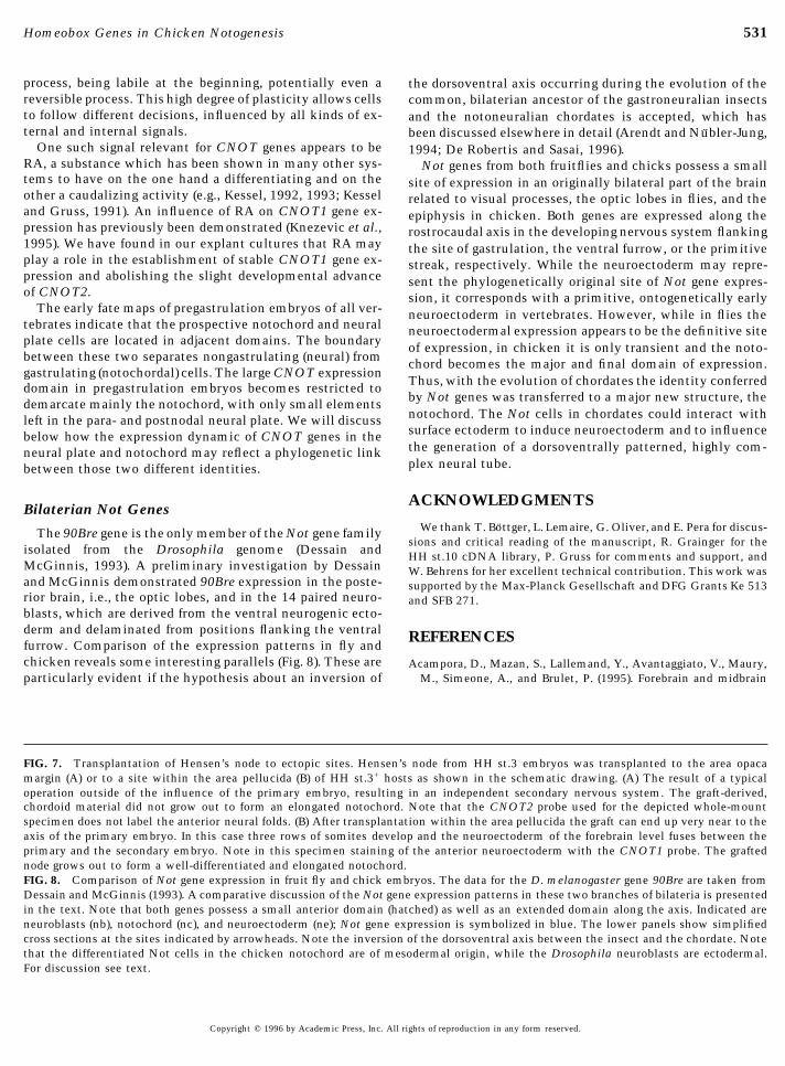
FIG. 7. Transplantation of Hensen’s node to ectopic sites. Hensen’s node from HH st.3 embryos was transplanted to the area opacamargin (A) or to a site within the area pellucida (B) of HH st.3/ hosts as shown in the schematic drawing. (A) The result of a typicaloperation outside of the influence of the primary embryo, resulting in an independent secondary nervous system. The graft-derived,chordoid material did not grow out to form an elongated notochord. Note that the CNOT2 probe used for the depicted whole-mountspecimen does not label the anterior neural folds. (B) After transplantation within the area pellucida the graft can end up very near to theaxis of the primary embryo. In this case three rows of somites develop and the neuroectoderm of the forebrain level fuses between theprimary and the secondary embryo. Note in this specimen staining of the anterior neuroectoderm with the CNOT1 probe. The graftednode grows out to form a well-differentiated and elongated notochord.FIG. 8. Comparison of Not gene expression in fruit fly and chick embryos. The data for the D. melanogaster gene 90Bre are taken fromDessain and McGinnis (1993). A comparative discussion of the Not gene expression patterns in these two branches of bilateria is presentedin the text. Note that both genes possess a small anterior domain (hatched) as well as an extended domain along the axis. Indicated areneuroblasts (nb), notochord (nc), and neuroectoderm (ne); Not gene expression is symbolized in blue. The lower panels show simplifiedcross sections at the sites indicated by arrowheads. Note the inversion of the dorsoventral axis between the insect and the chordate. Notethat the differentiated Not cells in the chicken notochord are of mesodermal origin, while the Drosophila neuroblasts are ectodermal.For discussion see text.
Copyright q 1996 by Academic Press, Inc. All rights of reproduction in any form reserved.
AID DB 8375 / 6x14$$$181 12-10-96 10:29:43 dbas

532 Stein, Niß, and Kessel
regions are deleted in otx2(0/0) mutants due to a defective ante- anterior-restricted homeobox gene progressively activated in therior neuroectoderm specification during gastrulation. Develop- prechordal plate, anterior neural plate and Rathke’s pouch of thement 121, 3279–3290. mouse embryo. Development 122, 41–52.
Arendt, D., and Nubler-Jung, K. (1994). Inversion of dorsoventral Hynes, M., Porter, J. A., Chiang, C., Chang, D., Tessier-Lavigne,axis? Nature 371, 26. M., Beachy, P. A., and Rosenthal, A. (1995). Induction of midbrain
Bally-Cuif, L., Gulisano, M., Broccoli, V., and Boncinelli, E. (1995). dopaminergic neurons by sonic hedgehog. Neuron 15, 35 –44.c-otx2 is expressed in two different phases of gastrulation and is Izpisua-Belmonte, J. C., De Robertis, E. M., Storey, K. G., and Stern,sensitive to retinoic acid treatment in chick embryos. Mech. Dev. C. D. (1993). The homeobox gene goosecoid and the origin of49, 49–63. organizer cells in the early chick blastoderm. Cell 74, 645–659.
Bellairs, R. (1986). The primitive streak. Anat. Embryol. 174, 1– Joyner, A. L., and Martin, G. R. (1987). En-1 and En-2, two mouse14. genes with sequence homology to the Drosophila engrailed gene:
Blum, M., Gaunt, J., Cho, K. W. Y., Steinbeisser, H., Blumberg, B., Expression during embryogenesis. Genes Dev. 1, 29 –38.Bittner, D., and De Robertis, E. M. (1992). Gastrulation in the Jurand, A. (1962). The development of the notochord in chick em-mouse: The role of the homeobox gene goosecoid. Cell 69, 1097– bryos. J. Embryol. Exp. Morphol. 10, 602–621.1106. Kappen, C., Schughart, K., and Ruddle, F. H. (1993). Early evolution-
Bober, E., Baum, C., Braun, T., and Arnold, H. H. (1994). A novel ary origin of major homeodomain sequence classes. GenomicsNK-related mouse homeobox gene: Expression in central and pe- 18, 54 –70.ripheral nervous structures during embryonic development. Dev. Kessel, M. (1992). Respecification of vertebral identities by retinoicBiol. 162, 288–303. acid. Development 115, 487–501.
Catala, M., Teillet, M.-A., and Le Douarin, N. M. (1995). Organiza- Kessel, M. (1993). Reversal of axonal pathways from rhombomeretion and development of the tail bud analyzed with the quail- 3 correlates with extra Hox expression domains. Neuron 10, 379–chick chimaera system. Mech. Dev. 51, 51–65. 393.
Charlebois, T. S., Spencer, D. H., Tarkington, S. K., Henry, J. J., Kessel, M., and Gruss, P. (1991). Homeotic transformations of mu-and Grainger, R. M. (1990). Isolation of a chick cytokeratin cDNA rine vertebrae and concomitant alteration of Hox codes inducedclone indicative of regional specialization in early embryonic by retinoic acid. Cell 67, 89 –104.ectoderm. Development 108, 33–45. Knezevic, V., Ranson, M., and Mackem, S. (1995). The organizer-
De Robertis, E. M., and Sasai, Y. (1996). A common plan for dorso- associated chick homeobox gene, Gnot1, is expressed before gas-ventral patterning in bilateria. Nature 380, 37–40.
trulation and regulated synergistically by activin and retinoicDessain, S., and McGinnis, W. (1993). Drosophila homeobox genes. acid. Dev. Biol. 171, 458–470.
In ‘‘Advances in Developmental Biochemistry’’ (P. M. Wassar-Kozak, M. (1987). An analysis of 5*-noncoding sequences from 699man, Ed.), Vol. 2, pp. 1–55. JAI Press, Greenwich.
vertebrate messenger RNAs. Nucleic Acids Res. 15, 8125–8146.Devereux, J., Haeberli, P., and Smithies, O. (1984). A comprehen-
Krumlauf, R. (1994). Hox genes in vertebrate development. Cell 78,sive set of sequence analysis programs for the VAX. Nucleic191–201.Acids Res. 121, 387–395.
Lints, T. J., Parsons, L. M., Hartley, L., Lyons, I., and Harvey, R. P.Dias, M. S., and Schoenwolf, G. C. (1990). Formation of ectopic(1993). Nkx-2.5: A novel murine homeobox gene expressed inneuroepithelium in chick blastoderms: Age related capacities forearly heart progenitor cells and their myogenic descendants.induction and self-differentiation following transplantation ofDevelopment 119, 419 –431.quail Hensen’s node. Anat. Rec. 229, 437–448.
Matsuo, I., Kuratani, S., Kimura, C., Takeda, N., and Aizawa, S.Eyal-Giladi, H., and Kochav, S. (1976). From cleavage to primitive(1995). Mouse otx2 functions in the formation and patterning ofstreak formation: A complementary normal table and a new lookrostral head. Genes Dev. 9, 2646–2658.at the first stages of the development of the chick. I. General
Meier, S. (1981). Development of the chick embryo mesoblast: Mor-morphology. Dev. Biol. 49, 321–337.phogenesis of the prechordal plate and cranial segments. Dev.Gont, L. K., Fainsod, A., Kim, S. H., and De Robertis, E. M. (1996).Biol. 83, 49–61.Overexpression of the homeobox gene Xnot-2 leads to notochord
New, D. A. T. (1955). A new technique for the cultivation of theformation in Xenopus. Dev. Biol. 174, 174–178.chick embryo in vitro. J. Embryol. Exp. Morphol. 3, 320–331.Gont, L. K., Steinbeisser, H., Blumberg, B., and De Robertis, E. M.
Noll, M. (1993). Evolution and role of Pax genes. Curr. Opin. Genet.(1993). Tail formation as a continuation of gastrulation: The mul-Dev. 3, 595–605.tiple cell populations of the Xenopus tailbud derive from the late
Paterson, B. M., and Eldrigde, J. D. (1984). a-cardiac actin is theblastopore lip. Development 119, 991–1004.major sarcomeric isoform expressed in embryonic avian skeletalHalpern, M. E., Thisse, C., Ho, R. K., Thisse, B., Riggleman, B.,muscle. Science 224, 1436–1438.Trevarrow, B., Weinberg, E. S., Postlethwait, J. H., and Kimmel,
Placzek, M. (1995). The role of the notochord and floor plate inC. B. (1995). Cell-autonomous shift from axial to paraxial meso-inductive interactions. Curr. Opin. Genet. Dev. 5, 499–506.dermal development in zebrafish floating head mutants. Devel-
Ranson, M., Tickle, C., Mahon, K. A., and Mackem, S. (1995).opment 121, 4257–4264.Gnot1, a member of a new homeobox gene subfamily, is ex-Hamburger, V., and Hamilton, H. L. (1951). A series of normalpressed in a dynamic, region-specific domain along the proximo-stages in the development of the chick embryo. J. Morphol. 88,distal axis of the developing limb. Mech. Dev. 51, 17–30.49–92.
Rivera-Perez, J. A., Mallo, M., Gendron-Maguire, M., Gridley, T.,Hanks, M., Wurst, W., Anson-Cartwright, L., Auerbach, A. B., andJoyner, A. L. (1995). Rescue of the en-1 mutant phenotype by and Behringer, R. R. (1995). Goosecoid is not an essential compo-replacement of en-1 with en-2. Science 269, 679–682. nent of the mouse gastrula organizer but is required for craniofa-
cial and rib development. Development 121, 3005–3012.Hermesz, E., Mackem, S., and Mahon, K. A. (1996). Rpx - a novel
Copyright q 1996 by Academic Press, Inc. All rights of reproduction in any form reserved.
AID DB 8375 / 6x14$$$181 12-10-96 10:29:43 dbas

533Homeobox Genes in Chicken Notogenesis
Rosenquist, G. C. (1983). The chorda center in Hensen’s node of genes related to the Drosophila empty spiracles gene are ex-pressed in the embryonic cerebral cortex. EMBO J. 11, 2541–the chick embryo. Anat. Rec. 207, 349–355.2550.Sasai, Y., Lu, B., Steinbeisser, H., and De Robertis, E. M. (1995).
Spemann, H., and Mangold, H. (1924). Uber die Induktion von Em-Regulation of neural induction by the Chd and Bmp-4 antagonis-bryoanlagen durch Implantation artfremder Organisatoren. Rouxtic patterning signals in Xenopus. Nature 376, 333–336.Arch. Entwicklungsmech. 100, 599–638.Sasai, Y., Lu, B., Steinbeisser, H., Geissert, D., Gont, L. K., and De
Spratt, N. T. (1952). Localization of the prospective neural plate inRobertis, E. M. (1994). Xenopus Chordin—A novel dorsalizingthe early chick blastoderm. J. Exp. Zool. 120, 109–130.factor activated by organizer-specific homeobox genes. Cell 79,
Stein, S., Fritsch, R., Lemaire, L., and Kessel, M. (1996). Checklist:779–790.Vertebrate homeobox genes. Mech. Dev. 55, 91–108.Sausedo, R. A., and Schoenwolf, G. C. (1993). Cell behaviors under-
Stein, S., and Kessel, M. (1995). A homeobox gene involved in node,lying notochord formation and extension in avian embryos:notochord and neural plate formation of chick embryos. Mech.Quantitative and immunocytochemical studies. Anat. Rec. 237,Dev. 49, 37–48.58–70.
Talbot, W. S., Trevarrow, B., Halpern, M. E., Melby, A. E., Farr, G.,Schoenwolf, G. C. (1992). Morphological and mapping studies ofPostlethwait, J. H., Jowett, T., Kimmel, C. B., and Kimelman,the paranodal and postnodal levels of the neural plate duringD. (1995). A homeobox gene essential for zebrafish notochordchick neurulation. Anat. Rec. 233, 281–290.development. Nature 378, 150–157.Schughart, K., Kappen, C., and Ruddle, F. (1989). Duplication of
von Dassow, G., Schmidt, J., and Kimelman, D. (1993). Inductionlarge genomic regions during the evolution of vertebrate homeo-of the Xenopus organizer: Expression and regulation of Xnot, abox genes. Proc. Natl. Acad. Sci. USA 86, 7067–7071.novel FGF and activin regulated homeobox gene. Genes Dev. 7,Seifert, R., Jacob, M., and Jacob, H. J. (1993). The avian prechordal355–366.head region: A morphological study. J. Anat. 183, 75 –89.
Wilkinson, D. G. (1992). ‘‘In Situ Hybridisation: A PracticalSelleck, M. A., and Stern, C. D. (1991). Fate mapping and cell lin-Approach.’’ Oxford Univ. Press, London.eage analysis of Hensen’s node in the chick embryo. Develop-
Wurst, W., Auerbach, A. B., and Joyner, A. L. (1994). Multiple devel-ment 112, 615–626.opmental defects in Engrailed-1 mutant mice: An early mid-Shawlot, W., and Behringer, R. R. (1995). Requirement for Lim1 inhindbrain deletion and patterning defects in forelimbs and ster-head-organizer function. Nature 374, 425–430.num. Development 120, 2065–2075.Simeone, A., Acampora, D., Pannese, M., D’Esposito, M., Stornaiu-
Yamada, G., Mansouri, A., Torres, M., Stuart, E. T., Blum, M.,olo, A., Gulisano, M., Mallalaci, A., Kastury, K., Druck, T.,Schultz, M., De Robertis, E. M., and Gruss, P. (1995). TargetedHuebner, K., and Boncinelli, E. (1994). Cloning and characteriza-mutation of the murine goosecoid gene results in craniofacialtion of two members of the vertebrate Dlx gene family. Proc.defects and neonatal death. Development 121, 2917–2922.Natl. Acad. Sci. USA 91.
Simeone, A., Gulisano, M., Acampora, D., Stornaiuolo, A., Ram- Received for publication July 15, 1996Accepted August 20, 1996baldi, M., and Boncinelli, E. (1992). Two vertebrate homeobox
Copyright q 1996 by Academic Press, Inc. All rights of reproduction in any form reserved.
AID DB 8375 / 6x14$$$181 12-10-96 10:29:43 dbas