human homeobox hoxa7 regulates keratinocyte
TRANSCRIPT
1
Human Homeobox HOXA7 Regulates Keratinocyte Transglutaminase Type 1
and Inhibits Differentiation
Peter T La Celle* Renata R Polakowska
University of Rochester School of Medicine and Dentistry
Department of Dermatology, Box 697 601 Elmwood Avenue
Rochester, New York 14642 USA
Universite du Droit et de la Sante
Faculte de Medecine INSERM U 459
1 Place de Verdun 59045 LILLE CEDEX
France
* Author to whom inquiries should be directed: [email protected] Phone (716) 275-9400 FAX (716) 273-1346
Running Title: Homeobox HOXA7 Regulates Transglutaminase 1
Copyright 2001 by The American Society for Biochemistry and Molecular Biology, Inc.
JBC Papers in Press. Published on July 2, 2001 as Manuscript M104598200 by guest on M
arch 26, 2018http://w
ww
.jbc.org/D
ownloaded from
2
SUMMARY Keratinocyte proliferation and differentiation result from expression of specific groups of genes
regulated by unique combinations of transcription factors. In order to better understand these
regulatory processes, we studied HOXA7 expression, and its regulation of differentiation-specific
keratinocyte genes. We isolated the homeobox transcription factor HOXA7 from keratinocytes
through binding to a differentiation-dependent viral enhancer, and analyzed its effect on
endogenous differentiation-dependent genes, primarily transglutaminase 1. HOXA7
overexpression repressed transglutaminase 1-reporter activity. HOXA7 message markedly
decreased, and transglutaminase RNA increased, upon phorbol ester-induced differentiation, in a
protein kinase C-dependent manner. Overexpression of HOXA7 attenuated the transglutaminase
1 induction by phorbol ester, demonstrating that HOXA7 expression is inversely related to
keratinocyte differentiation, and to transglutaminase 1 expression. Antisense HOXA7 expression
activated transglutaminase 1, involucrin, and keratin 10 message and protein levels,
demonstrating that endogenous HOXA7 downregulates multiple differentiation-specific
keratinocyte genes. In keeping with these observations, EGF receptor activation stimulated
HOXA7 expression. HOX genes function in groups, and we found that HOXA5 and HOXB7
were also downregulated by phorbol ester. These results provide the first example of protein
kinase C-mediated homeobox gene regulation in keratinocytes, and new evidence that HOXA7,
potentially in conjunction with HOXA5 and HOXAB7, silences differentiation-specific genes
during keratinocyte proliferation, that are then released from inhibition in response to
differentiation signals.
by guest on March 26, 2018
http://ww
w.jbc.org/
Dow
nloaded from
3
INTRODUCTION
The epidermis provides a protective barrier that undergoes constant renewal.
Keratinocytes of the innermost or basal layer withdraw from the cell cycle and become displaced
outwardly, differentiating to form layers of flattened, interconnected envelopes of cross-linked
proteins, packed with keratin filament bundles. Suprabasal cells inactivate genes expressed in
basal cells, such as keratins 5 and 14, and activate differentiation-specific genes such as keratins
1 and 10 (1), cornified envelope proteins such as involucrin, loricrin, and small proline-rich
proteins, and the enzyme required for envelope cross-linking, transglutaminase type 1 (reviewed
in (2-4)). EGF-receptor activation triggers keratinocytes proliferation (5,6) and inactivates
differentiation-specific genes (7). Rising extracellular calcium concentration, thought to be a key
physiological mediator, activates differentiation-specific gene expression and morphological
changes (8,9). Calcium-induced differentiation of cultured keratinocytes is protein kinase C
(PKC)-dependent, and markers of differentiation can be induced by PKC activators such as TPA
(10,11). Recent studies suggest that several differentiation-specific keratinocyte genes are
regulated by the integrated action of DNA-binding factors, including members of the AP-1, AP-
2, Sp1, and ets families (reviewed in (12)), and, perhaps least understood, the homeobox family
of transcription factors (13,14).
Homeobox genes encode a family of transcription factors sharing a conserved 60 amino
acid homeodomain (15). Duplication of gene clusters first described in Drosophila, has produced
four conserved mammalian AHox@ clusters, A-D (16), with capitalized names indicating the
human homologs. Their importance in segment identity, pattern formation, and cell fate
determination during development (16,17) suggests that Hox factors regulate batteries of genes
by guest on March 26, 2018
http://ww
w.jbc.org/
Dow
nloaded from
4
culminating in differentiation (18). Both Hox and non-Hox homeobox factors have also been
implicated in regulation of differentiation in adult tissues, such as blood (19), and skin (13,20).
In mouse skin, numerous non-Hox and Hox homeodomain genes are differentially
expressed during development (21-24). The non-Hox POU (Pit-Oct-Unc) homeodomain gene
Oct-11, or Skn-1a, message is associated with basal mouse epidermal cells by one study (25), but
with suprabasal cells in another study (26). The Drosophila Distal-less-related non-Hox
homeodomain gene Dlx3 is transcribed in differentiating keratinocytes (27). Ectopic expression
inhibits growth and induces the expression of differentiation-associated proteins, suggesting a
role in regulation of differentiation (13). In human keratinocytes, Oct-11/Skn-1a activates
expression of the differentiation markers K10 (26) and the small proline-rich envelope protein
SPRR2A (28), and Oct-6 inhibits expression of the proliferation-associated K5 and K14 genes
(29), suggesting activation of differentiation. However, others observed Oct-6 RNA in all living
layers of normal epidermis (29), and several POU family members, including Skn-1a, are capable
of inhibiting the differentiation-dependent involucrin promoter (30). HOX cluster genes have
also been implicated in the regulation of keratinocyte differentiation. HOXC4 message and
HOXB6 protein correlate with differentiation in normal skin, and HOXA4 is absent in
proliferative basal cell carcinomas (14,31). Stelnicki et.al. identified predominantly HOXA4,
HOXA5 and HOXA7 expression in suprabasal fetal human epidermis, with expression persisting
in the adult epidermis, but not in the dermis (32).
HOX transcription factors recognize similar DNA sequences in vitro. The diversity of
their effects and targets in vivo is believed to result from modulation by cofactors that affect
binding, or function. For example, multiple copies of the Drosophila Ultrabithorax (Ubx) bind
by guest on March 26, 2018
http://ww
w.jbc.org/
Dow
nloaded from
5
cooperatively to target gene sites (33), and HOX factors exhibit altered DNA sequence
recognition and cooperative DNA binding with either of two homeodomain proteins, PBX1
(34,35), via a conserved hexapeptide motif, or MEIS-1 (36). Further, HoxB7 exhibits no change
in DNA binding, but is transcriptionally coactivated by the histone acetylase CBP (CREB
binding protein) (37).
Another mechanism underlying specificity of function is the combined action of unique
sets of Hox genes. In development, Hox genes act in groups comprising paralogous genes
(corresponding genes from different clusters), as well as nearby genes within clusters, to bring
about target regulation. This mechanism may also underlie the expression of predominantly
HOXA4, HOXA5, HOXA7, but also HOXB6 and HOXB7 in a similar differential manner in
human skin (32). In this study we isolated and sequenced the HOXA7 cDNA from keratinocytes
through binding to a differentiation-dependent HPV-16 E6/E7 enhancer fragment. HOXA7 also
bound to a regulatory fragment of the differentiation-specific transglutaminase 1 gene, and
repressed transglutaminase 1-reporter activity. HOXA7, HOXA5 and HOXB7 were
downregulated in keratinocytes induced to differentiate with TPA, and overexpressed HOXA7
inhibited transglutaminase 1 expression during TPA-induced differentiation. Antisense HOXA7
expression upregulated keratinocyte differentiation markers and slowed growth, and HOXA7
message was upregulated in growth-activated keratinocytes. These results indicate that HOXA7,
potentially in conjunction with related family members, functions in silencing differentiation-
specific genes, prior to its own PKC-mediated downregulation during differentiation. A transient
increase in HOXA7 expression observed as cultured cells reach confluence may act as a brake
by guest on March 26, 2018
http://ww
w.jbc.org/
Dow
nloaded from

6
initially to limit the rate at which differentiation progresses, as cells become exposed to
differentiation signals.
by guest on March 26, 2018
http://ww
w.jbc.org/
Dow
nloaded from
7
EXPERIMENTAL PROCEDURES
Isolation of HOXA7 cDNA. An epidermal cDNA expression library (Clontech, Palo Alto, CA),
prepared in λgt11 from human keratinocyte messenger RNA, was screened for expressed
proteins that recognize a 232 bp HPV-16 E6/E7 enhancer DraI fragment (bp 7524-7756) by a
standard method (38). Briefly, phage plaques on cultures of infected E. coli were overlayed with
nitrocellulose filters pre-treated with isopropylthio-β-D-1-galactopyranoside, and dried.
Expressed, adsorbed proteins were denatured with 6M guanindine-HCl in Tris-buffered saline,
and renatured with washes in 6 decrements to zero denaturing agent. Filters were blocked with
5% non-fat dried milk, and 100 µg/ml salmon sperm DNA Tris-buffered saline with 0.5% Triton
X-100, and expressed proteins were probed with a 32P-labeled 232 bp DraI fragment (bp 7524-
7756), containing the CK element, a small cytokeratin homology motif, from the HPV-16 E6/E7
enhancer, or a neighboring 209 bp DraI control fragment (bp 7286-7495). Bound probe was
visualized by autoradiography. Positive plaques underwent two further cycles of E. coli infection,
plating, induction of protein expression and probing. Clone 124, containing the full length
HOXA7 cDNA, was subcloned into pCDNAI and sequenced.
Tissue culture, vectors and cell transfection. Neonatal human keratinocytes (NHK) were
obtained from human foreskin by overnight dispase digestion, followed by trypsinization of the
separated epidermis, and plating in Keratinocyte Serum-Free Medium (GibcoBRL, Rockville,
MD), containing recombinant human EGF and bovine pituitary extract. ME180 epidermoid
carcinoma cells were obtained from the American Tissue Culture Collection (ATCC) and grown
in RPMI1640 (Biowhitaker, Walkersville, MD) with 8% fetal calf serum (FCS, Sigma, St. Louis,
by guest on March 26, 2018
http://ww
w.jbc.org/
Dow
nloaded from

8
MO). HaCaT spontaneously immortalized keratinocytes were a generous gift from Dr. N. E.
Fusenig, and were cultured in Dulbecco=s modified Eagle=s medium with 8% FCS.
The full-length HOXA7 cDNA (forward primer 5'-
GTCGCCATGGGTTCTTCGTATTATGTG-3', generating a Kozak translation start sequence,
and vector reverse primer) and a 5' fragment from the translation start to just 5' of the
hexapeptide domain (Figure 1) (5'-ATACTCGAGTAGATGCGGAAATTGG-3' reverse primer),
were PCR amplified by pwo polymerase (Roche Molecular Biochemicals, Indianapolis, IN),
subcloned into the vector pCR2.1 (Invitrogen, Carlsbad, CA), then moved to the eukaryotic
expression vectors pCS2 (pCS2-HOXA7), pCDNA3 (Invitrogen) (p3-HOXA7, p3-HOXA7frag),
and pEF3 (pEF3-124fragRev) in the forward and reverse orientation, and sequenced. The
expression vector pEF3 was constructed by subcloning the EF-1α promoter, released from the
vector pEF6/HisA (Invitrogen) by partial digestion with HindIII/BglII, into the pCDNA3
(Invitrogen) backbone in place of the cytomegalovirus promoter. The 1.7 kb transglutaminase 1
upstream regulatory region K3, isolated previously (39,40), was subcloned into the enhancerless
and promoterless reporter vectors pCAT-Basic (pCAT-K3), and pGL3-B (pGL3-K3) (Promega),
for transient transfection. The control reporter pHβA-LacZ was constructed by inserting the lacZ
cDNA from pCH110 (Amersham Pharmacia Biotech) into the human β-actin promoter-driven
expression vector pHβAPr-1 (41).
Third passage NHK, and ME180 were transfected with Fugene 6 (Roche Molecular
Biochemicals, Indianapolis, IN), and HaCaT keratinocytes were transfected with Exgen 500
(Fermentas, Hanover, MD) under the recommended conditions. Cells were transiently
cotransfected with expression and reporter vectors (either HOXA7 or K3-containing,
by guest on March 26, 2018
http://ww
w.jbc.org/
Dow
nloaded from
9
respectively, or empty vector), and with the pHβA-LacZ control vector. Lysates obtained 2 days
after transfection were assayed for chloramphenicol acetyltransferase activity using 14C-
chloramphenicol and acetyl-CoA, by thin layer chromatography and autoradiography, and by
xylene extraction and scintillation counting, presented as cpm acetyl-14C-chloramphenicol per
mg lysate protein. Determinations of luciferase activity (Promega luciferase substrate), β-
galactosidase activity (Tropix, Applied Biosystems, Foster City, CA) and total protein
concentration (BCA, Pierce, Rockford, IL) were made according to the reagent suppliers=
instructions. Results are expressed as relative light units (RLU) per mg protein. Cotransfection of
expression vectors with empty reporters resulted in low, background level reporter signals in all
cases (not shown). Transfection efficiency was determined as the relative number of β-
galactosidase stained cells. Cells were fixed for 5 minutes with an ice-cold equivolume mixture
of acetone and methanol, washed in phosphate buffered saline, and stained for 16 hours at 37EC
with 1 mg/ml 5-bromo-4-chloro-3-indolyl β-D-galacropyranoside in 40 mM citrate-phosphate
buffer pH 7.5 with 5 mM ferro- and ferricyanide, 2 mM MgCl2 and 150 mM NaCl.
Stably transfected HaCaT were selected starting 48 hours after transfection with 500
ug/ml geneticin (Gibco-BRL), and maintained in 300 ug/ml geneticin. For analysis of growth
rate, 1.5 X 10⁵ antisense HOXA7, or vector control-transfected HaCaT were plated in 35mm
dishes. Triplicate samples of unattached cells, and attached cells released by trypsinization, at 24
hour intervals were counted. Data are reported as cells per well X 10-4, +/- standard deviation.
Statistical differences were determined by analysis of variance and T-test.
by guest on March 26, 2018
http://ww
w.jbc.org/
Dow
nloaded from
10
Electrophoretic Mobility Shift Assay. To verify binding to the HPV-16 E6/E7 enhancer, HOXA7
was expressed in λgt11 transformed 1090 E.coli by induction with 2 mM isopropylthio-β-D-1-
galactopyranoside for 3 hours, followed by sonication in the presence of phenylmethylsulfonyl
fluroide and centrifugation. For electrophoretic mobility shift assay, 232bp (bp 7524-7756) and
209 bp (bp 7286-7495) DraI fragments of HPV-16 were released by restriction digestion, purified
and 32P-labeled. Binding reactions containing 4x10⁵ cpm probe (about 1 ng DNA), and 5 µg of
HOXA7 bacterial phage expression lysate or non-transformed bacterial lysate, and 2 µg of poly-
dIdC in 20 mM N-2-hydroxyethyl-piperazine-N=-2-ethanesulfonic acid, pH 7.5, 50 mM
potassium chloride, 1 mM dithiothreitol, 6% glycerol, and 0.2 mM phenylmethylsulfonyl
fluoride, were incubated at room temperature for 20 minutes, separated on 5 % polyacrylamide
gels with 0.5X trishydroxymethyl aminomethane/borate/ethylenediaminetetraacetic acid at 4EC
for 4 hours, dried and autoradiographed.
To investigate HOXA7 binding to the TGM1 upstream regulatory region (K3),
recombinant HOXA7 was expressed by in vitro trancription/translation using the ATNT coupled
reticulocyte system@ from Promega according to the included protocol, using 1 µg template DNA
per reaction. In control synthesis reactions, 35S-methionine was added, and expression was
monitored by SDS-PAGE and autoradiography. For electrophoretic mobility shift assay, a 264 bp
K3 fragment (5' end at -710), containing a CK element similar to that found in the 232 bp DraI
HPV-16 fragment, and a 212 bp neighboring K3 fragment (5'end at -444), were prepared by
PCR (bp -709 to +90), digested with BamHI and HpaII, purified, 32P-end-labeled, and shifted
with 5 µg of the HOXA7 in vitro translation lysate as described above.
by guest on March 26, 2018
http://ww
w.jbc.org/
Dow
nloaded from
11
RNase Protection Assay. Total RNA was isolated using Trizol reagent (Gibco BRL) from third
passage neonatal human keratinocytes treated with 50 ng/ml TPA (Calbiochem, San Diego, CA).
32P-UTP-labeled RNA probes were synthesized using T7 polymerase (Ambion, Austin, TX) from
the linearized plasmid DNA template pCDNA3-HOXA7fragRev, containing a non-homologous
5' fragment of the HOXA7 cDNA (Figure 1), and pBSII-GAPDH (Stratagene, La Jolla, CA),
containing a 360 bp human glyceraldehydephosphate dehydrogenase cDNA fragment, both in the
reverse orientation. Following DNase I digestion for 15 minutes at 37EC, the probes were
electrophoretically purified in a 5 % polyacrylamide gel containing 1X TBS and 8 M urea
(Sequagel, National Diagnostics, Atlanta, GA), eluted (60 minutes, 37EC in 0.5 M ammonium
acetate, 1 mM ethylenediaminetetraacetate, 0.1% sodium dodecylsulfate and 50 µg/ml yeast RNA
(Ambion)), precipitated (1 M ammonium acetate and three volumes 2-propanol), dissolved in
H2O, and scintillation counted. For RNase protection, labeled probe was ethanol/ammonium
acetate precipitated together with 12 µg normal human keratinocyte or yeast RNA, redissolved in
hybridization buffer (Ambion), denatured (90EC, 4 minutes), and hybridized overnight at 42EC.
Single stranded RNA was digested using 1 part in 100 RNaseA1/RNaseT1 in digestion buffer
(Ambion) for 30 minutes at 37EC. Protected RNA was separated by denaturing polyacrylamide
gel electrophoresis as described for probe purification, and visualized by autoradiography.
For detection of antisense HOXA7 RNA in stably transfected HaCaT, total RNA was
hybridized as above, without the addition of probe, digested with RNase A/T for various times,
and phenol-chloroform extracted and precipitated, before detection of 5' HOXA7 RNA by RT-
PCR. The PCR product, spanning bases 291-451 (Figure 1), was produced using the HOXA7
by guest on March 26, 2018
http://ww
w.jbc.org/
Dow
nloaded from

12
forward primer described below under "RT-PCR", and the reverse primer 5'-
CTCGTCCGTCTTGTCGCAGG-3'.
RT-PCR
To determine the range of target DNA concentrations giving rise to a linear relationship
between target concentration and product concentration, restriction fragments of HOXA7,
TGM1, and β-Actin cDNA were purified, and 100-100,000 molecules were PCR amplified using
Taq DNA polymerase (Promega) and PCR buffer (Sigma), in the presence of 1.8 mM MgCl2,
400 nM primers, 400 µM each dNTP (Roche Molecular Biochemicals), and 5 µCi 32P-dCTP.
Samples were removed after 30 cycles of 94EC, 20 seconds, 57EC, 20 seconds, 72EC, 30
seconds, electrophoresed in 6% acrylamide, 0.5X trishydroxymethyl aminomethane-borate-
ethylenediaminetetraacetic acid buffered gels, and autoradiographed. Quantification of product
bands, using Scion Image (www.scioncorp.com), indicated a linear relationship between the
amount of target cDNA and the amount of PCR product formed, across the range of target
concentrations tested.
Total RNA (0.75 µg) from control, or 50 ng/ml TPA-treated NHK, denatured at 68EC for
10 minutes in the presence of 2 µM oligo-dT primer (Invitrogen) and 1.25 mM each dNTP served
as the template for cDNA synthesis by Superscript II reverse transcriptase (GibcoBRL) in the
supplied buffer, supplemented with 0.5 U/µl RNase inhibitor (Promega) and 10 mM
dithiothreitol, at 42EC for 60 minutes. The reaction was stopped with ethylenediaminetetraacetic
acid, and heating for 15 minutes at 70EC. cDNA diluted 1:10 with H2O, and added at 1/10th
volume to a 30 cycle PCR reaction under the above conditions gave rise to a signal intensity
by guest on March 26, 2018
http://ww
w.jbc.org/
Dow
nloaded from
13
corresponding to a target concentration for HOXA7 and TGM1 within the linear range of the
PCR assay. PCR using β-actin primers generated a signal equivalent to standards in the linear
range of PCR after 18 cycles. HOXA5 and HOXB7 were also amplified for 30 PCR cycles, and
involucrin, K5 and K10 were amplified for 25 cycles. Sham reactions in which no reverse
transcriptase was added produced no PCR bands. The HOXA7 and transglutaminase 1 PCR
primer sequences span intron splice sites to distinguish products arising from amplification of
cDNA. Treatment of RNA samples with 2 units DNaseI (Ambion) followed by heat inactivation
before cDNA synthesis had no affect on the PCR products formed. Primers of the following
sequence (5' to 3') were used: HOXA7 forward: CTTATACAATGTCAACAGCC, and reverse:
TCCTTATGCTCTTTCTTCC and TCTTCTTCATCATCGTCCTCCTCG; TGM1 forward:
TCTGTGGGTCCTGTCCCATCCATCCTGACC, and reverse:
CCCCAACGGCCCACATCGGAACGTGGCCCATCCATCATGC; human β-actin forward:
CAGGCTGTGCTATCCCTGTAC and reverse: CACGCACGATTTCCCGCTCGG; human
cytokeratin K10 forward: GGCTCTGGAAGAATCAAACTATGAGC and reverse:
GGATGTTGGCATTATCAGTTGTTAGG; involucrin forward:
TGTTCCTCCTCCAGTCAATACCC and reverse: ATTCCTCATGCTGTTCCCAGTGC;
keratin K5 forward: CTGTCTCCCGCACCAGCTTCACCTCC and reverse:
CTCCACAAGCACCCGCAAGGCTGACC; HOXA4 forward:
GGCGCTGACATGGATCTTCTTCATCC, and reverse:
CAACTACATCGAGCCCAAGTTCCCTCC; HOXA5 forward:
CCTCTCTGCTGCTGATGTGGGTGC, and reverse: ACGGCTACGGCTACAATGGCATGG;
by guest on March 26, 2018
http://ww
w.jbc.org/
Dow
nloaded from
14
HOXB7 forward: AAGTTCGGTTTTCGCTACCGGAGCC, and
CGCGCAGTGCATGTTGAAGG.
Transglutaminase Assay
HaCaT keratinocytes stably transfected with pCDNA3 (control) or pCDNA3-
HOXA7fragRev (HOXA7 antisense), carrying a 5' HOXA7 cDNA fragment (Figure 1) insert in
the reverse orientation, grown to 75 % confluence in 100 mm dishes, were washed in 10 ml and
then scraped up in 0.3 ml of 50 mM Tris pH 7.4, 150 mM NaCl, 0.2 mg/ml bovine serum
albumin, 20 mM ethylenediaminetetraacetic acid, and complete protease inhibitor cocktail
(Roche Molecular Biochemicals), microfuged 30 seconds at 4EC, forming the cell supernatant,
and resuspended in the same buffer with 20 mM ethylenediaminetetraacetic acid. Cells were
lysed by sonication, and centrifuged at 100,000 X g for 45 minutes at 4EC, forming the cytosolic
supernatant and transglutaminase 1-containing particulate pellet (42). TGaseI activity was
extracted from the disrupted pellet for 30 minites on ice in the lysis buffer supplemented with 1%
Triton X-100, and centrifuged at 10,000 X g for 15 minutes at 4EC, with the supernatant
designated the particulate fraction. The cell supernatant and cytosolic fractions were made 1 % in
Triton X-100 corresponding to the detergent level in the particulate fraction. Transglutaminase
activity of cellular fractions relative to total protein content was determined essentially as
described (42), by incorporation of 20 µCi/ml (0.2 umol/ml) 14C-putrescine into 2 mg/ml
dimethylcasein in the presence of 20 mM Tris pH 7.4, 60 mM NaCl, 20 mM CaCl2, 5 mM
dithiothreitol, and 0.4 % Triton X-100 for 45 minutes at 37EC, followed by trichloroacetic acid
precipitation and scintillation counting.
by guest on March 26, 2018
http://ww
w.jbc.org/
Dow
nloaded from

15
Western Blot
Control and antisense HOXA7-transfected HaCaT cells were scraped up in sodium
dodecyl sulfate sample buffer, electrophoresed in 8.5% polyacrylamide gels with a discontinuous
Tris-glycine buffer system, transferred to nitrocellulose in Tris-glycine with 20% methanol at 300
mA for 1 hour, blocked for 1 hour with 4% dry milk or bovine serum albumin in Tris buffered
saline with 0.1% Tween-20, and incubated 1 hour at 37EC with primary antibody, diluted 1:1000
in blocking solution, against human involucrin (BioTechnologies Inc., Staughton, MA), human
K1 (1:1:1000) or mouse K5 (1:200,000, cross-reacts with human), (BAbCo, Richmond, CA).
Bound antibody was detected by incubating filters for 45 minutes with a 1:5,000 dilution of
HRP-conjugated goat-anti-rabbit IgG (DAKO, Carpinteria, CA) in Tris-buffered saline with
0.1% Tween-20, and exposing autoradiographic film after treatment with luminescent substrate
(Amersham Pharmacia).
Northern Blot
Northern analysis of antisense HOXA7 RNA in stably transfected HaCaT keratinocytes
was performed according to standard methods. Total RNA (25 µg) from 80% confluent vector
control and antisense HOXA7 expressing HaCaT cells was isolated using Trizol following the
provided instructions (Life Technologies, Inc.), separated on a 1.2% agarose/MOPS/1.1%
formaldehyde gel, and transferred to a nylon membrane. The blot was hybridized with a 32P-
labeled riboprobe prepared as recommended (Maxiscript, Ambion) from the 5' region of the
HOXA7 cDNA inserted in the reverse orientation in the vector pCDNA3, using SP6 phage RNA
by guest on March 26, 2018
http://ww
w.jbc.org/
Dow
nloaded from
16
polymerase. The blot was washed at high stringency (0.1% SSC, 0.1% SDS at 68ºC), and
hybridized probe was detected by autoradiography. The blot was stripped and reprobed with a
GAPDH control probe 32P-labeled by random priming (Life Technologies, Inc.), washed as
above, and autoradiographed.
by guest on March 26, 2018
http://ww
w.jbc.org/
Dow
nloaded from
17
RESULTS
Isolation of the HOXA7 cDNA through binding to the HPV16 E6/E7 enhancer-
Epidermal differentiation results from the coordinated expression of keratinocyte genes, perhaps
by the action of a specific set(s) of transcription factors. The human papilloma virus-16 (HPV-
16) E6/E7 enhancer (p91) exhibits keratinocyte, and differentiation-specific activation. To take
advantage of any functional cis-acting elements pirated by the HPV-16 p91 enhancer from the
differentiation-specific keratinocyte gene regulation system, we used a 232 bp DraI fragment of
the HPV-16 E6/E7 enhancer, to screen a keratinocyte cDNA expression library. This fragment
encompasses an AAPuCCAAA motif (CK element) also found within the transglutaminase 1
(TGM1) gene upstream regulatory region (K3) (40,43), keratins K1 and K14 (44,45), and
involucrin upstream regulatory regions (46). A phage clone expressing a binding protein that did
not adhere to a neighboring 209 bp enhancer fragment was isolated, as verified by electrophoretic
mobility shift assay (EMSA) (data not shown).
Sequence analysis revealed a 954 bp cDNA encoding HOXA7, a class I homeobox
transcription factor of 230 amino acids, with a homeodomain extending from amino acid 130 to
189, a conserved six amino acid (hexapeptide) sequence just upstream of the homeodomain, and
an acidic C-terminal domain (Figure 1). The homeodomain and hexapeptide amino acid
sequences of the human HOXA7, and its mouse homolog Hoxa7 (formerly Hox1.1), are
identical in spite of some divergence at the nucleic acid level. The proteins also share overall
similarity, including an acidic C-terminal domain. The mammalian hexapeptide and homeobox
sequences represent remarkable conservation, varying by only one amino acid from the
Drosophila antennapedia, but sequences outside the homeodomain exhibit no obvious sequence
by guest on March 26, 2018
http://ww
w.jbc.org/
Dow
nloaded from
18
similarity to antennapedia. Examination of the genomic sequence indicates a structure similar to
the mouse Hoxa7 gene (47), with a 944 bp intron separating the two translated exons, between
the hexapeptide and homeodomain coding sequences (arrow in Figure 1).
HOXA7 transrepresses TGM1 gene upstream regulatory region (K3)-reporters in NHK, but
transactivates K3-reporters in the epidermoid carcinoma line ME180, and binds specifically to a
K3 fragment in vitro- To determine whether the highly conserved HOXA7, recognized by the
HPV-16 enhancer, might regulate differentiation-specific keratinocyte genes such as TGM1, we
examined the affect of transiently overexpressed HOXA7 on TGM1-K3 reporter activity.
HOXA7 over-expressed as a c-myc fusion (pCS-HOXA7) in primary neonatal keratinocytes
(NHK) repressed transcriptional activity of K3 (pCAT-K3) relative to the empty vector (Figure
2a). To rule out any effect of the c-myc portion of the fusion peptide, the HOXA7 cDNA was
subcloned into the eukaryotic expression vector pCDNA3; the K3 regulatory DNA was
subcloned into the promoterless, enhancerless pGL3B. As before, HOXA7 expression
inactivated K3 transcription relative to empty pCDNA3 in NHK. Interestingly, HOXA7 had the
opposite effect in the epidermoid carcinoma cell line ME180, and transactivated K3 relative to
vector control (Figure 2b).
Since HOXA7 affected TGM1 K3 reporters by transient transfection, we tested the
hypothesis that HOXA7 may recognize a potential binding site in the K3 regulatory region near
the CK element by electrophoretic mobility shift assay. The in vitro transcribed and translated
HOXA7 protein electrophoretically retarded a 264 bp 5= K3 fragment (Figure 3a, lanes 5, 7), but
the empty-vector control lysate did not (lane 4). Binding was abolished by competition with cold
by guest on March 26, 2018
http://ww
w.jbc.org/
Dow
nloaded from
19
probe (lane 6), but not by competition with a neighboring 212 bp K3 fragment (lane 7). Protein
synthesis was monitored in control reactions with 35S-methionine by SDS-PAGE and
autoradiography (Figure 3c). These results indicate that HOXA7 can specifically bind K3 and
regulate K3 transcriptional activity, and suggest that HOXA7 regulates TGM1 gene activity in
keratinocytes, via a mechanism altered in ME180 carcinoma cells.
HOXA7, HOXA5, and HOXB7 expression are repressed, and TGM1 is activated, in NHK
stimulated to differentiate with TPA by a PKC-dependent mechanism- With the potential to
transrepress the differentiation-specific gene TGM1 in keratinocytes, we investigated the level of
HOXA7 expression in keratinocytes induced to differentiate with TPA, which activates TGM1
gene expression. The concentration of HOXA7 RNA was markedly reduced, as measured by
RNase protection assay, by 2.5 hours after treatment of NHK with TPA, reaching a minimum at
5 hours, and remaining in decline for at least 10 hours (Figure 4a). The concentration of HOXA7
RNA was also reduced as measured by reverse transcription-polymerase chain reaction (RT-
PCR) analysis (Figure 4b, middle panel). HOXA7 message was reduced by treatment with as
little as 0.5 ng/ml TPA for 10 hours (not shown). TPA treatment also resulted in a decline in
HOXA5 and HOXB7 message levels (Figure 4c), suggesting co-regulation with HOXA7. The
HOXA7 autoradiographic bands represent processed mRNA only, as the primer hybridization
sites span the intron splice site (Figure 1, arrow). HOXA4 message was detected only at trace
levels by RT-PCR (not shown). RNA pretreatment with DNase1 had no affect on the bands
produced by RT-PCR, and sham RT samples yielded no detectable PCR product. RNA isolated
from untreated NHK at each time point exhibited a constant level of HOXA7 message (not
by guest on March 26, 2018
http://ww
w.jbc.org/
Dow
nloaded from
20
shown).
As expected, TPA treatment of normal human keratinocytes caused an increase in TGM1
message level (Figure 4b, top panel), which reached a maximum at a time later than the
maximum drop in HOXA7 message level. The TPA induction of TGM1 was transient, as
previously described (11), as was the inhibition of HOXA7. The β-actin message level remained
relatively constant over the time course tested (Figure 4b and 4c, bottom panels). The number of
PCR cycles was varied for each primer pair to maintain linearity of the relationship between the
number of target molecules and the amount of PCR product formed, as determined using purified
linear cDNA.
As expected, we found that TPA modulation of HOXA7, like that of TGM1, occurs
downstream of protein kinase C (PKC) activation. The marked decline in HOXA7 message
levels with TPA treatment of cultured NHK was blocked by the PKC inhibitor
bisindolylmaleimide (bIM) (Figure 5, top row). Treatment with bisindolylmaleimide alone had
no effect on the HOXA7 message level in non-confluent cells, and increased the HOXA7
message level in post-confluent cells (not shown), demonstrating that the decline of HOXA7
seen after keratinocytes reach confluence is PKC-mediated. β-actin message levels remained
unaffected by TPA and bisindolylmaleimide treatment.
HOXA7 expression is also repressed, and TGM1 is activated in NHK stimulated to
differentiate with calcium- NHK cultured in low-calcium medium remain undifferentiated (8).
Raising the calcium concentration above 0.1 mM. induces differentiation-associated proteins and
morphological changes, and TGM1 expression rises in proportion to the extracellular calcium
concentration (10,11). We found that NHK demonstrated a drop in HOXA7 message levels by 5
by guest on March 26, 2018
http://ww
w.jbc.org/
Dow
nloaded from
21
hours after raising the extracellular calcium concentration to 1.8 mM (Figure 6, top row). High
calcium treatment also stimulated a rise in the TGM1 message level within 10 hours (center
row), while β-actin message was unaffected. The HOXA7 message level in calcium-treated cells
continued to fall at 48 hours (not shown), but as seen in Figure 5, HOXA7 message levels decline
and TGM1 message levels rise even in untreated cells with the passing of successive days post-
confluence, as the cells contact inhibit and begin to differentiate. It is therefore difficult to
attribute changes solely to calcium signaling. Together, these data indicate that stimulation of
differentiation, either following cell-cell contact, or by TPA or calcium treatment, or by a
combination, results in a drop in HOXA7 and an increase in TGM1 message levels.
EGF stimulation of keratinocyte proliferation activates HOXA7 gene expression-
HOXA7 expression is downregulated in differentiating keratinocytes, and overexpression
represses the differentiation marker gene TGM1. These observations suggest that TGM1
expression occurs upon differentiation when HOXA7 expression is low, and that the higher level
of HOXA7 functions to block TGM1 expression during proliferation. We therefore tested
whether HOXA7 expression is activated under conditions promoting keratinocyte proliferation.
Adding back EGF to non-confluent, EGF-starved NHK cultured in SFM stimulated HOXA7
expression relative to β-actin, as measured in total RNA by RT-PCR (Figure 7, upper left
panels). The increase was blocked by pretreatment with the selective EGF receptor tyrosine
kinase activity inhibitor AG1478 (48) (Figure 7, upper right panels). Control cells that received
no EGF (lower left panels), or AG1478 alone (lower right panels), exhibited no detectable
HOXA7 message.
by guest on March 26, 2018
http://ww
w.jbc.org/
Dow
nloaded from
22
Overexpression of HOXA7 attenuates TPA-induced TGM1 expression, and antisense
HOXA7 activates expression of differentiation-associated genes at the RNA and protein level in
non-confluent keratinocytes- Our data indicate that HOXA7 binds to the TGM1 upstream
regultory region K3, transrepresses exogenous TGM1 reporters in NHK, is turned off prior to
TGM1 gene activation in differentiating cells, and is turned on in proliferating cells at a time
when TGM1 is inactivated. In order to test whether modulation of HOXA7 would also affect
endogenous TGM1 gene activity, NHK were transiently transfected with a HOXA7 expression
vector, or an antisense HOXA7 expression construct, and then treated with TPA. Figure 8a
shows that cells overexpressing HOXA7 (AHOXA7 +@) exhibited a reduced TPA activation of
TGM1 compared to control vector transfected cells (AHOXA7 -A). The relatively small
attenuation is believed to reflect the transfection efficiency of 20%, as determined by β-
galactosidase staining (not shown).
As seen in Figure 8b, non-confluent NHK transfected with a vector expressing antisense
HOXA7 showed a dose-dependent increase in TGM1 message level. The 5' fragment of the
HOXA7 cDNA chosen for antisense expression (Figure 1, shaded sequence), is not homologous
to other human homeobox transcription factor sequences, or open reading frames of other known
human genes, affirming that the TGM1 gene activation is the result of HOXA7 message
targeting.
Since antisense HOXA7 upregulated TGM1 in transiently transfected normal human
keratinocytes, the effect of antisense HOXA7 expression in stably transfected cells was studied.
Immortalized HaCaT keratinocytes were transfected with the HOXA7 antisense expression
construct, selected, and cell lines were cloned and analyzed for expression of differentiation
by guest on March 26, 2018
http://ww
w.jbc.org/
Dow
nloaded from
23
markers. Proliferating antisense cells showed an increase in the expression of the differentiation-
specific genes TGM1, involucrin and keratin K10, compared to vector-control HaCaT (Figure
9a). In contrast, the message level of the proliferating basal cell-associated keratin K5 increased
to only a minor degree, and the control β-actin message was not altered, in the antisense cells. As
seen in Figure 9b, involucrin and keratin K1 proteins were also greatly upregulated in the
antisense HOXA7-transfected cells, while the keratin 5 protein concentration remained relatively
constant. Proliferating antisense-transfected cells also contained an increased amount of TGase 1
enzyme activity compared to vector control cells (Figure 9c), measured as putrescine
incorporation into dimethyl casein. The TGase 1 enzyme is membrane anchored, and is found in
the pellet after keratinocyte sonication and centrifugation. TGase activity in the cytosolic fraction
represents the ubiquitous type 2 enzyme, which is downregulated in differentiating keratinocytes,
plus TGase 1 released from the plasma membrane by proteolytic cleavage. HOXA7 antisense
expressing HaCaT also grew at a slower rate, as seen in Figure 10. Expression of antisense RNA
in antisense HOXA7-transfected HaCaT keratinocytes was verified by Northern blot analysis
(Figure 11a), and by RNase protection (Figure 11b).
Taken together, these results demonstrate that the HOXA7, HOXA5, and possibly
HOXB7, genes are downregulated during differentiation, and HOXA7 is upregulated upon
stimulation of proliferation. Further, endogenous HOXA7 regulates differentiation-associated
genes and affects cell growth rate in transfected cells, suggesting that these HOX factors
represent an important component in the control of proliferation versus differentiation in
keratinocytes.
by guest on March 26, 2018
http://ww
w.jbc.org/
Dow
nloaded from
24
DISCUSSION
Keratinocytes differentiate as cells withdraw from the cell cycle and migrate suprabasally
in association with expression of differentiation-specific genes. Induction of keratinocyte
differentiation in vitro is also associated with differentiation marker expression. This report
establishes that keratinocyte expression of HOX genes HOXA7, HOXA5, and possibly HOXB7,
is downregulated upon TPA-induced differentiation in vitro. HOXA7 is also downregulated upon
differentiation triggered by cell-cell contact, or by raising the extracellular calcium concentration.
Further, antisense HOXA7 expression upregulates the differentiation-associated genes TGM1,
involucrin, and keratins 1 and 10 in non-confluent cells, and slows growth. HOXA7 protein
binds to a TGM1 regulatory DNA fragment in vitro, and transrepresses a TGM1-reporter
construct. These results provide evidence that HOXA7, likely in combination with other HOX
transcription factors, functions in downregulation of differentiation-associated keratinocyte genes
prior to the onset of differentiation, and that part of the differentiation program involves release
of these genes from HOX inhibition.
We analyzed the role of HOXA7 in regulation of differentiation-specific keratinocyte
genes. HOXA7 physically interacted with a fragment of the differentiation-specific TGM1 gene
promoter in vitro, and transrepressed a TGM1 promoter fragment (40) linked to a reporter in
normal human keratinocytes. Basal TGM1-reporter activity in the absence of the HOXA7 cDNA
(Figure 2) may represent saturation by the reporter construct of a limiting pool of inhibitory
endogenous HOXA7 protein, or depletion of a corepressor. Conversely, HOXA7 turned on the
TGM1-reporter in ME180 carcinoma cells, which exhibit a high level TGM1 expression (49) in
association with abnormal growth. This observation demonstrates the potential of HOXA7 to act
by guest on March 26, 2018
http://ww
w.jbc.org/
Dow
nloaded from
25
as a transcriptional activator. Whether a transcription factor transactivates or represses depends
on the nature of its functional domains (DNA binding, protein-protein interaction, transcriptional
activation and repression), on the number, sequence, and relative positions of cis elements in the
target, and on the presence of cofactors. The murine Hoxa7, over 90 % identical to the human
HOXA7 (Figure 1), comprises both transcriptional activating and inhibitory domains, and the
whole molecule has demonstrated both transcriptional repression (50) and activation (51).
Multiple copies of Hoxa7, like other Hox proteins, exhibit cooperative binding and
transactivation of targets with multiple recognition sites (51). Cooperativity involves proteins
bound to nearby, as well as to distant sites, presumably through a DNA looping action (33). In
addition, HOX proteins physically interact with the homeodomain cofactor PBX or MEIS1, or
both, resulting in altered site recognition and cooperative DNA binding (35,36,52-54). Other
known homeobox cofactors include the acetylase/integrator CREB binding protein (37,55), IkB
and NF-kB (56), the glucocorticoid receptor (57,58), and serum response factor (59-61),
presaging the existence of more, potentially tissue specific, cofactors. ME180 may constitutively
express a HOXA7 cofactor that yields TGM1 transcriptional activation, or that masks an
inhibitory HOXA7 domain, such as the acidic C-terminal region (50).
In addition to repressing TGM1 reporter activity, HOXA7 also downregulated
endogenous TGM1 gene activity. HOXA7 overexpressed in normal keratinocytes attenuated the
TPA-induced expression of TGM1. The maximum observed attenuation is limited by the
transfection efficiency. Even if the overexpressed HOXA7 blocked all TGM1 expression in
transfected cells, the apparent attenuation would not exceed the measured transfection efficiency
of 20%. The relatively small observed decrease in TGM1 induction is therefore to be expected,
by guest on March 26, 2018
http://ww
w.jbc.org/
Dow
nloaded from
26
and nevertheless implies a large inhibition of TGM1 activation in those cells transfected. TPA
treatment activates PKC-α, and induces transcriptional activation by AP-1 factors (62), which
can regulate differentiation-specific genes such as TGM1 (63), involucrin (64), filaggrin (65),
keratins (reviewed in (66,67)), and HPV-16 and -18 (68,69). The observed HOX versus TPA
antagonism could result from mutually exclusive promoter binding and function by HOX and
AP-1 factors, as observed in the case of the POU factor Pit-1 gene autoregulation (70).
Alternatively, since CBP binding has been observed to be obligatory and limiting in AP-1
transactivation (71,72), and CBP also binds and enhances HOXB7 transactivation potential (37),
HOXA7 and AP-1 antagonism may represent competition for available CBP. Whether or not the
mechanism of the observed antagonism of TPA activity is so direct, HOXA7 may function to
prevent basal AP-1 levels from activating differentiation markers in proliferating cells, or to
delay expression until later stages of differentiation.
Since overexpression of the otherwise rare HOXA7 protein might affect transcription of
genes outside its normal sphere of regulation, we determined whether endogenous HOXA7
regulates TGM1 gene activity. Targeting endogenous HOXA7 by antisense expression resulted in
marked TGM1 gene activation, in both normal and immortalized human keratinocytes, in
agreement with our in vitro binding, and HOXA7 sense transfection data. The antisense sequence
is derived from a non-homologous 5' portion of the HOXA7 cDNA, excluding the conserved
hexapeptide, homeobox, and acidic C-terminal encoding regions (Figure 1), so that the results
reliably reflect selective HOXA7 transcript targeting. In this case, the observed TGM1 induction,
the product of the fold induction within transfected cells, times the fraction of cells transfected, is
not limited to the value of the transfection efficiency. That the observed induction (Figure 8b) is
by guest on March 26, 2018
http://ww
w.jbc.org/
Dow
nloaded from
27
several fold means that the fold induction in transfected cells was large, indicating the presence
of a TGM1 gene activation signal, balanced by HOXA7 inhibition, in proliferating cultured
keratinocytes. In immortalized HaCaT keratinocytes, stable transfection with antisense HOXA7
upregulated not only TGM1, but also involucrin, and keratin 1 and keratin 10 expression, and
resulted in slower growth compared to vector control cells, suggesting that HOXA7 regulates
multiple genes associated with differentiation.
Keratinocyte differentiation, induced by TPA treatment (Figure 4), by raising
extracellular calcium (Figure 6), or by prolonged high-density culture (Figure 5), downregulates
HOXA7 expression. We therefore investigated whether mitogenic activation stimulates HOXA7
message levels. We found that HOXA7 expression is induced by EGF receptor activation,
supporting the hypothesis that HOXA7 helps silence TGM1 during proliferation, until the
appropriate time during differentiation. Further investigation will be required to define the
relevant regulatory pathways in more detail. It will be interesting to determine whether the
HOXA7 and HOXA5 genes are downregulated by AP-1 factors activated in differentiating
keratinocytes. Also, our results indicate that HOXA7 modulates both the early differentiation
markers K1 and K10, and the later marker TGM1, and is itself regulated by both calcium and
TPA treatment, whereas TPA treatment of mouse keratinocytes activates late differentiation
markers but blocks calcium-induced expression of K1 and K10 (73).
Overlapping expression and function of Hox genes is a common theme in development.
In mice, disruption of Hoxb6, Hoxa7, Hoxb7, and Hoxb9 all contribute to first and second rib
defects. Hoxa7 disruption alone causes no defects, but adding Hoxa7 mutations markedly
increases the rate and severity of rib defects observed in Hoxb7-/- mice, suggesting that Hoxa7
by guest on March 26, 2018
http://ww
w.jbc.org/
Dow
nloaded from
28
has functional roles that were not revealed in the Hoxa7-/- mice, and that these two genes act
together (74). Hoxa7 may also have a functional role in the epidermis, requiring closer
examination of morphology and differentiation marker expression, requiring some additional
challenge, or requiring a different double mutant gene partner, such as Hoxa5, for manifestation.
Synergistic and overlapping function have also been observed with Hoxa3, Hoxb3 and Hoxd3,
and with Hoxa11 and Hoxd11 disruption in mouse development (reviewed in (75)). Overlapping
function of groups of HOX factors regulating sets of target genes may be retained, following
cessation of development, where continual proliferation and subsequent differentiation occur. In
keeping with this idea, Stelnicki et.al. detected predominantly HOXA4, HOXA5 and HOXA7,
but also HOXB7 and HOXC4 message in fetal and adult human epidermis, but not in the dermis,
by RT-PCR using a set of degenerate HOX gene primers (32). HOXA7 was consistently the
most frequently identified gene upon cloning and sequencing the PCR products, suggesting an
important role in epidermal development and homeostasis. These findings are in good agreement
with our results from neonatal human keratinocytes, where HOXA7 and HOXA5 were expressed
much more highly than HOXB7, although we detected only a weak HOXA4 signal. They
observed further a suprabasal expression pattern of HOXA4, HOXA5 and HOXA7 in neonatal
and adult epidermis. Interestingly, we measured a transient increase in HOXA7 message level as
cultured keratinocytes reach confluence (not shown), as well as compelling evidence of HOXA7
downregulation upon PKC activation, and upregulation with EGF receptor activation. The
apparent conflict is resolved if we postulate that HOXA7 functions as an inhibitor of
differentiation during proliferation, but also as a brake system during the onset of differentiation.
Small amounts of HOXA7 may suffice to inhibit expression of differentiation-specific genes
by guest on March 26, 2018
http://ww
w.jbc.org/
Dow
nloaded from
29
during proliferation, when differentiation signal-transduction pathways are nearly idle. HOXA7
expression may be activated progressively, in parallel with differentiation signals during early
stages of differentiation, to limit the rate and extent of potentially destructive elements of the
process, allowing completion of important intermediate steps involving protein synthesis and
vesicle transport. This would account for the reported increase in suprabasal expression by in situ
hybridization. HOXA7 expression may ultimately be silenced, as mimicked by our TPA-
treatment of keratinocytes, at a time appropriate for completion of the keratinization process.
We found that, in addition to HOXA7, the HOXA5, and possibly the HOXB7 gene (very
low expression was observed) is also downregulated in cultured keratinocytes induced to
differentiate with TPA. It would be interesting to ascertain whether combined HOXA5 and
HOXA7, and even HOXB7 disruption, or inducible ectopic epidermal coexpression, would yield
a pronounced skin defect. Interestingly, our HOXA5 RT-PCR product comprised 2 bands (figure
4), raising the possibility that like HOXB6 (14), HOXA5 exhibits differential expression of
alternatively spliced message, and perhaps protein, although the band of anomalous
electrophoretic migration rate may represent an RT-PCR artifact. These results support the
contention that regulation of keratinocyte differentiation involves multiple HOX gene family
members, including HOXA7 and HOXA5, and perhaps HOXB7.
It may be worth noting that expression of all three of these genes has been associated with
a non-differentiated state, or with cellular proliferation. Among HOX genes, HOXA7 and
HOXB7 are highly expressed in chemically induced papillomas (76), suggesting a shared
function in the etiology of growth deregulation. Murine Hoxa7 and Hoxa9 cooperativity with
Meis-1 has been implicated in murine myeloid leukemia (54). Overexpression of Hoxa5, Hoxa7,
by guest on March 26, 2018
http://ww
w.jbc.org/
Dow
nloaded from
30
or Hoxb7 leads to transformation and tumorigenicity in two fibroblast cell lines. HOXA5 and
HOXB7 are associated with hematopoietic progenitor proliferation (77,78), and Hoxa4 and
Hoxa5 are upregulated in association with inhibition of differentiation by retinoic acid in the
developing mouse lung (79). Further, HoxB7 is expressed in proliferating mammary epithelial
cells, and disappears with matrix-induced differentiation (80). Finally, HOXB7, normally
expressed in proliferating melanocytes, is upregulated in, and implicated in the enhanced growth
of, metastatic melanomas (81,82). However, these observations represent potential function,
since examples of induction of differentiation can also be cited, and further studies are required
to fully elucidate the role of HOX genes in regulation of keratinocyte proliferation and
differentiation.
We isolated HOXA7 from a human keratinocyte library, via specific binding to the
human papillomavirus 16 (HPV-16) epithelial-dependent enhancer, which restricts viral
oncogene E6/E7 expression to differentiating keratinocytes (44). The HPV-16 epithelial-
dependent enhancer may fall within the spectrum of differentiation-dependent genes repressed by
HOXA7. Inspection of the enhancer fragment suggests that HOXA7 may bind to a HOX
consensus core motif, preventing cooperative Oct-1/NF-1 transactivation (83) at an overlapping
site, thus adding to the repression mediated by YY1 steric inhibition of AP-1 and Sp1
transcription factors (84). Downregulation of HOXA7 may contribute to E6/E7 activation, in
combination with upregulation of HPV-16 transactivation factors skn-1a (26,85), AP-1 (86), and
Sp1 (84) . Although the HOXA7 message appears to be rare (32), the stability and steady-state
accumulation of HOXA7 protein in proliferating keratinocytes remains to be determined.
In summary, these results suggest that HOXA7, likely in combination with other HOX
by guest on March 26, 2018
http://ww
w.jbc.org/
Dow
nloaded from

31
genes, plays an important role in regulating keratinocyte TGM1 expression, and possibly more
generally in regulating the expression of differentiation-specific keratinocyte genes. These
properties have implications for HOX involvement in keratinocyte oncogenesis, and in
keratinocyte-specific viral activation, that warrant continued investigation.
by guest on March 26, 2018
http://ww
w.jbc.org/
Dow
nloaded from
32
REFERENCES 1. Fuchs, E., and Green, H. (1980) Cell 19, 1033-1042
2. Eckert, R. L. (1989) Physiol Rev 69(4), 1316-1346
3. Fuchs, E. (1990) Journal of Cell Biology 111(6), 2807-2814
4. Blumenberg, M., and Tomic-Canic, M. (1997) EXS 78, 1-29
5. Rodeck, U., Jost, M., Kari, C., Shih, D.-T., Lavker, R. M., Ewert, D. L., and Jensen, P. J.
(1997) Journal of Cell Science 110, 113-121
6. King, L. E. J., Gates, R. E., Stoscheck, C. M., and Nanney, L. B. (1990) J Invest
Dermatol 94(6 Suppl), 164S-170S
7. Dlugosz, A. A., Cheng, C., Denning, M. F., Dempsey, P. J., Coffey, R. J. J., and Yuspa,
S. H. (1994) Cell Growth Differentiation 5(12), 1283-1292
8. Hennings, H., Michael, D., Cheng, C., Steinert, P., Holbrook, K., and Yuspa, S. H. (1980)
Cell 19, 245-254
9. Pillai, S., Bikle, D. D., Mancianti, M.-L., Cline, P., and Hincenbergs, M. (1990) Journal
of Cellular Physiology 143(2), 294-302
10. Yuspa, S. H., Kilkenny, A. E., Steinert, P. M., and Roop, D. R. (1989) J Cell Biol 109(3),
1207-1217
11. Dlugosz, A. A., and Yuspa, S. H. (1994) Journal of Investigative Dermatology 102(4),
409-414
12. Eckert, R. L., Crish, J. F., Banks, E. B., and Welter, J. F. (1997) Journal of Investigative
Dermatology 109(4), 501-509
13. Morasso, M. I., Markova, N. G., and Sargent, T. D. (1996) Journal of Cell Biology
by guest on March 26, 2018
http://ww
w.jbc.org/
Dow
nloaded from
33
135(6), 1879-1887
14. Komuves, L. G., Shen, W. F., Kwong, A., Stelnicki, E., Rozenfeld, S., Oda, Y., Blink, A.,
Krishnan, K., Lau, B., Mauro, T., and Largman, C. (2000) Develop Dynamics 218(4),
636-647
15. McGinnis, W., Garber, R. L., Wirz, J., Kuroiwa, A., and Gehring, W. J. (1984) Cell 37,
403-408
16. McGinnis, W., and Krumlauf, R. (1992) Cell 68, 283-302
17. Boncinelli, E., Mallamaci, A., and Lavorgna, G. (1996) Genetica 94, 127-140
18. Levine, M., and Hoey, T. (1988) Cell 55, 537-540
19. Giampaolo, A., Sterpetti, P., Gulgarini, D., Samoggia, P., Pelosi, E., Valtieri, M., and
Peschle, C. (1994) Blood 84(11), 3637-3647
20. Scott, G. A., and Goldsmith, L. A. (1993) Journal of Investigative Dermatology 101(1),
3-8
21. Kanzler, B., Viallet, J. P., Le Mouellic, H., Boncinelli, E., Duboule, D., and Dhouailly, D.
(1994) Int J Dev Biol 38, 633-640
22. Bieberich, C. J., Ruddle, F. H., and Stenn, K. S. (1991) Ann NY Acad Sci 642, 346-354
23. Mathews, C. H. E., Detmer, K., Lawrence, H. J., and Largman, C. (1993) Differentiation
52(2), 177-184
24. Detmer, K., Lawrence, H. J., and Largman, C. (1993) Journal of Investigative
Dermatology 101(4), 517-522
25. Yukawa, K., Yasui, T., Yamamoto, A., Shiku, H., Kishimoto, T., and Kikutani, H. (1993)
Gene 133, 163-169
by guest on March 26, 2018
http://ww
w.jbc.org/
Dow
nloaded from

34
26. Andersen, B., Schonemann, M. D., Flynn, S. E., Pearse, R. V. I., Singh, H., and
Rosenfeld, M. G. (1993) Science 260, 78-82
27. Park, G. T., and Morasso, M. I. (1999) J Biol Chem 274(37), 26599-26608
28. Fischer, D. F., Gibbs, S., van de Putte, P., and Backendorf, C. (1996) Mol Cell Biol
16(10), 5365-5374
29. Faus, I., Hsu, H.-J., and Fuchs, E. (1994) Mol Cell Biol 14(5), 3263-3275
30. Welter, J. F., Gali, H., Crish, J. F., and Eckert, R. L. (1996) Journal of Biological
Chemistry 271(25), 14727-14733
31. Rieger, E., Bijl, J., van Oostveen, J. W., Soyer, H. P., Oudejans, C. B., Jiwa, N. M.,
Walboomers, J. M., and Meijer, C. J. (1994) Journal of Investigative Dermatology
103(3), 341-346
32. Stelnicki, E. J., Komuves, L. G., Kwong, A. O., Holmes, D., Klein, P., Rozenfeld, S.,
Lawrence, H. J., Adzick, N. S., Harrison, M., and Largman, C. (1998) Journal of
Investigative Dermatology 110(2), 110-115
33. Beachy, P. A., Varkey, J., Young, K. E., von Kessler, D. P., Sun, B. I., and Ekker, S. C.
(1993) Mol Cell Biol 13(11), 6941-6956
34. Shen, W.-F., Chang, C.-P., Rozenfeld, S., Sauvageau, G., Humphries, R. K., Lu, M.,
Lawrence, H. J., Cleary, M. L., and Largman, C. (1996) Nucleic Acids Research 24(5),
898-906
35. Phelan, M. L., Rambaldi, I., and Featherstone, M. S. (1995) Mol Cell Biol 15(8), 3989-
3997
36. Shanmugam, K., Green, N. C., Rambaldi, I., Saragovi, H. U., and Featherstone, M. S.
by guest on March 26, 2018
http://ww
w.jbc.org/
Dow
nloaded from
35
(1999) Mol Cell Biol 19(11), 7577-7588
37. Chariot, A., van Lint, C., Chapelier, M., Gielen, J., Merville, M.-P., and Bours, V. (1999)
Oncogene 18(17), 4007-4014
38. Kalionis, B., and O'Farrell, P. H. (1993) Mechanisms of Development 43, 57-70
39. Polakowska, R. R., Eikbush, T., Falciano, V., Razvi, F., and Goldsmith, L. A. (1992)
Proceedings of the National Acadamy of Science USA 89, 4476-4480
40. Polakowska, R. R., Graf, B. A., Falciano, V., and LaCelle, P. (1999) Journal of Cellular
Biochemistry 73(3), 355-369
41. Gunning, P., Leavitt, J., Muscat, G., Ng, S.-Y., and Kedes, L. (1984) Proc Natl Acad Sci
84, 4831-4835
42. Thacher, S. M., and Rice, R. H. (1985) Cell 40, 677-683
43. Saunders, N. A., Bernacki, S. H., Vollberg, T. M., and Jetten, A. M. (1993) Molecular
Endocrinology 7(3), 387-398
44. Cripe, T. P., Haugen, T. H., Turk, J. P., Tabatabai, F., Schmid, P. G. I., Durst, M.,
Gissmann, L., Roman, A., and Turek, L. P. (1987) EMBO J 6(12), 3745-3753
45. Marchuck, D., McCrohon, S., and Fuchs, E. (1985) Proceedings of the National Acadamy
of Science USA 82, 1609-1613
46. Blessing, M., Zentgraf, H., and Jorcano, J. L. (1987) EMBO J 6(3), 567-575
47. Colberg-Poley, A. M., Voss, S. D., Chowdhury, K., and Gruss, P. (1997) Nature 314,
713-718
48. Fry, D. W., Kraker, A. J., McMichael, A., Ambroso, L. A., Nelson, J. M., Leopold, W.
R., Connors, R. W., and Bridges, A. J. (1994) Science 265(5175), 1093-1095
by guest on March 26, 2018
http://ww
w.jbc.org/
Dow
nloaded from
36
49. Duvic, M., Nelson, D. C., Annarella, M., Cho, M., Esgleyes-Ribot, T., Remenyik, E.,
Ulmer, R., Rapini, R. P., Sacks, P. G., Clayman, G. L., Davies, P. J. A., and Thacher, S.
M. (1994) Journal of Investigative Dermatology 102(4), 462-469
50. Schnabel, C. A., and Abate-Shen, C. (1996) Mol Cell Biol 16(6), 2678-2688
51. Gross, M. K., and Gruss, P. (1994) Mol Cell Biol 14, 238-254
52. Lu, Q., Knoepfler, P. S., Scheele, J., Wright, D. D., and Kamps, M. P. (1995) Mol Cell
Biol 15(7), 3786-3795
53. Knoepfler, P. S., Calvo, K. R., Chen, H., Antonarakis, E., and Kamps, M. P. (1997)
Proceedings of the National Acadamy of Science USA 94(26), 14553-14558
54. Nakamura, T., Largaespada, D. A., Shaughnessy, J. D. J., Jenkins, N. A., and Copeland,
N. G. (1996) Nature Genetics 12(2), 149-153
55. Lorentz, O., Suh, E. R., Taylor, J. K., Boudreau, F., and Traber, P. G. (1999) J Biol Chem
274(11), 7196-7199
56. Chariot, A., Princen, F., Gielen, J., Merville, M.-P., Franzoso, G., Brown, K., Siebenlist,
U., and Bours, V. (1999) Journal of Biological Chemistry 274(9), 5318-5325
57. Wang, J. M., Prefontaine, G. G., Lemieux, M. E., Pope, L., Akimenko, M. A., and Hache,
R. J. (1999) Mol Cell Biol 19(10), 7106-7122
58. Subramaniam, N., Cairns, W., and Okret, S. (1998) J Biol Chem 273(36), 23567-23574
59. Newman, C. S., Reecy, J., Grow, M. W., Ni, K., Boettger, T., Kessel, M., Schwartz, R. J.,
and Krieg, P. A. (2000) Mechanisms of Development 9(1-2), 369-373
60. Chen, C. Y., Croissant, J., Majesky, M., Topouzis, S., McQuinn, T., Frankovsky, M. J.,
and Schwartz, R. J. (1996) Developmental Genetics 19(2), 119-130
by guest on March 26, 2018
http://ww
w.jbc.org/
Dow
nloaded from

37
61. Grueneberg, D. A., Simon, K. J., Brennan, K., and Gilman, M. (1995) Mol Cell Biol
15(6), 3318-26
62. AngelP., Imagawa, M., Chiu, R., Stein, B., Imbra, R. J., Rahmsdorf, H. J., Jonat, C.,
Herrlich, P., and Karin, M. (1987) Cell 49(6), 729-739
63. Yamada, K., Yamanishi, K., Kakizuka, A., Kibe, Y., Doi, H., and Yasuno, H. (1994)
Biochemistry and Molecular Biology International 34(4), 827-836
64. Efimova, T., La Celle, P. T., Welter, J. F., and Eckert, R. L. (1998) Journal of Biological
Chemistry 273(38), 24387-24395
65. Jang, S.-I., Steinert, P. M., and Markova, N. G. (1997) Journal of Biological Chemistry
271(39), 24105-24114
66. Rossi, A., Jang, S.-I., Ceci, R., Steinert, P. M., and Markova, N. G. (1998) Journal of
Investigative Dermatology 110(1), 34-40
67. Eckert, R. L., and Welter, J. F. (1996) Molecular Biology Reports 23, 59-70
68. Chan, W. K., Chong, T., Bernard, H. U., and Klock, G. (1990) Nucleic Acids Res 18(4),
763-769
69. Butz, K., and Hoppe-Seyler, F. (1993) J Virology 67(11), 6476-6486
70. Delhase, M., Castrillo, J. L., de la Hoya, M., Rajas, F., and Hooghe-Peters, E. L. (1996) J
Biol Chem 271(50), 32349-32358
71. Arias, J., Alberts, A. S., Brindle, P., Claret, F. X., Smeal, T., Karin, M., Feramisco, J.,
and Montminy, M. (1994) Nature 370(6486), 226-229
72. Kwok, R. P., Lundblad, J. R., Chrivia, J. C., Richards, J. P., Bachinger, H. P., Brennan,
R. G., Roberts, S. G., Green, M. R., and Goodman, R. H. (1994) Nature 370(6486), 223-
by guest on March 26, 2018
http://ww
w.jbc.org/
Dow
nloaded from
38
226
73. Dlugosz, A. A., and Yuspa, S. H. (1993) Journal of Cell Biology 120(1), 217-225
74. Oka, M., Nagai, H., Ando, H., Fukunaga, M., Matsumura, M., Araki, K., Ogawa, W.,
Miki, T., Sakaue, M., Tsukamoto, K., Konishi, H., Kikkawa, U., and Ichihashi, M. (2000)
J Invest Dermatol 115(4), 699-703
75. Capecchi, M. R. (1997) in Cold Spring Harbor Symposia on Quantitative Biology Vol.
62, pp. 273-281, Cold Spring Harbor Laboratory Press, Cold Spring Harbor
76. Chang, P. Y., Kozono, T., Chida, K., Kuroki, T., and Hu, N. (1998) Biochemical and
Biophysical Research Communications 248, 749-752
77. Crooks, G. M., Fuller, J., Petersen, D., Izadi, P., Malik, P., Pattengale, P. K., Kohn, D. B.,
and Gasson, J. C. (1999) Blood 94(2), 519-528
78. Care, A., Valtieri, M., Mattia, G., Meccia, E., Masella, B., Luchetti, L., Felicetti, F.,
Colombo, M. P., and Peschle, C. (1998) Oncogene 18(11), 1993-2001
79. Packer, A. I., Mailutha, K. G., Ambrozewicz, L. A., and Wolgemuth, D. J. (2000)
Developmental Dynamics 217(1), 62-74
80. Srebrow, A., Friedmann, Y., Ravanpay, A., Daniel, C. W., and Bissell, M. J. (1998)
Journal of Cellular Biochemistry 69(4), 377-391
81. Care, A., Silvani, A., Meccia, E., Mattia, G., Stoppacciaro, A., Parmiani, G., Peschle, C.,
and Colombo, M. P. (1996) Mol Cell Biol 16(9), 4842-4851
82. Care, A., Silvani, A., Meccia, E., Mattia, G., Peschle, C., and Colombo, M. P. (1998)
Oncogene 16(25), 3285-3289
83. O'Connor, M. J., and Bernard, H.-U. (1995) Virology 207, 77-88
by guest on March 26, 2018
http://ww
w.jbc.org/
Dow
nloaded from
39
84. Dong, X. P., and Pfister, H. (1999) Journal of General Virology 80(8), 2097-2101
85. Yukawa, K., Butz, K., Yasui, T., Kikutani, H., and Hoppe-Seyler, F. (1996) J Virology
70(1), 10-16
86. O'Connor, M. J., Tan, S.-H., Tan, C.-H., and Bernard, H.-U. (1996) J Virology 70(10),
6529-6539
by guest on March 26, 2018
http://ww
w.jbc.org/
Dow
nloaded from
40
FOOTNOTES Abreviations used:
1The abreviations used are: TGM1, transglutaminase 1 gene; TGase1, transglutaminase
type 1; K3, a 1.7 kb TGM1 gene upstream regulatory DNA fragment; PKC, protein kinase C;
NHK, primary neonatal human keratinocytes; TPA, 12-O-tetradecanoylphorbol-13-acetate; HPV-
16, human papillomavirus 16; EGF, epidermal growth factor; RT-PCR, reverse transcription-
polymerase chain reaction; AP-1, activator protein 1; AP-2, activator protein 2; Sp1, transcription
factor Sp1; ets,; SPRR2A, small proline rich-related peptide 2A; HOX, class 1 homeobox
transcription factors related to the Drosophila Antennapedia complex and Bithorax complex
genes; POU, Pit-Oct-Unc -related transcription factors containing a conserved homeodomain and
POU domain; Dlx3, Drosophila distal-less-like homeobox transcription factor; CBP, cAMP
regulated enhancer binding binding protein.
by guest on March 26, 2018
http://ww
w.jbc.org/
Dow
nloaded from
41
FIGURE LEGENDS
FIG. 1. The Human HOXA7 cDNA sequence exhibits high sequence homology with
the murine HoxA7 (Hox1.1). Dashes in the murine sequence indicate nucleotide identity with the
human; the predicted human amino acid sequence is shown above, with asterisks indicating
differences in the amino acid sequences. The non-homologous 5' fragment used for expression of
antisense RNA in transfected cells, and for RNA probe synthesis, is shaded. The conserved
hexapeptide, required for certain protein-protein interactions, and the DNA-binding homeodomain
are boxed, and the polyadenylation signal is underlined. The Intron 1 boundary is marked with an
arrowhead. The human HOXA7 sequence is listed in the Genbank database under accession
number AF026397.
FIG. 2. HOXA7 represses TGM1 K3 transcription in NHK, but stimulates K3
transcription in ME180 carcinoma cells. a. CAT reporter activity of NHK transfected with the
empty pCS vector, or pCS2-HOXA7 producing a c-myc-HOXA7 fusion peptide, and the reporter
pCATB-K3; results are expressed (duplicate lanes) as a thin layer chromatography autoradiograph,
and as cpm organically extracted acetyl-14C-chloramphenicol per mg lysate protein. b. RLU/mg
protein of NHK and ME180 transfected with pCDNA3 (p3), or pCDNA3-HOXA7 (p3-HOXA7),
and pGL3B-K3 (pGL-K3). Results are representative of at least three experiments.
FIG. 3. HOXA7 binds specifically to a fragment of the TGM1 Upstream Retulatory
Region. a. HOXA7 expressed by in vitro transcription/translation formed a complex with a 264 bp
transglutaminase 1 gene fragment containing the CK-8-mer, probe 1 (lane 5), but not with the
by guest on March 26, 2018
http://ww
w.jbc.org/
Dow
nloaded from

42
neighboring 212 bp control fragment, probe 2 (lane 2). The binding was abrogated by competition
with cold probe 1 (lane 6), but not cold probe 2 (lane 7). Lanes 1, 3: probes 1 and 2 alone. Lane 4:
Probe 1 with control reticulocyte lysate. b: Delineation of the TGM1 K3 region comprising probes
1 and 2. c. The 35S-methionine labeled HOXA7 product (center lane); control reaction containing
the empty vector pCDNA3 (left lane); Expression of the luciferase gene product (positive control)
(right lane). Two addional experiments produced comparable results.
FIG. 4. HOXA7 message falls in NHK induced to differentiate with TPA a. RNase
protection of HOXA7 message in total RNA from NHK treated with 50 ng/ml TPA, immediately
after reaching confluence, for the indicated times (top panel), relative to GAPDH (bottom panel).
Data are representative of four independent experiments. b. HOXA7 message (center panel),
transglutaminase 1 message (top panel) and β-actin message (bottom) in NHK as determined by
RT-PCR after treatment with 50 ng/ml TPA and isolation of total RNA, in one of six repeated
experiments. c. HOXA5 message (top panel) and HOXB7 message (middle panel) in NHK as
determined by RT-PCR after TPA treatment as in a. Bottom panel: β-actin control. Data reflect
results of 3 experiments.
FIG. 5. Repression of HOXA7 gene expression with TPA treatment is PKC-dependent.
On successive days after plating, NHK were pretreated for 30 minutes with 100 mM PKC inhibitor
bisindolylmaleimide (bIM) (+), or DMSO (-), then treated for 10 hours with 20 ng/ml TPA (+) or
DMSO (-). HOXA7(top row), TGM1 (middle row) and β-actin message levels were determined by
RT-PCR. Results from 60-70% confluent (non-confluent, left column), confluent (middle column),
by guest on March 26, 2018
http://ww
w.jbc.org/
Dow
nloaded from
43
and 2 days post-confluent cells are shown. Results respresent 3 independent experiments.
FIG. 6. Calcium treatment represses HOXA7 and stimulates TGM1 expression. NHK
grown to confluence in low calcium medium were treated with 1.8 mM CaCl2 (top three panels), or
maintained in low-calcium medium (bottom panel). Total RNA was isolated before, and at 2.5, 5,
10 and 24 hours after initiation of calcium treatment. HOXA7, TGM1, and β-actin expression were
determined by RT-PCR.
FIG. 7. EGF activates expression of HOXA7 in cultured keratinocytes. NHK were
cultured in SFM, and cells at low density were switched to EGF-free SFM for 30 hours with 1
change of medium. Selected groups of wells were pretreated for 30 minutes with 3 µM AG1478,
and some additionally with 5 ng/ml EGF, such that groups of wells received each of the following:
EGF alone (upper block, left column), AG1478 and EGF (upper block, right column), no treatment
(control, lower block, left column), or AG1478 alone (lower block, right column). Total RNA was
isolated from wells in each group at 2 hours and 5 hours after EGF treatment, followed by RT-
PCR, PAGE and autoradiography for HOXA7 (top rows) and β-actin (bottom rows) message
levels.
FIG. 8. Overexpression of HOXA7 attenuates the TPA-induced expression of TGM1
in NHK, and HOXA7 antisense overexpression causes a dose-dependent increase in TGM1
gene expression in untreated NHK. a. Non-confluent, proliferating NHK were transfected in 60
mm dishes with 16 µg pcDNA3 (HOXA7 -, left column), or pcDNA3-HOXA7 (HOXA7 +, right
by guest on March 26, 2018
http://ww
w.jbc.org/
Dow
nloaded from
44
column), and 28 µl lipofectamine. Cells were treated with 0.5 ng/ml TPA for 8 hours, beginning 16
or 40 hours after transfection. Total RNA was analyzed by RT-PCR for TGM1 (top row) and � -
actin (bottom row) message levels. b. Proliferating NHK in 6-well plates were transfected in
duplicate with 0 (left column), 2 (middle column), or 4 ug of the antisense-HOXA7 expression
construct pEF3-HOXA7fragRev, plus control vector (pEF3) for a total of 6 µg DNA, and 4 µl
Fugene 6 per well. Relative amounts of TGM1 (top row) and β-actin message were assayed in total
RNA isolated after 48 hours after transfection by RT-PCR, PAGE, and autoradiography.
FIG. 9. Antisense HOXA7 transfected HaCaT keratinocyte cell lines express the
differentiation markers TGM1, involucrin, and keratin 10, at both the RNA and protein
level. a. Stably transfected HaCaT keratinocytes expressing the neomycin selection marker
introduced in the empty pCDNA3 vector (control, top row), or the HOXA7 antisense-containing
vector pCDNA3-124fragRev (antisense HOXA7, bottom row), were cultured at 60-70 %
confluence. Total RNA was analyzed for TGM1 (1st column), involucrin (INV, 2nd column),
keratin 10 (K10, 3rd column), keratin 5 (K5, 4th column), and β-actin message by RT-PCR, PAGE
and autoradiography. b. Control (c) and HOXA7 antisense (as) expressing cells were cultured as in
a, and SDS/antiprotease cocktail-containing cell lysates separated by 8.5 % polyacrylamide gel
electrophoresis were blotted and probed with antibody against involucrin (left panel), keratin 1
(center panel) or keratin 5. c. Transglutaminase type 1 enzyme activity was determined in control
and antisense HOXA7 cells grown to 60-70 % confluence, by 14C-putrescine incorporation into
dimethylcasein, followed by TCA precipitation and scintillation counting. Shown is a plot of pmol
cross-linked 14C-putrescine per mg protein from three experiments, +/- standard deviation. Open
by guest on March 26, 2018
http://ww
w.jbc.org/
Dow
nloaded from
45
bars: high speed supernatant after sonication (cytosol), containing mainly the ubiquitous
transglutaminase type 2. Filled bars: detergent extract of the high speed pellet after sonication
(particulate), containing the bulk of the keratinocyte-specific TGase1 activity. *, indicates a
statistically significant difference (p < 0.05) analysis of variance and t-Test. Similar results were
observed in at least three experiments.
FIG. 10. Antisense HOXA7 HaCaT lines are growth inhibited compared to vector control
cells. Antisense HOXA7 or vector control HaCaT were plated at 1.5 X 10⁵ cells per well in 35 mm
dishes. Detached (HOXA7 antisense - open squares; control HaCaT - filled squares) and trypsin
released adherent cells (HOXA7 antisense - open circles; control HaCaT - filled circles) were
counted every 24 hours. Plotted is cells per well X 10-4 from five experiments, +/- standard
deviation. *, indicates a statistically significant difference (p < 0.001) by t-Test.
FIG. 11. Antisense HOXA7 RNA is detected in antisense HOXA7-transfected HaCaT cells by
Northern analysis, and by RNase protection. RNA from antisense HOXA7-transfected HaCaT,
but not from vector control cells, hybridized a HOXA7 riboprobe (panel a). Total RNA from
antisense HOXA7 and control vector-transfected HaCaT was RNase A/T digested, and a HOXA7
sequence represented in the antisense expression construct was detected by RT-PCR (panel b).
by guest on March 26, 2018
http://ww
w.jbc.org/
Dow
nloaded from

46
Hu gtgctgcggcgagctccgtccaaaagaaaatggggtttggtgtaaatctgggggtgtaat 60Mu ----c------------------------------------------------------- 60
Hu gttatcatatatcactctacctcgtaaaaccgacactgaaagctgccggacaacaaatca 120Mu ---------------g-------------------------------------------- 120
Fragment for antisense1 M S S S Y Y V N A L F S K Y T AHu caggtcaaaattATGAGTTCTTCGTATTATGTGAACGCGCTTTTTAGCAAATATACGGCG 180Mu ------------------------------------------------------------ 180
*
17 G T S L F Q N A E P T S C S F A P N S QHu GGGACTTCTCTGTTCCAAAATGCCGAGCCGACTTCTTGCTCCTTTGCGCCCAACTCACAG 240Mu ---G-------------------------------------------------------- 240
* * * *
37 R S G Y G A G A G A F A S T V P G L Y NHu AGAAGCGGCTACGGGGCGGGCGCCGGCGCCTTCGCCTCGACCGTTCCGGGCTTATACAAT 300Mu ---------------C---CGC-G-C---------------------------------- 300
57 V N S P L Y Q S P F A S G Y G L G A D AHu GTCAACAGCCCCCTTTATCAGAGCCCCTTTGCGTCCGGCTACGGCCTGGGCGCCGACGCC 360Mu ------------------------------------------------------------ 360
*
77 Y G N L P C A S Y D Q N I P G L C S D LHu TACGGCAACCTGCCCTGCGCCTCCTACGACCAAAACATCCCCGGGCTCTGCAGTGACCTC 420Mu --- ------------------------------------------------------ 417
* * * *
97 A K G A C D K T D E G A L H G A A E A NHu GCCAAAGGCGCCTGCGACAAGACGGACGAGGGCGCGCTGCATGGCGCGGCTGAGGCCAAT 480Mu ---------------------G------------T---T--C---C----C—-A----G- 477
Hexapeptide117 F R I Y P W M R S S G P D R K R G R Q THu TTCCGCATCTACCCCTGGATGCGGTCTTCAGGACCTGACAGGAAGCGGGGCCGCCAGACC 540Mu -----------------------CAG---------C--------------A--------- 537
137 Y T R Y Q T L E L E K E F H F N R Y L THu TACACGCGCTACCAGACGCTGGAGCTGGAGAAGGAGTTCCACTTCAACCGCTACCTGACG 600Mu -----------------------A-----------A-----T------------------ 597
HOMEODOMAIN157 R R R R I E I A H A L C L T E R Q I K IHu CGGCGCCGCCGCATTGAAATCGCCCACGCGCTCTGCCTCACCGAGCGCCAGATTAAGATC 660Mu --------------C—G------T-----------------T-----------C------ 657
* * * *
177 W F Q N R R M K W K K E H K D E G P T AHu TGGTTCCAGAACCGCCGCATGAAGTGGAAGAAAGAGCATAAGGACGAAGGTCCGACTGCC 720Mu -----------T—-G--------------------------A—-T—-GA-C-A-G—-C-- 717
* * * * * *
197 A A A P E G A V P S A A A T A A A D K AHu GCCGCAGCTCCCGAGGGCGCCGTGCCCTCTGCCGCCGCCACTGCTGCCGCGGACAAGGCC 780Mu A-T-----C--G-—A-A---G--------C-TTT—-A-AG------A-AA—-CGG-C-AG 777
* * * *
217 D E E D D D E E E E D E E E -Hu GACGAGGAGGACGATGATGAAGAAGAGGAAGACGAGGAGGAATGAggggcc-gatccggg 840Mu --G—-A-----G—-A—-G—-G-----A-----G—-A-----G-A-a-----a-gca-a-- 837
Hu gccctctctgcaccggacagtcggaaaagcgtctttaagagactcactggttttacttac 900Mu a----gg------a-------t------------------------t—a------g---- 897
Hu aaaaatgggaaaaataaaagaaaatgtaaaaaacaaaaacaaaaacaaaaaagcc 955Mu ---------ggg—-at----tgt- 921
Figure 1
by guest on March 26, 2018
http://ww
w.jbc.org/
Dow
nloaded from
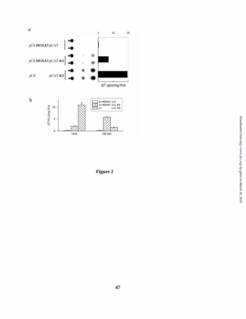
47
NHK ME180
105
RLU
/mg
Prot
0
5
10
p3-HOXA7 / pGLp3-HOXA7 / pGL-K3p3 / pGL-K3
b
0 10 20
1
2
3
105 cpm/mg Prot
a
pCS-HOXA7/pCAT
pCS-HOXA7/pCAT-K3
pCS /pCAT-K3
Figure 2
by guest on March 26, 2018
http://ww
w.jbc.org/
Dow
nloaded from
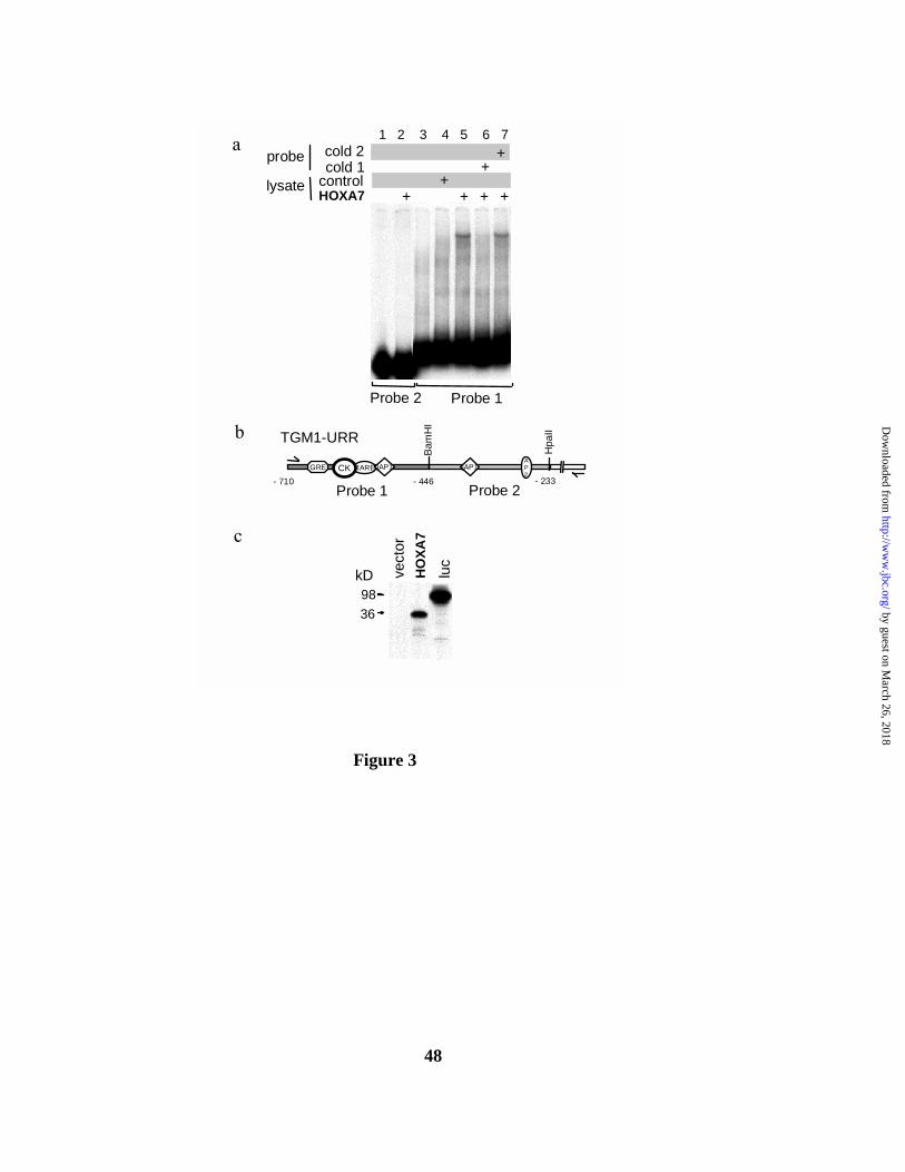
48
cold 2cold 1
controlHOXA7 + + + +
++
+
c
a
vect
orH
OXA
7
luc
9836
kD
Probe 2 Probe 1
b
Probe 1 Probe 2
GRE
Bam
HI
Hpa
II
RARECK- 710 - 446 - 233
jAP1 jAP1AP2
TGM1-URR
lysate
probe1 2 3 4 5 6 7
Figure 3
by guest on March 26, 2018
http://ww
w.jbc.org/
Dow
nloaded from

49
TGM1
HOXA7
β-Actin
TPA 0 2.5 5 10 24haHOXA7
GAPDH
b
c
TPA 0 2.5 5 10 24 48h
TPA 0 2.5 5 10 24h
HOXA5
HOXB7
β-Actin
Figure 4
by guest on March 26, 2018
http://ww
w.jbc.org/
Dow
nloaded from
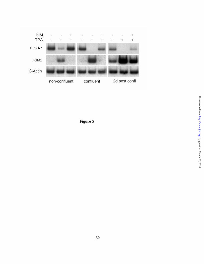
50
HOXA7
β-Actin
TGM1
non-confluent confluent 2d post confl
TPA - + + - + + - + +bIM - - + - - + - - +
Figure 5
by guest on March 26, 2018
http://ww
w.jbc.org/
Dow
nloaded from
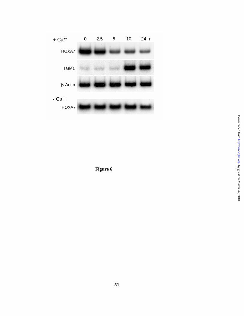
51
0 2.5 5 10 24 h
HOXA7
TGM1
β-Actin
+ Ca++
- Ca++
HOXA7
Figure 6
by guest on March 26, 2018
http://ww
w.jbc.org/
Dow
nloaded from
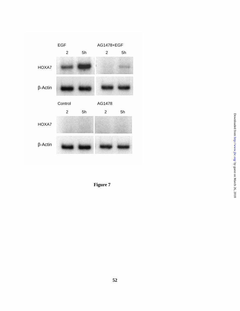
52
HOXA7
HOXA7
β-Actin
β-Actin
2 5h 2 5h
EGF AG1478+EGF
Control AG1478
2 5h 2 5h
Figure 7
by guest on March 26, 2018
http://ww
w.jbc.org/
Dow
nloaded from
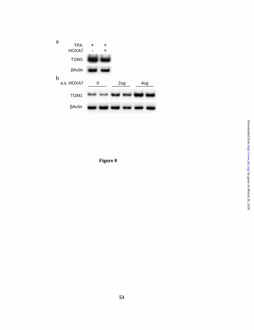
53
TGM1
βActin
βActin
TGM1
a.s. HOXA7 0 2ug 4ug
TPA + +HOXA7 - +
a
b
Figure 8
by guest on March 26, 2018
http://ww
w.jbc.org/
Dow
nloaded from

54
control
AntisenseHOXA7
TGM1 INV K10 K5 βactin
INV K1 K5
>>>
>
1118061
49
>
kD173
c c cas asas
a
b
c
0 200 400 600 800 1000 1200 1400
antisense
control
pmol X-linked 14C-Putrescine/mg Protein
*
Figure 9
by guest on March 26, 2018
http://ww
w.jbc.org/
Dow
nloaded from
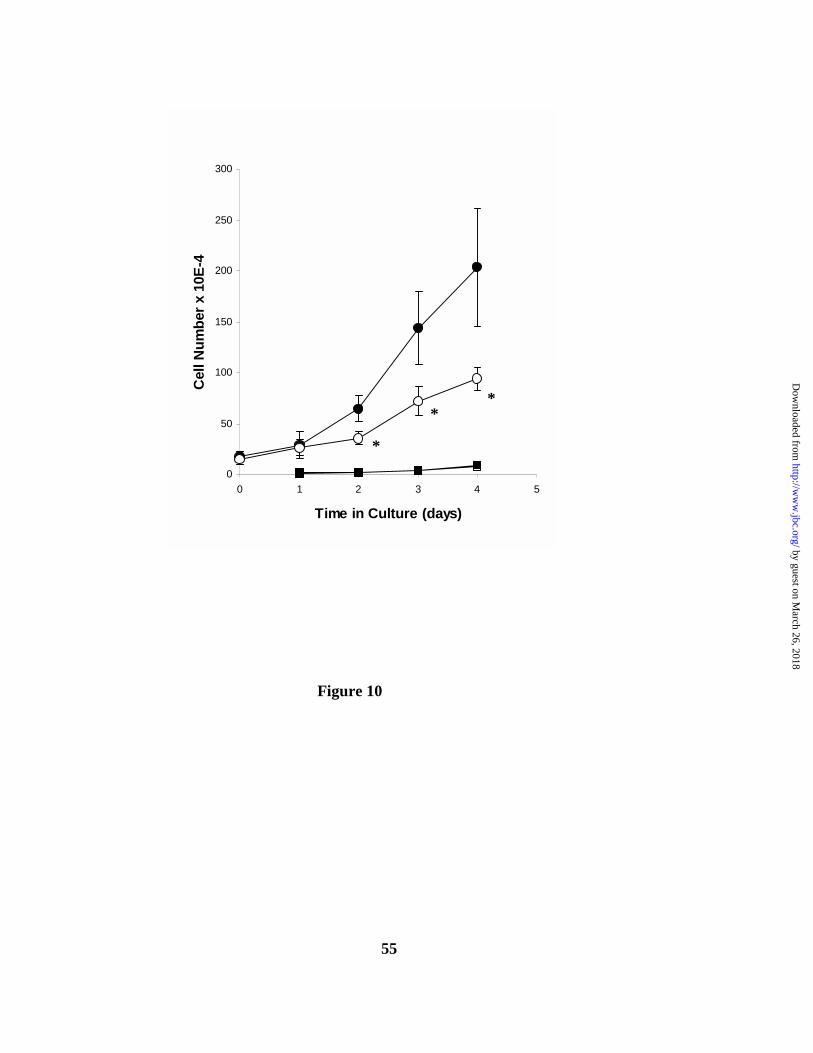
55
0
50
100
150
200
250
300
0 1 2 3 4 5
Time in Culture (days)
Cel
l Num
ber x
10E
-4
**
*
Figure 10
by guest on March 26, 2018
http://ww
w.jbc.org/
Dow
nloaded from

56
GAPDHa.s. HOXA7
a
b
β-Actin
HOXA7 frag
RNase 0 5 15 30 0 5 15 30’
Control a.s. HOXA7
C Cas as
Figure 11
by guest on March 26, 2018
http://ww
w.jbc.org/
Dow
nloaded from

Peter T. LaCelle and Renata R. Polakowskainhibits differentiation
Human homeobox HOXA7 regulates keratinocyte transglutaminase type 1 and
published online July 2, 2001J. Biol. Chem.
10.1074/jbc.M104598200Access the most updated version of this article at doi:
Alerts:
When a correction for this article is posted•
When this article is cited•
to choose from all of JBC's e-mail alertsClick here
by guest on March 26, 2018
http://ww
w.jbc.org/
Dow
nloaded from