diagnosis of nonalcoholic fatty liver disease in children ... · diagnosis of nonalcoholic fatty...
TRANSCRIPT

Co
CONSENSUS STATEMENT
Diagnosis of Nonalcoholic Fatty Liver Disease in Children
and Adolescents: Position Paper of the ESPGHAN
Hepatology Committee
� y z § jj
Pietro Vajro, Selvaggia Lenta, Piotr Socha, Anil Dhawan, Patrick McKiernan,e Lacaille, zzValerie alerio Nobili #Ulrich Baumann, ��Ozlem Durmaz, yyFlorencpyright 2012 by ESPGHAN and NASPGHAN. Unauthorized repro
in children ages 10 years and older. The first diagnostic step in these children
should be abdominal ultrasound and liver function tests, followed by
only liver histology canmatory changes from N
Received December 6, 2011; accepted February 23, 2012.From the �Department of Pediatrics, Medical School, University of
Salerno, Salerno, Italy, the yDepartment of Pediatrics, University ofNaples ‘‘Federico II,’’ Naples, Italy, the zDepartment of Gastroenter-ology, Hepatology, and Eating Disorders, the Children’s MemorialHealth Institute, Warsaw, Poland, the §Liver Unit, King’s College,London, the jjLiver Unit, Birmingham Children’s Hospital, Birming-ham, UK, the �Hepatometabolic Unit, ‘‘Bambino Gesu’’ Children’sHospital, Rome, Italy, the #Division of Pediatric Gastroenterology andHepatology, Hannover Medical School, Hannover, Germany, the��Department of Pediatrics, Istanbul Medical Faculty, University ofIstanbul, Turkey, the yyHopital Necker-Enfants Malades, Paris, France,and the zzDepartment of Pediatrics, University of Geneva Hospital,Geneva, Switzerland
Address correspondence and reprint requests to Pietro Vajro, MD, Chair ofPediatrics, University of Salerno, Via Allende, 84081 Baronissi(Salerno), Italy (e-mail: e-mail [email protected])
Drs Vajro, Socha, Dhawan, McKiernan, and Nobili are members of theNAFLD Group of the ESPGHAN Hepatology Committee. Drs Bau-mann, Durmaz, Lacaille, and McLin are other members of the ESP-GHAN Hepatology Committee. Dr Lenta is an invited expertparticipating in this ESPGHAN panel.
Conflicts of interest for the writing group appear on the ESPGHAN Web site(www.espghan.med.up.pt).
Copyright # 2012 by European Society for Pediatric Gastroenterology,Hepatology, and Nutrition and North American Society for PediatricGastroenterology, Hepatology, and Nutrition
DOI: 10.1097/MPG.0b013e318252a13f
700 JP
McLin, and �V
ABSTRACT
Nonalcoholic fatty liver disease (NAFLD) is the most common cause of
chronic liver disease in children and adolescents in the United States, and
most probably also in the rest of the industrialized world. As the prevalence
of NAFLD in childhood increases with the worldwide obesity epidemic,
there is an urgent need for diagnostic standards that can be commonly used
by pediatricians and hepatologists. To this end, we performed a PubMed
search of the adult and pediatric literature on NAFLD diagnosis through May
2011 using Topics and/or relevant Authors as search words. According to the
present literature, NAFLD is suspected based on the association of fatty liver
combined with risk factors (mainly obesity), after the exclusion of other
causes of liver disease. The reference but imperfect standard for confirming
NAFLD is liver histology. The following surrogate markers are presently
used to estimate degree of steatosis and liver fibrosis and risk of progression
to end-stage liver disease: imaging by ultrasonography or magnetic
resonance imaging, liver function tests, and serum markers of liver fibrosis.
NAFLD should be suspected in all of the overweight or obese children and
adolescents older than 3 years with increased waist circumference especially
if there is a NAFLD history in relatives. The typical presentation, however, is
exclusion of other liver diseases. Overweight/obese children with normal
ultrasonographic imaging and normal liver function tests should still be
monitored due to the poor sensitivity of these tests at a single assessment.
Indications for liver biopsy include the following: to rule out other
treatable diseases, in cases of clinically suspected advanced liver disease,
before pharmacological/surgical treatment, and as part of a structured
intervention protocol or clinical research trial.
Key Words: children, histology, imaging, liver biopsy, nonalcoholic fatty
liver disease, nonalcoholic steatohepatitis, noninvasive biomarkers, obesity-
related liver disease
(JPGN 2012;54: 700–713)
O besity is a major public health concern. The rise in theincidence of obesity diffusion is paralleled by that of its
comorbidities, including nonalcoholic fatty liver disease (NAFLD)(1). The latter includes a spectrum of clinicopathological entitiesranging from simple steatosis through nonalcoholic steatohepatitis(NASH) to cirrhosis and end-stage liver disease (Table 1).The nomenclature is inconsistent, with NAFLD being both thesummarizing term for the entire spectrum of the condition and thedescriptor of the more benign forms of simple steatosis and mildinflammation in contrast to NASH. The histopathological definitionof steatohepatitis requires at least 5% of liver cells with micro- ormacrovesicular fatty infiltration. NAFLD has become the mostcommon chronic hepatopathy both in adults and children. Itshistologically proven prevalence in children in the United States(as revealed at autopsy after accidents) ranges from 9.6% in normal-weight individuals up to 38% in obese ones (2). Due to its tendencyto progress through this spectrum in childhood (3) or after transitioninto adulthood (4), early diagnosis and treatment are importantissues at all ages (5). Treatment should address not only the liverdisease itself but also the entire spectrum of comorbidities toimprove overall survival and quality of life (6).
Available diagnostic procedures include a set of clinicalsigns and symptoms, laboratory and radiological imaging tests,and a combination of clinical parameters and blood test results (7,8).Although several of these markers are commonly used for thediagnostic evaluation of a patient with suspected NAFLD, noneof them seems to have a high specificity and sensitivity capableof definitely excluding another underlying liver disease. With therising prevalence of childhood obesity, the proportion of childrenwith both an underlying primary liver disease, such as autoimmuneliver disease or Wilson disease, and additional NAFLD increases,so it becomes essential not to miss a treatable condition. Also,
duction of this article is prohibited.
distinguish simple steatosis or mild inflam-ASH and determine the presence and stage
GN � Volume 54, Number 5, May 2012

Co
TABLE 1. Definitions of the spectrum of clinicopathological entities of nonalcoholic fatty liver
Simple steatosis At least 5% of liver cells with micro- or macrovesicular fatty infiltration
NAFLD The more benign form of simple steatosis and mild inflammationorThe summarizing term for the entire spectrum of the condition
NASH ‘‘Adult type’’: steatosis with ballooning degeneration and lobular inflammation,with or without perisinusoidal fibrosis, and without portal inflammation
‘‘Pediatric type’’: macrovesicular hepatocellular steatosis with portal inflammation,with or without portal fibrosis, in the absence of ballooning degeneration andperisinusoidal fibrosis
Cirrhosis The most advanced stage of fibrosis (stage 3¼ bridging fibrosis, stage 4¼ cirrhosis)
NAFLD¼ nonalcoholic fatty liver disease; NASH¼ nonalcoholic steatohepatitis.
JPGN � Volume 54, Number 5, May 2012 Diagnosis of NAFLD in Children and Adolescents
of fibrosis (9). At present, liver biopsy remains the ‘‘imperfect’’reference standard for NAFLD diagnosis (10), but it representsan impractical screening procedure because it is both expensiveand invasive.
METHODSWe performed a PubMed search by Topics and/or relevant
Authors up to May 2011 of the adult and pediatric literatureon NAFLD diagnosis using the following terms: ‘‘fatty liver,NAFLD, NASH, obesity-related liver disease, liver steatosis’’ with‘‘diagnosis, genetics, obesity, imaging, liver biopsy, histology,noninvasive biomarkers, liver tests.’’ We selected articles examin-ing conventional and novel diagnostic options in both adults andchildren. When pediatric data were not available, adult studies werereviewed. The data were written and reviewed by panelists of theEuropean Society for Pediatric Gastroenterology, Hepatology, andNutrition NAFLD working group and by the members of theEuropean Society for Pediatric Gastroenterology, Hepatology,and Nutrition Hepatology Committee.
GENETIC FACTORSNAFLD is considered a multifactorial disease with a sub-
stantial genetic component. Several studies have shown thatsome single-nucleotide polymorphisms of genes involved ininsulin sensitivity, lipid metabolism, and inflammation/fibrosismay influence both the mechanism and the extent of hepaticsteatosis and its progression to NASH and cirrhosis (11). A non-synonymous single-nucleotide polymorphism (rs738409) in thegene PNPLA3 (the gene for adiponutrin, an insulin-regulatedphospholipase) is associated with hepatic steatosis but not withinsulin sensitivity or inflammatory changes at histology in adultand pediatric populations. The 148-mol/L variant has been reportedto be associated early in life with increased levels of alaninetransaminase (ALT)/aspartate aminotransferase (AST) in a cohortof obese children, with liver steatosis more prevalent in carriers oftwo 148-mol/L alleles (12–14).
Polymorphisms of interleukin-6 (174G/C) (15) and tumornecrosis factor (TNF)-a (16), both involved in inflammationand insulin resistance, have been associated with NASH. A splicemutation in the tumor suppressor gene Kruppel-like factor 6(KLF 6) has been identified in patients with NAFLD with liverfibrosis (17). It has been shown that variants in the UGT1A1 gene
pyright 2012 by ESPGHAN and NASPGHAN. Un
(Gilbert syndrome) contribute to increased bilirubin levels, thusreducing the risk for NAFLD onset or development (18).
www.jpgn.org
Executive Summary
A combination of genetic and environmental factors is likelyresponsible for both the development of NAFLD and its progressionfrom simple steatosis to NASH. Several genes involved in lipo-genesis and inflammation have been found to have significantlyaltered expression levels in adults and children with NAFLD, andsome polymorphisms in regulatory cascades may be pathogenic.Presently, genetic factors are not relevant in the clinical approachto children with NAFLD.
RISK FACTORS AND CLINICAL ANDLABORATORY FEATURES
NAFLD is a diagnosis of exclusion requiring carefulconsideration of demographic, anthropometric, clinical, and labora-tory features. Table 2 illustrates the large spectrum of causes of fattyliver in children (7,19,20).
Risk Factors
NAFLD prevalence is higher in overweight (sex- and age-specific body mass index [BMI] >85th percentile) or obese(>95th percentile) peripubertal male children compared withnormal-weight age-matched pairs, and higher in male comparedwith female age-matched individuals of the same BMI (2,21).Although studies support the association between obesity andNAFLD, rates of obesity and overweight among children with aclinical diagnosis of NAFLD remain variable: Hispanic origin is arisk factor (2,21,22), whereas black race seems to be protective (23).
Familial clustering of obesity, insulin resistance, NAFLD, ortype 2 diabetes mellitus is frequent and should raise suspicion ofNAFLD in children from such families (24,25). The prevalence ofNAFLD is higher in children older than 10 years than it is inyounger ones (2), although they do not seem more prone to NASH/fibrosis (22). Unlike in NAFLD, male sex is not a risk factor forNASH (26). Rapid weight increase may be a risk factor for NAFLDin obese children (27).
Low birth weight combined with early catch-up growth isassociated with early obesity and is a risk factor for NAFLD (28),whereas breast-feeding seems to reduce the risk (29). Consumptionof (rich in fructose) soft drinks seems to be associated with NAFLD,independent of the metabolic syndrome (30). Obstructive sleepapnea (OSA) is associated with insulin-resistant NAFLD, but
authorized reproduction of this article is prohibited.
it does not seem to be related to the severity of steatohepatitis(NASH) (31).
701

Co
TABLE 2. Causes of fatty liver disease in children
General or systemic Genetic-metabolic causesOther rare hereditary
genetic disorders Drugs’ hepatotoxicity
Acute systemic disease Cystic fibrosis and Shwachmansyndrome
Alstrom syndrome Ethanol
Acute starvation Wilson disease Bardet-Biedl syndrome Ecstasy, cocaineProtein energy malnutrition a1-Antitrypsin deficiency Prader-Willi syndrome NifedipineTotal parenteral nutrition Galactosemia Cohen syndrome DiltiazemObesity/metabolic syndrome Fructosemia Cantu syndrome (1p36 deletion) EstrogensPolycystic ovary syndrome Cholesteryl ester storage disease Weber-Christian disease CorticosteroidsObstructive sleep apnea Glycogen storage disease
(types I and VI)Amiodarone
Rapid weight loss Mitochondrial and peroxisomaldefects of fatty acid oxidation
Perhexiline
Anorexia nervosa Madelung lipomatosis CoralgilCachexia Lipodystrophies TamoxifenInflammatory bowel disease Dorfman-Chanarin syndrome MethotrexateCeliac disease Abeta or hypobetalipoproteinemia PrednisoloneHepatitis C a- and b-oxidation defects ValproateNephrotic syndrome Porphyria cutanea tarda VitaminType 1 diabetes mellitus
and Mauriac syndromeHomocystinuria L-asparaginase
Thyroid disorders Familial hyperlipoproteinemias Zidovudine andHIV treatments
Hypothalamo-pituitary disorders Tyrosinemia type 1 SolventsBlind loop (bacterial overgrowth) Bile acids synthesis defects Pesticides
Congenital disorders of glycosylationTurner syndromeOrganic acidosisCitrin deficiencyHFE (hemochromatosis)
Modified from (7,19,20). Exclusions should be adjusted to age and clinical presentation. In infants and young children, NAFLD is hardly to be expected,whereas genetic, metabolic, syndromic, and systemic causes should be primarily considered guided by clinical signs and symptoms. In children older than10 years, NAFLD is expected when�1 features of the metabolic syndrome are present; still, Wilson disease and a1-antitrypsin deficiency should be excluded
Vajro et al JPGN � Volume 54, Number 5, May 2012
Clinical Features
Clinically, most pediatric patients with NAFLD/NASHhave nonspecific symptoms. Some complain of fatigue, malaise,or vague abdominal pain (42%–59% of cases), especially in theright upper quadrant, which has been associated with the moreprogressive form of NASH (2). Acanthosis nigricans is a clinicalmarker of hyperinsulinemia and has been observed in one-third tohalf of the children with biopsy-proven NAFLD (23). Hepatome-galy can be frequently detected (up to 50% of cases) (21,22).
Anthropometric Features
Visceral adiposity, which may be related to a state of insulinresistance, is a major contributor to fatty liver, representing a moreinfluential component than BMI in predicting liver steatosis.Unfortunately, indirect measurements of visceral adiposity usedin adult studies such as waist-to-hip ratio are not appropriate forchildhood because they change with age and have poor correlationwith measures of adiposity measured by DEXA (32,33). In children,
and autoimmune hepatitis should be considered.
pyright 2012 by ESPGHAN and NASPGHAN. Un
waist circumference alone represents a practical anthropometricparameter to identify central adiposity and it may predict increased
702
risk for insulin resistance and the metabolic syndrome (34). Specificpercentiles have been developed for children ages 5 to 16 years (35)and 11 to 18 years (36). The importance of waist circumferencemeasurement in childhood NAFLD is well established (37).Lin et al (37) showed that in obese children and adolescents, forevery 5-cm increase in waist circumference, there was an odds ratioof 1.4 for predicting ultrasonographic liver steatosis, but no detailson percentiles were given. Increased waist circumference is alsoassociated with increased hepatic fibrosis (38). There is a need forstandard international waist circumference charts.
Laboratory Tests
In clinical practice the diagnosis of NAFLD is usuallysuggested by finding elevated serum hepatobiliary enzymes(mostly ALT and g-glutamyl transpeptidase [GGT]), and/or evi-dence of a bright liver on ultrasound (US), most frequently amongoverweight/obese children (27,39,40).
Serum ALT activity is a widely available and inexpensive testfor the screening and initial evaluation of NAFLD. The sensitivity of
authorized reproduction of this article is prohibited.
this biochemical marker, however, remains low because a number ofadult and pediatric patients may present ALT values in the normal
www.jpgn.org

Co
range (41). In fact, determining an ALT cutoff for NAFLD has beenthe subject of some debate. In a study involving 72 obese childrenwith NAFLD, an ALT >35 IU/L had a sensitivity of 48% andspecificity of 94% for detecting steatosis >5% as measured bymagnetic resonance imaging (MRI) (42). More recently, the Screen-ing ALT for Elevation in Today’s Youth (SAFETY) study has shownthat in American laboratories conventional ALT cutoff values are settoo high for the reliable detection of pediatric chronic liver disease,including NAFLD. In the National Health and Nutrition ExaminationSurvey (NHANES) study, the 95th percentile levels for ALT inhealthy weight, metabolically normal, liver disease–free patientswere 25.8 U/L (boys) and 22.1 U/L (girls) (43). Comparable con-clusions were also reached in the European pediatric population (44).
It is now widely accepted that the degree of ALT elevationdoes not correlate with the presence (42,45) or severity of histo-logical findings of NAFLD (40). A number of children with normalALT or minimal serum ALT elevation may have advanced fibrosison liver biopsy. The natural history of the disease is not yet welldetermined in children, but at times ALT tends to fluctuate and mayeven normalize (22,41). High serum levels of GGT represent a riskfactor for advanced fibrosis in NAFLD (46).
NAFLD may be considered the hepatic manifestation of themetabolic syndrome, which is defined by the presence of visceralobesity, hypertension, insulin resistance or diabetes, dyslipidemia,and hyperuricemia. Hyperinsulinemia, due to insulin resistance,most probably represents the first pathogenetic hit of NAFLD (47).It is a sensitive but nonspecific predictor of NAFLD (48), and henceunsuitable as a single indicator of NAFLD; however, it may be apredictor for progressive hepatic fibrosis (21,26). Abnormalities inthe oral glucose tolerance test also may suggest NAFLD (49).
Hypertriglyceridemia is another biochemical marker fre-quently reported in obese children with NAFLD (21). Oliveiraet al (50) showed a positive correlation between ALT and trigly-ceride values among pediatric patients. Others showed that amongpatients with suspected steatohepatitis, ALT concentration wassignificantly higher in subjects with elevated triglycerides (51).Finally, in children with NAFLD, an atherogenic lipid profile
JPGN � Volume 54, Number 5, May 2012
pyright 2012 by ESPGHAN and NASPGHAN. Un
correlates with severity of liver injury (52). High levels of serumuric acid have been reported in the majority of subjects with the
TABLE 3. Laboratory workup in children with suspected NAFLD
Metabolic function and liver testsBasic profile: blood counts, standard liver function tests, fasting glu
ALT/AST ratioLipid profile (cholesterol, triglycerides, HDL-cholesterol, LDL-cholGlucose tolerance test (OGTT), glycosylated hemoglobinCalculation of HOMA-IR, ISI-gly as markers for insulin resistanceThyroid function tests
Tests for exclusion of other main causes of hepatic steatosisSerum lactate, uric acid, iron, ferritin, pyruvateSerum copper, ceruloplasmin levels, 24-hour urinary copperSweat testAntibodies against tissue transglutaminase IgA and total IgAa1-Antitrypsin levels and phenotype when indicatedAmino and organic acidsPlasma-free fatty acids and acyl carnitine profileUrinary steroid metabolitesOther specific tests as suggested by history and examination (eg, vi
Modified from (7,8). ALT¼ alanine aminotransferase; AST¼ aspartate ammodel assessment; INR¼ international normalized ratio; ISI-gly¼ insulin stolerance test.
www.jpgn.org
metabolic syndrome, and has been proposed as an independentpredictor of NAFLD both in adults (53) and children (49), probablyas a marker of high fructose consumption that correlates with theprogression of fibrotic changes (30). Serum IgA level is elevatedin about 25% of cases of NAFLD-NASH, but its meaning anddiagnostic value are still not clear. High levels of IgA antibodiesagainst tissue transglutaminase have been reported in several chronicliver diseases, including NAFLD (54). Silent celiac disease and fattyliver have been reported to coexist in unrecognized obese children(55).
Increased titers of serum nonorgan-specific autoantibodies(particularly anti-nuclear antibody and anti-smooth muscle anti-body) have been reported in up to one-third of all of the investigatedpatients, both in adults (56) and children (26), and may requireimmediate definitive further investigation to rule out associatedautoimmune hepatitis. Table 3 summarizes the biochemicalmarkers routinely performed in clinical practice in suspectedpediatric NAFLD (7,8).
Executive Summary
The panel agreed that careful consideration of a series ofanthropometric, demographic, clinical, and laboratory featuresmay offer a clue to the identification of NAFLD risk. Acanthosisnigricans and increased waist circumference are warning signs forNAFLD. ALT in combination with liver ultrasound is an indicatorof NAFLD, but normal ALT does not exclude liver steatosis orits progression to severe fibrosis and cirrhosis. Insulin resistanceand increased triglyceride concentration are additional risk factorsof NAFLD. These factors were identified based on observationalstudies that associated NAFLD with clinical, anthropometric, andlaboratory parameters.
DIFFERENTIAL DIAGNOSISAbnormal serum aminotransferases in overweight or obese
patients are not diagnostic of NAFLD/NASH. Other causes of
Diagnosis of NAFLD in Children and Adolescents
authorized reproduction of this article is prohibited.
muscle (57) and treatable liver disease should be ruled out, withspecial emphasis on celiac disease–related hepatopathy (55),
cose and insulin, urea and electrolytes, coagulation, INR,
esterol), lipoproteins
ral hepatitis panel, serum immunoglobulins, liver autoantibodies)
inotransferase; HDL¼ high-density lipoprotein; HOMA-IR¼ homeostaticensitivity index; LDL¼ low-density lipoprotein; OGTT¼ oral glucose
703

Co
Wilson disease (58), and autoimmune hepatitis (56,26). ALT serumlevels alone are a useful tool, but they are not adequate as a singlemarker for diagnosing NAFLD. The presence of hepatomegaly orsplenomegaly is suggestive of advanced liver disease, which callsfor a rapid and complete assessment, including early liver biopsy toexclude other etiologies (19).
The differential diagnosis of NAFLD/NASH is detailed inTable 2 and the proposed workup is outlined in Table 3. NAFLDusually does not occur in extremely young children (younger than3 years) and is rare in children younger than 10 years. Differentialdiagnosis should be based first on clinical features, then on bloodtests, and finally liver biopsy must be considered (Fig. 1).
Executive Summary
The panel indicates a high suspicion of metabolic disordersas cause of fatty liver in young children. NAFLD hardly occurs inchildren younger than 3 years and is rare in children younger than10 years. Thus, young children require a detailed diagnostic work-up to exclude other etiologies. In older children and teenagers, somemetabolic disorders should also be considered for differentialdiagnosis. Obesity per se does not justify making the diagnosisof NAFLD in patients with increased transaminase activity.
THE REFERENCE STANDARD: LIVER BIOPSY
Vajro et al
pyright 2012 by ESPGHAN and NASPGHAN. Un
Liver biopsy is the test with the highest discrimination forexcluding other potentially treatable conditions. It is also the only
Obese children and adol
Perform LFTs and sonogra
If US hyperec
Consider age,
Infants & children <3 y(NAFLD less probable)
NAbvi
If
Consider liver biopsy after completionof laboratory diagnostic workup C
Suspect first other disease: genetic/metabolic, syndromic, systemic
causes workupNAFLD diagnosis must be critically
questioned
If normal
Follow for central obesity &consider fatty liver at MRI if clinical
signs of insulin resistance (IR)
Nonobese childrenadolescentswith hyper-
transaminasemiaand hyperechogenic
liver
FIGURE 1. Overall management algorithm for children with susdisease; LFTs¼ liver function tests; MRI¼magnetic resonance im
704
single test that can reliably distinguish between simple steatosis(NAFLD) and NASH. As summarized in Table 1, it providesimportant information regarding the degree of liver damage,changes in the liver architecture, and severity of inflammatoryactivity and fibrosis (59). Normal liver function tests do not excludeany degree of NAFLD-related liver injury (41). Furthermore,evaluation of liver biopsy may be essential for detecting coexistingdiseases (eg, for diagnosis of autoimmune hepatitis).
The principal histological features of NASH include thepresence of macrovesicular fatty changes of hepatocytes withdisplacement of the nucleus to the edge of the cell, ballooningdegeneration of hepatocytes, and a mixed lobular inflammation.Other features, such as perisinusoidal-pericellular fibrosis, Malloryhyaline, megamitochondria, acidophil bodies, and glycogenatednuclei, can be present but are not mandatory to establish thediagnosis of NASH (60).
In an effort to standardize the histological criteria, theNational Institute of Diabetes and Digestive and Kidney Diseasesponsored the NASH Clinical Research Network to developthe NAFLD activity score (NAS) (59). This score is based onthe classification proposed earlier by Brunt et al (61) and consistsof an unweighted sum for each of the following lesions: steatosis(0–3), lobular inflammation (0–3), and hepatocellular ballooning(0–2). A score�5 is strongly suggestive of NASH, whereas a score<3 is largely consistent with the absence of NASH; however, theNAS cannot replace a pathologist’s diagnosis of steatohepatitis.
JPGN � Volume 54, Number 5, May 2012
authorized reproduction of this article is prohibited.
Furthermore, its utility in assessing the response to therapeuticintervention remains to be determined.
escents
phy in all
hogenicity or increased AST/ALT
history, and physical examination
Children >10 y(NAFLD more probable)
Children 3-10 y(NAFLD less probable)
FLD diagnosis should beased after exclusions ofral, toxic, metabolic (eg,WD), systemic causes
(eg, CD) workup
If central obesity, IR andno clinical signs of
progressive liver disease
Trial of weight reductionand lifestyle changes for
3-6 months
If persistenthypertransaminasemiaor hyperechogenicity at
US
Perform laboratoryworkup of exclusion of
other causes
If sustainedhypertransaminasemia orhyperechogenicity at US
negative or inconclusive
onsider early liver biopsy Consider liver biopsy Consider early liver biopsy
Family histoty of NASHHepartosplenomegalyComorbiditiesHypothalamicExpansive processesMarkedhypertransaminaserniaElevated fibrosisserum markers
pected nonalcoholic fatty liver disease (NAFLD). CD¼ celiacaging; US¼ultrasound; WD¼Wilson disease.
www.jpgn.org

Copyright 2012 by ESPGHAN and NASPGHAN. Unauthorized reproduction of this article is prohibited.
TA
BLE
4.
Featu
res
of
non
inva
sive
imag
ing
dia
gn
ost
icm
eth
od
sin
ad
ult
sw
ith
non
alc
oh
olic
fatt
yliv
er
dis
ease
US
CT
MR
IM
RS
MR
Sel
asto
gra
ph
y(1
03
)F
ibro
scan�
Ass
essm
ent
Qu
alit
ativ
eQ
ual
itat
ive
and
qu
anti
tati
ve
Qu
alit
ativ
ean
d
qu
anti
tati
ve
Qu
alit
ativ
ean
d
qu
anti
tati
ve
Qu
alit
ativ
ean
d
qu
anti
tati
ve
Qu
alit
ativ
ean
d
qu
anti
tati
ve
Co
stL
ow
Med
ium
Ex
pen
sive
Ex
pen
sive
Ex
pen
sive
Lo
w
Met
ho
dV
isua
lco
mp
aris
on
Liv
erat
ten
uat
ion,
liv
er-s
ple
en
atte
nuat
ion
rati
o
T1
wei
gh
ted
gra
die
nt-
echo
in/o
ut
of
ph
ase
sequen
ce,
T2-f
at
satu
rate
d
Sp
ectr
osc
op
y,m
easu
rem
ent
of
area
un
der
the
lip
id
reso
nan
cep
eak
Modifi
edphas
e-co
ntr
ast
MR
Ise
quen
ceto
vis
ual
ize
pro
pag
atin
g
shar
ew
aves
inti
ssu
es
US
tran
sdu
cer
pro
be
that
mea
sure
liv
er
stif
fnes
str
ou
gh
pro
pag
atio
no
fel
asti
c
shar
ew
ave
Acc
ura
cyS
ensi
tiv
ity
:
60
%–
96
%70,7
1S
ensi
tiv
ity:
82
%77
Sen
siti
vit
y:1
00
%80
Sen
siti
vit
y:8
7%
–1
00
%83
Sen
siti
vit
y:
NE
Sen
siti
vit
y:
81%
–85%
89
Sp
ecifi
city
:
84
%–
10
0%
70,7
1S
pec
ifici
ty:
10
0%
77
Sp
ecifi
city
:9
0.4
%80
Dia
gn
ost
icp
reci
sio
n:
80
%–
85
%83
Spec
ifici
ty:
NE
Spec
ifici
ty:
74%
–78%
89
Lim
its:
op
erat
or
dep
ende
nt
Lim
its:
ion
izin
g
rad
iati
on
PP
V:
50
%81
Lim
its:
fib
rosi
sco
nfu
sed
wit
hst
eato
sis
ifB
MI
>2
8
NP
V:
10
0%
81
Over
all
accu
racy
91.2
%81
Acq
uis
itio
n
tim
e
Var
iab
le,
5–
20
min
,
dep
ends
on
op
erat
or
<5
min
10
–1
5m
in1
0–
15
min
10
–1
5m
in<
5m
in
Av
aila
bil
ity
Wid
ein
mo
sth
osp
ital
sW
ide
inm
ost
ho
spit
als
So
ftw
are
no
tav
aila
ble
inal
lM
RI
un
its
So
ftw
are
no
tav
aila
ble
in
all
MR
Iu
nit
s
Soft
war
enot
avai
lable
inal
lM
RI
un
its
No
tw
ides
pre
adin
mo
st
ho
spit
als
Thre
shold
of
det
ecti
on
Pat
ien
tsw
ith
>2
0%
stea
tosi
s
Pat
ien
tsw
ith>
30
%
stea
tosi
s
Det
ect
gra
de
Ist
eato
sis
(5%
–3
0%
)
Det
ect
gra
de
Ist
eato
sis
(5%
–3
0%
)
Det
ect
gra
de
Ist
eato
sis
(5%
–3
0%
)
Fib
rosi
s
Dis
ting
uis
hes
NA
SH
from
NA
FL
D
No
No
No
No
No
No
Mo
difi
edfr
om
(69
).C
T¼
com
pu
ted
tom
ogr
aph
y;
MR
I¼m
agn
etic
reso
nan
ceim
agin
g;
MR
S¼
mag
net
icre
son
ance
spec
trosc
op
y;N
AF
LD¼
no
nal
coh
oli
cfa
tty
liv
erd
isea
se;
NA
SH¼
no
nal
coh
oli
c
stea
tohep
atit
is;
NE¼
no
tev
alua
ted
;U
S¼
ult
raso
nog
rap
hy.
�F
ibro
scan
mea
nval
ues
of
sensi
tivit
yan
dsp
ecifi
city
inped
iatr
icN
AF
LD
wer
e97%
–100%
and
91%
–100%
,re
spec
tivel
y.M
ost
accu
rate
val
ues
wer
eobta
ined
inca
ses
wit
hh
igh
erfi
bro
sis
(70
).
JPGN � Volume 54, Number 5, May 2012 Diagnosis of NAFLD in Children and Adolescents
www.jpgn.org 705
Co
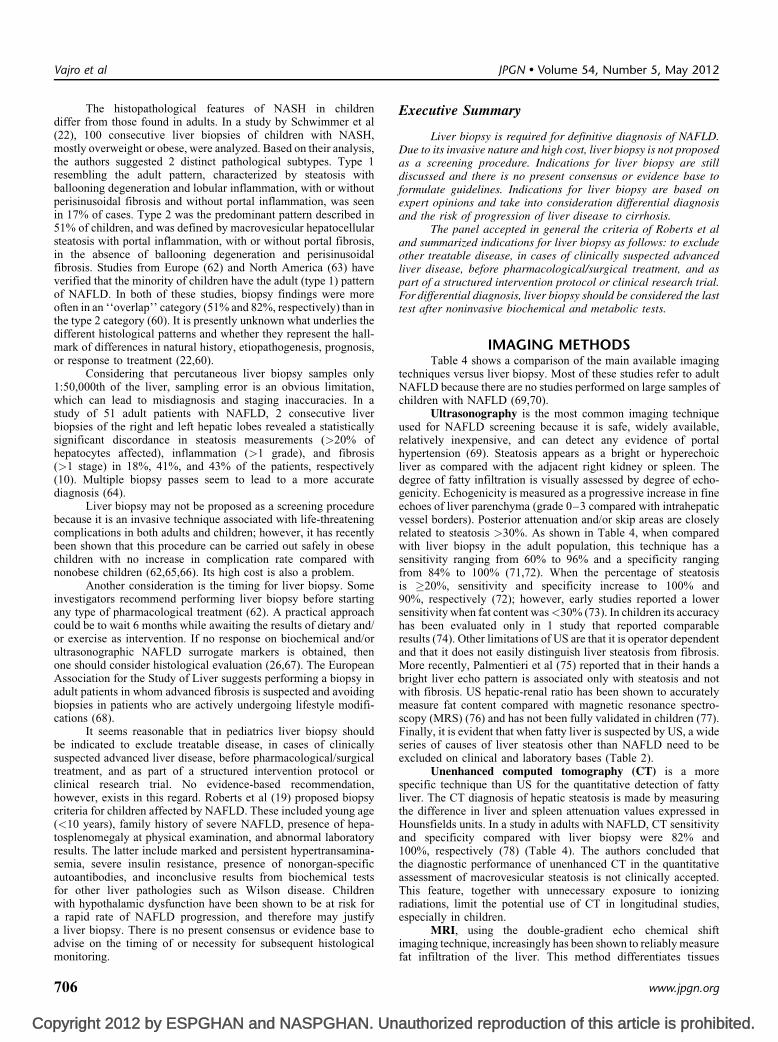
The histopathological features of NASH in childrendiffer from those found in adults. In a study by Schwimmer et al(22), 100 consecutive liver biopsies of children with NASH,mostly overweight or obese, were analyzed. Based on their analysis,the authors suggested 2 distinct pathological subtypes. Type 1resembling the adult pattern, characterized by steatosis withballooning degeneration and lobular inflammation, with or withoutperisinusoidal fibrosis and without portal inflammation, was seenin 17% of cases. Type 2 was the predominant pattern described in51% of children, and was defined by macrovesicular hepatocellularsteatosis with portal inflammation, with or without portal fibrosis,in the absence of ballooning degeneration and perisinusoidalfibrosis. Studies from Europe (62) and North America (63) haveverified that the minority of children have the adult (type 1) patternof NAFLD. In both of these studies, biopsy findings were moreoften in an ‘‘overlap’’ category (51% and 82%, respectively) than inthe type 2 category (60). It is presently unknown what underlies thedifferent histological patterns and whether they represent the hall-mark of differences in natural history, etiopathogenesis, prognosis,or response to treatment (22,60).
Considering that percutaneous liver biopsy samples only1:50,000th of the liver, sampling error is an obvious limitation,which can lead to misdiagnosis and staging inaccuracies. In astudy of 51 adult patients with NAFLD, 2 consecutive liverbiopsies of the right and left hepatic lobes revealed a statisticallysignificant discordance in steatosis measurements (>20% ofhepatocytes affected), inflammation (>1 grade), and fibrosis(>1 stage) in 18%, 41%, and 43% of the patients, respectively(10). Multiple biopsy passes seem to lead to a more accuratediagnosis (64).
Liver biopsy may not be proposed as a screening procedurebecause it is an invasive technique associated with life-threateningcomplications in both adults and children; however, it has recentlybeen shown that this procedure can be carried out safely in obesechildren with no increase in complication rate compared withnonobese children (62,65,66). Its high cost is also a problem.
Another consideration is the timing for liver biopsy. Someinvestigators recommend performing liver biopsy before startingany type of pharmacological treatment (62). A practical approachcould be to wait 6 months while awaiting the results of dietary and/or exercise as intervention. If no response on biochemical and/orultrasonographic NAFLD surrogate markers is obtained, thenone should consider histological evaluation (26,67). The EuropeanAssociation for the Study of Liver suggests performing a biopsy inadult patients in whom advanced fibrosis is suspected and avoidingbiopsies in patients who are actively undergoing lifestyle modifi-cations (68).
It seems reasonable that in pediatrics liver biopsy shouldbe indicated to exclude treatable disease, in cases of clinicallysuspected advanced liver disease, before pharmacological/surgicaltreatment, and as part of a structured intervention protocol orclinical research trial. No evidence-based recommendation,however, exists in this regard. Roberts et al (19) proposed biopsycriteria for children affected by NAFLD. These included young age(<10 years), family history of severe NAFLD, presence of hepa-tosplenomegaly at physical examination, and abnormal laboratoryresults. The latter include marked and persistent hypertransamina-semia, severe insulin resistance, presence of nonorgan-specificautoantibodies, and inconclusive results from biochemical testsfor other liver pathologies such as Wilson disease. Childrenwith hypothalamic dysfunction have been shown to be at risk fora rapid rate of NAFLD progression, and therefore may justifya liver biopsy. There is no present consensus or evidence base to
Vajro et al
pyright 2012 by ESPGHAN and NASPGHAN. Un
advise on the timing of or necessity for subsequent histologicalmonitoring.
706
Executive Summary
Liver biopsy is required for definitive diagnosis of NAFLD.Due to its invasive nature and high cost, liver biopsy is not proposedas a screening procedure. Indications for liver biopsy are stilldiscussed and there is no present consensus or evidence base toformulate guidelines. Indications for liver biopsy are based onexpert opinions and take into consideration differential diagnosisand the risk of progression of liver disease to cirrhosis.
The panel accepted in general the criteria of Roberts et aland summarized indications for liver biopsy as follows: to excludeother treatable disease, in cases of clinically suspected advancedliver disease, before pharmacological/surgical treatment, and aspart of a structured intervention protocol or clinical research trial.For differential diagnosis, liver biopsy should be considered the lasttest after noninvasive biochemical and metabolic tests.
IMAGING METHODSTable 4 shows a comparison of the main available imaging
techniques versus liver biopsy. Most of these studies refer to adultNAFLD because there are no studies performed on large samples ofchildren with NAFLD (69,70).
Ultrasonography is the most common imaging techniqueused for NAFLD screening because it is safe, widely available,relatively inexpensive, and can detect any evidence of portalhypertension (69). Steatosis appears as a bright or hyperechoicliver as compared with the adjacent right kidney or spleen. Thedegree of fatty infiltration is visually assessed by degree of echo-genicity. Echogenicity is measured as a progressive increase in fineechoes of liver parenchyma (grade 0–3 compared with intrahepaticvessel borders). Posterior attenuation and/or skip areas are closelyrelated to steatosis >30%. As shown in Table 4, when comparedwith liver biopsy in the adult population, this technique has asensitivity ranging from 60% to 96% and a specificity rangingfrom 84% to 100% (71,72). When the percentage of steatosisis �20%, sensitivity and specificity increase to 100% and90%, respectively (72); however, early studies reported a lowersensitivity when fat content was<30% (73). In children its accuracyhas been evaluated only in 1 study that reported comparableresults (74). Other limitations of US are that it is operator dependentand that it does not easily distinguish liver steatosis from fibrosis.More recently, Palmentieri et al (75) reported that in their hands abright liver echo pattern is associated only with steatosis and notwith fibrosis. US hepatic-renal ratio has been shown to accuratelymeasure fat content compared with magnetic resonance spectro-scopy (MRS) (76) and has not been fully validated in children (77).Finally, it is evident that when fatty liver is suspected by US, a wideseries of causes of liver steatosis other than NAFLD need to beexcluded on clinical and laboratory bases (Table 2).
Unenhanced computed tomography (CT) is a morespecific technique than US for the quantitative detection of fattyliver. The CT diagnosis of hepatic steatosis is made by measuringthe difference in liver and spleen attenuation values expressed inHounsfields units. In a study in adults with NAFLD, CT sensitivityand specificity compared with liver biopsy were 82% and100%, respectively (78) (Table 4). The authors concluded thatthe diagnostic performance of unenhanced CT in the quantitativeassessment of macrovesicular steatosis is not clinically accepted.This feature, together with unnecessary exposure to ionizingradiations, limit the potential use of CT in longitudinal studies,especially in children.
MRI, using the double-gradient echo chemical shift
JPGN � Volume 54, Number 5, May 2012
authorized reproduction of this article is prohibited.
imaging technique, increasingly has been shown to reliably measurefat infiltration of the liver. This method differentiates tissues
www.jpgn.org

Co
containing only water from those containing both fat and water.This noninvasive and nonirradiating technique is of great interestfor use in children. At the moment, limited data are availableregarding the utility of hepatic fat quantification in pediatricNAFLD (79). A study involving 50 obese children with NAFLDshowed good correlation between MRI and US (P< 0.0001),especially in patients with moderate and severe steatosis (80).In that study, however, there was no comparison with liver biopsy.In adults, sensitivity and specificity of MRI compared withhistopathological findings are 100% and 90.4%, respectively(81) (Table 4). Furthermore, MRI is not subject to interobservervariation and may be more useful than US for monitoring childrenwith fatty liver. It can prove useful in identifying fat regressionor progression, especially when the grade is mild (82). Novelapproaches have been proposed to improve the accuracy of thetechnique (83).
An emerging imaging modality for the quantitative assess-ment of hepatic steatosis is 1H-MR spectroscopy (1H-MRS).This technique grades hepatic triglyceride content by directlymeasuring protons in the acyl groups of the liver tissue triglycerides.Sensitivity and diagnostic precision in adults range from 87% to100% and from 80% to 85%, respectively (84). MRS has beenapplied successfully in a pediatric pilot study to measure hepatic fatcontent in patients with biopsy-proven NASH before and afterpharmacological treatment (85). At present, it is the most accuratemethod in which fat content is <10%, and a recent meta-analysisconfirmed that MRI and 1H-MRS are the techniques of choice foraccurate evaluation of steatosis (86); however, MRS is not widelyperformed because it is time-consuming and requires off-scananalysis by an expert. Because of these limitations, MRS at presentseems to be most appropriate for research studies at specializedcenters and is not suitable for widespread use.
The fibroscan is a new medical device using transientelastography to evaluate liver fibrosis based on stiffness in anoninvasive, rapid, painless, and reproducible way. Promisingresults were first shown in adults with chronic hepatitis C (87).Fibroscan has correlated well with hepatic histology both in adults(88) and in children (70) with chronic liver disease includingNAFLD. In adults, sensitivity ranges from 81% to 85%, andspecificity from 74% to 78%. (89) (Table 4). The limitation ofthis technique is that fibrosis may be mistaken for steatosis in adultpatients with a BMI >28 (89). In addition, this technique is not yetperformed in everyday clinical practice. Finally, its resolution is notsufficient to detect changes in fibrosis over time and after treatment.
A few studies have demonstrated the accuracy of this tech-nique in assessing hepatic fibrosis in children with NAFLD.Unfortunately, at the present time, the probe size is not appropriatefor smaller children (70,88). Most recently normal values of liverstiffness in children ages 7.5 to 8.6 years, using a pediatric probe,have been proposed (90). The results showed that a wide differenceexists between values obtained using adult and pediatric probes.Further validation of transient elastography is necessary before itcan be adopted for the stratification of NAFLD in childhood eitheralone or in association with other noninvasive approaches.
Magnetic resonance elastography (MRE) uses a modifiedphase-contrast MRI sequence to visualize propagating sharewaves in tissues. It could be a convenient complement to MRSto estimate noninvasively the degree of steatosis and degree of liverstiffness; however, further studies are required before MRE can beintroduced in clinical practice. Its association with laboratorytests (eg, AST/platelet ratio index, GGT levels) may improve thespecificity and sensitivity of the noninvasive estimation of liverfibrosis (46,91). At the moment there are no data concerning the
JPGN � Volume 54, Number 5, May 2012
pyright 2012 by ESPGHAN and NASPGHAN. Un
use of MRE in children. Therefore, we consider this technique tobe experimental.
www.jpgn.org
Executive Summary
Diagnosis of NAFLD in Children and Adolescents
mon
au
hasadu
Accurate noninvasive imaging techniques to diagnose anditor NAFLD are being developed, as follows:
1. U
ltrasounds are safe, but they are limited by the inability toquantify steatosis or fibrosis.MRI is not cost-effective, even if with certain modifications 2.i t could enable rapid, reproducible measurements of steatosisand fibrosis.Fibroscan has been used in children with NAFLD, but in its 3.present state of development it is not yet suitable for widespreaduse in these patients.NOVEL NONINVASIVE LABORATORYASSESSMENT OF NAFLD STAGES AND GRADES
Despite the high prevalence of pediatric NAFLD, fewstudies have evaluated noninvasive markers for the prediction ofhepatic steatosis and progression to steatohepatitis in children.Logical markers would reflect the most common pathogenicmechanisms underlying NAFLD. Some of these are highlightedhere.
Serum Markers of Hepatic Inflammation
An imbalance between several proinflammatory (TNF-a;resistin, interleukin-6) and anti-inflammatory cytokines (adipo-kines [eg, adiponectin]) seems to be involved in the progressionfrom simple steatosis to NASH (92). Therefore, a variety ofbiomarkers of hepatic inflammation have been proposed assurrogate markers for diagnosing NASH both in adult and pediatricages.
Low serum levels of adiponectin have been reported inpediatric patients with NASH with normal levels of proinflam-matory cytokines, suggesting that adiponectin may play a role inboth the pathogenesis and disease progression (93). As such, it mayserve as a noninvasive biomarker.
Serum leptin has been implicated in disease progressionbecause of a direct profibrotic effect (94). High levels of serumTNF-a (a major inflammatory cytokine that is also secreted byadipose tissue, and antagonizes the effects of adiponectin) andserum leptin correlated well with histological features of steato-hepatitis in a cohort of histologically proven NAFLD in children(95).
Retinol-binding protein 4, another adipokine that is associ-ated with insulin resistance, has been shown in a pediatric cohort of59 children with biopsy-proven NAFLD to be inversely correlatedto degree of liver damage (96).
A recent study in a pediatric cohort with biopsy-provenNAFLD showed that serum levels of endotoxin and plasminogenactivator inhibitor-1 may serve as a reliable marker of NASH,suggesting that endotoxin may participate in the progression fromNAFLD to NASH (97).
Although several groups have investigated circulatingcytokine levels and their correlation with disease severity, thereare no data at the present time supporting their generalized use in thediagnosis of NASH in adults and children.
Another inflammatory biomarker that seems involved inNAFLD progression is C-reactive protein, an acute-phase reactantsynthesized in the liver. This marker is frequently elevated insubjects with metabolic syndrome and represents an independentpredictor of NAFLD (32). High-sensitivity C-reactive protein
thorized reproduction of this article is prohibited.
been reported as a potential marker of severity of fibrosis inlt NASH (98).
707

Co
Increased plasma ferritin levels are seen in 20% to 50% ofadults with NAFLD and elevated transferrin saturation in 5% to10% (99). These data also have been reported in pediatric patientswith NAFLD as a marker of systemic inflammation rather than amarker of iron overload (32). An association with a heterozygosityfor human hemochromatosis (HFE) gene has been described both inadults (100) and in children (32).
Another important mediator of hepatic insulin resistance,fetuin-A, has been described in children with metabolic syndromeand fatty liver. Fetuin-A was significantly higher in obese childrenwith NAFLD versus controls and decreased considerably in thosewho lost weight, suggesting its role as a potential biomarker fordiagnosis and assessment of treatment response (101).
Markers of Oxidative Stress
Enhanced oxidative stress (OS) has long been recognized as amechanism involved in liver damage and disease progression inhuman and animal models of NASH. Several oxidation pathwaysmay play a role in the overproduction of reactive oxygen species.A number of studies have attempted to elucidate whether measure-ment of systemic markers of OS may reflect the levels of OS presentin the liver (102).
Chalasani et al (103) measured circulating levels of lipidperoxidation products and their metabolic and nutritional correlatesin 21 adults with NASH and 19 matched controls. Although patientswith NASH had higher serum levels of oxidized low-densitylipoprotein and thiobarbituric acid–reacting substance, thesedifferences were not significant in multivariate analyses and totalantioxidant status did not differ between groups. In 59 childrenwith NAFLD versus controls and hepatitis C virus, hepatic lipidperoxidation (studied as hepatic malondialdehyde) was increased,but without differences in NAFLD versus NASH (104). In anotherstudy involving 36 children with NAFLD, increased serum levels ofprotein glutathionylation were detected, showing a correlation ofthis OS marker with histological steatohepatitis and liver fibrosis(105). Similar results were seen in 40 children with biopsy-provenNAFLD in which OS was evaluated with serum protein carbonyls,hepatic expression of 8-hydroxy-2-deoxyguanosine, and circulatingantibody against malondialdehyde-adducted human serum albumin(106).
Bilirubin has known antioxidant properties, and variantsin the UGT1A1 gene (Gilbert syndrome) contribute to increasedbilirubin levels and reduced risk for onset or development ofNAFLD (16).
Markers of Apoptosis
Several groups have reported that serum markers ofhepatocyte apoptosis can discriminate NASH from benign steatosis.Cytokeratin 18 (CK18) is an intracellular intermediate filamentprotein expressed at high levels by hepatocytes. Caspase-cleavedCK18 fragments represent an indirect measure of cell death; highserum levels have been reported in adults with NASH and theselevels correlate well with disease stage on biopsy (107). In apediatric study, Vos et al (108) showed that CK18 levels areelevated in patients with NAFLD compared with normal-weightcontrols and obese individuals without liver involvement. Levelsin NASH were higher than in simple steatosis without reachingstatistical significance. A more recent work showed insteadthat CK18 M30 levels were significantly higher in patients withNAFLD than in controls (median 288 vs 172 IU/L) and in those
Vajro et al
pyright 2012 by ESPGHAN and NASPGHAN. Un
with steatohepatitis (median 347 IU/L) versus simple steatosis(NAS <3; median 191 IU/L). Significant fibrosis (�F2) could be
708
differentiated from no/minimal fibrosis (<F2) (median 393 vs243 IU/L) (109). Further studies in the pediatric population arenecessary to confirm the usefulness of this marker in stratifyingdisease severity with its potential use in the longitudinal monitoringof disease progression in pediatric NAFLD.
Markers of Hepatic Fibrosis
The increasing number of children with NAFLD is anti-cipated to lead sooner or later to the increased prevalence ofliver cirrhosis and hepatocellular carcinoma. Several noninvasiveapproaches have been proposed as a replacement for, or to be usedwith, the histopathological analysis of liver biopsy.
A combination of several demographic, anthropometric,and laboratory features has been assessed both in adults andchildren as predictors of liver fibrosis. Among demographic factors,age was significantly associated with the presence of fibrosis ina pediatric study (62), but it was not in a previous study (21).In these studies, children with (perisinusoidal) fibrosis tended to bemore obese than children without fibrosis. Children with moderatefibrosis may have a greater degree of insulin resistance than thosewith mild fibrosis (22,26).
Nonspecific Markers of Liver Fibrosis
An AST/ALT ratio >1 may indicate advanced fibrosis,but sensitivity is poor (26). High AST/platelet ratio index ispromising in adults (110) but requires further validation in childrenwith NAFLD. As previously mentioned, high levels of GGT areassociated with histological findings of advanced fibrosis (22,26).Recently, waist circumference is the only component of the meta-bolic syndrome to have been shown to contribute to liver fibrosis inchildren with NASH (38).
An increasing number of serum fibrosis markers panelshave been introduced in the last several years, reflecting the lackof validated noninvasive measures of hepatic fibrosis in NAFLD(111). Most of the studies were conducted in adults. The mostwidely validated fibrosis marker panel is the FibroTest (includingtotal bilirubin, GGT, a2 macroglobulin, apolipoprotein A1,haptoglobin, corrected for age and sex; Biopredictive, Paris,France) (112). This panel, originally described in patients withchronic hepatitis C virus, has shown good correlation with liverbiopsy results in patients with NAFLD (89). Another panel, theFIB4 index (based on age, AST and ALT levels, and plateletcounts), has been shown to be superior to 7 other noninvasivemarkers of fibrosis in adult patients with NAFLD (113). A non-invasive index called the pediatric NAFLD fibrosis index calculatedbased on age, waist circumference, and triglycerides seems topredict the presence of fibrosis in children with NAFLD, but stillneeds to be cross-validated, especially for longitudinal assessmentof fibrotic changes (114,115) (Table 5).
Fibrosis Markers
The European liver fibrosis (ELF) panel, which includeshyaluronic acid (HA), amino-terminal propeptide of type IIIcollagen, and tissue inhibitor of metalloproteinase I, has been pro-posed as a screening test for progressive fibrosis, with a goodpredictive capability. A study assessed ELF score in predictingliver fibrosis in children with NAFLD. It showed a high degreeof sensitivity and specificity, compared with liver biopsy (115)(Table 5). The combination of ELF panel and NAFLD fibrosis index
JPGN � Volume 54, Number 5, May 2012
authorized reproduction of this article is prohibited.
in children was significantly better in differentiating any fibrosis fromno fibrosis with an AUC of 0.944 (95% CI 0.917–0.99) (116).
www.jpgn.org

Co
TABLE 5. Fibrosis markers panels evaluated in children with NAFLD
Test (reference) Age Components ProprietaryAUROC for
F2–4 vs F0–1 Sensitivity Specificity Comparison
PNFI in pediatricage (114)
P Age, waist circumference,triglycerides
No 0.85 (0.80–0.90) PPV 98.5%(PNFI >9)
NPV 44.5%(PNFI >9)
Histology
ELF in pediatricage (115)
P Hyaluronic acid, TIMP-1,PIII-NP
No 0.98 (0.96–1.00) 94% 93% Histology
AUROC¼ area under receiver operator curve; ELF¼European liver fibrosis; P¼ pediatric patients; PNFI¼ pediatric NAFLD fibrosis Index; PIII-NP¼rote
JPGN � Volume 54, Number 5, May 2012 Diagnosis of NAFLD in Children and Adolescents
In children, serum HA and/or laminin has already beenshown to be promising for the prediction of hepatic fibrosis inchildren with other chronic hepatopathies (117). It has been shownthat the serum levels of HA alone are predictors of the degree ofhepatic fibrosis in children with NAFLD (118). If these data areconfirmed by further studies, HA may allow a simple and efficientscreening of patients at risk for progressive liver disease needingfurther investigation, including the execution of liver biopsy, inspecialized centers. The combination of HA and tissue inhibitor ofmetalloproteinase I with clinical variables (age) seemed to representa reliable noninvasive serum marker of fibrosisþ inflammation thatpredicted the presence of NASH in a pilot cohort of adults withNAFLD (119).
OTHER BIOCHEMICAL PREDICTORSPoynard et al (120) reported a combination of markers for
steatosis referred to as the Steatotest. This test combines 10 bloodcomponents readily available in every laboratory (cholesterol,triglycerides, glucose, AST, ALT, GGT, bilirubin, haptoglobin,a2 macroglobulin, and apolipoprotein A1) with age, sex, andBMI. Concordance of this test with biopsy for steatosis detectionhas been demonstrated to be higher than US (P¼ 0.02) and moresensitive for the follow-up of treated patients. Recently, the samegroup also developed the NashTest, which includes AST in additionto the components of the SteatoTest. This panel has demonstratedgood sensitivity and high specificity in a large cohort of adultpatients for the diagnosis of NASH versus no NASH or borderlinecases as compared with the NAS (121). A study with 2 concomitantpercutaneous liver biopsies in patients with NAFLD showed a highrisk of misdiagnosis of NASH and fibrosis staging with a lack ofaccuracy versus liver biopsy, probably also because the histologicallesions could be unevenly distributed throughout the liver parench-yma (10). Finally, the FibroMax Test (FibroTestþSteato-SteatoTestþNashTest) (122) has been introduced in adults as asimple, noninvasive marker that indirectly estimates histologicalfindings. At the moment it needs further investigation to establishits accuracy, especially in the pediatric age group. These predictivetests (generally reaching an AUC of approximately 0.80 � 0.10 inadults) could limit the need for liver biopsy (115) because of itsinherent risk of sampling error.
Executive Summary
Development of noninvasive methods is needed to identifychildren with NAFLD and predict those at increased risk forprogression to NASH.
Markers of inflammation, OS, apoptosis, and fibrosis havebeen reported from several groups trying to discriminate NASH
N-terminal peptide of procollagen III; TIMP I¼ tissue inhibitor of metallop
pyright 2012 by ESPGHAN and NASPGHAN. Un
from benign steatosis. The panel agreed that larger groups areneeded to validate these reported diagnostic test characteristics in
www.jpgn.org
pediatric fatty liver disease before they can be applied in clinicalpractice.
FUTURE RESEARCH DIRECTIONSAny future research direction should take account of
proteomic methodologies that include the isolation, identification,and quantification of proteins by means of surface-enhanced laserdesorption/ionization time-of-flight mass spectrometry (123). Theidentification of novel proteins (eg, related to double-charged ionsof a- and b-hemoglobin subunits) by proteomic profiling predictiveof NASH in obesity will probably be helpful for the definition ofNAFLD disease subphenotypes and for the response to therapy. In astudy involving 69 patients with varying stages of NAFLD among>1700 identified serum proteins, expression levels of 55 and15 proteins changed significantly between the simple steatosisand NASH F3/F4 group and the overall NASH plus NASHF3/F4 group, respectively. Classification of proteins with signifi-cant changes showed an involvement in immune system regulationand inflammation, coagulation, cellular and extracellular matrixstructure and function, and roles as carrier proteins in the blood.Furthermore, many of these proteins are synthesized exclusively bythe liver and could serve as diagnostic biomarkers for identifyingand staging NAFLD (124).
It is hoped that growing information on the underlyingsusceptibility of genetic factors to NASH and/or type of prognosisin affected individuals will lead to a reduction in the rate ofliver biopsies to screen for NASH and will probably prove usefulin the management of at-risk obese children for early diagnosis,monitoring, and tailored treatments.
Executive Summary
NASH is a complex multifactorial metabolic disease as aconsequence of a wide variety of insults. Improved understandingof pathogenesis through genetic and familial studies is achallenge for the near future. Strategic information will come fromlarge population-based epidemiological studies ongoing aroundthe world. Newly developed techniques in the proteomic field mayhelp us to identify validated criteria in the selection of childrenwho should undergo more extensive evaluation.
CONSIDERATIONS AND CONCLUSIONSNAFLD is still underdiagnosed in children. Its recognition is
based on detection of fatty liver combined with risk factors (mainlycentral obesity/overweight) and exclusion of other liver diseases.The reference but imperfect standard for confirming liver steatosisand/or NASH is liver biopsy, which has important limitations,including its risks/complications, cost, and possible sampling error.
inase I.
authorized reproduction of this article is prohibited.
Some noninvasive surrogate markers have been developed aimed atassessing the degree of steatosis, inflammation and fibrosis, and the
709

Co
risk of progression to end-stage liver disease. Among these,liver imaging (ultrasonography or MRI), liver function tests(ie, transaminases ratio, GGT levels), and specific or nonspecificmarkers of liver fibrosis are used increasingly. Their diagnosticaccuracy compared with liver biopsy has been evaluated in childrenrarely and further studies are needed to establish the sensitivityand specificity of these methods in this age range. In the future, theincreasing understanding of the pathogenic mechanisms underlyingdisease progression of NAFLD will, it is hoped, result in thedevelopment of new, specific markers. Such a development couldrepresent a valid alternative to liver biopsy at initial assessmentand during follow-up, and should facilitate individualized medicaltreatment.
For clinical purposes, the diagnosis of NAFLD is at presentusually based on presence of�1 features of the metabolic syndrome(mostly in children older than 10 years), ultrasound imaging of theliver showing liver brightness, and eventually increased trans-aminase activity. Exclusions of other steatotic or nonsteatotic liverdiseases are mandatory in pediatrics, and the performed tests andprocedures should be adjusted to age and clinical presentation.Therefore, it is difficult to create a simple diagnostic algorithm as itwas proposed for adults (69,125). Still, some major indicationsshould be provided to pediatricians taking care of obese/overweightchildren. A stepwise diagnostic approach based on the hithertoexamined studies is proposed below.
Diagnostic Algorithm
NAFLD should be suspected in overweight/obese childrenand adolescents, especially if there is familial clustering, if theyconsume drinks with high fructose content, and if their waistcircumference is >>95th percentile for age and sex. AbdominalUS and liver function tests should be performed in this group ofchildren. Finding increased transaminase activity and/or bright liveron US mandate further investigations to exclude other major causesof liver disease, including infectious hepatitis, autoimmunehepatitis, liver toxic injury, Wilson disease, and a1-antitrypsindeficiency. Increased serum ALT and GGT raise the suspicionof NAFLD in children at risk for more severe disease. Overweight/obese children with normal US imaging and normal liver functiontests still should be studied in obesity clinics, and any abnormalfindings on physical examination indicating liver disease,appearance of other complications of obesity, and increasingobesity should be considered for further investigation for NAFLD.Although NAFLD is less common at younger ages, US imaging andliver function tests should also be performed in obese childrenages 3 to 10 years. Because severe liver injury due to NAFLD israre at this age, abnormal imaging or liver function tests warrantthorough workup and exclusion of age-specific diagnoses.
Overweight/obesity below 3 years of age usually does notproduce liver steatosis. Consequently, it is our opinion thatchildren in this age group do not need to be screened for NAFLD.Still, brightness of the liver or increased aminotransferases inthis age group requires a detailed workup including the many raremetabolic or systemic diseases presenting with fatty liver (the so-called NASH trash bin) (20,126,127). The proposed diagnosticapproach to children with overweight/obesity and/or abnormal liverfunction tests is presented in a flowchart (Fig. 1).
The major consideration of a clinician is the right time andindications for liver biopsy, which were also described in theflowchart. We recommend following the indications of Robertset al (19) for liver biopsy in children possibly affected by NAFLD.
Vajro et al
pyright 2012 by ESPGHAN and NASPGHAN. Un
These included young age (younger than 10 years), family history ofsevere NAFLD, presence of hepatosplenomegaly at physical
710
examination, and abnormal laboratory results. Among these arepersistent hypertransaminasemia, insulin resistance (measured byHOMA-IR), nonorgan-specific autoantibodies, and inconclusiveresults from biochemical tests for severe/progressive liver diseasessuch as Wilson disease. Among the other criteria warranting liverbiopsy, one may consider the association of other liver diseasessuch as chronic viral hepatitis and a1-antitrypsin deficiency. Further-more, hypothalamic expansive processes have been shown to beassociated with a rapid rate of NAFLD progression and therefore mayjustify a liver biopsy. Finally, it is accepted that biopsies should beperformed before initiating pharmacological treatment.
Fibrosis markers evaluated at the same time as liver biopsymay be useful in the near future to follow-up fibrotic changeswithout the need for repeating histology.
REFERENCES1. Loomba R, Sirlin CB, Schwimmer JB, et al. Advances in pediatric
nonalcoholic fatty liver disease. Hepatology 2009;50:1282–93.
2. Schwimmer JB, Deutsch R, Kahen T, et al. Prevalence of fatty liver inchildren and adolescents. Pediatrics 2006;118:1388–93.
3. Feldstein AE, Charatcharoenwitthaya P, Treeprasertsuk S, et al.The natural history of nonalcoholic fatty liver disease in children:a follow-up study for up to 20 years. Gut 2009;58:1538–44.
4. Molleston JP, White F, Teckman J, et al. Obese children with steato-hepatitis can develop cirrhosis in childhood. Am J Gastroenterol2002;97:2460–2.
5. Socha P, Horvath A, Vajro P, et al. Pharmacological interventions fornonalcoholic fatty liver disease in adults and in children: a systematicreview. J Pediatr Gastroenterol Nutr 2009;48:587–96.
6. Kistler KD, Molleston J, Unlap A, et al. Symptoms and quality of lifein obese children and adolescents with nonalcoholic fatty liver disease.Aliment Pharmacol Ther 2010;31:396–406.
7. Socha P, Wierzbicka A, Dhawan A. Non alcoholic fatty liver disease inchildren. In: Jara P, ed. International Hepatology Updates: LiverDiseases in Children. Barcelona, Spain: Perlmayer; 2009:147–161.
8. De Bruyne RM, Fitzpatrick E, Dhawan A. Fatty liver disease inchildren: eat now pay later. Hepatol Int 2010;4:375–85.
9. Wieckowska A, McCullough AJ, Feldstein AE. Noninvasive diagnosisand monitoring of nonalcoholic steatohepatitis: present and future.Hepatology 2007;46:582–9.
10. Ratziu V, Charlotte F, Heurtier A, et al. Sampling variability of liverbiopsy in nonalcoholic fatty liver disease. Gastroenterology 2005;7:1898–906.
11. Younossi ZM, Baranova A, Ziegler K, et al. A genomic and proteomicstudy of the spectrum of nonalcoholic fatty liver disease. Hepatology2005;42:665–74.
12. Santoro N, Kursawe R, D’Adamo E, et al. A common variant in thepatatin-like phospholipase 3 gene (PNPLA3) is associated with fattyliver disease in obese children and adolescents. Hepatology 2010;4:1281–90.
13. Romeo S, Sentinelli F, Cambuli VM, et al. The 148 M allele of thePNPLA3 gene is associated with indices of liver damage early in life.J Hepatol 2010;53:335–8.
14. Valenti L, Alisi A, Galmozzi E, et al. I148 M patatin-like phos-pholipase domain-containing 3 gene variant and severity of pediatricnonalcoholic fatty liver disease. Hepatology 2010;4:1274–80.
15. Carulli L, Canedi I, Rondinella S, et al. Genetic polymorphisms in non-alcoholic fatty liver disease: interleukin-6-174G/C polymorphism isassociated with non-alcoholic steatohepatitis. Dig Liver Dis 2009;41:823–8.
16. Tokushige K, Takakura M, Tsuchiya-Matsushita N, et al. Influence ofTNF gene polymorphisms in Japanese patients with NASH and simplesteatosis. J Hepatol 2007;46:1104–10.
17. Miele L, Beale G, Patman G, et al. The Kruppel-like factor 6 genotypeis associated with fibrosis in nonalcoholic fatty liver disease.Gastroenterology 2008;135:282–91.
18. Lin YC, Chang PF, Hu FC, et al. Variants in the UGT1A1 gene and
JPGN � Volume 54, Number 5, May 2012
authorized reproduction of this article is prohibited.
the risk of pediatric nonalcoholic fatty liver disease. Pediatrics 2009;124:e1221–7.
www.jpgn.org

Co
�
19. Roberts EA. Pediatric nonalcoholic fatty liver disease (NAFLD):a ‘‘growing’’ problem? J Hepatol 2007;46:1133–42.
20. Cassiman D, Jaeken J. NASH may be trash. Gut 2008;57:141–4.
21. Schwimmer JB, Deutsch R, Rauch JB, et al. Obesity, insulin resistance,and other clinicopathological correlates of pediatric nonalcoholic fattyliver disease. J Pediatr 2003;143:500–5.
22. Schwimmer JB, Behling C, Newbury R, et al. Histopathology ofpediatric nonalcoholic fatty liver disease. Hepatology 2005;42:641–9.
23. Louthan MV, Theriot JA, Zimmerman E, et al. Decreased prevalenceof nonalcoholic fatty liver disease in black obese children. J PediatrGastroenterol Nutr 2005;4:426–9.
24. Willner IR, Waters B, Patil SR, et al. Ninety patients with non alcoholicsteatohepatitis: insulin resistance, familial tendency, and severity ofdisease. Am J Gastroenterol 2001;96:2957–61.
25. Schwimmer JB, Celedon MA, Lavine JE, et al. Heritability of nonalcoholic fatty liver disease. Gastroenterology 2009;136:1585–92.
26. Patton HM, Lavine JE, Van Natta ML, et al. Clinical correlatesof histopathology in pediatric nonalcoholic steatohepatitis. Gastro-enterology 2008;135:1961–71.
27. Franzese A, Vajro P, Argenziano A, et al. Liver involvement inobese children. Ultrasonography and liver enzyme levels at diagnosisand during follow-up in an Italian population. Dig Dis Sci 1997;42:1428–32.
28. Nobili V, Alisi A, Panera N, et al. Low birth weight and catch-up-growth associated with metabolic syndrome: a ten year systematicreview. Pediatr Endocrinol Rev 2008;6:241–7.
29. Nobili V, Bedogni G, Alisi A, et al. A protective effect of breastfeedingon the progression of NAFLD. Arch Dis Child 2009;94:801–5.
30. Abdelmalek MF, Suzuki A, Guy C, et al. Increased fructose consump-tion is associated with fibrosis severity in patients with nonalcoholicfatty liver disease. Hepatology 2010;51:1961–71.
31. Ahmed MH, Byrne CD. Obstructive sleep apnea syndrome andfatty liver: association or causal link? World J Gastroenterol 2010;16:4243–52.
32. Mandato C, Lucariello S, Licenziati MR, et al. Metabolic, hormonal,oxidative, and inflammatory factors in pediatric obesity-related liverdisease. J Pediatr 2005;147:62–6.
33. Taylor RW, Jones IE, Williams SM, et al. Evaluation of waistcircumference, waist-to-hip ratio, and the conicity index as screeningtools for high trunk fat mass, as measured by dual-energy X-rayabsorptiometry, in children aged 3–19 y. Am J Clin Nutr 2000;72:490–5.
34. Moreno LA, Pineda I, Rodrıguez G, et al. Waist circumference for thescreening of the metabolic syndrome in children. Acta Paediatr2002;91:1307–12.
35. McCarthy HD, Jarrett KV, Crawley HF. The development of waistcircumference percentiles in British children aged 5.0–16.9 years.Eur J Clin Nutr 2001;55:902–7.
36. Katzmarzyk PT. Waist circumference percentiles for Canadian youth11–18 y of age. Eur J Clin Nutr 2004;58:1011–5.
37. Lin YC, Chang PF, Yeh SJ, et al. Risk factors for liver steatosis in obesechildren and adolescents. Pediatr Neonatol 2010;51:149–54.
38. Manco M, Bedogni G, Marcellini M, et al. Waist circumferencecorrelates with liver fibrosis in children with non-alcoholic steato-hepatitis. Gut 2008;9:1283–7.
39. Vajro P, Fontanella A, Perna C, et al. Persistent hyperaminotrans-ferasemia resolving after weight reduction in obese children. J Pediatr1994;125:239–41.
40. Nobili V, Reale A, Alisi A, et al. Elevated serum ALT in childrenpresenting to the emergency unit: relationship with NAFLD. Dig LiverDis 2009;41:749–52.
41. Manco M, Alisi A, Nobili V. Risk of severe liver disease inNAFLD with normal ALT levels: a pediatric report. Hepatology2008; 48:2087–8.
42. Burgert TS, Taksali SE, Dziura J, et al. Alanine aminotransferaselevels and fatty liver in childhood obesity: associations with insulinresistance, adiponectin, and visceral fat. J Clin Endocrinol Metab2006;91:4287–94.
43. Schwimmer JB, Dunn W, Norman GJ, et al. SAFETY study: alanine
JPGN Volume 54, Number 5, May 2012
pyright 2012 by ESPGHAN and NASPGHAN. Un
aminotransferase cutoff values are set too high for reliable detection ofpediatric chronic liver disease. Gastroenterology 2010;138:1357–64.
www.jpgn.org
44. England K, Thorne C, Pembrey L, et al. Age- and sex-related referenceranges of alanine aminotransferase levels in children: European Pae-diatric HCV Network. J Pediatr Gastroenterol Nutr 2009;49:71–7.
45. Fishbein MH, Miner M, Mogren C, et al. The spectrum of fatty liver inobese children and the relationship of serum aminotransferases toseverity of steatosis. J Pediatr Gastroenterol Nutr 2003;36:54–61.
46. Tahan V, Canbakan B, Balci H, et al. Serum gamma-glutamyl-transpeptidase distinguishes non-alcoholic fatty liver disease at highrisk. Hepatogastroenterology 2008;55:1433–8.
47. Sundaram SS, Zeitler P, Nadeau K. The metabolic syndrome andnonalcoholic fatty liver disease in children. Curr Opin Pediatr 2009;21:529–35.
48. Cambuli VM, Incani M, Pilia S, et al. Oral glucose tolerance test inItalian overweight/obese children and adolescents results in a very highprevalence of impaired fasting glycaemia, but not of diabetes. DiabetesMetab Res Rev 2009;25:528–34.
49. Sartorio A, Del Col A, Agosti F, et al. Predictors of non-alcoholic fattyliver disease in obese children. Eur J Clin Nutr 2007;61:877–83.
50. Oliveira AM, Oliveira N, Reis JC, et al. Triglycerides and alanineaminotransferase as screening markers for suspected fatty liver diseasein obese children and adolescents. Horm Res 2009;71:83–8.
51. Engiz O, Berberoglu M, Siklar Z, et al. Risk factors for non-alcoholicfatty liver disease in obese children. Horm Res 2009;72:63–4.
52. Nobili V, Alkhouri N, Bartuli A, et al. Severity of liver injury andatherogenic lipid profile in children with nonalcoholic fatty liverdisease. Pediatr Res 2010;67:665–70.
53. Lonardo A, Loria P, Leonardi F, et al. Fasting insulin and uric acidlevels but not indices of iron metabolism are independent predictors ofnon-alcoholic fatty liver disease. A case-control study. Dig Liver Dis2002;34:204–11.
54. Germenis AE, Yiannaki EE, Zachou K, et al. Prevalence and clinicalsignificance of immunoglobulin A antibodies against tissue transglu-taminase in patients with diverse chronic liver diseases. Clin DiagnLab Immunol 2005;12:941–8.
55. Franzese A, Iannucci MP, Valerio G, et al. Atypical celiac diseasepresenting as obesity-related liver dysfunction. J Pediatr GastroenterolNutr 2001;33:329–32.
56. Adams LA, Lindor KD, Angulo P. The prevalence of autoantibodiesand autoimmune hepatitis in patients with nonalcoholic fatty liverdisease. Am J Gastroenterol 2004;99:1316–20.
57. Veropalumbo C, Del Giudice E, Capuano G, et al. Duchenne andBecker muscular dystrophy presenting as nonalcoholic fatty liverdisease. J Pediatr Gastroenterol Nutr 2011;53:463–4.
58. Spindelboeck W, Deutschmann A, Lackner K, et al. Obesity andWilson disease in childhood and adolescence. J Pediatr GastroenterolNutr 2009;48(suppl 3):E63.
59. Kleiner DE, Brunt EM, Van Natta M, et al. Design and validation ofa histological scoring system for nonalcoholic fatty liver disease.Hepatology 2005;41:1313–21.
60. Brunt EM. Pathology of nonalcoholic fatty liver disease. Nat RevGastroenterol Hepatol 2010;7:195–203.
61. Brunt EM, Kleiner DE, Wilson LA, et al. Portal chronic inflammationin nonalcoholic fatty liver disease (NAFLD): a histologic markerof advanced NAFLD-clinicopathologic correlations from the non-alcoholic steatohepatitis clinical research network. Hepatology2009;49:809–20.
62. Nobili V, Marcellini M, Devito R, et al. NAFLD in children: aprospective clinical-pathological study and effect of lifestyle advice.Hepatology 2006;44:458–65.
63. Carter-Kent C, Yerian LM, Brunt EM, et al. Nonalcoholic steato-hepatitis in children: a multicenter clinicopathological study.Hepatology 2009;48:1113–20.
64. Vuppalanchi R, Unalp A, Van Natta M, et al. Effects of liver biopsysample length and number of readings on sampling variability innonalcoholic fatty liver disease. Clin Gastroenterol Hepatol 2009;4:481–6.
65. Pietrobattista A, Fruwirth R, Natali G, et al. Is juvenile liver biopsyunsafe? Putting an end to a common misapprehension. Pediatr Radiol2009;39:959–61.
66. Harwood J, Bishop P, Liu H, et al. Safety of blind percutaneous liver
Diagnosis of NAFLD in Children and Adolescents
authorized reproduction of this article is prohibited.
biopsy in obese children: a retrospective analysis. J Clin Gastroenterol2010;44:e253–5.
711

Co
67. Manco M, Bottazzo G, DeVito R, et al. Nonalcoholic fatty liver diseasein children. J Am Coll Nutr 2008;27:667–76.
68. Ratziu V, Bellentani S, Cortez-Pinto H, et al. A position statementon NAFLD/NASH based on the EASL 2009 special conference.J Hepatol 2010;53:372–84.
69. Roldan-Valadez E, Favila R, Martınez-Lopez M, et al. Imagingtechniques for assessing hepatic fat content in nonalcoholic fatty liverdisease. Ann Hepatol 2008;7:212–20.
70. Nobili V, Vizzutti F, Arena U, et al. Accuracy and reproducibility oftransient elastography for the diagnosis of fibrosis in pediatric nonalcoholic steatohepatitis. Hepatology 2008;48:442–8.
71. Joseph AE, Saverymuttu SH, al-Sam S, et al. Comparison of liverhistology with ultrasonography in assessing diffuse parenchymal liverdisease. Clin Radiol 1991;43:26–31.
72. Dasarathy S, Dasarathy J, Khiyami A, et al. Validity of real timeultrasound in the diagnosis of hepatic steatosis: a prospective study.J Hepatol 2009;51:1061–7.
73. Saadeh S, Younossi ZM, Remer EM, et al. The utility of radiologicalimaging in nonalcoholic fatty liver disease. Gastroenterology 2002;123:745–50.
74. Shannon A, Alkhouri N, Carter-Kent C, et al. Ultrasonographicquantitative estimation of hepatic steatosis in children with NAFLD.J Pediatr Gastroenterol Nutr 2011;53:190–5.
75. Palmentieri B, de Sio I, La Mura V, et al. The role of bright liver echopattern on ultrasound B-mode examination in the diagnosis of liversteatosis. Dig Liver Dis 2006;38:485–9.
76. Mancini M, Prinster A, Annuzzi G, et al. Sonographic hepatic-renalratio as indicator of hepatic steatosis: comparison with [1]H magneticresonance spectroscopy. Metabolism 2009;58:1724–30.
77. Vajro P, Mandato C, Licenziati MR, et al. Effects of Lactobacillusrhamnosus strain GG in pediatric obesity-related liver disease.J Pediatr Gastroenterol Nutr 2011;52:740–3.
78. Park Sh, Kim Pn, Kim KW, et al. Macrovesicular hepatic steatosisin living liver donors: use of CT for quantitative and qualitativeassessment. Radiology 2006;239:105–12.
79. Radetti G, Kleon W, Stuefer J, et al. Non-alcoholic fatty liverdisease in obese children evaluated by magnetic resonance imaging.Acta Paediatr 2006;95:833–7.
80. Pacifico L, Celestre M, Anania C, et al. MRI and ultrasound forhepatic fat quantification: relationships to clinical and metaboliccharacteristics of pediatric nonalcoholic fatty liver disease. ActaPaediatr 2007;96:542–7.
81. Kim SH, Lee JM, Han JK, et al. Hepatic macrosteatosis: predictingappropriateness of liver donation by using MR imaging-correlationwith histopathologic findings. Radiology 2006;240:116–29.
82. Pozzato C, Verduci E, Scaglioni S, et al. Liver fat change in obesechildren after a 1-year nutrition-behavior intervention. J PediatrGastroenterol Nutr 2010;51:331–5.
83. Vajro P, Mandato C, Licenziati MR, et al. Effects of Lactobacillusrhamnosus strain GG in pediatric obesity-related liver disease.J Pediatr Gastroenterol Nutr 2011;5:740–3.
84. Kim H, Taksali SE, Dufour S, et al. Comparative MR study ofhepatic fat quantification using single-voxel proton spectroscopy,two-point Dixon and three-point IDEAL. Magn Reson Med 2008;59:521–7.
85. Schwimmer JB, Middleton MS, Deutsch R, et al. A phase 2 clinicaltrial of metformin as a treatment for non-diabetic paediatric non-alcoholic steatohepatitis. Aliment Pharmacol Ther 2005;21:871–9.
86. Bohte AE, van Werven JR, Bipat S, et al. The diagnostic accuracy ofUS, CT, MRI and 1H-MRS for the evaluation of hepatic steatosiscompared with liver biopsy: a meta-analysis. Eur Radiol 2011;21:87–97.
87. Castera L, Vergniol J, Foucher J, et al. Prospective comparisonof transient elastography, Fibrotest, APRI, and liver biopsy for theassessment of fibrosis in chronic hepatitis C. Gastroenterology 2005;128:343–50.
88. de Ledinghen V, Le Bail B, Rebouissoux L, et al. Liver stiffnessmeasurement in children using FibroScan: feasibility study andcomparison with Fibrotest, aspartate transaminase to platelets
Vajro et al
pyright 2012 by ESPGHAN and NASPGHAN. Un
ratio index, and liver biopsy. J Pediatr Gastroenterol Nutr 2007;45:443–50.
712
89. Fukuzawa Y, Ohashi T, Matsumoto EJ, et al. Efficacy of non-invasivehepatic fibrosis quantification by liver elasticity measurement inNASH—comparison of ultrasonic transient elastography and histo-pathological diagnosis. Gastroenterology 2006;130:A-79 [abstract].
90. Francavilla R, Cecinati V, Castellaneta S, et al. Normal values oftransient elastography (fibroscan) in children without evidence of liverdisease: comparison of adult versus pediatric probe. Dig Liver Dis2009;41S3:202–3 [abstract].
91. Gulizia R, Ferraioli G, Filice C. Open questions in the assessment ofliver fibrosis using real-time elastography. Am J Roentgenol2008;190:W370–1.
92. Tilg H, Hotamisligil GS. Nonalcoholic fatty liver disease: cytokine–adipokine interplay and regulation of insulin resistance. Gastroentero-logy 2006;131:934–45.
93. Lebensztejn DM, Wojtkowska M, Skiba E, et al. Serum concentrationof adiponectin, leptin and resistin in obese children with non-alcoholicfatty liver disease. Adv Med Sci 2009;54:177–82.
94. Nobili V, Manco M, Ciampalini P, et al. Leptin, free leptin index,insulin resistance and liver fibrosis in children with non-alcoholic fattyliver disease. Eur J Endocrinol 2006;155:735–43.
95. Manco M, Marcellini M, Giannone G, et al. Correlation of serum TNF-alpha levels and histologic liver injury scores in pediatric nonalcoholicfatty liver disease. Am J Clin Pathol 2007;127:954–60.
96. Nobili V, Alkhouri N, Alisi A, et al. Retinol-binding protein 4: apromising circulating marker of liver damage in pediatric non alcoholicfatty liver disease. Clin Gastroenterol Hepatol 2009;7:575–9.
97. Alisi A, Manco M, Devito R, et al. Endotoxin and plasminogen activatorinhibitor-1 serum levels associated with nonalcoholic steatohepatitis inchildren. J Pediatr Gastroenterol Nutr 2010;50:645–9.
98. Yoneda M, Mawatari H, Fujita K, et al. High-sensitivity C-reactiveprotein is an independent clinical feature of nonalcoholic steato-hepatitis (NASH) and also of the severity of fibrosis in NASH.J Gastroenterol 2007;42:573–82.
99. Adams PC, Passmore L, Chakrabarti S, et al. Liver diseases inthe hemochromatosis and iron overload screening study. Clin Gastro-enterol Hepatol 2006;4:918–23.
100. Nelson JE, Bhattacharya R, Lindor KD, et al. HFE C282Y mutationsare associated with advanced hepatic fibrosis in caucasians withnonalcoholic steatohepatitis. Hepatology 2007;46:723–9.
101. Reinehr T, Roth CL. Fetuin-A and its relation to metabolic syndromeand fatty liver disease in obese children before and after weight loss.J Clin Endocrinol Metab 2008;93:4479–85.
102. Bonnefonnt-Rousselot D, Ratziu V, Giral P, et al. Blood oxidativestress markers are unreliable markers of hepatic steatosis. AlimentPharmacol Ther 2006;23:91–8.
103. Chalasani N, Deeg MA, Crabb DW. Systemic levels of lipid per-oxidation and its metabolic and dietary correlates in patients withnonalcoholic steatohepatitis. Am J Gastroenterol 2004;99:1497–502.
104. Bell LN, Molleston JP, Morton MJ, et al. Hepatic lipid peroxidationand cytochrome p-450 2e1 in pediatric nonalcoholic fatty liver diseaseand its subtypes. J Clin Gastroenterol 2011;45:800–7.
105. Piemonte F, Petrini S, Gaeta LM, et al. Protein glutathionylationincreases in the liver of patients with non-alcoholic fatty liver disease.J Gastroenterol Hepatol 2008;23:e457–64.
106. Nobili V, Parola M, Alisi A, et al. Oxidative stress parametersin paediatric non-alcoholic fatty liver disease. Int J Mol Med 2010;26:471–6.
107. Yilmaz J, Kedrah AE, Ozdogan O. Cytokeratin-18 fragments andbiomarkers of metabolic syndrome in non alcoholic steatohepatitis.World J Gastroenterol 2009;15:4387–91.
108. Vos MB, Barve S, Joshi-Barve S, et al. Cytokeratin 18, a marker of celldeath, is increased in children with suspected nonalcoholic fatty liverdisease. J Pediatr Gastroenterol Nutr 2008;47:481–5.
109. Fitzpatrick E, Mitry RR, Quaglia A, et al. Serum levels of CK18 M30and leptin are useful predictors of steatohepatitis and fibrosis inpaediatric NAFLD. J Pediatr Gastroenterol Nutr 2010;51:500–6.
110. Cales P, Laine F, Boursier J, et al. Comparison of blood tests for liverfibrosis specific or not to NAFLD. J Hepatol 2009;50:165–73.
111. Vizzutti F, Arena U, Nobili V. Non-invasive assessment of fibrosis
JPGN � Volume 54, Number 5, May 2012
authorized reproduction of this article is prohibited.
in non-alcoholic fatty liver disease (NAFLD). Ann Hepatol 2009;8:89–94.
www.jpgn.org
Co
�
112. Ratziu V, Massard J, Charlotte F, et al. Diagnostic value of biochemicalmarkers (Fibro Test-Fibro SURE) for the prediction of liver fibrosis inpatients with non-alcoholic fatty liver disease. BMC Gastroenterol2006;6:1–14.
113. Shah AG, Lydecker A, Murray K, et al. Comparison of noninvasivemarkers of fibrosis in patients with non alcoholic fatty liver disease.Clin Gastroenterol Hepatol 2009;7:1104–12.
114. Nobili V, Alisi A, Vania A, et al. The pediatric NAFLD fibrosis index:a predictor of liver fibrosis in children with non-alcoholic fatty liverdisease. BMC Med 2009;7:21.
115. Nobili V, Parkes J, Bottazzo G, et al. Performance of ELF serummarkers in predicting fibrosis stage in pediatric non-alcoholic fattyliver disease. Gastroenterology 2009;136:160–7.
116. Alkhouri N, Carter-Kent C, Lopez R, et al. A combination of thepediatric NAFLD fibrosis index and enhanced liver fibrosis testidentifies children with fibrosis. Clin Gastroenterol Hepatol 2011;9:150–5.
117. Vajro P, De Chiara C, D’Armiento M, et al. Serum hyaluronic acid andlaminin in children with chronic hepatitis B. Eur J GastroenterolHepatol 1994;6:1023–6.
118. Nobili V, Alisi A, Torre G, et al. Hyaluronic acid predicts hepaticfibrosis in children with non-alcoholic fatty liver disease. Transl Res2010;156:229–34.
119. Miele L, Forgione A, La Torre G, et al. Serum levels of hyaluronic acidand tissue inhibitor of metalloproteinase-1 combined with age predictthe presence of nonalcoholic steatohepatitis in a pilot cohort of
JPGN Volume 54, Number 5, May 2012
pyright 2012 by ESPGHAN and NASPGHAN. Un
154:194–201.
www.jpgn.org
120. Poynard T, Ratziu V, Naveau S, et al. The diagnostic value ofbiomarkers (SteatoTest) for the prediction of liver steatosis. CompHepatol 2005;4:10.
121. Poynard T, Ratziu V, Charlotte F, et al. Diagnostic value of bio-chemical markers (NashTest) for the prediction of non alcoholosteatohepatitis in patients with non-alcoholic fatty liver disease. BMCGastroenterol 2006;6:1–16.
122. Munteanu M, Ratziu V, Morra R, et al. Noninvasive biomarkers for thescreening of fibrosis, steatosis and steatohepatitis in patients withmetabolic risk factors: FibroTest-FibroMax experience. J GastrointestLiver Dis 2008;17:187–91.
123. Trak-Smayra V, Dargere D, Noun R, et al. Serum proteomic profilingof obese patients: correlation with liver pathology and evolution afterbariatric surgery. Gut 2009;58:825–32.
124. Bell LN, Theodorakis JL, Vuppalanchi R, et al. Serum proteomicsand biomarker discovery across the spectrum of non-alcoholic fattyliver disease. Hepatology 2010;51:111–20.
125. Loria P, Adinolfi LE, Bellentani S, et al. Practice guidelines for thediagnosis and management of nonalcoholic fatty liver disease.A decalogue from the Italian Association for the Study of the Liver(AISF) Expert Committee. Dig Liver Dis 2010;4:272–82.
126. Ratziu V, Bellentani S, Cortez-Pinto H, et al. A position statementon NAFLD/NASH based on the EASL 2009 special conference.J Hepatol 2010;2:372–84.
127. Vajro P, Veropalumbo C. Citrin deficiency: learn more and don’t forget
Diagnosis of NAFLD in Children and Adolescents
to add it to the list of neonatal cholestasis and the NASH trash bin.
subjects with nonalcoholic fatty liver disease. Transl Res 2009;authorized reproduction of this article is prohibited.
J Pediatr Gastroenterol Nutr 2010;50:578–9.
713